DNA damage in TpT induced by very low energy electrons
Par
Ghazal Khorsandgolchin
Department of Nuclear Medicine and Radiobiology
Mémoire présenté à la Faculté de médecine et des sciences de la santé en vue de l’obtention du grade de maître ès sciences (M. Sc.)
en Sciences des radiations et imagerie biomédicales
Sherbrooke, Québec, Canada June, 2019
Membres du jury d’évaluation
J.Richard Wagner, Co-director, Department of Nuclear Medicine and Radiobiology, Faculty of Medicine and health Sciences, Université de Sherbrooke
Léon Sanche, Co-director, Department of Nuclear Medicine and Radiobiology, Faculty of Medicine and health Sciences, Université de Sherbrooke
Benoit Paquette, internal evaluator of program, Department of Nuclear Medicine and Radiobiology, Faculty of Medicine and health Sciences, Université de Sherbrooke Yi Zheng, external evaluator of program, Research Institute of Photocatalysis, State Key
Laboratory of Photocatalysis on Energy and Environment, Fuzhou University c
DNA damage in TpT induced by very low energy electrons Par
Ghazal Khorsandgolchin
Department of Nuclear Medicine and Radiobiology
Mémoire présenté à la Faculté de médecine et des sciences de la santé en vue de l’obtention du grade de maître ès sciences (M. Sc.) en Sciences des radiations et imagerie
biomeédicales, Faculté de médecine et des sciences de la santé, Université de Sherbrooke, Sherbrooke, Québec, Canada, J1H 5N4
Nous nous concentrons sur les dommages de l’ADN induits par les électrons de très basse énergie (VLEE ∼ 1.8 eV) en utilisant des méthodes d’irradiation à l’état solide et l’analyse LC-MS / MS des produits de dégradation.Trois principaux types de dommages sont pro-duits lors de l’irradiation du TpT avec des VLEE : 1) clivage de la liaison C-O (similaire à une rupture de brin) ; 2) libération de thymine non modifiée ; et 3) réduction de la thymine en 5,6-dihydrothymine. La formation de chaque type de produit est linéaire en fonction de la dose et les rendements sont à peu près égaux, tel que déduit par les analyses LC-MS / MS. Le clivage de la liaison C-O peut se produire dans le TpT au niveau des terminaisons phosophodiester des extrémités 30 ou 50. Lorsque la réaction se produit en position 30, les fragments sont la thymidine 50-monophosphate et la 30, 20- didésoxythymidine. Lorsque la réaction se produit en position 50, les fragments sont la thymidine 30-monophosphate et la 50, 20-didésoxythymidine. Il est intéressant de constater que le rendement en thymi-dine monophosphate est supérieur à celui du fragment didéoxythymithymi-dine correspondant, suggérant que d’autres réactions contribuent au clivage de la liaison phosphodiester. Sur la base du rendement des produits, le clivage C-O aux extrémités 30est deux fois plus efficace que celui aux extrémités 50. En ce qui concerne les autres types de dommages, la libération de thymine non modifiée peut s’expliquer soit par le clivage de la liaison N-glycosidique induite par le attachement dissociatif (DEA), soit par la formation de radicaux centrés sur le fragment 2-désoxyribose. Nous n’avons pas observé de rendements équivalents de sites abasiques (TpT sans résidu T) suggérant que le clivage N-glycosidique induit par DEA est indirect ou donne une chimie compliquée à l’état solide. Enfin, nous avons observé une quantité relativement importante de TpT contenant de la 5,6- dihydrothymine : le produit de réduction de la thymine. Cette réaction implique probablement l’addition de l’électron sur la double liaison 5,6 de la thymine. En variante, les atomes d’hydrogène générés par DEA au niveau d’autres sites de la molécule peuvent ensuite réagir avec la thymine pour produire la 5,6-dihydrothymine. En résumé, nous montrons que les électrons à très basse énergie (1.8 eV) induisent des dommages aux composés simples, constituants de l’ADN,
par des processus initiés par DEA.
DNA damage in TpT induced by very low energy electrons By
Ghazal Khorsandgolchin
Program: Radiation sciences and biomedical imaging
Thesis presented at the Faculty of Medicine and Health Sciences for the obtention of Master degree diploma in Radiation sciences and biomedical imaging, Faculty of Medicine and Health Sciences, Université de Sherbrooke, Sherbrooke, Québec, Canada,
J1H 5N4
We focus on DNA damage induced by very low energy electrons (VLEEs ∼1.8 eV) us-ing solid-state irradiation methods and LC-MS/MS analysis of the degradation products. Three major types of damage are produced upon irradiation of TpT with vLEEs: 1) C-O bond cleavage (similar to a strand break); 2) release of non-modified thymine; and 3) reduction of thymine to 5,6-dihydrothymine. The formation of each type of product was linear as a function of dose and the yields were about equal as inferred by LC-MS/MS analyses. C-O bond cleavage can occur in TpT at either the 30 or 50 phosophodiester termini. When the reaction occurs at the 30 position, the fragments are thymidine 50 -monophosphate and 30,20-dideoxythymidine. When the reaction occurs at the 50 posi-tion, the fragments are thymidine 30-monophosphate and 50,20-dideoxythymidine. Inter-estingly, the yield of thymidine monophosphate was higher than that of the corresponding dideoxythymidine fragment, suggesting other reactions contribute to phosphodiester bond cleavage. Based on the yield of products, C-O cleavage at 30termini was two fold more efficient than that at 50 termini. As for the other types of damage, the release of non-modified thymine may be explained by either DEA-induced N-glycosidic bond cleavage or formation of C-centered radicals at the 2-deoxyribose moiety. We did not observe equivalent yields of abasic sites (TpT without a T residue) suggesting that DEA mediated N-glycosidic cleavage is indirect or gives complicated chemistry in the solid state. Lastly, we observed a relatively large amount of TpT containing 5,6-dihydrothymine: the reduc-tion product of thymine. This reacreduc-tion likely involves addireduc-tion of the electron onto the 5,6-double bond of thymine. Alternatively, hydrogen atoms generated by DEA at other sites of the molecule may subsequently react with thymine to produce 5,6-dihydrothymine. In summary, we show that vLEEs induce damage to DNA model compounds by DEA-mediated processes.
T
ABLE DES MATIÈRES
Résumé iii
Summary v
Table des matières vi
Liste des figures viii
Liste des tableaux xi
1 Introduction 1
1.1 Is radiation necessary for life? . . . 1
1.2 Caution radiation area! . . . 1
1.3 Radiation exposure and health effects . . . 2
1.4 How cancer develops in the human body . . . 5
1.4.1 Types of DNA damage . . . 7
1.5 Why study low-energy electron-induced reactions . . . 7
1.6 Principles of the interaction of LEEs with molecules . . . 8
1.7 DNA damage induced by ionizing radiation . . . 10
1.7.1 Report of prior results by our group . . . 12
1.8 Our laboratory methods and LEE irradiator system . . . 14
1.8.1 Spin Coating System . . . 14
1.8.2 Apparatus for low energy electron production . . . 15
1.8.3 DNA damage detection by LC-MS/MS technique . . . 16
1.9 The brief explanation of the research project (Objective) . . . 18
2 Article 19 2.1 Résumé . . . 20
2.2 Abstract . . . 21
2.3 Introduction . . . 22
2.4 Methods and Materials . . . 24
2.5 Results and Discussion . . . 25
2.5.1 Analysis of products from vLEE-induced modification of TpT . . 25
2.5.2 DEA-mediated C-O bond cleavage of TpT . . . 27
2.5.3 DEA-mediated C-N bond cleavage of TpT . . . 31
2.5.4 Reduction of thymine to 5,6-dihydrothymine by vLEE . . . 33
2.6 Conclusions . . . 34
2.7 Acknowledgements . . . 34
3 Discussion 48 3.1 Fragments and mechanisms induced by vLEE . . . 48 3.1.1 First part: N-glycosidic bond cleavage . . . 48 3.1.2 First part: Phosphodiester bond cleavage (at 30 C-O bond and 50
C-O bond) . . . 49 3.1.3 First part: Base damage (Conversion of thymine to 5,6-dihydrothymine
moeity) . . . 51 3.1.4 Second part: Damage induced on monomers by vLEE . . . 52 3.1.5 Comparison of our findings to previous studies . . . 54
4 Conclusions and Perspectives 57
L
ISTE DES FIGURES
1.1 Electromagnetic spectrum separating non-ionizing and ionizing radiation. 2 1.2 Direct and indirect action of radiation in biological systems . . . 3 1.3 Time scale of radiolysis of water (Alizadeh et Sanche, 2012) . . . 5 1.4 DNA forms a double stranded helix, and adenine pairs with thymine and
cytosine pairs with guanine. . . 6 1.5 (a) Schematic energy distribution of secondary electrons generated
dur-ing a primary ionizdur-ing event which means the energy distribution of the secondary electrons demonstrate that the majority of these electrons have energies below 10 eV. (b) cross section for electron-induced dissociation for a typical molecule; (c) dissociation yield as a function of electron en-ergy for a typical molecule. . . 10 1.6 (reprinted from Kumar et Sevilla (2012)). Schematic diagram showing the
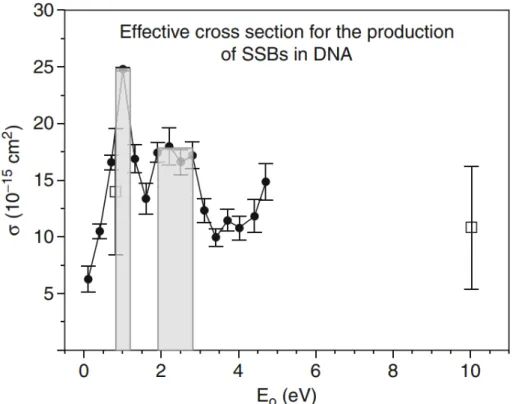
electronic configuration of a neutral (a) and transient negative ion (TNI) (b, c). The interacting electron initially captures into the unoccupied MOs of the neutral molecule resulting in TNI formation via: (a) shape reso-nance or (c) core-excited resoreso-nance. . . 11 1.7 Effective cross sections (σ ) for the formation of SSB in plasmid DNA
by 0.1– 4.7 eV electrons. The shaded portion corresponds to shape reso-nances at 1 eV and around 2.5 eV.(Reprinted from Panajotovic et al. (2006)). 12 1.8 Schematic diagram of the spin-coating system. I—vacuum chamber, J—tube
holder, K— sample substrate, L1 and L2—ball-bearing shafts, M—magnetic coupling, N—electric motor, O—Teflon space. . . 14 1.9 I. The schematic diagram of the experimental setup for irradiating DNA.
(A) electron gun, (B) linear drive, (C) rotatable disk used as cylinder sup-port, (D) electron current detector, (E) cylindrical sample substrate, (F) quick access port, (G) glove box sealed under a N2 atmosphere, (H)
Tan-talum Plate. IIa. Illustation of electon trajectories. IIb. Multi-detector. . . 15 1.10 Schematic design of LC-MS. This diagram demonstrates the technique
that combines the physical separation capabilities of liquid chromatogra-phy (HPLC) with the mass analysis capabilities of the mass spectrometer. 17 1.11 Schematic of the multiple reaction monitoring (MRM) scanning technique
on a triple quadrupole mass spectrometer. The targeted parent ion (shown in yellow) is selected in the first quadrupole (Q1) and enters the second quadrupole (Q2) where it undergoes collision induced dissociation (CID). The resulting “product ions” are mass analyzed using the third quadrupole (Q3) . . . 18
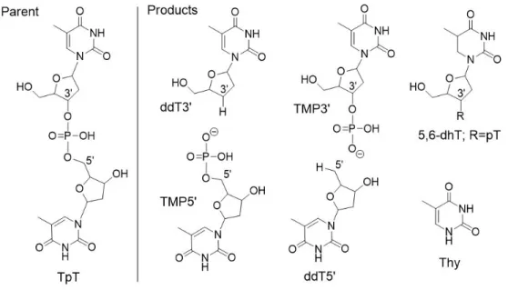
2.1 Structures of compounds under study. vLEEs irradiation of the parent
compound (TpT) gave 20,30-dideoxythymidine (ddT30), thymidine 50-monophosphate (TMP50), 20,50-dideoxy-thymidine (ddT50) thymidine 30 monophosphate
(TMP30), thymine (Thy) and 5,6-dihydro-20-deoxythymidine (5,6-dhT). . 26 2.2 LC-MS/MS analysis of products from C30-O and C50-O cleavage of TpT.
The formation of ddT30and ddT50increased considerably from non-irradiated controls (a) to irradiated samples (c). Likewise, the formation of TMP50 and TMP30increased upon irradiation with vLEEs (chromatograms b and d). . . 27 2.3 LC-MS/MS analysis of products from C10-N1 cleavage and base
reduc-tion from TpT. The formareduc-tion of thymine increased when comparing non-irradiated control to non-irradiated samples (chromatograms a and c) as in-dicated by the ratio of isotopically internal standard (red line) and natu-ral product present in the sample (blue line). Likewise, the formation of TMP50and TMP30increased upon irradiation with vLEEs (chromatograms b and d). . . 28 2.4 Formation of major products as a function of electron fluence. Error bars
show the standard deviation from five independent experiments. Data were fitted by linear regression analysis (red line). Regression coefficient (r2) was greater than 0.99 except for Thy (r2=0.92). Percent error in stan-dard derivation of the slope was 5.1% for TMP50, 3.6% for ddT30, 4.0% for TMP30, 4.7% for ddT50, 2.0% for ddT50, and 13% for Thy. All slopes were statistically significant (P < 0.001). . . 29 2.1 Proposed mechanism for the formation of C30-O and C50-O cleavage
prod-ucts. . . 30 2.2 Proposed mechanism for the release of non-altered thymine. . . 31 S1 Diagram depicting the irradiation of target molecules. A system used
in numerous experiments for electrons with energies between 4-20 eV (Zheng et al., 2004b) was modified for electrons with lower energies (1.8 eV) by placing an electrode at the exit of the cylinder to reflect vLEEs back toward the surface. Red lines emanating from the electron gun (top center) show the simulated trajectory of vLEEs. A multi-detector (shown on the right) was inserted in place of the cylinder to adjust the electron flux so that electrons were evenly distributed along the inside surface of the cylinder. . . 35 S2 Calibration curve for labeled (m/z + 4) and non-labeled thymine. Linear
regression of the data gave y = 35.9 − 4.1 (r2> 0.99) for labeled Thymine (Thy) and y = 25.4x − 2.0 (r2> 0.99) for non- labeled Thymine (Thy). . . 37 S3 Calibration of isotopic standard for the quantification of thymine. Linear
regression gave: y = 0.83x + 1.05 (r2> 0.99). . . 38 S4 Calibration curve for ddT50and ddT30. Linear regression of the data gave
y= 14.1x − 1.7 (r2> 0.99) for ddT50and 10.6x − 1.2 (r2> 0.99) for ddT30. 38 S5 Calibration curve for 5,6-dhT. Linear regression of the data gave y =
S6 Calibration curve for TMP30 and TMP50. Linear regression of the data gave y = 1.76x − 0.68 (r2> 0.99) TMP30and 0.86x − 0.3 (r2> 0.99) for
TMP50. . . 40
S7 Formation of 50-X1pT-30from irradiated 50-TpT-30. . . 41
S8 Formation of 50-X2pT-30from irradiated 50-TpT-30. . . 41
S9 Formation of ddT50from irradiated TMP50. . . 42
S10 Formation of 5,6-dhT from irradiated TMP50. Note that the dinucleotide was enzymatically digested to the nucleoside before analysis. . . 43
S11 Formation of Thy from irradiated TMP50. . . 43
S12 Formation of ddT30from irradiated TMP30. . . 44
S13 Formation of 5,6-dhT from irradiated TMP30. . . 44
S14 Formation of Thy from irradiated TMP30. . . 45
S15 Formation of ddT30from irradiated thymidine. . . 46
S16 Formation of ddT50from irradiated thymidine. . . 46
S17 Formation of 5,6-dhT from irradiated thymidine. . . 47
S18 Formation of Thy from irradiated thymidine. . . 47
D1 A proposed mechanism for thymidine N-glycosidic bond break by LEE bombardment . . . 49
D2 Proposed mechanism of C-O bond cleavage and released products by LEEs 51 D3 A proposed pathway for formation of 5,6-dihydrothymine (base damage). 52 D4 There are 3 main channels for TpT, II) For simpler molecule such as dThd (dT) fewer channels are available, thus, the electron has an increased prob-ability to go to the other side. . . 54
L
ISTE DES TABLEAUX
2.1 Summary of product yields for different molecular targets showing the ef-ficiency of vLEE-induced damage in molecules of product per incident electrons (×10−4). The values were obtained from the slopes of a lin-ear regression for the formation of each product as a function of fluence (Fig.2.4 for TpT; Figs. S9-S11 for TMP50; Figs. S12-S14 for TMP30; and Figs. S15-S18 for thymidine (dThd). All slopes were statistically signifi-cant (P<0.05). . . 30 S1 LC-MS/MS properties of vLEE-induced products of TpT, TMP30, TMP50
and thymidine. The products include thymine non labeled (NL), thymine labeled (L), 5,6-dihydro-20- deoxythymidine (5,6-dhT), 20,30-dideoxythymidine (ddT30), 20,50-dideoxythymidine (ddT50), thymidine 50-monophosphate (TMP50), thymidine 30-monophosphate (TMP30), 5.-X1pT-30where X1= 10,20-dideoxyribose, and 50-X2pT-30 where X2= 20-deoxyribose. Products (a): LC separation
was carried out with a Phenomenex Luna Omega 1.6 um Polar C18 100 x 2.1 mm (i.d.) column protected by a pre-column of the same material. The products were eluted with solvent A (0.05% formic acid) using a gra-dient of solvent B (90% acetonitrile) from an initial 1% to a final 30% in 8 min followed by a short wash (2 min) and re-equilibration (3 min). The nucleoside of 5,6-dhT was measured in separate runs after enzymatic digestion of the sample into its component nucleosides The analysis of TMP30 and TMP50was also carried out in a separate run under the same conditions as above except the MS was operated in negative mode. The dwell time of the MS was 100 ms and the analytes were measured using unit resolution. The complete analysis of products was carried out in three stages. Products (b): LC separation was carried out with a Acquity UPLC HSS T3; 1.8 µm particle size; 100 x 2.1 mm (i.d.) protected by a Van-GuardTM pre-column. The products were eluted with solvent A (5 mM formate buffer (pH 5.0) using a gradient of solvent B (80% acetonitrile) from an initial 1% to a final 12% in 8 min followed by a short wash (2 min) and re-equilibration (3 min). . . 36
L
ISTE DES ABRÉVIATIONS
Ade Adenine C Cytosine
◦C Degree celcius
C-O bond Sugar-phosphate bond CI Collision Induced Dissociation cm Centimeter
Cyt Cytosine
DEA Dissociative electron attachment ddT30 20,30-dideoxythymidine
ddT50 20, 50-dideoxythymidine dTh deoxythymidine
DFT Density functional theory
5,6-dHT 5,6-dihydro-20-deoxythymidine DNA Deoxyribonucleic acid
DSB Double strand break(s) E Incident electron energy ESI electrospray ionization e− Electron
Ea Activation energy
EC electrochemical detection
ec− Continuum electron E. coli Escherichia coli
ESIMS Electrospray ionization mass spectrometry et− Transter electron
eV Electon volt Fe2+ Ferrous ion Fe3+ Ferric ion
FWHM Full width half maximum G Guanine
GC/MS Gas chromatography/ mass spectrometry Gua Guanine h Hour(s) •H Hydrogen radical H- hydride anions H2 Hydrogen gas H2O∗ Excited water H2O+ Water cation H2O2 Hydrogen peroxide H3O+ Hydronium ion
HOMO Highest occupied molecular orbital HPLC High performance liquid chromatography IR Infrared
keV Kilo electron volt (103 electron volt) LBL Layer-by-layer
LC-MS/MS Liquid chromatography-mass spectrometry/ mass spectrometry LEE(s) Low-energy electron(s)
LET Linear energy transfer
LUMO Lowest unoccupied molecular orbital M Molar
MeV Mega electron volt (106 electron volts) MRM Multiple reaction monitoring
min Minute(s) N2 Nitrogen gas
nm Nanometer(n) (10-9 meter) N2O Nitrous oxide
O2 Oxygen gas
•OH Hydroxyl radical
O(3P) Oxygen atom in the triplet 3P ground state P Phosphate group
pT Thymidine-50-monophosphate pTp Thymidine-30,50-diphosphate pTpTp 50-pTpTp-30
QQQ Triple quadrupole in mass spectrometer QTOF Quadrupole time-of-flight
R Deoxyribose sugar
ROS Radical oxidative species RPM Revolutions per minute
SATP Standard ambient of temperature and pressure SE Secondary electrons
SSB Single strand break(s) Thy Thymine
TNI Transient negative ions TMI Transient molecular anion TMP50 thymidine 50-monophosphate TMP30 Thymidine-30-monophosphate TOF Time-of-flight Tp Thymidine-30-monophosphate TpT Thymine-phosphate-thymine TTT 50-TpTpT-30 TXT 50-TpXpTp-30(X= T, C, A, G) UHV Ultra high vacuum
UMO unoccupied molecular orbital UV Ultraviolet
1 I
NTRODUCTION
1.1 Is radiation necessary for life?
These days, developments in science and technology are fundamentally altering the way people live and have increased the willingness of humans to know more and increase their understanding of the physical world. One of the most significant discoveries in the 19th century was radiation. At that time nobody thought that certain kinds of matter can emit radiation, especially when submitted to a high voltage, but in 1896 a German physics professor, Wilhelm Conrad Roentgen, discovered the main properties of X-rays. He found that the X-ray would pass through the tissue of humans leaving the bones and metals visible. This feature was enough to change the medical world forever. Soon afterward, the penetrating properties of the rays began to be exploited for medical purposes (Reed, 2011).
In the past few decades, many researchers and physicians have developed radiation sci-ence in a variety of different ways. It has been used for different reasons and applications. For example, some people used it wisely to produce positive outcomes in medical sci-ence, however, others utilized it for destructive purposes such as nuclear weapons like those used in the atomic bombings of Hiroshima and Nagasaki in 1945 that demonstrated the grave biological response to radiation. Therefore, it has both beneficial and harmful effects simultaneously. However, due to people’s tendency to remember negative events or traumatic experiences they might have had, they face unnecessary or irrational fear. These are one of the reasons that some people are afraid of radiation, and we called this Radiophobia (Koslov, 1982).
1.2 Caution radiation area!
We live and breathe in a world that is like an invisible sea of radiation. In recent years, people have learned to fear the consequences of radiation. They do not want to live near nuclear reactors. They are shocked by reports of connections between excessive exposure to sunlight and skin cancer. They are worried about the leakage from microwave ovens or the radiation produced by their television sets but what is radiation and how hazardous is it? Radiation can be explained as energy or particles that travel through space or other mediums. Many types of interactions can take place when radiation strikes an object.
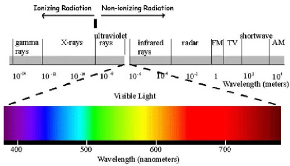
Overall, two things can occur if radiation is absorbed by matter: excitation or ionization. We can generally classify radiation as either ionizing or non- ionizing based on whether it has sufficient energy to remove an electron from the atom that it interacts with. Ioniz-ing radiation is mostly X-ray and Gamma-rays (Figure 1.1) that can remove tightly bound electrons from the orbit of an atom and cause the atom to become charged or ionized. The resulting secondary species can react with molecules, such as DNA inside the cells, along the radiation track, which can be applied for the treating human health problems. Non-ionizing radiation can be considered less dangerous than ionizing radiation. Overex-posure to non-ionizing radiation can cause health issues, for instance, from power lines, microwaves, radio waves, infrared radiation, visible light, and lasers.
Figure 1.1 – Electromagnetic spectrum separating non-ionizing and ionizing radiation.
Excitation is the transfer of an electron by absorbing energy from a lower electronic energy level to a higher energy level, thus, it causes an atom to move from a ground state to an excited state. The principal difference between excitation and ionization is that excitation illustrates the movement of an electron from a lower electronic energy level to a higher energy level while ionization involves the complete removal of an electron from the atom or molecule.
1.3 Radiation exposure and health effects
Ionizing radiation harms living things at the molecular level by ionizing molecules inside microscopic cells that make up the human body. The use of radiation, especially ionizing
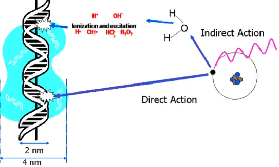
radiation, is currently attracting considerable attention in the field of medical sciences, thus, it is essential to know and understand the role of ionizing radiation. The first way radiation affects our health is through the breakage of DNA molecules by direct or indi-rect effects. If an X-ray or ionizing radiation interacts with the DNA molecule, this is considered a direct effect, whereas, most of the damage to DNA molecules from X-ray is achieved through the indirect effect because when X-rays or ionizing radiation enter a cell, they are much more likely to ionize a water molecule because water is the most abun-dant molecule in cells; water consist of 70% or more of total cell mass (Frohlinde, 1986; O’Neill et al., 2002).
Overall, ionization can happen in any molecule in the cell forming a radical cation and an ejected electron. The ejected electron can attack another molecule or it can become solvated before additional reactions. The radical fragment can be transferred to another nearby molecule. Simultaneously, the radical cations can react and become neutralized by giving up a proton, such is the case of the radical cation produced by ionization of water. H2O+ reacts with an adjacent water molecule immediately (10-14s) to create the
hydroxyl radical •OH. As a consequence of these reactions, cellular DNA can be damaged in several ways, including direct ionization of the DNA, reactions between the DNA and electrons, or solvated electrons, •OH or H2O+, or other radicals ( Figure 1.2 ) (Han et Yu,
2009).
Figure 1.2 – Direct and indirect action of radiation in biological systems
biolog-ical changes. Therefore, it is vital to recognize whether the equal doses of different types of radiation provide the same effect when they are absorbed in biological material. For instance, the rate of ionizing particle energy loss along their tracks for X-ray and electrons is not the same, thus, it is necessary to compare the detected radiation effects with the rate of energy loss, the idea of linear energy transfer (LET) was proposed (Swiderek, 2006). Each type of radiation has a specific structure and can be delivered over a range of different energies. When the radiation interacts with matter, it loses its energy through interactions with the atoms. The average amount of energy is spread over a determined distance, for example, the energy deposited in cells, tissues, and organs is known as the LET. The par-ticular unit regularly used for this quantity is kilo electron volt per micrometer (keV/µm) of unit density material. LET is used to classify radiation as High LET radiation and Low LET radiation. High LET radiation is a type of ionizing radiation that deposits a signifi-cant amount of energy in a small distance, e.g. neutrons and alpha particle, whereas Low LET radiation deposits less amount of energy along the track or has widely spaced ion-izing events. e.g. X-rays and Gamma-rays. If we take water as an example, High LET radiation ionizes water into OH•and H•radicals over a very short track but Low LET ra-diation also ionizes water molecules over a much longer track giving a rara-diation-induced decomposition of water molecules by ionization and excitation. This pathway is called radiolysis of water which can also contribute to the destruction of cells.
For understanding the biological effects which are produced by ionizing radiation, it is better to understand the physical and chemical stages provided by ionizing radiation in liquid water since mammalian cells typically consist of ∼70-85% water, ∼10-20% pro-teins, ∼10% carbohydrates, and ∼2-3% lipids (Turner, 2008).
The physical stage is the initial phase that is produced by radiation in water and thereby forms the ionized and excited molecules, e.g. H2O+, H2O∗take place in less than 10−15
s. After, the initial radicals such as H−, OH−, H2O•−, e−, H•, OH• are generated by
a physio-chemical process within 10−15 to 10−12 s. The radical-radical reaction of two OH• or H•give stable molecules (H2O2 and H2). In the following stage which depends
on LET, the radicals species which are produced in the last step begin to come adequately close to react with each other as their diffusion and distribution in water advance. Chem-ical changes due to bond breakage result from the reaction of those radChem-icals with target molecules in the biological stage causing lots of damage in the cell at various times. That is how cancer develops over time in the human body, by acquiring more and more DNA mutations until they reach a hazardous point at which the cell eventually becomes cancer-ous in the human body (Figure 1.3).
Figure 1.3 – Time scale of radiolysis of water (Alizadeh et Sanche, 2012)
1.4 How cancer develops in the human body
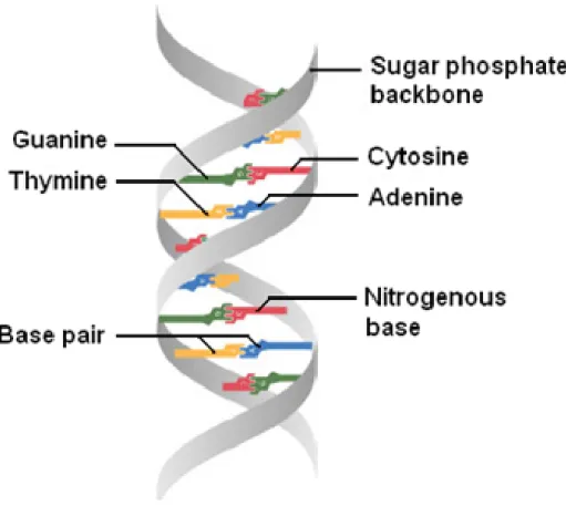
If radiation is absorbed in biological material, ionization and excitations will happen and is not distributed at random but favors localization along the tracks of single charged particles in a way that depends on the dose and type of radiation. Depending on the geometry of the tracks, the biological effects of radiation can vary widely. Some quickly occur while others may take years to become evident. DNA damage induced by radiation can cause critical health effects. Deoxyribonucleic acid, more generally known as DNA, is a complex molecule that contains all of the information required to build and maintain an organism. It consists of two long polymeric strands made of four types of nucleotide subunits which include five-carbon sugar attached to one or more phosphate groups and a nitrogen- containing base which includes purine ( guanine and adenine) and pyrimidine ( cytosine and thymine). Each of these chains is known as a DNA chain or a DNA strand. The sugar and phosphate groups, which build the backbone of each strand, are placed on the surface of DNA whereas the bases are on the inside of the helix (Fig. 1.4). Hydrogen bonds between the complementary base pairs of each strand, connect the two strands of the helix (i.e. between A and T and between C and G), and lead to the pairing of bases that holds the two strands together and gives stability to the DNA polymer (Alberts et al., 2002).
If ionizing radiation interacts with a cell, it can break the DNA strands. In this situation, three things can happen:
Figure 1.4 – DNA forms a double stranded helix, and adenine pairs with thymine and cytosine pairs with guanine.
function or leave no damage
• DNA repair deficiency can lead to mutation. This indicates that cells can lose their ability to reproduce themselves correctly and then transfer the genetic abnormality on to other cells through reproduction, prompting the biophysical change in cells. • Cell death occurs when the damaged on the cell cannot be repaired. In this case,
cells permanently lose their proliferating ability and their functions. This process occurs all the time in everyone. In reality, people are exposed to nearly 10000 to 100000 toxic damage per cell every day due to thermal depurination, oxidation, and alkylation (Lindahl et al., 1993) but the probability of this type of harmful effect is proportionate to the dose and it improves with increasing the radiation dose. If the cell structure changes because it repairs itself inappropriately, this modification could have no additional consequence or the effect could appear later in life. Thus, cancer and genetic defects may or may not succeed (Jackson et Bartek, 2009)
1.4.1 Types of DNA damage
It is accepted that radiation can create a significant amount of DNA lesions including dam-age to the nucleotide bases ( Base damdam-age) or DNA single and double-strand breaks. Base damage occurs via radiation when there is a chemical modification in one of the four base pairs or breakage in the inner rings of the DNA ladder due to several processes. One of them is oxidation by Reactive Oxygen Species (ROS) products. There are various sources of ROS that can cause the formation of oxidative damage. Radiolysis of water by ionizing radiation is one of the sources of ROS products involved in base damage mostly prompted by the reactive species formed from water radiolysis including hydroxyl radicals, solvated electrons, and H atoms (Bauer et al., 2015). The damaged base can be repaired through various base excision repair pathways. Basically, it just removes the damaged nucleotide and replace it with a normal nucleotide. If the abstraction of a hydrogen atom from de-oxyribose moiety occurs, a single strand break will be formed and the polymer is broken into two fragments after this interaction. Although most of this type of damage produced by radiation can easily and quickly be repaired, approximately in 5 minutes, while the other class is clustered lesions which includes DSBs and other multiple lesions involving strand breaks and base damages, are much more complicated to repair and also results in potentially dangerous DNA damage responses. DSBs induced by ionizing radiation can cause abnormality in the chromosomes. Chromosomes are found in the nucleus of most living cells, providing genetic information in the forms of genes. Therefore radiation can harm many genes causing failure and death in cells (Han et Yu, 2010). DSB is the common lesion produced by ionizing radiation where the phosphate backbone of two corresponding DNA strands are broken and generate very cytotoxic forms of damage (Mehta et Haber, 2014). Recently, another feature of radiation damage has been identified as clustered DNA damage which occurs when two or more lesions are formed within one or two helical turns of the DNA through the reactive species produced by the track of a charged particle via direct and indirect effects.
The LET of radiation defines the rate and complexity of clustered damages, in view of several modeling studies, as estimated to be about 30% and 70% of DSBs induced by low and high LET radiation respectively (Cannon, 2016).
1.5 Why study low-energy electron-induced reactions
When high energy radiation (such as X-rays and Gamma-rays) and fast charged particles interact with matter, copious numbers of electrons will be produced by the ejection of electrons from molecules during the initial ionization process. There are two processes
which dominate when the matter absorbs energetic photons. At low energies, typically the primary process is the photoelectric absorption (E< 0.5 MeV), a photon transfers all its energy to an inner-shell electron in an atom resulting in the ejection of that electron from its shell. In turn, the electron can pass through the surrounding matter to cause additional ionization.
Briefly, photoelectric reactions are most probable to occur with low-energy photons and elements with high atomic numbers provided the photons have adequate energy to over-come the forces binding in electrons in their cells. Photoelectric absorption facilitates the measurement of the energy of a Gamma-ray photon and this interaction can also lead to the creation of X-ray fluorescence.
At higher energies (approximately 0.7-10 MeV), if the incident X-ray photon is deflected from its initial path by interaction with an electron, it will cause the ejection of that electron from its orbital position. Thus, the X-ray photon loses energy because of the interaction but continues to travel through the material along an altered path. It can subsequently be involved in further interactions leading to the production of a large number of relatively fast electrons because after scattering, the photon has less energy so it is more probable to produce the photoelectric effect which these fast electrons can further excite or ionize other atoms in the medium giving a large amount of the secondary LEE before the electron is thermalized. In both of these processes, nearly all of the absorbed photon energy turns into kinetic energy when an X-ray produces a photoelectron this electron is a primary electron and after secondary LEE.
The reason why these LEEs are so important is that they are produced in large amounts. Approximately 4 × 10⁴ per 1 MeV of the primary photon changes into many reactive species, e.g. radicals, ions, and excited molecules which were created through the ionizing radiation track. Since the optical oscillator strength for small (e.g., H2O) and large (e.g.,
DNA) biomolecules is greatest at an energy of about 22 eV, when this primary interaction leads to ionization, the distribution of electrons has a maximum below 15 eV.’The majority of the energy of LEEs is distributed below 30 eV with the most probable energy of around 9–10 eV. These electrons are referred to as secondary LEE (Sanche, 2003).
1.6 Principles of the interaction of LEEs with molecules
To get the idea that how LEEs induce damage to DNA and generate radiobiological dam-age such as strand breaks and other fragments, it is crucial to understand and precisely explain the detailed pathways of reactions and mechanisms involving low-energy elec-trons (LEEs) interaction with DNA.
Generally, low-energy electron collisions with molecules can be classified into two main types: elastic and inelastic. During elastic collisions, the loss of electron energy to the target is negligible, therefore, there is no loss of energy. In contrast, inelastic collisions lose a significant amount of electron energy to the target. Thus, it may create electronically excited states followed by other reactions. The elastic collisions are less significant as no energy is deposited, in contrast to the inelastic collision. The inelastic collisions of low-energy electrons with molecules and atoms lead to several energetic species that are the primary reasons for the wide variety of radiation-induced chemical reactions. Because of the numerous inelastic collisions, these secondary electrons become thermalized in nearly one picosecond.
There are several processes in this study initiated by electron interactions with matter and terminating with DNA damage by the initial formation of a transient molecular anion (TMA) or transient negative ion (TNI) of a DNA localized subunit. TMA is one of the most important features illustrating how LEEs induce DNA damage. TMA is formed by the initial capture of an electron by a molecule i.e. the incoming electron temporarily occupies a previously unfilled orbital of a molecule. Upon TMA formation, an extra electron is captured into the unoccupied molecular orbital (UMO) of the neutral molecule resulting in shape or core-excited resonance (Kumar et Sevilla, 2008). A shape resonance or a single-particle state occurs if the additional electron occupies a previously unfilled orbital (such as a lowest unoccupied molecular orbital or LUMO which is an electron in an excited state that skips from the ground state after the energy of a photon of sufficient energy is transferred to the electron in the HOMO) of a molecule in its ground state. Usually, this happens at low energies (0–4 eV) with lifetimes in the range 10−15 to 10−10 s. Core-excited resonance or two-particle states, formed when an interacting electron excites one of the core electrons of the molecule from the ground state (when the incident electron has higher energy, usually E > 3eV) giving a configuration consisting of an electron in a LUMO and an electron-hole in HOMO. Core-excited resonances occur typically above 4 eV, are highly energetic, and have been suggested to play a role in double-strand breaks in DNA (Figure 1.6) (Alizadeh et al., 2016).
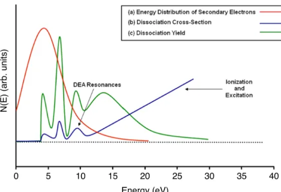
The other process, which is also the most important process, is called dissociative electron attachment (DEA). In this process, if the TMA state is dissociative and the resonance lifetime is higher than about half of the vibration period of the anion, it can dissociate into natural and anionic fragments. The dissociation possibility rises with increasing incident electron energy. The dissociation yield is highest at low incident electron energies (E > 10 eV) owing to the abundance of secondary electrons at those energies. In contrast, electron
Figure 1.5 – (a) Schematic energy distribution of secondary electrons generated during a primary ionizing event which means the energy distribution of the secondary electrons demonstrate that the majority of these electrons have energies below 10 eV. (b) cross section for electron-induced dissociation for a typical molecule; (c) dissociation yield as a function of electron energy for a typical molecule.
impact excitation and the electron impact ionization regularly take place at energies above 6 eV and 10 eV respectively (Figure 1.5)(Arumainayagam et al., 2010)
1.7 DNA damage induced by ionizing radiation
As briefly described in Section 1.6, low-energy electrons (LEE) are produced copiously during high-radiation events and are increasingly being considered as important DNA-damaging agents. The primary series of experiments by Sanche and coworkers (Li et al., 2008; Zheng et al., 2004a; Abdoul-Carime et al., 2001) determined that LEEs in the 0–10 eV range produce single- and double-strand breaks in DNA. Indeed, they found that LEEs are several times more damaging than photons of comparable energy.
In 2000, a key publication (Boudaïffa et al., 2000) concerned the formation of resonances from the interreaction of DNA with low-energy electrons. In this study, plasmid DNA (pGEM 3Zf(-)) obtained from E. coli was irradiated with monoenergetic LEE beams (+/− 0.5 eV) with kinetic energies in the range between 3–20 eV. They found that electrons having energy below the ionization limit of DNA (ca. 7.5–10 eV (Hush et Cheung, 1975;
Figure 1.6 – (reprinted from Kumar et Sevilla (2012)). Schematic diagram showing the electronic configuration of a neutral (a) and transient negative ion (TNI) (b, c). The inter-acting electron initially captures into the unoccupied MOs of the neutral molecule resulting in TNI formation via: (a) shape resonance or (c) core-excited resonance.
Orlov et al., 1976) ) were able to create SSB and DSB. From these studies, the yield of damage was demonstrated for both SSB and DSB, and calculated as 8.2 × 10⁻⁴, and 2 × 10⁻⁴strand breaks per incident electron, respectively, for 10 eV electrons. The yields of SSB and DSB in DNA depended on the energy of the interacting electron and were proposed to be produced by the rapid fragmentation reactions of transient molecular res-onances localized on DNA components i.e. base and sugar moieties, and the phosphate backbone.
Sanche and co-workers obtained further insights into the mechanisms of LEE-induced DNA damage (Panajotovic et al., 2006). There are several key results shown in this study. The first one is that 0–4.7 eV electrons are able to generate SSB in plasmid DNA with related yields to those obtained at higher energies. The second key finding is that the cross- section for SSB formation at 1 eV is greater than that observed at 10 eV. The last main point that this group also found was that 0.1–4.5 eV electrons induce only SSB by the involvement of resonances for causing damage at 0.8 and 2.2 eV. Due to the low electron energies, these resonances were defined as shape resonances(Figure 1.7).
The findings from strand breaks in plasmid DNA induced by 3–100 eV electrons and cor-relation with shape resonances of the bases identified by Burrow et al in the gas phase indicated that, below 5 eV, LEE-induced SSB takes place through dissociative electron attachment (DEA) via shape resonances (Boudaïffa et al., 2000; Huels et al., 2003; Mar-tin et al., 2004; Panajotovic et al., 2006; Brun et al., 2009)whereas, between 5 and 15 eV, the core-excited resonances induce SSB and DSB. Also, several theoretical studies were
Figure 1.7 – Effective cross sections (σ ) for the formation of SSB in plasmid DNA by 0.1– 4.7 eV electrons. The shaded portion corresponds to shape resonances at 1 eV and around 2.5 eV.(Reprinted from Panajotovic et al. (2006)).
undertaken to explain the mechanism of LEE induction of DNA strand breaks soon after the discovery that low- energy (3–20 eV) electrons caused both single- and double-strand breaks in plasmid DNA (Boudaïffa et al., 2000). The authors used density functional the-ory (DFT) and theoretical simulations to predict LEEs interactions with DNA (Bao et al., 2006; Berdys et al., 2004; Gu et al., 2005; Kumar et Sevilla, 2007; Simons, 2007). Since the calculation of the complete DNA molecule at the ab initio or DFT level is presently restricted to small molecule size, fragments of DNA structure were usually modeled in-cluding a base, sugar, and phosphate moieties attached at 30- and 50- ends of the sugar ring.
1.7.1 Report of prior results by our group
DNA damage induced by irradiation has been studied for many decades. Such studies en-able us to have a better understanding of the dangers caused by radiation and to improve the efficiency of the radiotherapies that are used to combat cancer. Regarding this idea, the fundamental interactions of LEEs with nucleobases, 2-deoxyribose derivatives, oligonu-cleotides, and plasmid DNA have been investigated (Ptasi´nska et al., 2005; Huels et al.,
1998; Abdoul-Carime et al., 2001; Breton et al., 2004; Huels et al., 2004; Lepage et al., 1998; Ptasi´nska et al., 2004; Park et al., 2006; Ray et al., 2005; Martin et al., 2004). First, in 2004, Zheng et al. studied the interaction of thymidine with LEEs. They showed that cleavage of the N-glycosidic bond of thymidine leading to the release of thymine was a significant product (Zheng et al., 2004a). This means that glycosidic bond cleavage created within the formation of a transient anion state is formed by low-energy electrons localizing in the antibonding orbitals of the glycosidic bond. Hence, the conclusion was that LEEs are involved in glycosidic bond cleavage. Later, this group focused on the for-mation of products by monoenergetic LEEs from bigger molecules: two tetramers (CGTA and GCAT) (Zheng et al., 2005). They observed the formation of numerous products in-cluding non-modified nucleobase, nucleoside, and nucleotide fragments associated with the cleavage of the phosphodiester C–O bonds between the sugar and phosphate bonds to-gether with the N-glycosidic bond between the base and sugar group within each tetramer. These results demonstrate the pathway of bond breaking via electron promotion into an antibonding orbital of the phosphate group or an antibonding orbital of the DNA base, from where the electron can be transferred to the phosphate group through bond trans-fer followed by cleavage of the C-O bond. The breaking of the C-O bond is pretrans-ferred owing to the very high electron affinity of the phosphate group (Swiderek, 2006). This hy-pothesis was further supported by Simon and co-workers who showed, using theoretical calculations, that the excess electron is initially captured in an orbital located in the nu-cleobase and then transferred remotely to the phosphodiester bond (Simons, 2006). Such experiments play a significant role in obtaining a fundamental understanding of radiation-induced DNA damage in a living cell.
There are several investigations about the effect of the phosphate group reported by Li and co-workers. First, they focus on the effect of the terminal phosphate group using different sizes of DNA model compounds including monomers (pT, Tp, pTp), dinucleotides (pTpT, TpTp, pTpTp), and trinucleotides (TpTpT). Based on their experiments, the presence of terminal phosphate groups dramatically affected the distribution of low-energy electron-induced damage in DNA model compounds. In addition, Li and co-workers studied the effect of base sequences in a series of oligonucleotide trimers including TXT where X can be one of four standard DNA bases (C, T, A, G). The analysis of damage remaining within the nonvolatile condensed phase was determined following irradiation by the low-energy electron (10 eV). They found that the initial low-energy electron capture and subsequent bond breaking within the transient negative anion depended on the sequence and electron affinity of the bases, with the highest damage attributed to the most electronegative base,
thymine (Li et al., 2010).
In later years, different experiments have led to a better understanding of the effects of low-energy electrons. Surakan and co-workers investigated the products induced by both LEE and ionization. They deposited prepared dry films of linear double-stranded DNA on glass and tantalum substrates and then, after LEE bombardment, they measured damage as base modification and non-modification. Their results indicated the formation of both base release and base modification products by LEE-induced, DEA-mediated processes. The nature and yields of products were very similar, but not identical to, those arising from ionization. The yield of non-modified bases, as well as base modifications, increased by 20- 30% when DNA was deposited on a tantalum substrate which generated low-energy electrons compared to that on a glass substrate which mimicked direct ionization (Choo-fong et al., 2016).
1.8 Our laboratory methods and LEE irradiator system
1.8.1 Spin Coating System
In order to prepare samples for bombardment with LEE, a spin-coating system was de-veloped in our lab several years ago (Zheng et al., 2004a). The biomolecules were spin-coated onto the inner surface of the tantalum cylinders (Figure 1.8). Seven tantalum cylin-ders were bound together with Teflon spacers, and they can contain a certain amount of the solution individually onto the inner surface of each cylinder. The cylinders were in-stalled into a UHV chamber and rotated magnetically outside the chamber to an angular velocity up to around 1500 rpm under low pressure. Through this method, the sample can be expected to be distributed uniformly onto the inner surface of the cylinders resulting in a thin coat of controllable thickness depending on the initial amount of molecules added.
Figure 1.8 – Schematic diagram of the spin-coating system. I—vacuum chamber, J—tube holder, K— sample substrate, L1 and L2—ball-bearing shafts, M—magnetic coupling, N—electric motor, O—Teflon space.
1.8.2 Apparatus for low energy electron production
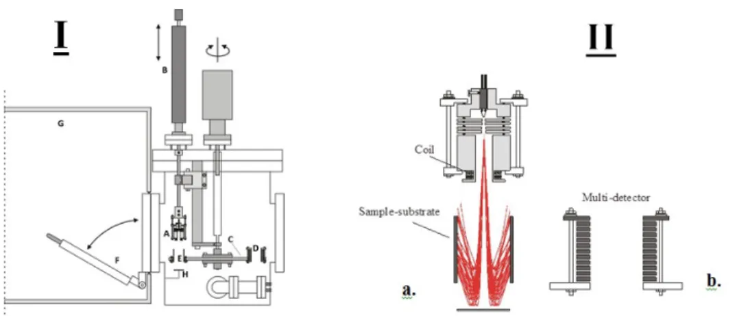
Irradiation with a low-energy electron is accomplished using an electron gun apparatus. Our group developed this novel system several years ago. The LEE gun irradiator was set in an ultra-high vacuum (UHV) chamber driven by an oil-free turbo molecular pump enabling the chamber to be evacuated rapidly from atmospheric pressure to the 109 Torr range (Figure 1.9). The assembly includes the electron gun (A) fixed on a linear drive (B) and a rotatable circular platform (C) such that rotation of this platform enables the sample to be bombarded individually through the electron gun at precise energy and current for a given time. A cylindrical multiple-electrode detector can support the energy distribution of an electron (D) in the inner surface of seven tantalum cylinders (E). The port is used for quick access to the UHV chamber (F) from the inside of a glove box (G) which is kept under a dry N2 atmosphere. Therefore, using this equipment, the gun can irradiate the
inner surface (26 cm2) of a tantalum cylinder with 3-130 eV electrons having an entire energy spread of 0.5 eV full width at half maximum.
Lately, our group modified this system to produce electrons below an energy of 3 eV by placing a tantalum plate (H) directly below the cylindrical sample substrate to repel the incident electron toward the inner surface of tantalum cylinders (Figure 1.9).
Figure 1.9 – I. The schematic diagram of the experimental setup for irradiating DNA. (A) electron gun, (B) linear drive, (C) rotatable disk used as cylinder support, (D) electron current detector, (E) cylindrical sample substrate, (F) quick access port, (G) glove box sealed under a N2atmosphere, (H) Tantalum Plate. IIa. Illustation of electon trajectories.
1.8.3 DNA damage detection by LC-MS/MS technique
In the past, numerous analytical techniques were used to measure and detect DNA dam-age. These included acid hydrolysis of DNA, comet assay, enzymatic digestion of DNA, high- performance liquid chromatography (HPLC) with electrochemical detection (ECD), GC/MS, and LC/MS.
These days, among all these techniques, LC-Mass spectrometry (LC-MS) has become the most popular technology compared to others due to its ability for sensitivity and specificity but, with growing experience, researchers improved this technique as a significant discov-ery in the field of clinical research. Instruments have been transformed from complicated, high- cost, highly advanced research tools to robust, easy-to-use routine detectors.
Moreover, as the instruments have been improved, more applications have been devel-oped. The progress of tandem MS or MS/MS in this field is mainly because of its higher sensitivity (up to the ppt range) which is suitable for the accurate detection of a variety of reductively and oxidatively modified bases of DNA.
The LC-MS/MS performs with a combination of chromatography (LC) and multiple quadrupole mass spectrometers (MS). The chromatographic system first separates the various compo-nents, concentrating the amount of each single component entering the mass spectrometer. Division of the sample components is achieved through an HPLC column where the ana-lytes display differential separation between the mobile phase (eluent) and the stationary phase (coated onto a support material and packed into the column). The principle of mass spectrometry, which is directly connected to electrospray, is the alteration of the separated analyte molecules to a charged (ionized) state following the analysis of the ions and any fragment ions that are formed through the electrospray ionization (ESI) process.Because the ions travel through a magnetic or electrical field, their movement is determined by their m/z ratio, therefore, ions are separated based on their m/z ratio in the MS analyzer (Gross, 2006). In this system, m and z stand for mass and charge of the detected ions respectively (Figure 1.10).
There are several very popular mass analyzers used for LC-MS and they differ in the primary way in which they separate species on a mass-to-charge basis (Tretyakova et al., 2013). The first one is the Quadrupole Mass Analyzer. The ions are first filtered by the electrostatic potentials applied to the elements of the mass analyzers and then they focus ions for analysis depending on their mass to charge ratio. The second one is the Time of Flight (TOF) Mass Analyzer. Ion filtration can be done by the time it takes for a flight from one point (start) to another point (end) because higher m/z ions require more time to
Figure 1.10 – Schematic design of LC-MS. This diagram demonstrates the technique that combines the physical separation capabilities of liquid chromatography (HPLC) with the mass analysis capabilities of the mass spectrometer.
flight compared to the low m/z ions. The last one is the Trapped-Ion Mass Analyzer which operates by sorting ions using direct current (DC) and radio frequency (RF) electric filed to trap ions. The ions can be collected or manipulated in various ways, such as isolation or fragmentation, and are then driven from the cell in m/z sequence. Ion traps are available in linear and 3-D configurations. This gives some unique capabilities such as extended MS/MS experiments, very high resolution, and high sensitivity.
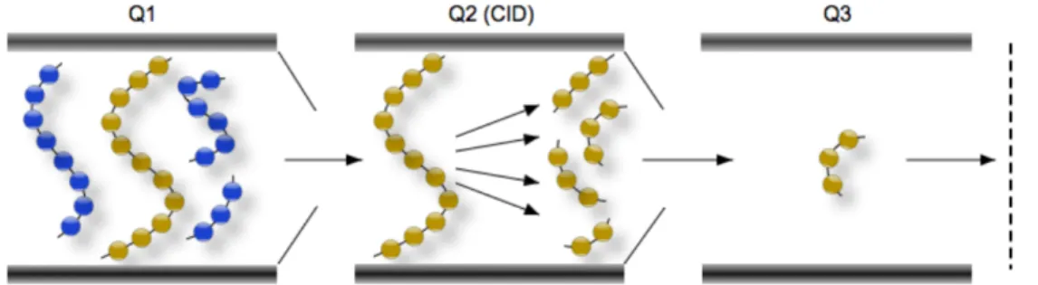
In this section, I will focus on the Triple Quadrupole Mass Analyzer and will describe it for a better understanding of my project. Triple Quadrupole Mass Spectrometer (QQQ) Analyzers are often used when higher sensitivity and specificity is needed. They consists of a series of three quadrupoles (Q1-Q3) together with several modes of operation resulting in different information. Therefore, the first quadrupole (Q1) selects ions of interest that were generated in the ion source. The second quadrupole (Q2) is typically filled with nitrogen and is used as a collision chamber to create fragment or daughter ions by a process called Collision Induced Dissociation (CID). The third quadrupole (Q3) is used to monitor the specified fragment ions which are related to the molecular structure of analyte ions and, thus, provides the characteristic structural information of the molecule.
Triple Quadrupole MS systems can be performed in a tandem MS/MS called Selected Multiple Reaction Monitoring (MRM) mode which is the most common method for quan-titation of analytes by LC/MS/MS (Dass, 2007). In our experiment, we detect DNA dam-age products with this method. MRM mode acts like a double mass filter which drastically reduces noise and increases selectivity. The first quadrupole filters a particular precursor ion of interest (Q1) and then enters the second quadrupole (Q2) known as a collision cell. The parameters are optimized to produce fragments from the neutral collision gas, such as nitrogen, and create product ions that are transferred into the third quadrupole where
only a specific m/z is permitted to pass (Q3) This is a very sensitive method and used for quantitation (Keshishian et al., 2007)(Figure 1.11).
Figure 1.11 – Schematic of the multiple reaction monitoring (MRM) scanning technique on a triple quadrupole mass spectrometer. The targeted parent ion (shown in yellow) is selected in the first quadrupole (Q1) and enters the second quadrupole (Q2) where it undergoes collision induced dissociation (CID). The resulting “product ions” are mass analyzed using the third quadrupole (Q3)
1.9 The brief explanation of the research project (Objective)
With the purpose of better understanding the mutagenic and lethal effects of ionizing ra-diation, Our group has recently begun studying the low-energy electron induced process in biomolecular films. Our recent studies have shown that low-energy secondary electrons produced in biological tissues through high-energy radiation therapy can create single-and double-strsingle-and DNA breaks single-and site-specific cleavage of DNA bases.
Our main goal for the present project is to understand the chemical mechanism of dam-age by a very low-energy electron (below 5 eV) using chemical analyses of damdam-aged molecules and model compounds of DNA in particular dinucleotides. We chose to study thymidine dinucleotide (TpT) as a simple model of DNA because we have much experi-ence with thymine decomposition and because previous studies suggested that it was the most sensitive of DNA bases to low-energy electron induced damage. For experiments, we deposited the thin film of TpT into the inner surface of tantalum cylinders to be exposed to a very low- energy electron (∼1.8 eV) under SATP (Standard Ambient Temperature and Pressure) surrounded by the N2atmosphere. The DNA damage such as single-strand
breaks, base release (Thy), and base modification of Thy caused by vLEE are detected by LC-MSMS by MRM mode.
2 A
RTICLE
Strand breaks induced by very low energy electrons: Product analysis
and mechanistic insight into the reaction with TpT
Authors: Ghazal Khorsandgolchin, Léon Sanche, Pierre Cloutier, J. Richard Wagner.
Article status: Submitted to Journal of the American Chemical Society, March 26, 2019. I did most of the experiments and all the calculation regarding the final results in this paper. Prof. J. Richard Wagner and Mr. Pierre Cloutier did the optimization and calibration of products as given in part in the supplementary information of the paper. Alos, Prof. J. Richard Wagner taught me how to use the instruments of HPLC and LC-MS/MS. Mr. Pierre Cloutier helped me to work with the LEE irradiation source and repaired it when it had problems. Prof. Wanger and Sanche wrote the manuscript. I helped in preparing the methods, materials and the results sections.
2.1 Résumé
De nombreuses études expérimentales montrent que les électrons de 5-15 eV induisent des ruptures de brin dans l’ADN même à des énergies inférieures au seuil d’ionisation des composants de l’ADN. Dans cette gamme d’énergie, les dommages à l’ADN résultent principalement de la formation d’ions négatifs transitoires et dissociatifs (attachement dis-sociatif d’électron, DEA) et d’excitation électronique d’états disdis-sociatifs. Ici, nous avons effectué une analyse LC-MS / MS des produits de dégradation résultant de l’irradiation de TpT, un composé modèle de l’ADN, bombardé avec des électrons de très basse én-ergie (1,8 ± 0,3 eV). La formation de thymidine 50-monophosphate (TMP50) avec la 20, 30-didésoxythymidine (ddT30) peut être expliquée par le clivage direct de la liaison C30-O de TpT, alors que la thymidine 30-monophosphate (TMP30) et la 20, 50-didésoxythymidine (ddT50) sont formées par clivage de la liaison C50-O. La formation de ddT30 et de ddT50 diminue lors de l’irradiation de TMP50 ou de TMP30, et même plus dans le cas de la thymidine, soulignant le rôle critique du groupe phosphate. Il est intéressant de noter que les rendements en TMP50et TMP30étaient supérieurs à ceux des produits correspon-dants ddT30et ddT50, suggérant des destins alternatifs pour les radicaux sucre centré C30 et C50. En revanche, la libération de thymine était faible (<20%) et n’a pas entraîné la formation de produits attendus à partir du clivage induit par la DEA au niveau de la liai-son N-glycosidique. Enfin, les électrons de 1,8 eV ont induit la conversion de la thymine en 5,6-dihydrothymine (5,6-dhT) au sein de TpT, une réaction impliquant vraisemblable-ment des radicaux anions thymine. En résumé, nous montrons qu’une des voies majeures de dégradation implique le clivage, induit par la DEA, des liaisons C30-O et C50-O de TpT, ce qui entraîne la formation de fragments spécifiques, qui représentent une rupture d’un seul brin de l’ADN.
Mots clés: rayonnements ionisants, lésions de l’ADN, la spectrométrie de masse, élec-trons secondaires
2.2 Abstract
Numerous experimental studies show that 5-15 eV electrons induce strand breaks in DNA at energies below the ionization threshold of DNA components. In this energy range, DNA damage arises principally by the formation of transient negative ions, decaying into dis-sociative electron attachment (DEA) and electronic excitation of disdis-sociative states. Here, we carried out LC- MS/MS analysis of the degradation products arising from bombar-ment of TpT, a DNA model compound, irradiated with very low energy electrons (vLEEs; ∼1.8 eV). The formation of thymidine 50-monophosphate (TMP50) together with 20,30 -dideoxythymidine (ddT30) can be explained by direct cleavage of the C30-O bond of TpT, whereas thymidine 30-monophosphate (TMP30) and 20,50-dideoxythymidine (ddT50) are formed by cleavage of the C50-O and bond. The formation of ddT30and ddT50decreased upon irradiation of either TMP50 or TMP30, and even further in the case of thymidine, underlining the critical role of the phosphate group. Interestingly, the yield of TMP50 and TMP30 was higher than that of the corresponding ddT30 and ddT50 products, sug-gesting alternative fates of C30 and C50-centered sugar radicals. In contrast, the release of thymine was minor (<20%) and did not result in the formation of expected products from DEA-mediated cleavage at the N-glycosidic bond. Lastly, vLEE induced the conver-sion of thymine to 5,6-dihydrothymine (5,6-dhT) within TpT, a reaction likely involving thymine anion radicals. In summary, we show that a major pathway of vLEEs involves DEA-mediated cleavage of the C30-O and C50-O bonds of TpT resulting in the formation of specific fragments, which represent a single strand break in DNA.
2.3 Introduction
The genotoxic effects of ionizing radiation have largely been attributed to the reaction of hydroxyl radicals with DNA (the indirect effect) and the direct ionization of DNA compo-nents, both giving oxidative DNA modifications, including base damage and strand breaks (Sonntag, 2006; Cadet et Wagner, 2014). In contrast, the effects of electrons that are ejected during the ionization process have been neglected because they are believed to either ionize the medium further or rapidly undergo hydration to solvated electrons. The latter species react with oxygen to give unreactive superoxide anions, or react with DNA to give base damage but no direct strand breaks (Nabben et al., 1982). There is over-whelming evidence today that a third process also contributes to DNA damage, namely the reaction of low energy electrons (LEEs; 0-20 eV). About 3 × 10⁴ LEEs are released during the ionization process for every MeV absorbed along a radiation track (Pimblott et LaVerne, 2007). Numerous experiments demonstrate the ability of LEEs to break va-lence bonds in model compounds and isolated DNA under a variety of conditions (Sanche, 2009; Alizadeh et Sanche, 2012; Alizadeh et al., 2015). LEE bombardment stimulates the desorption of various ionic fragments (H−, O−, CN−) from solid films of DNA model compounds below the ionization threshold (Ptasi´nska et Sanche, 2006). Upon LEE im-pact with solid films of plasmid DNA, numerous types of macromolecular damage are produced, such as strand breaks and DNA crosslinks, as inferred by the analysis of DNA fragments by gel electrophoresis (Brodeur et al., 2018; Luo et al., 2014). In the condensed phase, the maximum yield of ion desorption as well as strand breaks occurs in the region between 5-12 eV. Similar results with a maximum around 7 eV for strand breaks were also observed in oligonucleotides using a novel DNA origami platform approach (Schür-mann et al., 2017). Although it is clear that LEEs induce strand breaks in condensed films of oligonucleotides and plasmid DNA, there is a paucity of information about the chem-ical steps from initial bond cleavage to the formation of stable products. It is important to clarify these chemical steps and the corresponding intermediate and final products, be-cause the structure of the final products ultimately dictates the biochemical consequences of damage in cellular DNA.
Previously, our approach has been to irradiate short oligonucleotides (e.g., GpCpApT, TpTpT, TpT) with monoenergetic LEEs (5-15 eV) as a dry condensed film under ultra-high vacuum (Zheng et al., 2005, 2006a,b; Li et al., 2011, 2010, 2008; Park et al., 2011, 2013). The non-volatile products are recovered from the surface and subsequently sub-jected to LC-MS/MS analysis. Using this approach, the main products of DNA model targets were identified as non-altered nucleobases; 2) nucleotide fragments bearing a
terminal phosphate group; and 3) base modifications (e.g., 5,6-dihydrothymine and 5-hydroxymethyluracil). The formation of these products is explained by LEE-mediated dissociative electron attachment (DEA) involving C10-N1 bond cleavage (nucleobase re-lease); phosphodiester C-O bond cleavage (fragments bearing a terminal phosphate group), and either C-H or N-H bond cleavage (base modifications). Recently, we extended these studies to longer oligonucleotides and purified DNA exposed to LEEs with an average energy of 5.8 eV under standard temperature and pressure (Choofong et al., 2016). Ex-perimentally, the mechanism of strand break formation is mainly based on the appearance of fragments bearing a terminal phosphate; for example, Tp and pT from the irradiation of TpTpT with 10 eV electrons (Li et al., 2010, 2008; Park et al., 2011, 2013). However, var-ious reactions of the sugar moiety result in the formation of fragments bearing a terminal phosphate group, such as the direct ionization of sugar moieties, photoexcitation of base cation radicals and others (Adhikary et al., 2014). Thus, one of the goals in the present study is to confirm the mechanism of C-O cleavage by obtaining a more complete balance sheet of associated stable fragments using a relatively simple target, TpT.
Cleavage of the C10-N1, C30-O and C50-O bonds in DNA has been extensively exam-ined by molecular computational methods (Carsky et Curik, 2011; Gu et al., 2012; Kumar et Sevilla, 2017; Kohanoff et al., 2017). Although theory and experiment are largely in agreement, there is still much debate concerning the nature of the shape and core-excited resonances and in particular, the sites of initial electron capture and cleavage. The energy range between 0-4 eV is very interesting, since available reaction channels are limited to specific shape resonances decaying exclusively into DEA. This condition eliminates other more complex pathways arising from the attachment of 4-12 eV electrons. These include the mixed possibility of decay of core-excited resonances via DEA or autoioniza-tion leading to dissociaautoioniza-tion of electronically excited states. Core-excited resonances as well as some high-energy shape resonances are excluded for vLEEs in the 0-4 eV regime. Many theoretical investigations predict that the most favorable pathway to strand break in the vLEE regime involves the initial capture of electrons by the base moiety followed by transfer to an antibonding orbital and cleavage of the C30-O30 bond as part of the sugar phosphate backbone (Aflatooni et al., 1998; Simons, 2006; Bao et al., 2006; Wang et al., 2010; Smyth et Kohanoff, 2012; Chen et al., 2014). In contrast, there is considerable the-oretical support for DEA occurring via initial capture of vLEEs by the phosphate group, leading directly to strand breaks (Li et al., 2003; Kumar et Sevilla, 2007, 2008, 2009; Bhaskaran et Sarma, 2014; Cauët et al., 2013). Notwithstanding, there have been only a few experiments providing evidence for strand cleavage in this low energy regime (Mar-tin et al., 2004; Liu et al., 2016; Solomun et al., 2009; Koˇcišek et al., 2018). To clarify the
chemical steps in the mechanism of formation of DNA strand breaks, we have examined in greater detail the formation of products from TpT upon impact with vLEE (∼1.8 eV). 2.4 Methods and Materials
The irradiation system and protocol has been employed in numerous experiments with electrons of energies between 4-20 eV, as previously described in detail (Zheng et al., 2004b). The target molecules are uniformly deposited by spin-coating on the inside sur-face of tantalum cylinders using a rotary device under reduced pressure. The system holds seven cylinders for individual irradiation with 11 µg of target molecules per cylinder, which corresponds to approximately 5 monolayers taking the average density of DNA (1.7 g cm−3). The advantage of this technique is that relatively large quantities of target molecules can be irradiated, permitting a more comprehensive chemical analysis of the stable products. Utilization of this apparatus for electrons below 4 eV, however, was not efficient because most electrons in this energy range were deflected away from the tar-get due to space charges. To remedy this situation, an additional electrode was placed at the exit of the cylinder to reflect vLEEs back toward the irradiation surface (Fig. S1). By choosing the right distance between the cylinder and the electrode and by adjusting the magnitude of the magnetic field collimating the electron beam, the system was now able to efficiently irradiate the inside surface of the cylinder with vLEEs ( ∼1.8 eV). The dispersion of electrons was adjusted with the aid of a multi-array charge detector to as-sure that the beam was uniform over the entire inner surface of the cylinder as described previously (Zheng et al., 2004b).
The analysis of DNA modifications was carried out by ultra-high-performance liquid chro-matography (LC) on line with tandem mass spectrometry (LC-MS/MS). The LC system (Nexera X2, Shimadzu) consisted of dual pumps (Model 30AD), an autosampler (SIL 30AC), a column oven (CTO-10AC) and an UV/Vis detector (SPD-20A). The products were separated using a Phenomenex Luna Omega 1.6 um Polar C18 100 × 2.1 mm (i.d.) column protected by a pre-column of the same material. The temperature of the column was maintained at 30◦C. For product separation, the mobile phase consisted of a gradient of 0.05% (v/v) formic acid in water (pH 3.5; solvent A) and 90% acetonitrile in water (sol-vent B). The mobile phase composition increased from 2% to 20% of sol(sol-vent B in 10 min followed by a 2 min wash cycle with 100% of solvent B and a 3 min equilibration back to the initial conditions of 2% solvent B. For the detection of products, the eluent from the LC was first directed into a UV detector for quantification of the parent compound at 260 nm and then into the tandem mass spectrometer system (API 3000, AB-Sciex, Concorde,