AVIS
Ce document a été numérisé par la Division de la gestion des documents et des archives de l’Université de Montréal.
L’auteur a autorisé l’Université de Montréal à reproduire et diffuser, en totalité ou en partie, par quelque moyen que ce soit et sur quelque support que ce soit, et exclusivement à des fins non lucratives d’enseignement et de recherche, des copies de ce mémoire ou de cette thèse.
L’auteur et les coauteurs le cas échéant conservent la propriété du droit d’auteur et des droits moraux qui protègent ce document. Ni la thèse ou le mémoire, ni des extraits substantiels de ce document, ne doivent être imprimés ou autrement reproduits sans l’autorisation de l’auteur.
Afin de se conformer à la Loi canadienne sur la protection des renseignements personnels, quelques formulaires secondaires, coordonnées ou signatures intégrées au texte ont pu être enlevés de ce document. Bien que cela ait pu affecter la pagination, il n’y a aucun contenu manquant.
NOTICE
This document was digitized by the Records Management & Archives Division of Université de Montréal.
The author of this thesis or dissertation has granted a nonexclusive license allowing Université de Montréal to reproduce and publish the document, in part or in whole, and in any format, solely for noncommercial educational and research purposes.
The author and co-authors if applicable retain copyright ownership and moral rights in this document. Neither the whole thesis or dissertation, nor substantial extracts from it, may be printed or otherwise reproduced without the author’s permission.
In compliance with the Canadian Privacy Act some supporting forms, contact information or signatures may have been removed from the document. While this may affect the document page count, it does not represent any loss of content from the document.
,
How real is movement in virtual environments?
par
Luiz Alberto Manfré Knaut
Département de Sciences Biomédicales, École de réadaptation Faculté de médecine
Thèse présentée à la Faculté des études supérieures en vue de l'obtention du grade de Maître ès sciences (M.Sc.)
en sciences biomédicales option réadaptation
Juillet, 2008
Cette thèse intitulée :
How real is movement in virtual environments?
présentée par : Luiz Alberto Manfré Knaut
a été évaluée par un jury composé des personnes suivantes:
Dr C. Elaine Chapman, président-rapporteur Dr Mindy F. Levin, directeur de recherche
Dr Daniel Bourbonnais, co-directeur Dr Sophie J. De Serres, examinateur externe
Résumé
La réalité virtuelle (VR) en réadaptation est une intervention innovatrice qui permet d'incorporer les éléments nécessaires au rétablissement moteur chez les personnes ayant eu un accident vasculaire cérébral (A VC). Cependant, il n'est pas très bien connu si les mouvements exécutés dans les environnements virtuels (YE) complètement immersifs sont similaires à ceux exécutés dans les environnements physiques (PE). L'objectif de cette étude était de comparer la cinématique des mouvements de pointage réalisés dans un VE à ceux faits dans un PE. Les pointages dans le VE étaient générés dans un casque à réalité virtuelle (HMD) à 3 dimensions. Quinze sujets adultes avec hémiparésie chronique (4 femmes et Il hommes, âgés de 59 ± 15,4 ans) à la suite d'un ACV, avec un score entre 3/7 et 6/7 pour la section du bras du Chedoke-McMaster (indiquant un déficit moteur de modéré à sévère), ont participé à l'étude. Les participants ont été recrutés dans 3 établissements associés au Centre de recherche interdisciplinaire en réadaptation du Montréal Métropolitain (CRIR). Des sujets sains (6 femmes et 6 hommes, âgés de 53,3 ±
17,1 ans) ont aussi participé à l'étude. La cinématique du bras et du tronc a été enregistrée dans le VE et le PE avec le système d'analyse de mouvement Optotrak (6 marqueurs, 100 Hz, 5 s). La tâche expérimentale consistait à réaliser des mouvements de pointage le plus rapidement et le plus précisément possible vers 6 cibles (12 essais pour chaque cible, dans une séquence aléatoire) placées dans différentes positions devant le participant. Cela a exigé différents patrons de mouvement du bras et présentait différents niveaux de difficulté. Les deux environnements ont été construits de la façon la plus similaire possible. Les mouvements ont été analysés au niveau du patron de mouvement du bras et du tronc (amplitudes de mouvement du coude et de l'épaule, coordination interarticulaire entre le coude et l'épaule, déplacement et rotation du tronc) et de la performance du mouvement du bras (précision, trajectoire, vitesse maximale de l'extrémité). L'analyse statistique a été faite en utilisant une ANOV A 2 x 2 x 6 multivariée avec les facteurs environnement (physique, virtuel) et groupe (sujets sains, sujets hémiparétique) comme variables
indépendantes et avec le facteur posItIon de la cible (ipsilatérales, centrales et controlatérales dans les rangées supérieures et inférieures) comme variable dépendante. Les résultats ont montré que, chez le groupe des sujets sains, les mouvements de pointage dans le VE complètement immersif ont été similaires à ceux dans le PE pour toutes les variables mesurées au niveau du patron de mouvement. Des différences significatives ont été observées au niveau de la précision de l'atteinte et dans la trajectoire de l'extrémité quand les mouvements de pointage ont été exécutés vers les cibles controlatérales et au niveau de la vitesse maximale pour toutes les cibles. Chez les sujets ayant subi un ACV, les amplitudes de mouvement du coude et de l'épaule et la vitesse maximale ont été similaires dans les deux environnements. Dans ce groupe, des différences ont été observées au niveau du déplacement et de la rotation du tronc, ainsi que pour la trajectoire et la précision du pointage, et ceci seulement dans les cas de mouvements vers les cibles controlatérales. De plus, la coordination interarticulaire entre le coude et l'épaule a été différente entre les deux environnements lors de la performance des mouvements de pointage vers la cible ipsilatérale inférieure. Aucune interaction entre les facteurs de groupe et d'environnement n'a été observée. Ces résultats indiquent que les mouvements dans les VEs en 3D sont assez similaires aux mouvements en PEs, donc nous pouvons considérer que ces environnements sont valides en ce qui concerne les interventions cliniques en réadaptation et au niveau des études sur le contrôle moteur.
Abstract
Virtual reality (VR) for rehabilitation is an innovative intervention that incorporates the necessary elements to induce motor recovery in patients following stroke. However, it is not very weIl known whether movements performed in fully immersive VR environments (VE) are similar to those performed in physical environments (PE). The objective of the current study was to compare the kinematics of pointing movements performed in a 3D VE displayed through head-mounted display (HMD) to those of movements performed in PE. Fifteen adults with chronic hemiparesis (4 female and Il
male aged 59
±
15.4 years old) due to stroke and Chedoke-McMaster Arm Scores ranging from 3-6 out of 7, indicating moderate to severe motor impairment, were recruited from 3 establishments associated with the Centre for Interdisciplinary Research in Rehabilitation of Montreal (CRIR). Healthy subjects (6 female and 6 males aged 53.3±
17.1 years old) were also recruited. Arm and trunk kinematics were recorded in both VE and PE with an Optotrak Motion Analysis System (6 markers, 100 Hz, 5 s). The experimental task was to point as quickly and as accurately as possible to 6 targets (12 trials per target, in randomized sequence) placed in different areas in front of the participant, requiring different arm movement patterns and levels of difficulty. Both environment conditions were arranged to be as similar as possible to each other. Movements were analyzed in terms of arm and trunk movement patterns (elbow and shoulder ranges of motion, elbow/shoulder coordination, as weIl as trunk displacement and rotation) and performance outcome measures (endpoint precision, trajectory and peak velocity). Statistical analyses were done using a multivariate 2 x 2 x 6 ANOV A with environment (physical, virtual) and group (healthy, stroke) conditions as independent variables and with target placement (ipsi, middle and contralateral targets in the upper and lower rows) as the dependent variable. Results indicated that, in the healthy subject group, pointing in the fully immersive VE and in the PE were similar for all movement pattern outcomes. Differences were observed in terms of precision and trajectory straightness when pointing to contralateral targets and inthe peak velocity for all targets. In the stroke patient group, elbow and shoulder ranges of motion and movement peak velocity were the same in both environments. For this group, differences in trunk displacement and rotation, trajectory and precision were found only for inovements to contralateral targets and in elbow/shoulder coordination only when pointing to the lower ipsilateral target. There were no group by environment interactions. The present findings show that movements in 3D virtual environments are sufficiently similar to movements in a physical environment to consider them as val id environments for clinical rehabilitation intervention and motor control studies.
Table of Contents
Chapter 1. Literature Review ... 16
1.1. Introduction ... 16
1.2. Post-stroke motor impairments ... 17
1.3. Motor recovery post-stroke ... 20
1.4. Feedback ... 23
1.5. Psycho1ogical Factors ... 25
1.6. Virtual rea1ity training environments ... 26
Chapter 2. Rationale, Objective and Hypothesis ... 31
2.1. Rationale for the study ... , ... 31
2.2. Objective ... 32
2.3. Hypothesis ... 32
Chapter 3. Methods ... 33
3.1. Study Sample ... 33
3.2. Inclusion and exclusion criteria ... 35
3.3. Recruitment of participants ... 36
3.4. Experimental protoco1 ... 36
3.5. Data analyses ... 48
3.6. Statistical analyses ... 50
Chapter 4. Results ... 52
4.1. Kinematics of pointing movements ... 52
4.2. Presence Questionnaire ... 65
Chapter 5. Discussion ... 68
References. . ... 74
List of Tables
Table 1.
Demographic and clinicat description of participants with stroke. 'F' female, 'M' male. 'MC artery' middle cerebral artery ... 34Table 2.
Comparison (p values) between movements made in the virtual and physical environments of each variable for each target obtained with the multivariate ANOV As. Significant p values are bolded ... 65List of Figures
Figure 1. Figure 2. Figure 3. Figure 4. Figure S.IRED placement and task start position ... 38
Physical environ ment setup ... 40
Target arrangement on coronal (A) and transversal (B) planes ... 41
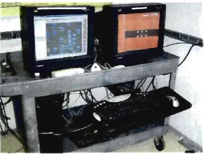
PC computer used to create the VE ... 42
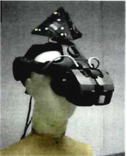
HMD used to display the VE to the user; and the.rigid body with .the IREDs used to reproduce the position and orientation of the head in the VE ... 43
Figure 6. Endpoint visual representation in VE (blue dot; A); and the visual command used in VE to indicate the beginning of the trial (B) ... 43
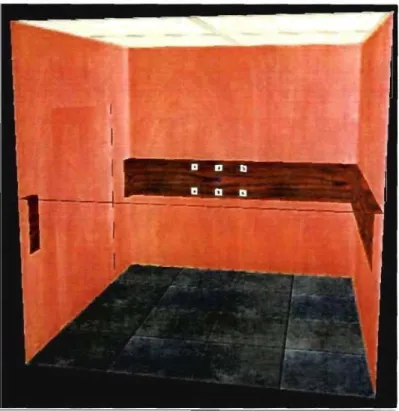
Figure 7. Virtual environment (i.e. virtual elevator) ... 44
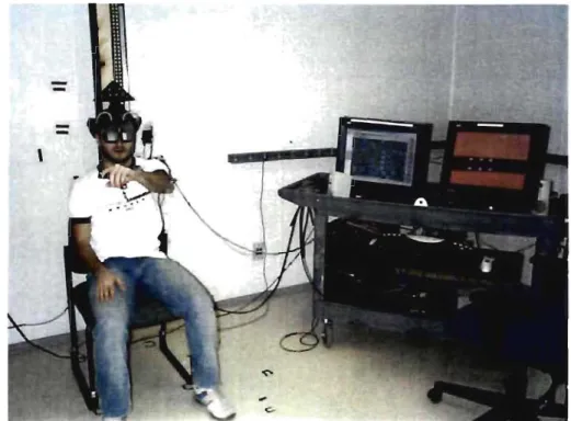
Figure 8. Subject performing the pointing movement toward to a contraIaterai target in the VE ... 46
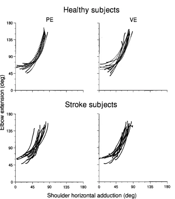
Figure 9. Elbow extension and shoulder horizontal adduction coordination of the pointing movement executed toward the lower middle target in 12 healthy subjects and 15 stroke patients in physical (PE) and virtual (VE) environments ... 53
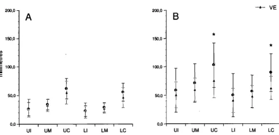
Figure 10. Trunk displacement means and standard deviations of healthy subjects (A) and stroke patients (B) for the 6 targets in physical (PE) and virtual (VE) environments ... 54
Figure 11. Trunk rotation means and standard deviations of healthy subjects (A) and stroke patients (B) for the 6 targets in physical (PE) and virtual (VE) environments. 55 Figure 12. Elbow extension means and standard deviations of healthy subjects (A) and stroke patients (B) for the 6 targets in physical (PE) and virtual (VE) environments. 56 Figure 13. Shoulder flexion means and standard deviations of healthy subjects (A) and stroke patients (B) for the 6 targets in physical (PE) and virtual (VE) environments. 57 Figure 14. Shoulder horizontal adduction means and standard deviations of healthy subjects (A) and stroke patients (B) for the 6 targets in physical (PE) and virtual (VE) environments ... 58
Figure 15. Elbow extension/shoulder horizontal adduction coordination means and standard deviations of healthy subjects (A) and stroke patients (B) for the 6 targets in physical (PE) and virtual (VE) environments ... 59 Figure 16. Endpoint trajectories toward the 6 targets in physical (PE) and virtual environments (VE) obtained from 1 healthy subject and 1 stroke patient. ... 60 Figure 17. Endpoint trajectory straightness (index of curvature IC) means and standard deviations of healthy subjects (A) and stroke patients (B) for the 6 targets in physical (PE) and virtual (VE) environments ... 61 Figure 18. Endpoint precision means and standard deviations of healthy subjects (A) and stroke patients (B) for the 6 targets in physical (PE) and virtual (VE) environments ... 62 Figure 19. Endpoint peak velocity means and standard deviations of healthy subjects (A) and stroke patients (B) for the 6 targets in physical (PE) and virtual (VE)
environments ... 63 Figure 20. The most frequent responses in the questionnaire obtained from 10 healthy subjects and 15 stroke patients. The least agreement was indicated by response 1, while the most agreement was indicated by response 8 ... 67
List of Abbreviations
2D 3DADL
ANOVA AVM CAREN CIMT CLRC CNS CRIR CVA EBRSR EMG FOR FOVHDS
HMD
IRED IRMnIFE
JRH Two dimensional Three dimensional Activities of Daily living Analysis of VarianceArteriovenous malformations
Computer Assisted Rehabilitation Environment Constraint lnduced Movement Therapy
Constance-Lethbridge Rehabilitation Centre Central Nervous System
Centre for lnterdisciplinary Research in Rehabilitation of Greater Montreal Cerebrovascular accident
Evidence-based review of stroke rehabilitation Electromyography
Field of regard Field of view
Hamilton Depression Scale Head-mounted displays Infrared-emitting diode
Institut de réadaptation de Montréal Johns Hopkins Functioning Examination Jewish Rehabilitation Hospital
KP KR LC
LE
LI LM Ml PE PEDro RCT RMS ROM SD SPSS SR SSS TEMPA TS UC UE UI UM Knowledge of PeIformance Knowledge of ResuIts Lower contralateral target Lower ExtremityLower ipsilateral target Lower middle target Motor cortex
Physical Environment
Physiothempy Evidence Database Scale Randomized controlled trial
Root-mean-squared Range of motion Standard Deviation
Statistical Package for the Social Sciences Stretch reflex
Scandinavian Stroke Study
Test d'Evaluation des Membres Superieurs de Personnes Agées Task -specifie
Upper contralateral target Upper Extremity
Upper ipsilateral target Upper middle target
VE VR WCPT WHO Virtual Environment Virtual Reality
World Confederation for Physical Therapy Congress World Health Organization
1 would like to dedicate this master thesis to my parents Edmundo and Ester, my sister Michele and my brother Luiz Guilhenne. 1 am the happiest person in the world because 1 have them in my life.
Acknowledgements
Ficst of aU, 1 would like to thank God for giving me the vigour and perseverance to accomplish this thesis. This work would never be possible without the support of my family, my friends and especially, Sibele Melo who has been more than a friend.
1 am grateful to my research director Dr. Mindy F. Levin who shared her knowledge and experience with me with a lot of patience; to Dr. Anatol G. Feldman who a1lowed me to work in his research laboratory; and to Amy Henderson, Christian Beaudoin, Eric Johnston and Sandeep Subramanian for their technical assistance on this project.
Finally, 1 express my sincere gratitude to the "Faculté des études supérieures" of the "Université de Montréal" for the "Bourse d'exemption des droits de scolarité supplémentaires" .
1.1.
Introduction
Cerebrovascular accident (CV A or stroke) is an ensemble of symptoms caused by the interruption of blood suppl Y to, at least, part of the brain. The interruption of the blood supply has, usually, two origins: ischemic or hemorrhagic. Ischemie strokes are characterized by an obstruction of the blood flow in the brain due to a thrombus (thrombotic stroke) or an embolus (embolie stroke) and represents, approximately 80% of the cases. Less frequent, in about 20% of cases, hemorrhagic strokes occur when a blood vessel on the brain ruptures. Aneurysms and arteriovenous malformations (A VM) are common reasons for those ruptures (Heart and Stroke Foundation of Canada, 2006).
Fifteen million cases of stroke are estimated to occur annually in the world. Of those, 5.5 million people die. Stroke is the third most important worldwide cause of death (10%), after coronary heart disease (13%) and cancer (12%) (World Health Organization-WHO, 2002). In Canada, specifically, it is estimated around 40,000-50,000 new cases of stroke each year (Heart and Stroke Foundation of Canada, 2002).
Besides the high incidence of death, stroke results in another significant consequence Worldwide, five million people post-stroke become permanently disabled every year (WHO, 2002). In North America, stroke represents the principal cause of physical disability in adults (American Heart Association, 2006) and hemiparesis is the most cornmon impairment found on those patients (Krakauer 2005).
The sudden loss of brain function after stroke results in different types and levels of neurological impairments. Jorgensen et al. (1995) observed that, in the first week after stroke, the level of patients' neurological impairments was: very severe for 9%, severe for 12%, moderate for 29% and mild for 50%. These numbers were obtained using the Scandinavian Neurological Stroke Scale (SSS - Scandinavian Stroke Study Group, 1985; (Lindestrom et al. 1991), which takes into account several factors varying frorn level of consciousness to gait. Deficits after the stroke result in difficulties for patients to perform
varied activities. Only 21 % of patients are able to perform activities of daily living (ADL) independently and the dependency persists in 48% of patients for at least one year post-stroke (Carod-Artal et al. 2(00). Stroke has a significant impact on the patient's and their family's daily life, as weIl as on society. According to the American Heart Association, the 4.8 million post-stroke patients. living in the United States indirectly cost, in 2004, US $20;6 billion, due to the loss of their productivity (American Heart Association, 2004).
Considering upper (UE) and lower extremities (LE) separately, stroke survivors are more dependent for tasks involving the UE. OIsen (1990) noted that, three months after stroke, 32% of patients regained independence of their hemiparetic 1eg whi1e only 21 % regained independence of the hemiparetic arrn. These consequences place stroke survivors as the biggest users of rehabilitation services (American Heart Association, 2006) and make disability of the UE an important obstacle to the re-establishment of personal autonomy.
1.2.
Post-stroke motor impairments
Motor impairments in stroke patients include: spasticity, abnonnal patterns of synergy, incoordination, weakness and loss of sensation. The degree of importance of each factor on movement deficits is not precisely known; however it seems to he variable for every patient.
Spasticity refers to a disorder on the motor system where certain muscles are continuous1y contracted, producing stiffness or tightness. It is characterized by the combination of hyper-tonicity (increased muscle tone), c10nus (a series of rapid muscle contractions) and exaggerated deep tendon reflex es (Levin and Hui-Chan 1992). By definition, spasticity is increased muscle tone caused by the velocity-dependent hyper-excitability of the stretch reflex (SR; Lance 1980). This impainnent has been cited as one of the factors that contribute. to the voluntary rnovement deficit in the more affected arrn foUowing the stroke (Levin et al. 2000; Musampa et al. 2007). Sorne studies in
single-(Jobin and Levin 2000; Levin et al. 20(0) and double-joint systems (Mihaltchev et al. 2005; Musampa et al. 2(07) have suggested that spasticity is one of the consequences of a d~ficit
on the regulation of the SR threshold (Levin and Fe1dman 1994; Feldman and Levin 1995). Deficits in the regulation of SR thresholds results in an inability to relax muscles in different articular ranges (Levin et al. 2(00). Moreover, clinical measures of spasticity (Composed Spasticity Index) were significant1y correlated with SR threshold measures (Levin et al. 2000).
Muscle incoordination is frequently present in stroke survivors (Bourbonnais et al. 1992). For sorne authors, the presence of ab normal muscle coordination patterns is considered to be the primary source of motor dysfonction or global disability in stroke survivors (Dewald et" al. 2(01). By definition, muscle incoordination is the difficulty to activate theadequate muscles, in a selective way (i.e. spatial recruitment), in the opportune moment (i.e. temporal recruitment) and at an optimal intensity according to the motor task to accomplish. As a consequence, stereotypical movements may occur when stroke survivors attempt to produce an effort with the paretic limb (Brunnstrom 1970). For example, in the upper limb, the flexor synergy consists of forearm supination and elbow flexion associated with shoulder flexion, abduction and external rotation. The extensor synergy is characterized by pronation and elbow extension combined with shoulder extension, adduction and internal rotation.
The elaboration of coordinated movements is accomplished by the progressive mastering of the redundant degrees of freedom available to achieve a desired trajectory and the development of more controllable and stable segments (i.e. trunk and limb; Bernstein 1967). After stroke, patients often present a different inteIjoint coordination when compared with healthy subjects (Levin 1996; Cirstea and Levin 2000; Levin et al. 2002). InteIjoint coordination between elbow and shoulder movements is disrupted when movements are performed by hemiparetic patients into or out of the typical extensor or flexor synergies (Levin 1996). In addition, incoordination has been correlated with the level of motor impairment (Fugl-Meyer Assessment, UE section; Cirstea et al. (2003a). In terms
of intersegmental coordination, healthy and stroke subjects present a similar. stereotyped sequential recruitment of the arm and trunk. in that the trunk began moving simultaneously with or before the hand and stopped moving after the end of hand movement. However, the contribution of the trunk movement to the endpoint displacement is substantially higher in the hemiparetic patients and occurs earlier in the reach. One of the reasons for these differences is the incoordination between hand and trunk in stroke patients (Levin et al. 2(02). Muscle weakness has also been studied as a crucial element of the motor impairment in stroke survivors. In the literature, muscle weakness following a stroke is not only descrihed as a loss of maximal strength (Adams et al. 1990; Bohannon 1995), but also as an increased delay of force production (Bohannon 1992; Canning et al. 1999), earlier onset of fatigue (Ingles et al. 1999), an increased perception of effort (Gandevia 1982) as well as a . . difficulty to generate the optimal force for a specific task (Beer et al. 1999). Furthermore, weakness has also heen correlated with deficits on SR threshold regulation, leading to the inability to activate muscles in different joint ranges (Levin et al. 2000).
Although the relation between the UE muscle weakness and the level of motor performance (e.g., Box and Blocks test) in hemiparetic subjects had been described by Mercier and Bourbonnais (2004), the relationship between performance and force production seems to he greater on the lower extremity (Bohannon 2007). In a single-blind, randomized controlled trial, Bourbonnais et al. (2002) observed that a treatment based on force feedback improves LE deficits (gait velocity over a 12-m distance and the longest distance in 2 min) but not on UE deficits (TEMPA, Box and Blocks test, and finger-to-nose test). One of the possible reasons for this result is the relevance of strength of the LE for functional activity performance as compared with UE (Bohannon 2(07). For example, to bring food to the mouth requires little strength (Bohannon et al. 1991) and would he expected to improve rapidly with small increases in strength as long as adequate h~d
dexterity and upper extremity coordination are present.
We cannot exclude the impact of sensory deficits in motor function. Impairment of cutaneous and proprioceptive sensation contributes to the loss of motor function in about 30
to 60% of subjects following a stroke (Shah 1978; Carey et al. 1993; Win ward et al. 1999). Proprioception (i.e perception of position and/or movement) is strongly correlated with motor recovery in the pàretic limb. Moreover, it is a reliable prognosticsign of motor recoveryin the long term (Wadell etal. 1987; Desrosiers et al. 2003). Cutaneous sensation (e.g. vibration sense) is one of the tirst somatosensory modalities to he affected and to return to normal following a stroke (Boivie et al. 1989; Pause and Freund 1989; Holmgren et al. 1990). For these reasons, the recovery of sensory function is of utmost importance for the complete restoration of motor function.
As a result of the sensorimotor deticits described above, movements are affected at the motor pattern (e.g. range of motion, trunk compensation) and performance levels (e.g., endpoint precision, velocity and trajectory) during pointing movements (Cirstea & Levin, 2000). However, motor deticits and their consequences can be attenuated or even reversed during spontaneous recovery and by physical rehabilitation.
1.3.
Motor recovery post-stroke
Physiological or spontaneous recovery is responsible for early neurological functional improvement after the stroke. This process results from resolution of local edema, resorption of local toxins, improved local circulation, and recovery of partially damaged ischemic neurons. The time of spontaneous recovery varies from 1 to 6 months, depending on the severity of the lesion (Teasell et al. 2006). In this sub-acute phase, recovery is relatively rapid and motor improvements are more evident (Nakayama et al. 1994; Jorgensen et al. 1995).
Many studies have suggested that patients reach their maximal physical and functional recovery levels in the sub-acute phase. After this, a recovery plateau is achieved, revealing that motor impairments and function become stable. Sorne of the most influential studies supporting this idea are those of The Copenhagen Stroke Study- started on 'the
1990's. Nakayama et al.(l994) found that patients with mild to severe paresis were not able to improve their UE motor disability (measured with the Barthel Index subscores for feeding and grooming) after the 6th and the ll'h weeks, respectively, following the stroke. Jorgensen et al. (1995) noted that recovery of activities of daily living (ADL) did not improve signiticantly after the 131h month post-stroke.
More recently, however, recovery during the chronic phase (more than 6 months) after stroke has been demonstrated in several studies (Nudo 2003; Michaelsenet al. 2006; Teasell et al. 2006). Later recovery may be possible because of the inherent capacity of the brain to develop new synapses, reorganizing the cortex, in response to learning and experience. This phenomenon is called neuroplasticity (Le. plasticity) and allows the undamaged region of the cortex to assume the lost function of the damaged cortex. For Nudo (2003), post-stroke neuroplasticity is based on three main concepts. First, the acquisition of skilled movements in a normal animal will induce predictable functional changes within the motor cortex. Second, injury to the motor cortex, as might occur in stroke, induces functional changes in the cortical tissue spared by the in jury. Third, these two events interact so that after a cortical in jury, the reacquisition of motor skills influences the type and quality of functional plasticity that occurs in the intact, undamaged cortex.
Post-stroke neuroplasticity is possible when cortical areas adjacent and/or remote to the infarct are preserved. However, this is not the only condition in which this phenomenon takes place. A sequence of studies by Nudo and colleagues indicated that enlargement of the adjacent cortical representation occurs when the hemiparetic limb is stimulated through repetitive meaningful movements while disuse ofthis limb decreases the size of.the cortical representation. In the tirst study, by Nudo et al. (1996a), animals (i.e. squirrel monkeys) were not stimulated to use their more-affected limb and their cortical map was examined before and 3 months after an ischemic infarct in the primary motor cortex (MI). The authors noted that, in those animals, the lack of use of the. more-affected hand contributed to the reduction of the digit representation in the intact adjacent cortex. This result supports the result of Liepert et al. (1995), where patients had a diminished representation of the
motor cortical area of the anterior tibial muscle after immobilization of the ankle joint. In a second study, Nudo et al. (1996b) restrained the use of the more-affected arrn and provided animals with a dailyl-hour rehabilitative program. In contrast to the previous study, the intervention resulted in an en largement of. the hand cortical representation. In a similar study, monkeys with ischemic infarcts on the hand area of Ml had their more-affected arrn immobilized with a restrictive jacket; however the animais did not receive any rehabilitative training (Friel et al. 2000). The changes observed in Friel's study were similar to those noted in Nudo et al. (1996a). in which monkeys did not have their more-affected arrn restrained; however the changes in the cortical representations were significantly different from those observed by Nudo et al (l996b). The Mt mapping showed a decreased hand representation 1 month after the stroke. The authors suggest that. in addition to limb constraint, task repetition that requires skill re-acquisition is necessary to induce reorganization on the intact motor cortex after stroke.
In faet, repetitive task-specific (TS) practice has been suggested as an important element to improve movement outcomes (Butefisch et al. 1995; Cirstea et al. 2003b; Blennerhassett and Dite 2004; Michaelsen et al. 2006; Wolf et al. 2006). In Blennerhassett & Dite' s study, patients undergoing stroke rehabilitation were separated into 2 groups. One group of patients received additional TS practice for the UE while the second group received additional training for the LE. After 4 weeks of additional practice, only the UE group had a significant improvement on the scores ofthe Jebsen Taylor Hand Function Test (Jebsen et al. 1969) and the Motor Assessment Scale (upper arrn and hand items; Carr et al. 1985), suggesting a motor function improvement on the paretic UE. In another study, the efficacy of a rehabilitation program based on repetitive TS practice was demonstrated by Wolf et al. (2006). One-hundred-six stroke patients (3 to 9 months post-infarct) that received the Constraint Induced Movement Therapy (CIMT; Taub et al. 1993) were compared to t16 patients treated with usual and customary care. In the CIMT group, patients hadtheir less-affected arrn restrieted during 90% of their waking hours aver a 14 day period. During this period. patients performed functional task repetitively for, at least, 6
hours per day. Following the training, the motor function improvement (measured with the Wolf Motor Function Test and the Motor Activity Log) was significantly greater for the CIMT group than for the control group with the improvements persisting for 12 months in the experimental group.
Although effective in improving UE motor function, repetitive TS practice, when not weIl managed, may not promote motor recovery (Cirstea et al. 2003b; Michaelsen and Levin 2004; Michaelsen et al. 2006). In Cirstea et al's (2003b) study, chronic stroke patients performed reaching movement repetitively during a single session, consisting of 70 trials. Kinematics were recorded before, during and 10 minutes after the session (retention test). The results showed that after a short-term series of repetitions, patients with mild-to-moderate hemiparesis (Fugl-Meyer score::: 50) executed movements faster, more precisely, more smoothly (less segmentation) and with less variability. For patients with moderate-to-severe hemiparesis (Fugl-Meyer score
<
50), movement time, segmentation and movement time variability were also decreased. However, for these patients, improvements in motor performance were accompanied by increased trunk recruitment (compensation) even in a situation where such recruitment was not required for the task. Previous studies have argued that compensatory trunk use may be maladaptive in that it may actually limit the potential for arm motor recovery. Thus the authors suggested that practice alone without particular attention to compensatory strategies may not be sufficient to optimize motor recovery on those patients.1.4.
Feedback
Whether or not a movement is effective can be signaled to the performer via two types of feedback: intrinsic and extrinsic feedback. Intrinsic feedback (i.e. inherent feedback) refers to a person's own sensory-perceptual information. Several sensory processes, including vision, proprioception, touch, pressure and audition, can mediate this information. Intrinsic feedback helps to formulate a person' s internal representation of the
movement goal he or she is trying to achieve. Extrinsic feedback (i.e. augmented feedback) is infonnation additional to intrinsic feedback that comes from an outside source, such as comments from a therapist or changes in the environment (van Dijk et al. 2005; van Yliet and Wulf 2(06).
After stroke, intrinsic feedback systems may be compromised in some patients, making stroke survivors more dependent on extrinsic feedback to guide and to improve their motor perfonnance (Sabari 2001; Flinn and Radomski 2002). The importance of extrinsic feedback on motor learning after stroke has been suggested by some authors (Newell 1991; Schmidt and Lee 1999) and augmented feedback combined wi th rehabilitation techniques has been investigated in se v eral studies (Armagan et al. 2003; Cirstea and Levin 2007).
Annagan et al. (2003) evaluated the efficacy of electromyographic (EMG) biofeedback treatment in the functional recovery of the hemiplegic hand. In addition to an exercise program using Brunnstrom's neurophysiologic approach, participants were treated with EMG biofeedback or with placebo EMG biofeedback. Both treatments were applied five times a week for a period of 20 days. The results showed significant improvements in impainnent and functional measures for both groups after the treatment. However, improvements in the wrist active range of motion and surface EMG potentials of wrist muscles were significantly greater in the EMG biofeedback group.
In another study, by Cirstea and Levin (2007), chronic stroke patients were separated in 2 groups. Both groups practiced repetitive pointing movements during 1 hour
per day, for 2 weeks. ln addition, the first group received terminal extrinsic fcedback focusing on movement precision (i.e. knowledge of results - KR) and the second group received concurrent extrinsic feedback focusing on arm pattern of movement (i.e. knowledge of perfonnance - KP). After the training, only the group that received KP improved significantly in shoulder horizontal adduction and flexion range of movement and coordination between shoulder and elbow movement. These results suggested that motor
leaming in post-stroke patients is influenced not. only by the· presence of extrinsic infonnation received during the. motor training, but also by type and delivery of this infonnation. Finally, the type of feedback should be adjusted for the stage of learnÎng of the subjeçt (Gentile 1987) and should be able to sustain the patient's motivation during the rehabilitation proœss. whkh is another very important factor in motor recovery (Sol mon . . and Boone 1993).
1.5.
Psychological Factors
Motivation is usually associated with the participants' active engagement in a treatmentltraining intervention (Madean et al. 2000) and this engagement is essential to
achieve positive rehabilitation results (Chen et al. 1999). Although liule research has been carried out on motivation in patients with stroke, in sorne cases, motivation has been even used as a detenninant of rehabilitation outcome (Madean et al. 2000). The impact of depression on the recovery of ADL functions was demonstrated by Chemerinski et al. (2001). In their study, 171 patients following stroke were evaluated with psychiatrie (Hamilton Depression Scale - HDS; Hamilton 1960) and motor function test (Johns Hopkins Functioning Examination - JHFE; Robinson & Szetela 1981) in the second week and 3 or 6 months after the stroke onset. A positive correlatÎon between improvements in HDS and in JHFE tests was observed, suggesting that beUer motor funetional reeovery depends also on the patient's mood.
This importance of motÎvation to re-establish motor function has also been studicd in stroke survivors. Barker and Brauer (2005) investigated upper limb recovery t'rom the stroke survivors' perspeçtive. The goal of the study was to determine factors other than medical diagnosis and co-morbidities that contribute to recovery. Twenty-one sub-acute and chronic stroke patients and 9 spouses participated ta face to face forums and group or individual interviews, where they were encouraged to comment or answer sentences and questions like: Think about someone who had a 'good recovery' (or a 'bad recovery') and
explciin why.; What factors influence. recovery?; How do you think we can maximise
recovery?; .etc. In contrast to what they. usually learn from rehabilitation professionals.
patients believe that motor performance can improve even many years after stroke. For them, recoveiy stops only if the patient "gives up". Practicing exercises regularly, intensively, appropriately and continually and. using the arm in.everyday tasks are seen by the patients as the means to reach maximal physical recovery. However, according to them, the maintenance of motivation to exercise over a long period of time is a problem. They believe that to overcome such obstacles, it is important to be surrounded by relatives; friends and health professionals who are positive and encouraging. In addition, stroke patients emphasize the importance of feedback received from rehabilitation professionals to keep them motivated to con.tinue training. With this information, the authors ~uggested that to better promote UE recovery, rehabilitation services need to consistently implement their training program and in sorne cases use innovative interventions and services.
·1.6.
Virtual reality training envh70nments
An innovative intervention that incorporates the necessary elements to induce motor recovery (Le. repetition, task-specificity, augmented feedback and motivation)is virtual reality (VR - i.e. virtual environment, VE). By definition, VR is a multisensorial experience in which a person is immersed in a computer-generated environment. The term "VR" was firstly used by Jaron Lamier, a computer scientist, in 1986 (Riva 2003), however, its history started in the 1 960s (Sutherland 1965). In stroke rehabilitation, the use ofVR was firstly discussed only in the 1990s (Wilson et al. 1997). Since then, the Iiterature about the use of VR has advanced from articles which primarily described its potential benefits, to articles that describe the development of actual working systems, testing of prototypes, and early clinical results with patients trained in such environments (Holden 2005).
'The dynamic and extensive adjustability of VR environinents and events make il
more advantageous for sensorimotor rehabilitation when compared to conventional environments (Le. physical environment, PE; Weiss et al. 2004; Holden 2005). Using VR, it is possiblè to create different tasks thar are not easily constructed in PE. It is also possible to ptovide specific extrinsic feedback in a precise and flexible manner using environmental changes and visual cues (Todorov et al. 1997; Holden 2005). In addition, in VR, environments and tasks can he easily and quickly individualized to patient's motor abilities and preferences, as weil as to the goal of therapy (Sveistrup 2004). VR is also a reliabJe tool that can provide quantitative and qualitative information about the patient's rehabilitation progress (Kenyon et al. 2004). It allows physiotherapists and occupational therapists to hetter identify the appropriate moment to increase the tasks' level of difficulty for the patients. Allied to the task and environinent relevance, the challenging situations provided in VR, where patients receive scores about their performance and think they are playing a video game, are very useful to enhance their degree of interaction with the therapy. This degree of interaction is very important for positive rehabilitation outcome since the level of a patient's commitment to therapy is negatively influenced by boredom, fatigue, and lack of enthusiasm(Tinson 1989). Thus VR seems to bea valuable tool· in clinical setups where rehabilitation professionals·need to create motivating interventions for theirpatients. Finally, besides being advantageous for the performance of repetitive tasks in motivating environments; VRalso allows the patients to perform tasks in· safe conditions where they are not exposed to risks presented in sorne conventional tasks (Sveistrup 2004).
Virtual experience is possible only because of the use of special hardware and softwàre. Input interfaces such as tracking systems (e.g. Optotrak Motion Capture System Northem Digital; Fastrak .:..- Polhemus Corp; Cyberglove Immersion Corp:) allow movements to he tracked so that users can interact with objects in the VE. Users percei ve the VEwith output interfaces such as head-mounted displays (HMD), flat screen displays, audio speakers etc (Riva et al. 2004). Although outputs are possible for aIl the senses,
visual and auditory stimuli are the most frequently used in VR systems (Weiss and Katz 2004). Finally, computers and software are necessary to integrate ail those equipments.
In the literature, different VR systems have been described, varying according to the level of immersion delivered to the users. In more immersive VR systems, users can better experience a neuropsychological phenomenon known as presence (Riva 2003; Holden 2(05). Presence is defined as the "sense of being there" and is suggested as. an essential
element for the transfer of leaming. from VE to PE (Stanney et al. 1998; Riva 2oo3). Since vision is the most important sense for immersion in the virtual experience, the meaning of visual displays is very significant. Thus, VR systems such as those where visual çmtput is displayed in 20 by desktop, flat.screen or projection systems are considered non-immersive or less-immersive. In con!Iast, anHMO system is a fully-immersive VR system where the user sees only the computer-generated image (Keshner 2004). In addition to permitting the subject to interact with a 30 VE, this system allows users to take advantage of stereoscopie vision so that the distance between objects can be perceived (Riva et al. 2004). Also, the HMD system provides à. bigger field of rçgard (FOR) when compared to less immersive systems. It is achievable because the head position and orientation tracking relies on changes in the VR viewpoint when the user moves herlhis head (Riva et al. 2004). CA VE™ systems (Cruz-Neira et al. 1992) and the video capture VR system (Weiss et al. 2004) are other examples of immersive VR systems.
Although there are only a few clinical studies to date using VR therapy, it has been suggested that training in VR may improve UE motor function in patients following stroke (Foley et al. 2007; Henderson et al. 2007). In a systematic review, Henderson investigated the evidence of using immersive and non-immersive VR to increase UE motor performance and function in patients with acute, sub-acute or chronic hemiparesis following stroke. A total of 6 articles met the inclusion criteria (Le. he published in English-Ianguage scientific literature; have an element of retraining of arm movements.and not hand movements alone; do not use other types of training interfaces performed in non-virtual environments), including two randomized controlled trials (RCTs; Piron et al. 2003; Jang et al. 2005), one
single subject design (Broeren et al. 2004), and three pre-post design studies (Holden et al. 1999; Holden and Dyar 2002; Piron et al. 2005). Results indicated evidence from one good quality RCT (PEDro score 2:6) and one single subject study suggesting a greater benefit from training in immersive VR compared to no therapy. On the other hand, for training in non-immersive VR compared to no therapy, conflicting evidence was observed from three studies using a pre-post design. Although limited, the results are sufficiently encouraging to justify further research efforts in this area.
Evidence of the effectiveness of training in VR was also evaluated by Teasell and colleagues in the Evidence-based review of stroke rehabilitation (EBRSR). However, in
this review, in addition to the studies analysed in Henderson's study, studies were also included where only hand movements were trained (Jack et al. 2001; Merians et al. 2002; Merians et al. 2006; Fischer et al. 2007). The analyses indicated strong evidence (indicated by at least two good-to-excellent quality RCTs - PEDro score 2:6) that VR treatment can improve UE motor function in the chronic stages of stroke.
Even if the results presented above are encouraging, more studies about behaviour and movement characteristics in VR are necessary to better understand the applicability of this tool in clinical rehabilitation and motor control studies. Viau et al. (2004), compared movement kinematics of identical tasks made in PE and VB. The goal was to validate a non-immersive VR as a tool for studying reaching and grasping in healthy subjects and in individuals with hemiparesis. In both environments, participants grasped a baIl (real or virtual) from the edge of a table (real or virtual), reached forward by leaning the trunk and then placed the baIl within a target (real or virtual). The representation and orientation of the subject's hand in the 2D environment was obtained using a Cyberglove (Immersion Corp.) and a Fastrak (polhemus Corp.) electromagnetic sensor. Also, prehension force feedback was provided to the participants by a Cybergrasp (Immersion Corp.). The movements were evaluated in terms of endpoint path curvature, maximal grip aperture, trajectory length, angular ranges of joint motion and elbow-shoulder interjoint coordination. The results presented a certain similarity in movement kinematics between physical and
virtual reaching and grasping. However, both healthy and stroke panicipanls usee! significantly less wrist extension and more elbow extension in VE clunng the hall transportation and release phases. One of the reasons indieatcd for those changes Oll the movement pattern was the absence of depth perception in the VR condition. For lhe authors, the participants could not estimate the correct distance bctween them 10 the wall 011 the 2D VE, leading to movement compensations. In addition. Viau ct al argued lhal differences found in this study would not exist in 3D immersive VR whell: stereoscopic vision is provided, such as those visualized through a HMD.
2.1.
Rationale for the study
As summarized in the literature review section, stroke i~ one of the major causes of physical disability in adults worldwide. After stroke, the hemiparetic upper extremity (liE)
remains an obstacle to the re-establishment of the patient's autonomy. The lack 01 independence of stroke survivors has a significant impact on the patient's and thcir family's daily life, as weil as on society. It places those patients as the biggest Wiers of rehabilitalion services (American Heart Association, 2006).
Motor impairments like spasticity, abnormal patterns of synergy, incoordination. weakness and loss of sensation affect what motor patterns are used to produce movcmcnt (e.g. range of motion, trunk compensation) and motor performance variables (e.g .. endpoint precision, velocity and trajectory). These sensorimotor deficits can be attenuatcd or evcn reversed after the stroke by experience-dependent plasticity in the CNS that can be induced through motor training tasks even 6 months post lesion (Le. chronic phase). However, sorne elements such as task-specificity and relevance, task repetition, feedback and motiVai ion must be incorporated into motor training approaches to make them more effective.
An innovative intervention in which these elements can be easily inlegratcd IS virtual reality (VR). VR is a multisensorial experience in which a person is immer~ed in u computer-generated environment. In VR, environments and ta<;ks are simply and quickly individualized to patient's motor abilities and preferences, as weil as 10 the thcrapeutic goal. In addition, VR enhances the degree of interaction between the patient and therapy. This degree of interaction is important for increasing the efficacy of rehabilitation and is negatively influenced by boredom, fatigue, lack of enthusiasm and lack of cooperation.
The efficacy of VR as a tool to improve UE motor function of patients in the chronic stages of stroke has been supported in the literature (Foley et al. 2007: Hcndcrson et al. 2007). However, it is not very weil known whether movements performed in VR arc sirnilar to those performed in physical training environments. This information is neccssary
to better understand the applicability of this tool in clinicai rehabilitation and motor control studies. In a previous study (Viau et al. 2004), the kinematics of reaching and grasping movements performed in a two dimensional virtual environ ment (VE) presented sorne differences compared to those of the same movement performed in a physical cnvironment (PB). The probable reason for the differences was the absence of depth perception in the 2D VB which was displayed to the participants on a computer monitor. Considering the limitation of the VR system in the previous study, it is appropriate to investigate UE movement kinematics in. a 3D immersive VR where stereoscopie vision is provided, su ch as that visualized through a head-mounted display.
2.2.
Objective
The objective of the current study was to compare the kinematics of pointing movements performed in a 3D fully:immersive (HMD) VR system to those of movements performed in a PB (i.e. conventional condition) in healthy subjects and in subjects with motor deficits due to stroke-related brain damage. The purpose of this study was. not to determine the differences in arm kinematics of pointing movements between healthy subjects and stroke survivors since these differences have been previously well-documented.
2.3.
Hypothesis
Since a 3D immersive VB provides stereoscopie vision to the users (depth perception), \oYe hypothesized that there wou Id be no differences in the kinematics of pointing movements performed in a 3D immersive VB and a simiJar PE in healthy subjects or in subjects with motor deficits due to stroke-related brain damage.
3.1.
Study Sample
The differences hetween the physical andvirtual. environments ,(PE and VE, réspectively) were investigated in two different populations: 1) Patients following stroke and 2) healthy subjects. Fifteen stroke patients with hemiparesis (4 female and II male aged 59 ± 15.4 years old; Table 1) were recruited From three rehabilitation centers associated with the Centre for Inter.disciplinary Research in Rehabilitation of Greater Montreal (CRIR). Twelve hea1thy subjects (6 females and 6 males aged 53.3 ± 17.1 years old) also participated in the study. Ethics approval was obtained from the CRIR (Annex 1) and aIl subjects signed an informed consent form (Annex II) prior to participating, In order to he eligible for the study, ,the participants had to fit the inclusion and exclusion criteria detailed below.
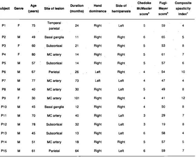
Table 1. Demographie and clinical description of participants with stroke. 'F' female, 'M' male. 'MC artery' middle cerebral artery
Chedoke Fugl- Composite
Age Du ration Hand SI de of
SubJect Genre Site of leslon McMaster Meyer spasticity
(years) (months) dominance hemlparesls
score· scoreb index·
Pl F 75 Temporal 24 Righi Lelt 5 59 4
parietal
P2 M 49 Basal gang lia 11 Righi Righi 6 65 5
P3 F 60 Subcortical 21 Righi Righi 5 53 8
P4 F 80 MC artery 14 Righi Righi 5 61 7
P5 M 57 Subcortical 14 Righi Righi 5 57 6
P6 M 67 Parielal 26 Left Righi 4 54 10
P7 M 77 MC artery 73 Left Left 4 47 4
P8 M 40 MC artery 30 Righi Left 5 49 8
P9 F 30 MC artery 101 Righi Righi 4 41 12
PlO M 45 Basal ganglia 12 Righi Righi 4 50 8
Pll M 70 MC artery 40 Righi Lelt 3 29 7
P12 M 78 Subcortical 32 Righi Lell 3 19 8
P13 M 45 Subcortical 13 Righi Lelt 6 58 4
P14 M 51 MC artery 18 Righi Righi 5 57 5
P15 M 61 Parielal 64 Righi Lelt 6 59 7
a Arm section of the Chedoke McMaster score (7
=
nonnal ann activity) b Upper Limb section of the Fugl-Meyer score (66 = nonnal ann function)3.2.
Inclusion and exclusion criteria
Inclusion criteria for the stroke patients were: 1. be between 18 and 81 years old;
2. have had a single stroke more than 6 months previously (i.e. chronic stroke); 3. have a score between 3 and 617 in the Arm Section of the Chedoke-McMaster Stroke Assessment ScaIe, indicating a moderate hemiparesis (Gowland et al. 1993).
Exclusion criteria for stroke patients were:
1. have a lesion in the cerebellum or the occipital lobe; 2. have marked apraxia or aphasia;
3. have an orthopedie or neuromuscular problem in the arm and/or trunk; 4. have attention deficits or uncorrected visual problems;
5. be unable to speak or understand English or French. Inclusion criterion for healthy subjects was:
1. be between 18 and 81 years old. Exclusion criteria for healthy subjects were:
1. have pain in the arm and/or trunk;
2. have an orthopaedic, neuromuscular or neurological problem in the arm and/or trunk;
3. have attention deficits or uncorrected visual problems; 4. he unable to speak or understand English or French.
3.3.
Recruitment of participants
Stroke patients
The recruitment of stroke subjects started by screening the medical charts from three rehabilitation centres: Jewish Rehabilitation Hospital (JRH), Institut de réadaptation
de Montréal (IRM), Constance-Lethbridge Rehabilitation Centre (CLRC). After potential
participants were identified (according to the inclusion and exclusion criteria), the clinical research coordinator from each rehabilitation centre contacted the patients through an informative letter about the project (Annex III). In the letter, the project was described to the patients and they were invited to contact one of the project team members if they were interested in participating or receiving more detailed information about the study. Following the conversation with the team member, interested individuals were invited to go to an initial screening assessment at the research centre of the JRH. Subjects meeting study criteria signed the consent form and an appointment was set up for the next laboratory visit.
Healthy subjects
Healthy subjects who were interested in participating responded to announcements (Annex N) that were posted on bulletin boards at the JRH and IRM. The same procedure for obtaining consent was followed.
3.4.
Experimental protocol
The experimental protocol consisted of clinical assessment (only in the stroke patients group) followed by the kinematic data collection, which was done in two environments (Le.
PE
and VE) and finally, of a questionnaire, filled in by the participants, about how they interacted with and appreciated the virtual experience.Clinical measurements
Prior to the experiment, all stroke subjects were assessed by research clinicians using a series of clinical tests to determine. the level of motor impairment and function of their affected upper limb. In total, these evaluations took around 30 minutes and were done at the JRH.
The motor recovery level of the hemiparetic upper extremity (UE) was evaluated with the Fugl-Meyer Upper Limb Scale (Duncan et al. 1992). This evaluation measures the capacity of the patient toproduce movements voluntarily, selectively, in a coordinated fashion and out of pathological synergies. According to this scale, UE motor function ~s
considered normal if the subject reached the maximal score of 66 points (Fugl-Meyer et al. 1975).
Spasticity of the elbow muscles was assessed using the CompositeSpasticity Index. This valid (Nadeau et al. 1998) and reliable test measures spasticity by: the resistance felt during stretch of the passive elbow flexors, the excitability of the biceps brachial tendon reflex, as weIl as wrist flexor muscle clonus. A score of 4116 indicates normal tonus, while a score of 16/16 means severe spasticity (Levin and Hui-Chan 1993).
Kinematic recording
Movement kinematics were recorded using the Optotrak Motion System Analysis (Northern Digital Corp., Type 3020) at a frequency of 100 Hz. This system is composed of markers (Le. infrared-emitting diodes; IREDs) and three optical cameras able to capture the information emitted by the markers in three dimensions (x, y and z planes).
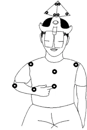
To record the participant's arm and truilk movements, 6 IREDs were placed on: tip of index (distal phalange of the index fiilger, Le., endpoint), wrist (styloid process at the
head of radius), elbow (lateral epicondyle), ipsilateral and contralateral shoulders (acromion processes) and trunk (middle of sternum) (Figure 1).
Data recording started at the sarne time that the participant received the command to begin the movement and lasted for 5 seconds.
Figure
1.
IRED placement and task start position.Physical environ ment
Given that the goal of the study was to compare kinematics of pointing movements in two different environments, the PE and VE were created to be as similar as possible to each other.
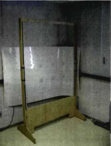
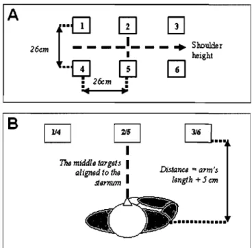
In the PE, six 6 x 6 cm square targets were attached to an adjustable support (Figure 2) and arranged in 2 rows and 3 columns. The squares represented the targets to which the participants should point. The top squares were labelled with the numbers l, 2 and 3 and the bottom targets were labelled with the numbers 4,5 and 6 (Figure 3). The grid of squares was positioned in front of the participant such that the middle squares (Le. targets 2 and 5) were aligned to the sternum of the participant and the midline between the top and bottom squares was aligned with the participant' s shoulders. The most important feature was that the distance between the participant and the midline point was equal to the length of the subject's arm (Le. from the acromion to the tip of the index) plus 5 cm. An additional5 cm was added to the arm length in order to avoid physical contact of the fingertip with the target. Finally, the distance between the centers of adjacent squares was 26 centimetres (Figure 3).
A
26cmB
1
..
~ ~
-
~
1-
-~ S~wœ,
height--8 0
0
- 26cm--..
~~
1
Th9 middl9 ta7 g9t $1
al ign9d t 0 th91
st971lUmrJ;6I
~--Di$ta1lC9 '" a7m'$ 1911gth +5 cmFigure 3. Target arrangement on coronal (A) and transversal (B) planes.
Virtual reality environment
The virtual reality environment (VE) consisted of a 3D environment generated by a PC computer (Dual Xeon 3.06 GHz, 2 GB RAM, 160 GB hard drive; Figure 4) and displayed to the user through a head-mounted display (HMD; Kaiser XL 50, resolution 1024 X 768, frequency 60 Hz; Figure 5). The head position and orientation in the virtual space were reproduced by an optical tracker (Le. Optotrak). One rigid body, composed of 6 IREDs was attached to the HMD (Figure 5). The endpoint was represented in the VE by a blue dot, obtained from the IRED on the tip of the index finger (Figure 6). This was the only body cue indicated to the users when they were immersed in the VE. The data created by these interfaces were integrated by CAREN software (Computer Assisted Rehabilitation Environment), developed by Motek BV. The system also included a dual-head Nvidia
The scene in the VE consisted of 6 targets of the same dimensions and ch\plilyeJ III
the sarne array as that described for the PE, except that they appeared as c)cvalor nUllolb
arranged on a virtual elevator wall. The scene was caJibrated so that the target location" ln
the 3D space were exactly the same as in the PE with respect to the distanœ frolll the
participant's body (Figure 7; Subramanian et al., 2007).
Figure 6. Endpoint visual representation in VE (blue dOL A); and the visualcOllllllélfl(J
Figure 7. Virtual environ ment (i,e, virtual clcvalur),
Experimental procedure
Subject Position
During the experiment. pal1lCipanl~ Wl'rl' l',lIl1ll1l'[;lhl\ ',',iI,'d "II ,1 ,Ii,111 \\ Il
approximately 90° of hip and knee flexion and wilh Ihl' !cel 'l1pP'Hü'd Ilil iii,' ~1"lllld l 'i 1111
to each trial, participants had to place the tlp ollhur Illdn lïll,L"'! "11 Ih,'!! \'1'llt'lll ,""" " so that the ann was in approximately .'i0" of shouldcr ahJudl\l11 :ll1d Il ,d Il,'\1''11 ,_'ri ,r elbow flexion and the forearm and the Wrt~t Werl' III Ill'Ull'dl PI"1111l11 l " ::l1l'(' 1 i
The task
Participants in each group were asked to perform the same task in both environments (Le. PE and VE). The task consisted of 72 trials (3 blocks of 24 trials) of pointing movements toward the 6 different targets (12 trials per target). The target sequence and the order of presentation of environments were randomized to avoid learning effects. The task was designed so that forward trunk displacement was not necessary, since the goal was to point to and not to touch the targets. This pointing movement task was chosen because it required the coordination of multiple arm joints, an ability that should be re-acquired during recovery from stroke (Cirstea et al. 2003a).
Prior to the beginning of the experiment, the participants were instructed to execute the movements as accurately and as fast as possible. While stroke patients performed the task with their more affected UE, healthy subjects used their non-dominant UE. We chose to investigate the non-dominant UE of the hea1thy subjects since this limb is less skilful than the dominant UE and so, more comparable to patient's condition.
The target arrangement caused the participants to produce movements of different Ievels of difficulty using different patterns of movement. For ex ample, the upper row of targets (Le. 1,2 and 3) required more shoulder flexion than the lower row. In addition, the targets placed on the ipsilateral side of the evaluated arm required shoulder horizontal abduction combined with elbow extension and those on the contralateral side required shoulder horizontal adduction combined with elbow extension (Figure 8).
The target to be pointed at was indicated at the beginning of each trial by an auditory go signal emitted by the computer (e.g., 'six' meaning 'point to target 6'). Information about successful pointing attempts in terms of precision (i.e., finger arrived within the 6"x6" target) and speed (within 5 s) were indicated to the subject by a 'ping' sound generated by the computer. In addition, in the VE, a concurrent visual command was ruso used to indicate the target to the participant (Figure 6). Following the start command, the participant had 5 seconds to complete the pointing trial. As soon as the trial was
completed or after 5 seconds, the participants had to resume the starting position and be
ready for the next trial.
Figure 8. the VE.
Subject performing the pointing movement toward a contralateral target in
Data collection
During the experiment, the pointing task was performed in both environments. Thus, every participant had to execute 72 trials in the PE and 72 trials in the VE, for a total of 144 trials. In order to avoid learning and fatigue effects, the order of the experimental environ ment was randomized. In addition, to avoid fatigue during or after the data