UNIVERSITE LILLE 2 DROIT ET SANTE
FACULTE DE MEDECINE HENRI WAREMBOURG
Année : 2020
THESE POUR LE DIPLOME D'ETAT DE DOCTEUR EN MEDECINE
Comment réduire les pH au scalp ? Une étude rétrospective évaluant la
valeur diagnostique de la stimulation au scalp fœtal pour prédire
l’acidose évaluée par le pH au scalp
Présentée et soutenue publiquement le vendredi 19 juin à 14h
au Pôle Recherche
Par Mélissa Gilbert
_______________
JURY
Président :
Monsieur le Professeur Damien SUBTIL
Assesseurs :
Madame le Docteur Louise GHESQUIERE
Monsieur le Docteur Paul BERVEILLER
Monsieur le Docteur Vassili FAGUE
Directeur de Thèse :
Monsieur le Professeur Charles GARABEDIAN
A
VERTISSEMENT
La Faculté n'entend donner aucune approbation aux opinions émises dans les thèses : celles-ci sont propres à leurs auteurs.
L
ISTE DES ABREVIATIONS
SSF : Stimulation au Scalp FœtalPS : pH au Scalp
RCF : Rythme Cardiaque Fœtal IC : Intervalle de Confiance IMC : Infirmité Motrice Cérébrale
ERCF : Enregistrement du Rythme Cardiaque Fœtal ARCF : Anomalie du Rythme Cardiaque Fœtal
NICE: National Institute For Health and Care Excellence AI : Auscultation Intermittente
Table des matières
Avertissement ... 2
Liste des abréviations ... 4
Résumé ... 6
Introduction ... 8
Material and Methods ... 15
Results ... 17
Discussion... 19
Conclusion ... 24
Tables and figures ... 32
R
ESUME
Objectif – Comparer la Stimulation au Scalp Fœtal (SSF) et le pH au scalp (PS) comme test de seconde ligne de l’évaluation du bien-être fœtal afin de diminuer l’utilisation du pH au scalp.
Design de l’étude – Étude rétrospective monocentrique de février à décembre 2019, Lille, France
Méthodes – Dans cette étude, 191 PS ont été réalisés pour Rythme Cardiaque Fœtal (RCF) non rassurant pendant le travail. Une stimulation digitale était réalisée pendant 15 secondes, 2 minutes avant chaque PS. Ce test était considéré comme positif quand une accélération et/ou une variabilité normale étai(en)t provoquée(s). Le PS était considéré comme normal quand le résultat était ³ 7.25.
Résultats – Parmi les 191 PS réalisés, 163 (85.3%) retrouvaient un résultat normal, 122 (63.9%) et 154 (80.6%) avaient, respectivement, une accélération et une variabilité normales post-SSF. Quand une accélération post-SSF était obtenue, le PS était normal dans 91,6% des cas (IC 95%, 85-95). Quand une variabilité normale, post-SSF était obtenue, le PS était normal dans 87.4% des cas (IC 95%, 81-92). Dix cas de nouveau-nés avec une SSF positive (accélération présente) mais un PS pathologique ont été rapportés : le pH était < 7.10 dans 2 cas mais aucun score d’Apgar n’était < 7.
Conclusion – Cette étude suggère que la SSF pourrait être une alternative intéressante au PS comme examen de seconde ligne puisqu’elle semble fiable, non-invasive et facile à réaliser. Ainsi, la SSF pourrait être réalisée en premier lieu en cas de RCF
non-rassurant afin de limiter l'utilisation du PS seulement en cas d’absence d’accélération post-SSF.
I
NTRODUCTION
L’enregistrement du rythme cardiaque fœtal (ERCF) intrapartum a été introduit en pratique clinique afin de réduire la mortalité périnatale ainsi que l’hypoxie néonatale (1). Cependant, son interprétation est soumise à une grande variabilité inter et intra-observateur ce qui pourrait affecter sa validité d’interprétation (2–5). Depuis, plusieurs systèmes de classifications ont émergé afin d’assister le clinicien dans sa décision et lui donner une méthodologie systématique d’interprétation du rythme cardiaque fœtal (RCF) en utilisant des critères tels que la ligne de base de la fréquence cardiaque, la variabilité, la présence d’accélération ou de décélération et les contractions utérines
(4,6,7). Le clinicien est alors supposé être en mesure de déterminer le risque fœtal d’hypoxie/acidose et de décider de la meilleure stratégie clinique. Malheureusement, les taux d’infirmité motrice cérébrale (IMC) et de mortalité néonatale n’ont pas diminué ces trente dernières années (3,8,9). De plus, même si certaines anomalies du RCF ont été associées au risque d’IMC, le taux de faux positifs d’interprétation de l’ERCF de 60% est élevé (1,8,9). En raison de ce taux de faux positifs et de l’augmentation du taux de césariennes, les obstétriciens ont cherché des tests de seconde ligne pour évaluer le bien-être fœtal tels que le prélèvement de sang au scalp fœtal (pour évaluer les lactates ou le pH) ou l’analyse du segment ST (6,10,11). Le pH au scalp (PS) est utilisé comme indicateur de l’acidose fœtale avec une valeur limite pour indiquer une intervention à 7,20 (7,20-7,24 étant la zone de pré-acidose) (12). Cependant, la place du pH au scalp est discutée notamment du fait d’un manque d’études randomisées, des nombreux facteurs influençant son résultat ainsi que du fait des contraintes liées à sa réalisation (13).
dernière consiste à stimuler le scalp fœtal en le frictionnant digitalement, en utilisant une pince sur la peau du scalp fœtal ou en utilisant de manière intermittente une stimulation vibroacoustic sur l’abdomen maternel (6). Une méta-analyse comparant ces différentes techniques (vibroacoustique, digitale, pince d’Allis ou piqure au niveau du scalp) a conclu qu’une accélération sur le RCF suivant la stimulation pourrait permettre d’exclure une acidose fœtale quand l’ERCF était pathologique (14). Cependant, il existe peu d’étude sur la SSF et sa fiabilité vis-à-vis du statut acido-basique. Dans notre centre, le PS est utilisé depuis de nombreuses années comme examen de seconde ligne. En lien avec les recommandations du National Institute for Health and Care Excellence et les récentes controverses sur le PS, nous avons modifié notre protocole : la SSF est systématiquement réalisée quand il existe des anomalies du RCF (ARCF) (15). L’objectif de notre étude était d’évaluer la valeur diagnostique de la SSF pour prédire l’acidose fœtale détectée par le PS.
How to reduce fetal scalp blood sampling? A retrospective study evaluating the diagnostic value of scalp stimulation to predict fetal acidosis assessed by scalp blood sampling.
Gilbert M1, Ghesquiere L1,2, Drumez E2,3, D. Subtil1,2, V. Fague4, P. Berveiller5, Garabedian C1,2
Affiliations:
1. CHU Lille, Department of obstetrics, F-59000, Lille, France
2. Univ. Lille, ULR 2694 METRICS, Évaluation des technologies de santé et des pratiques médicales, F-59000, Lille, France
3. CHU Lille, Department of biostatistics, F-59000, Lille, France
4. CH Valenciennes, Department of obstetrics, F-59300 Valenciennes, France 5. CH Poissy, Department of obstetrics, F-78300 Poissy, France
Auteur correspondant : Mélissa Gilbert
CHU Lille, Hôpital Jeanne de Flandre Department of obstetrics
2 Avenue Oscar Lambret, 59000, Lille, France +33 3 20 44 66 26
Abstract:
Objective - To compare the Fetal Scalp Stimulation (FSS) to Fetal Blood Sampling (FBS) as an adjunctive test of fetal wellbeing in labor in order to reduce Fetal Blood Sampling.
Methods – A retrospective monocentric study of 11 months including 191 FBS procedures performed for non-reassuring fetal heart rate during labor. A gentle digital scalp stimulation was performed for 15 seconds, two minutes before each FBS. It was considered as positive when accelerations and/or normal variability were elicited. The FBS was classified as normal when pH was ³ 7.25.
Results - Of the 191 FBS procedures, 163 (85.3%) found a normal pH result, 122 (63.9%) and 154 (80.6%) had an acceleration and a normal variability post-FSS, respectively. When accelerations were observed after FSS, FBS pH result was normal in 91.6% cases (95%CI, 85-95). When normal variability was observed after FSS, FBS pH result was normal in 87.4% cases (95% CI, 81-92). Ten cases of newborns with normal FSS (accelerations present) but pathological FBS were reported: arterial pH result was acidotic (pH < 7.10) in two cases but no Apgar score at 5 minutes was < 7. Conclusion - This study suggests that FSS could be an interesting alternative adjunctive test to FBS as it seems to be reliable, non-invasive and easy to perform. Thus, FSS could be performed in the first instance when non-reassuring fetal heart rate is observed in order to limit FBS only to absence of acceleration after FSS.
Key words: Fetal Blood sampling; Fetal Scalp Stimulation; Cardiotocography; Fetal
Abreviations:
FSS: Fetal Scalp Stimulation FBS: Fetal Blood Sampling CTG: Cardiotocography FHR: Fetal Heart Rate
NICE: National Institute For Health and Care Excellence SD: Standard Deviation
IQR: Interquartile Range CI: Confidence Interval
TOP: Termination Of Pregnancy IUFD: In Uteri fetal Death
Se: Sensitivity Sp: Specificity
PPV: Predictive Positive Value NPV: Negative Predictive Value IA: Intermittent Auscultation
Introduction
Intrapartum cardiotocography (CTG) has been introduced into clinical practice to reduce perinatal death and neonatal hypoxic brain injuries (1). However, it is submitted to intra and inter-observer variation which could affect the validity of cardiotocograph interpretation (2–5). Since then, several classification systems have emerged (4,6,7) to assist clinicians by giving them a systematic methodology of fetal heart rate (FHR) interpretation using criteria such as fetal heart rate baseline, variability, acceleration, deceleration and uterine contractions. The clinician is then supposed to be able to determine the fetal risk of hypoxia and/or acidosis and to decide of the best clinical management. Unfortunately, the rates of cerebral palsy and mortality have not decreased over the past 30 years (3,8,9). Moreover, even if some abnormalities of the FHR monitoring have been linked to cerebral palsy risk, the false positive rate of 60% is very high (1,8,9). Due to this high false positive rate and the increasing caesarean sections rate, obstetricians have been looking for adjunctive tests of fetal well-being such as fetal blood sampling (FBS) for assessment of lactates or pH and ST analysis (6,10,11). The FBS is used as an indicator of fetal acidosis with a cut-off value of pH for intervention at 7.20 (7.20-7.24 being a pre-acidosis range) (12). However, the place of FBS is discussed notably through the lack of randomized trials, the influencing factors in the pH value, as well as the limitations related to the technical procedure (13).
An alternative to FBS could be Fetal Scalp Stimulation (FSS). FSS involves stimulating the fetal scalp by rubbing it with the examiner's fingers or using forceps to clasp the fetal skin, or alternatively using vibroacoustic stimulation applied to the mother's abdomen (6). A meta-analysis comparing different FSS (vibroacoustic, digital, Allis clamp and scalp puncture) concluded that an acceleration following a FSS could
help to rule out fetal acidemia when the CTG was pathological (14). However, there are few studies on FSS and its reliability to fetal acid-base status. In our center, FBS is used for many years as a second line tool. As recommended by the NICE guidelines and due to recent controversies on FBS, we modified our protocol and FSS is systematically performed (15). Therefore, the objective of our study was to evaluate the diagnostic values of FSS to predict fetal acidosis detected by the FBS.
M
ATERIAL AND
M
ETHODS
A retrospective monocentric (CHU, Lille) cohort study was carried out from February to December 2019. The inclusion criteria were singleton pregnancy with gestational age of more than 36 weeks, cephalic fetal presentation and fetal heart rate abnormalities during labor with indication of FBS.
The protocol in our center is continuous recording of FHR during labor whatever the risk level of the patient. The risk of acidosis is assessed by the national guidelines which are classified in five groups: normal, almost normal, intermediate, pathological and pre-terminal (7). When fetal acidosis was classified as intermediate or pathological by the midwife, indication of FBS was discussed and performed if necessary in absence of fetal or maternal contraindication. Since February 2019, we modified our protocol and FSS was performed before FBS. A gentle digital scalp stimulation was first performed for 15 seconds. Then, the midwife could immediately install the amnioscope to perform the FBS sampled in pre-heparinized capillaries and analyzed immediately (ABL90FlexPlus, Radiometer®). The FBS was classified as normal (pH ≥7.25), borderline (pH 7.21-7.24) and abnormal (pH ≤7.20).
FHR variations after the FSS were analyzed by an expert. Response was considered positive in the presence of:
- Acceleration defined as an increase in the FHR above the baseline of at least 15 beats per minute for at least 15 seconds, for the 2 minutes following the FSS
- And/or variability defined as normal when variations of the baseline were between 5 and 25 beats per minute for at least 10 minutes following the FSS. FBS was performed whatever the result of FSS during this period. The primary
positive results on comparing FSS with FBS (pH < 7.25 on FBS and acceleration present on FSS) were reviewed individually in terms of the neonatal outcome.
Statistics
Categorical variables are expressed as numbers (percentage). Quantitative variables are expressed as mean ± standard deviation (SD) or median (interquartile range, IQR). Normality of distributions was assessed using histograms and Shapiro-Wilk test. Diagnostic values of accelerations and variability post FSS to predict pathological in utero pH were evaluated by calculating sensibility, specificity, positive predictive value, negative predictive value and their 95% confidence intervals (CIs). Diagnostic values were calculated using generalized linear mixed models (binomial distribution, logit function) to take into account repeated measurements per patient. All statistical tests were done at the two-tailed α level of 0.05 using SAS software, release 9.4 (SAS Institute, Cary, NC).
Ethics
The study was approved by the French Committee of Research in Obstetrics and Gynecology CEROG 2020-OBST-0503
R
ESULTS
:
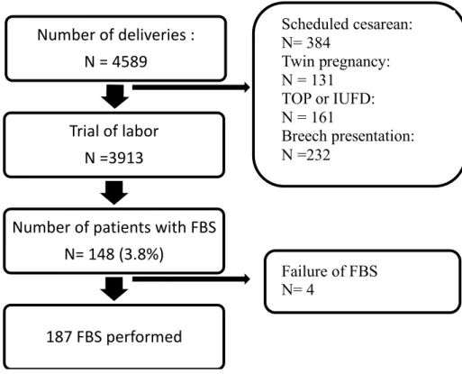
During the study period, 4589 deliveries occurred with 3913 trials of labor (Figure1). One hundred and ninety-one FBS were performed on 148 women. Table 1 represents the population and labor characteristics. Seventy-three (49.3%) of these women had an induction of labor and 68.9% were nulliparous. Almost every woman had an epidural during labor (95.3%). Among these 148 women, 62,2% had a vaginal delivery and 65,2% of them needed operative deliveries. The main indication for these instrumental deliveries were non-reassuring FHR (44/60). Mean arterial birth pH was 7.20 ± 0.1 and 19 (12.9%) were less than 7,10.
When the FBS were performed, the CTGs were classified as normal or almost normal (16.9%), intermediate (57.9%), pathological (24.7%) and preterminal (0.5%) (Table 2). Of the 191 completed procedures, 187 FBS results were obtained. 163 (85.3%) FBS pH results were normal (pH ³ 7.25). When the FSS was performed, we observed accelerations in 63.9% of cases, and variability in 80.6% of cases. 64.9% FBS were performed successfully at the first attempt and one operator was sufficient in 93.2% of cases. The median delay between the decision of performing FBS and the result was 17. 0 (12;22) minutes.
Table 3 reports the diagnostic values of FSS to predict the FBS by comparing normal FBS (pH ³ 7.25) with a normal FSS response based on accelerations or normal variability. When accelerations were observed after FSS, FBS pH result was normal in 91.6% cases (95%CI, 85-95). When normal variability was observed after FSS, FBS pH result was normal in 87.4% cases (95% CI, 81-92).
Table 4 reports the cases of false positive results with FSS (pH < 7.25 on FBS and accelerations after FSS). Six of these FBS pH results were pre-acidotic (7.20-7.24)
of acceleration during FSS and pathological FBS (respectively 7.11 and 7.19). No Apgar Score was less than 7 at 5 minutes and no transfer in Neonatal Intensive Care Unit was needed.
D
ISCUSSION
:
Main findings
To reduce our practice of FBS given the lack of evidence of its usefulness, we aimed to evaluate the diagnostic values of FSS to predict fetal acidosis detected by the FBS and we found that the presence of accelerations is associated in 91.6 % with a normal scalp pH. This study highlights the place of FSS to assess fetal well-being and as proposed by the NICE guidelines. Therefore, FSS could be the adjunctive technology performed in the first instance when non-reassuring FHR is observed in order to limit FBS use to only in case of a negative response after FSS (16).
Interpretation
Continuous cardiotocography is associated with no significant difference in reducing cerebral palsy, neonatal mortality or other standard measures of neonatal well-being(8,9). Its high false positive rate, however, lead to an increase of caesarean sections and operative deliveries (3). To avoid these limits, several adjunctive technologies have been developed such as FBS (pH and/or lactates). For many years, FBS was the most used second line test in labor ward in complement with cardiotocography and recommended in national guidelines (7). However, the reliability of this test is questioned. First of all, it is supposed to detect hypoxia and acidosis but the blood sample is taken from the fetus scalp, a peripheral tissue, when it is known that in cases of hypoxia, the oxygenated blood is diverted to essential organs (heart, brain, adrenal glands) (15). There are also influencing factors in the pH value such as
the scalp localization of the sample, the presence of amniotic fluid or uterine contraction during sample (17–19). The FBS result is quickly outdated and needs to be repeated. Moreover, a Cochrane systematic review showed no evidence of reducing caesarean sections rate nor neonatal seizures nor improvement in long-term neurological outcomes when FBS was practiced (20–22). FBS required having a trained team, a sufficient cervix dilatation and no contraindication (HIV, Herpes Simplex Virus or suspected fetal blood disorder)(6). It may be a time-consuming test with a median interval of 18 minutes between the decision to perform and the result (23). In about 10% of attempts no pH information is obtained owing to blood clotting within the capillary, insufficient blood obtained, air bubbles inside the capillary, or a blood gas measurer that is calibrating at the time the sample needs to be analyzed(6). Some countries have abandoned the FBS as it seemed that a properly interpreted CTG would be equal or superior to the fetal scalp pH to predict both good and bad fetal outcomes (24). Even if the FBS complications are very rare, they can be severe : cerebrospinal fluid leakage, fetal hemorrhagic shock, scalp abscess… (25,26). Some studies have assessed lactates in FBS and showed no significant difference with pH analysis for Apgar score at 5 minutes of life, nor in rates of operative delivery. The interest is the smaller amount of blood required for the analysis (10,11,27).
Because of all these questions about the interest of FBS, FSS returns to the spotlight after multiple studies published in the 1990s (14,28). Indeed, fetal heart rate acceleration in response to various stimuli has long been considered to be a reliable sign of fetal well-being in both the intrapartum and antepartum periods (14,29). In a meta-analysis, Skupski et al. evaluated eleven studies on FSS which included four kind of tests: fetal scalp puncture, Allis clamp scalp stimulation, vibroacoustic stimulation, and digital scalp stimulation (14). The Likehood ratio for both positive and
the different techniques. It appeared that these tests were valuable to predict both presence and lack of fetal acidemia. Digital scalp stimulation is the most chosen technique as it is easy to perform and less invasive (14). More recently, Mahmood et al. conducted a prospective study comparing FSS and FBS over 298 procedures (30). The consistency between FSS and FBS was “fair” so as the FSS and the FBS when compared to cord blood results respectively, suggesting that FSS could be a reliable alternative to FBS. Goodman et al. also conducted a prospective, non-randomized trial which aimed was to compare the diagnostic value of intermittent auscultation (IA) alone (n=251) versus IA and FSS (n=267) in a limited resource setting. It appeared that FSS improved the performance of IA for detecting severe acidemia (pH < 7.0) from 27 to 70% (p = 0.032) (31). The negative predictive value of intermittent auscultation was also improved with the fetal scalp stimulation test from 88 to 99% for mild (pH < 7.2) to severe fetal acidemia. Finally, Murphy and al. recently published the results of their feasibility study of a pilot randomized controlled trial comparing FSS to FBS as second line-tests for fetal well-being assessment (32). They highlighted that the feasibility for such a trial was acceptable for both groups (FSS and FBS). The randomized controlled trial protocol had a high rate of adherence in patients and clinicians. The cesarean section rate was high in both arms, as expected with a cohort of women requiring second-line tests for abnormal FHR monitoring, with a tendency of reduction in the FSS group vs FBS (5/25; 20% versus 13/25; 52%, p = .018) without increasing adverse fetal outcomes. Their estimates suggest that a sample of 2500 women is required to conduct a definitive randomized controlled trial.
Clinical implication
negative predictive value was for accelerations post-FSS, and highest specificity for presence of accelerations and/or normal variability. We then proposed to avoid FBS in case of accelerations (with or without modification of the variability). We choose the negative predictive value (NPV) as the most important result for assessing the diagnostic value of FSS for several reasons. First of all, the aim of our study was to reduce FBS use, and consequently to avoid invasive procedures when reassurance criteria were present without missing potential fetal acidemia. Second of all, the NPV is the most studied criteria in literature when it comes to evaluating FSS. In our study, when an acceleration or a normal variability was elicited, respectively 91.6% and 87.4% of FBS pH results were normal but the positive predictive value seemed to be limited. The cases of discordance between an abnormal FBS pH and normal FSS based on an elicited acceleration were associated with 2 cases of acidotic cord arterial pH but new born had a 5 minutes Apgar score of 10 and had no NICU transfer. In ten cases, FSS elicited an acceleration but the FBS pH result was abnormal which may suggest that FSS value could be questionable in certain CTG patterns when it is helpful when the CTG shows reduced variability(6). It also suggests that it relies on CTG interpretation which has limitations (1).
Strengths and limitations
The strength of this study is the sample of almost 200 consecutive FBS procedures with various experiences of obstetricians and midwives. Our results seem to be consistent with literature especially concerning the NPV of FSS and the small number of abnormal FBS pH results. It also shows that in almost 17% of cases, FBS is performed even if the CTG is classified as normal or quasi-normal. This may show the limitations of such a classification to assess fetal well-being. Indeed, the obstetrical
Moreover, the knowledge of the physiological fetal response to hypoxemia during labor is described by different authors to improve fetal monitoring (34–36).
Our study has a main limitation: we compared FSS to FBS which is abandoned in some countries because of its questioned reliability and the lack of controlled randomized trials (37). Indeed, FBS is considered as the gold standard in our study which could be discussed. Eventually, FSS response is subject to observer variability as it depends on CTG interpretation.
C
ONCLUSION
:
This study suggests that FSS could be an interesting alternative adjunctive test to FBS as it seems to be reliable, non-invasive and easy to perform. Thus, FSS could be performed in the first instance when non-reassuring FHR is observed in order to limit FBS only to absence of acceleration after FSS.
DISCLOSURE OF INTEREST
The authors have no conflicts of interest
CONTRIBUTION OF AUTHORSHIP
M.G collected data from patient files. She wrote the manuscript with assistance of C.G. E.D conducted all the statistical analysis and wrote the statistic methodology. D.S, P.B, L.G, V.F and E. D reviewed and corrected the manuscript.
FUNDING
No funding source was necessary for this work.
ACKNOWLEDGEMENTS
Discussion :
Afin de réduire nos pratiques de PS en raison du manque de données sur son utilité, nous avions pour objectif d’évaluer la valeur diagnostic de la SSF pour prédire l’acidose fœtale détectée par le PS. Nous avons mis en évidence que la présence d’une accélération post-SSF était associée dans 91,6% des cas à un pH normal. Cette étude souligne la place de la SSF pour évaluer le bien-être fœtal comme proposée par les recommandations NICE. Par conséquent, la SSF pourrait être l’examen de seconde ligne réalisé en premier lieu quand un RCF non-rassurant est mis en évidence afin de limiter l’utilisation du PS uniquement au cas de réponse négative (absence d’accélérations) à la SSF (16).
L’ERCF n’a pas montré de différence significative en termes de réduction d’IMC, de mortalité néonatale ni en termes d’évaluation du bien-être néonatal (8,9). Son taux élevé de faux positifs a, cependant, conduit à une augmentation du taux de césariennes en court de travail (3). Afin d’éviter ces limites, plusieurs examens de seconde ligne ont été développés tels que le PS. Depuis de nombreuses années, le PS est le test de seconde ligne, recommandé par le Collège National des Gynécologues et Obstétriciens Français, le plus utilisé en salle de naissance, en complément de l’ERCF (7). Cependant, sa fiabilité est discutée. Premièrement, il est supposé détecter l’hypoxie et l’acidose mais l’échantillon sanguin est prélevé sur le scalp fœtal qui est un tissu périphérique alors qu’en cas d’hypoxie, le sang oxygéné est redistribué en priorité aux organes essentiels (cœur, cerveau et glandes
pH tels que : la localisation du prélèvement sur le scalp, la présence de liquide amniotique ou les contractions utérines pendant le prélèvement (17–19). Le résultat du pH est rapidement dépassé et nécessite d’être répété. De plus, une revue de la littérature de la Cochrane n’a montré aucune preuve de diminution du taux de césarienne, ni du taux de convulsions néonatales ni d’amélioration à long terme du pronostic neurologique quand le PS était réalisé (20–22). Ce PS nécessite pour son prélèvement, une équipe entrainée, une dilatation cervicale suffisante et aucune contre-indication (Infection maternelle transmissible, suspicion de trouble de la coagulation fœtale) (6). Cet examen peut être chronophage avec une médiane du délai décision-résultat à 18 minutes (23). Dans environ 10% des cas, aucune information n’est obtenue en raison de la coagulation du prélèvement au sein du capillaire, de l’insuffisance de quantité de sang, de bulles d’air gênant l’analyse ou encore de la calibration de la machine effectuant l’analyse au moment du prélèvement (6). Certains pays ont abandonné le PS sur l’argument qu’une analyse correcte de l’ERCF pourrait être égale ou supérieure au PS pour prédire à la fois les issues favorables et défavorables (24). Même si, les complications du PS sont très rares, elles peuvent être sévères (fuite de liquide céphalo-rachidien, hémorragie fœtale, abcès du scalp…)
(25,26). Certaines études ont évalué l’analyse des lactates sur le prélèvement au scalp et n’ont montré aucune différence significative par rapport à l’analyse du pH au scalp en termes de score d’Apgar à 5 minutes ni en termes d’accouchement instrumental ou césarienne. L’intérêt des lactates est essentiellement la plus faible quantité de sang nécessaire pour l’analyse (10,11,27).
En raison de tous ces questionnements au sujet de l’intérêt de la réalisation du PS, la SSF revient sur le devant de la scène après plusieurs études publiées dans les
variés est considérée depuis longtemps comme un signe fiable de bonne vitalité fœtale à la fois en intra- et en ante-partum (14,29). La méta-analyse de Skupski et al. a évalué onze études sur la SSF qui évaluaient quatre types de stimulation : la piqure du scalp, la stimulation du scalp à la pince d’Allis, la stimulation vibroacoustique et la stimulation digitale (14). Les rapports de vraisemblance pour les tests à la fois positifs et négatifs (évalués par la présence ou l’absence d’accélération) étaient similaires entre les différentes techniques. Ces tests étaient utiles pour prédire à la fois la présence ou l’absence d’une acidose fœtale. La stimulation digitale est la plus choisie en raison de sa simplicité de réalisation et de sa technique moins invasive (14). Plus récemment,
Mahmood et al. ont conduit une étude prospective comparant la SSF et le PS en cas
d’ARCF sur 298 prélèvements (30). La concordance entre la SSF et le PS était acceptable et il en était de même lorsque la SSF et le PS étaient comparés respectivement au pH artériel du cordon à la naissance ce qui suggère que la SSF pourrait être une alternative fiable au PS. Goodman et al. ont également conduit une étude prospective, non randomisée dont le but était de comparer la valeur diagnostic de l’auscultation intermittente (AI) seule (n=251) versus l’AI associée à la SSF (n=267) dans un pays aux ressources limitées. Il a été mis en évidence que la SSF améliorait la performance de l’AI pour détecter l’acidose sévère ((pH < 7.0) de 27 à 70% (p = 0.032) (31). La valeur prédictive négative (VPN) de l’AI était également améliorée de 88% à 99% pour l’acidose modérée à sévère. Enfin, Murphy et al. ont publié récemment les résultats de leur étude de faisabilité d’essai pilote contrôlé et randomisé comparant la SSF au PS comme test de seconde ligne pour l’évaluation du bien-être fœtal (32). Ils ont mis en évidence une faisabilité acceptable pour les deux groupes comparés. Le protocole de cet essai contrôlé et randomisé avait un taux d’adhérence élevé chez les patientes et les praticiens. Le taux de césariennes était élevé dans
de seconde ligne pour ARCF, avec toutefois une tendance à la diminution de ce taux dans le groupe SSF vs PS (5/25; 20% versus 13/25; 52%, p = .018) sans augmenter les issues défavorables. Leurs estimations pour conduire une étude contrôlée, randomisée définitive suggèrent qu’il faudrait un échantillon de 2500 femmes.
La SSF semble être un test de seconde ligne intéressant lorsqu’il suscite une/des accélération(s) et/ou une bonne variabilité. Dans notre étude, nous avons observé que la valeur prédictive négative la plus élevée concernait les accélérations pst-SSF et la spécificité la plus élevée concernait la présence d’accélération et/ou variabilité normale. Nous avons choisi la VPN comme résultat principal pour évaluer la valeur diagnostique de la SSF pour plusieurs raisons. Premièrement, notre objectif était de réduire le nombre de PS et par conséquent d’éviter toute procédure invasive quand des critères de réassurance étaient présents sans manquer le diagnostic de potentielles acidoses fœtales. Deuxièmement, la VPN est le critère le plus étudié dans la littérature quand il s’agit d’étudier la SSF. Dans notre étude, quand une accélération ou une variabilité normale suivait une SSF, le résultat du PS était normal dans 91,6% et 87,4% des cas, respectivement mais la valeur prédictive positive semblait limitée. Les cas de discordances entre un PS anormal et une SSF positive (basée sur l’apparition d’accélération) étaient associés dans deux cas à une acidose (pH < 7,10) au pH artériel du cordon mais ces nouveau-nés avaient un score d’Apgar à 10 à 5 minutes de vie et n’ont pas nécessité de transfert en unité de soins intensifs néonataux. Dans 10 cas, la SSF a provoqué une accélération mais le résultat du PS était anormal ce qui pourrait suggérer que la valeur de la SSF pourrait être discutable pour certaines anomalies du RCF alors qu’elle semble utile quand l’ERCF met en évidence une variabilité réduite (6). Cela suggère aussi que la SSF est basée sur l’interprétation du RCF qui a elle-même des limites (1).
réalisées par des obstétriciens et des sages-femmes d’expériences variées. Nos résultats semblent concordants avec la littérature, particulièrement concernant la VPN de la SSF et le faible nombre de PS pathologiques. Nos résultats montrent que dans presque 17% des cas, le PS est réalisé alors que le tracé du RCF est classé comme normal ou quasi-normal. Cela montre les limites de telles classifications pour évaluer le bien-être fœtal (33). En effet, le contexte obstétrical (fœtal et maternel) n’est pas pris en compte dans ces différentes classifications. De plus, la connaissance de la réponse physiologique fœtale à l’hypoxie pendant le travail est décrite par plusieurs auteurs afin d’améliorer l’interprétation du RCF (34–36).
La principale limite de notre étude est due au fait que nous comparions la SSF au PS qui est un test abandonné dans certains pays en raison de sa fiabilité discutable et du manque d’essai randomisé (37). En effet, le PS est considéré comme le Gold Standard dans notre étude ce qui est discutable. Enfin, la réponse à la SSF est soumise à la variabilité inter-observateur en raison de son interprétation basée sur la lecture du RCF.
CONCLUSION :
Cette étude suggère que la SSF pourrait être une alternative intéressante au PS puisqu’elle semble fiable, non-invasive et facile à réaliser. Ainsi, la SSF pourrait être réalisée en premier lieu en cas de RCF non-rassurant afin de limiter la réalisation du PS uniquement en l’absence d’accélération post-SSF.
T
ABLES AND FIGURES
Figure 1: FlowchartTOP: Termination Of Pregnancy, IUFD: In Uteri Fetal Death, FBS: Fetal Blood Sample
Number of deliveries : N = 4589
Trial of labor N =3913
Number of patients with FBS N= 148 (3.8%) 187 FBS performed Scheduled cesarean: N= 384 Twin pregnancy: N = 131 TOP or IUFD: N = 161 Breech presentation: N =232 Failure of FBS N= 4
Table 1 - Population and labor characteristics (n= 148)
Age (years) 27 ± 6
Gestational age (weeks) 40 ± 1
Body mass index (kg/m2) 25.2 ± 5.2
Induction of Labor 73 (49.3) Nulliparous 102 (68.9) Scared uterus 17 (11.5) Pregnancy complication Gestational diabetes Preeclampsia Rupture of membranes > 24h Preexisting diabetes Growth restriction 50 (33.8) 2 (1.4) 10 (6.8) 2 (1.4) 7 (4.7) Vaginal delivery Operative delivery Forceps Vacuum Spatula Sequential instruments
Indication of operative delivery
Non reassuring fetal heart rate Failure of progress 92 (62.2) 60/92 (65.2) 25/60 (41.7) 24/60 (40) 5/60 (8.3) 6/60 (10) 44/60 (73.3) 16/60 (26.7) Cesarean Indications Dystocia
Unsuccessful instrumental delivery Fetal blood sample result
Non reassuring fetal heart rate
56 (37.8) 12/56 (21.4) 2/56 (3.6) 18/56 (32.1) 24/56 (42.9) Birth characteristics Umbilical cord pH Umbilical cord pH < 10 Lactates (mmol/L) Base excess (mmol/L)
Apgar Score < 7 at 5 minutes Respiratory distress
Neonatal Care Intensive Unit transfer Birth weight (g) 7.20 ± 0.1 19 (12.9) 6.0 ± 2.1 -6.0 ± 3.6 4 (2.7) 2 (1.3) 3 (2) 3425 ± 479
Table 2 - FBS characteristics (n =191)
Fetal heart rate (CNGOF Classification)
Normal Almost normal Intermediate Pathological Preterminal 3 (1.6) 29 (15.3) 111 (58.1) 47 (24.7) 1 (0.5) Labor characteristics Oxytocin Maternal fever Amniotic fluid: Clear Stained Meconium Absent Bloody 74 (38.7) 21 (11.1) 141 (73.8) 9 (4.7) 16 (8.4) 24 (12.6) 1 (0.5)
Fetal Scalp Stimulation
Acceleration Variability
122 (63.9) 154 (80.6)
Fetal Blood Scalp
Cervix dilatation (cm) Number of attempts/each FBS: 1 2 3 4 5 Number of operators/each FBS: 1 2 3 Failure of FBS
Delay between Decision and FBS result (mn) Delay between FBS and result (mn)
Delay between 2 FBS (mn)
Delay between FBS and Birth (mn)
6 ± 2 124 (64.9) 46 (24.1) 19 (9.9) 1 (0.5) 1 (0.5) 178 (93.2) 12 (6.3) 1 (0.5) 4 (2.1) 17.0 (12.0; 22.0) 9.0 (6.0; 13.0) 87.5 (57.5; 115.0) 99.5 (48.0 ; 177.0)
Fetal Blood Scalp result
Normal ³ 7.25 Pre-acidosis 7.20-7.24 Acidosis < 7.20 163 (85.3) 17 (8.9) 7 (3.7)
Results are presented as number (percentage), Mean +/– Standard deviation or Median and interquartile
Table 3 – Diagnostic values of accelerations and variability post FSS to predict pH < 7.25
Se (95% CI) Sp (95% CI) PPV (95% CI) NPV (95% CI)
Accelerations 58.3 (37-77) 67.6 (59-75) 20.9 (13-33) 91.6 (85-95) Variability 20.8 (8-43) 81.5 (74-87) 13.9 (5-31) 87.4 (81-92) Accelerations
and/or variability 20.8 (8-43) 83.5 (76-89) 15.2 (6-34) 87.6 (81-92)
Values are expressed as percentage (95% confidence interval)
Se = sensitivity, Sp = specificity, PPV = positive predictive value, NPV = negative predictive value
Table 4 - Cases of FSS false positive results (FBS value <7.25 and acceleration present on FSS) Case number FBS pH Cervical dilatation (cm)
Mode of delivery Cord artery pH Apgar Score (5min) 1 7.15 9 Cesarean 7.25 10 2 7.11 4 Cesarean 7.01 10 3 7.19 5 Cesarean 7.08 10 4 7.20 7 Forceps + Vacuum 7.16 10 5 7.14 5 Cesarean 7.22 10 6 7.24 4 Cesarean 7.12 9 7 7.21 10 Cesarean 7.22 10 8 7.24 6 Cesarean 7.16 10 9 7.23 7 Cesarean 7.19 10 10 7.24 8 Cesarean 7.17 8
BIBLIOGRAPHIE
1. Beard RW, Filshie GM, Knight CA, Roberts GM. The Significance of the changes in the continuous fetal rate in the first stage of labour. BJOG: An International Journal of Obstetrics & Gynaecology. 1 oct 1971;78(10):865-81.
2. Paneth N, Bommarito M, Stricker J. Electronic fetal monitoring and later outcome. Clin Invest Med. avr 1993;16(2):159-65.
3. Blackwell SC, Grobman WA, Antoniewicz L, Hutchinson M, Gyamfi Bannerman C. Interobserver and intraobserver reliability of the NICHD 3-Tier Fetal Heart Rate Interpretation System. Am J Obstet Gynecol. oct 2011;205(4):378.e1-5.
4. Ayres-de-Campos D, Spong CY, Chandraharan E, FIGO Intrapartum Fetal Monitoring Expert Consensus Panel. FIGO consensus guidelines on intrapartum fetal monitoring: Cardiotocography. International Journal of Gynecology & Obstetrics. 1 oct 2015;131(1):13-24.
5. Devane D, Lalor J. Midwives’ visual interpretation of intrapartum cardiotocographs: intra- and inter-observer agreement. J Adv Nurs. oct 2005;52(2):133-41.
6. Visser GH, Ayres-de-Campos D, FIGO Intrapartum Fetal Monitoring Expert Consensus Panel. FIGO consensus guidelines on intrapartum fetal monitoring: Adjunctive technologies. International Journal of Gynecology & Obstetrics. 1 oct 2015;131(1):25-9.
7. Collège National des Gynécologues et Obstétriciens Français. Journal de Gynécologie Obstétrique et Biologie de la Reproduction. sept 2005;34(5):513.
8. Nelson KB, Dambrosia JM, Ting TY, Grether JK. Uncertain value of electronic fetal monitoring in predicting cerebral palsy. N Engl J Med. 7 mars 1996;334(10):613-8.
9. Clark SL, Hankins GDV. Temporal and demographic trends in cerebral palsy— Fact and fiction. American Journal of Obstetrics and Gynecology. mars 2003;188(3):628-33.
10. Westgren M, Kruger K, Ek S, Grunevald C, Kublickas M, Naka K, et al. Lactate compared with pH analysis at fetal scalp blood sampling: a prospective randomised study. Br J Obstet Gynaecol. janv 1998;105(1):29-33.
11. Wiberg-Itzel E, Lipponer C, Norman M, Herbst A, Prebensen D, Hansson A, et al. Determination of pH or lactate in fetal scalp blood in management of intrapartum fetal distress: randomised controlled multicentre trial. BMJ. 7 juin
2008;336(7656):1284-7.
12. Bretscher J, Saling E. pH values in the human fetus during labor. American Journal of Obstetrics and Gynecology. 1 avr 1967;97(7):906-11.
13. Chandraharan E. Fetal scalp blood sampling during labour: is it a useful diagnostic test or a historical test that no longer has a place in modern clinical obstetrics? BJOG: An International Journal of Obstetrics & Gynaecology. août 2014;121(9):1056-62.
14. Skupski DW, Rosenberg CR, Eglinton GS. Intrapartum fetal stimulation tests: a meta-analysis. Obstet Gynecol. janv 2002;99(1):129-34
15. National Collaborating Centre for Women’s and Children’s Health (UK). Intrapartum Care: Care of Healthy Women and Their Babies During Childbirth [Internet]. London: National Institute for Health and Care Excellence (UK); 2014 16. Intrapartum care for healthy women and babies [Internet]. London: National Institute for Health and Care Excellence (UK); 2017 [cité 4 mai 2020]. (National Institute for Health and Care Excellence: Clinical Guidelines)
17. Odendaal H. Factors influencing the pH value of foetal scalp blood with special reference to caput succedaneum. S Afr Med J. 12 janv 1974;48(2):59-62.
18. Carbonne B, Cudeville C, Maillard F, Goffinet F, French Study Group on Fetal Pulse Oximetry. Predictive value of pulse oximetry and fetal scalp blood pH in the case of meconium-stained amniotic fluid. Eur J Obstet Gynecol Reprod Biol. 1 juill 2003;109(1):27-32
19. Renou P, Newman W, Lumley J, Wood C. Fetal scalp blood changes in relation to uterine contractions. J Obstet Gynaecol Br Commonw. juin 1968;75(6):629-35 20. Alfirevic Z, Devane D, Gyte GM, Cuthbert A. Continuous cardiotocography (CTG) as a form of electronic fetal monitoring (EFM) for fetal assessment during labour. Cochrane Pregnancy and Childbirth Group, éditeur. Cochrane Database of Systematic Reviews [Internet]. 3 févr 2017 [cité 23 oct 2018];
21. Haverkamp AD, Orleans M, Langendoerfer S, McFee J, Murphy J, Thompson HE. A controlled trial of the differential effects of intrapartum fetal monitoring. Am J Obstet Gynecol. 15 juin 1979;134(4):399-412.
22. Al Wattar BH, Lakhiani A, Sacco A, Siddharth A, Bain A, Calvia A, et al. Evaluating the value of intrapartum fetal scalp blood sampling to predict adverse neonatal outcomes: A UK multicentre observational study. Eur J Obstet Gynecol Reprod Biol. sept 2019;240:62-7.
23. Tuffnell D, Haw WL, Wilkinson K. How long does a fetal scalp blood sample take? BJOG. mars 2006;113(3):332-4.
25. Schaap TP, Moormann KA, Becker JH, Westerhuis MEMH, Evers A, Brouwers HAA, et al. Cerebrospinal fluid leakage, an uncommon complication of fetal blood sampling: a case report and review of the literature. Obstet Gynecol Surv. janv 2011;66(1):42-6.
26. Sabir H, Stannigel H, Schwarz A, Hoehn T. Perinatal hemorrhagic shock after fetal scalp blood sampling. Obstet Gynecol. févr 2010;115(2 Pt 2):419-20.
27. East CE, Leader LR, Sheehan P, Henshall NE, Colditz PB. Intrapartum fetal scalp lactate sampling for fetal assessment in the presence of a non-reassuring fetal heart rate trace. Cochrane Database Syst Rev. 17 mars 2010;(3):CD006174.
28. Clark SL, Gimovsky ML, Miller FC. The scalp stimulation test: A clinical alternative to fetal scalp blood sampling. American Journal of Obstetrics and Gynecology. févr 1984;148:274-7.
29. Clark SL, Gimovsky ML, Miller FC. Fetal heart rate response to scalp blood sampling. American Journal of Obstetrics and Gynecology. 15 nov 1982;144(6):706-8. 30. Mahmood UT, O’Gorman C, Marchocki Z, O’Brien Y, Murphy DJ. Fetal scalp stimulation (FSS) versus fetal blood sampling (FBS) for women with abnormal fetal heart rate monitoring in labor: a prospective cohort study. The Journal of Maternal-Fetal & Neonatal Medicine. 3 juill 2018;31(13):1742-7.
31. Goodman DM, Mlay P, Thielman N, Small MJ, Schmitt JW. Using fetal scalp stimulation with Doppler ultrasonography to enhance intermittent auscultation in low-resource settings: a diagnostic trial from Tanzania. BMC Pregnancy and Childbirth. 13 févr 2019;19(1):71.
32. Hughes O, Murphy DJ. Comparing second-line tests to assess fetal wellbeing in Labor: a feasibility study and pilot randomized controlled trial. The Journal of Maternal-Fetal & Neonatal Medicine. 12 janv 2020;0(0):1-9.
33. Garabedian C, Berveiller P, Maisonneuve E. Moyen mnémotechnique pour l’interprétation du rythme cardiaque fœtal : un « conte de bavards ». Gynécologie Obstétrique Fertilité & Sénologie [Internet]. 25 sept 2019 [cité 16 mai 2020]
34. Ayres-de-Campos D, Arulkumaran S, FIGO Intrapartum Fetal Monitoring Expert Consensus Panel. FIGO consensus guidelines on intrapartum fetal monitoring: Physiology of fetal oxygenation and the main goals of intrapartum fetal monitoring. Int J Gynaecol Obstet. oct 2015;131(1):5-8.
35. Ugwumadu A. Are we (mis)guided by current guidelines on intrapartum fetal heart rate monitoring? Case for a more physiological approach to interpretation. BJOG. août 2014;121(9):1063-70.
36. Turner JM, Mitchell MD, Kumar SS. The physiology of intrapartum fetal compromise at term. Am J Obstet Gynecol. 2020;222(1):17-26.
37. Caughey AB, Cahill AG, Guise J-M, Rouse DJ. Safe prevention of the primary cesarean delivery. American Journal of Obstetrics and Gynecology. 1 mars 2014;210(3):179-93.
Date de Soutenance : 19 juin 2020
Titre de la Thèse : Comment réduire les pH au scalp ? Une étude rétrospective évaluant la valeur diagnostique de la stimulation au scalp fœtal pour prédire l’acidose évaluée par le pH au scalp
Thèse - Médecine - Lille 2020
Cadre de classement : GYNECOLOGIE OBSTETRIQUE DES + spécialité : GYNECOLOGIE OBSTETRIQUE
Mots-clés : Stimulation au Scalp Fœtal, pH au Scalp, Cardiotocographie, Rythme
Cardiaque Fœtal
Résumé :
Objectif – Évaluer la valeur diagnostique de la stimulation au scalp fœtal (SSF) pour prédire l’acidose fœtale détectée par le pH au scalp (PS)
Design de l’étude – Étude rétrospective monocentrique de février à décembre 2019, Lille, France
Méthodes – Dans cette étude, 191 PS ont été réalisés pour Rythme Cardiaque Fœtal (RCF) non rassurant pendant le travail. Une stimulation digitale était réalisée pendant 15 secondes, 2 minutes avant chaque PS. Ce test était considéré comme positif quand une accélération et/ou une variabilité normale étai(en)t provoquée(s). Le PS était considéré comme normal quand le résultat était ³ 7.25.
Résultats – Parmi les 191 PS réalisés, 163 (85.3%) retrouvaient un résultat normal, 122 (63.9%) et 154 (80.6%) avaient, respectivement, une accélération et une variabilité normales post-SSF. Quand une accélération post-SSF était obtenue, le PS était normal dans 91,6% des cas (IC 95%, 85-95). Quand une variabilité normale, post-SSF était obtenue, le PS était normal dans 87.4% des cas (IC 95%, 81-92). Dix cas de nouveau-nés avec une SSF positive (accélération présente) mais un PS pathologique ont été rapportés : le pH était < 7.10) dans 2 cas mais aucun score d’Apgar n’était < 7.
Conclusion – Cette étude suggère que la SSF pourrait être une alternative intéressante au PS comme examen de seconde ligne puisqu’elle semble fiable, non-invasive et facile à réaliser. Ainsi, la SSF pourrait être réalisée en premier lieu en cas de RCF non-rassurant afin de limiter l'utilisation du PS seulement en cas d’absence d’accélération post-SSF.
Composition du Jury : Président : Pr D. SUBTIL
Assesseurs : Dr L. GHESQUIERE, Dr P. BERVEILLER, Dr V. FAGUE Directeur de thèse : Pr C. GARABEDIAN