HAL Id: dumas-02053132
https://dumas.ccsd.cnrs.fr/dumas-02053132
Submitted on 1 Mar 2019HAL is a multi-disciplinary open access
archive for the deposit and dissemination of sci-entific research documents, whether they are pub-lished or not. The documents may come from teaching and research institutions in France or abroad, or from public or private research centers.
L’archive ouverte pluridisciplinaire HAL, est destinée au dépôt et à la diffusion de documents scientifiques de niveau recherche, publiés ou non, émanant des établissements d’enseignement et de recherche français ou étrangers, des laboratoires publics ou privés.
Faut-il opérer les patients porteurs d’une maladie de
Cushing avec IRM hypophysaire normale ou douteuse ?
Étude rétrospective de 196 patients dont 88 sans image
d’adénome
Justine Cristante
To cite this version:
Justine Cristante. Faut-il opérer les patients porteurs d’une maladie de Cushing avec IRM hypophy-saire normale ou douteuse ? Étude rétrospective de 196 patients dont 88 sans image d’adénome. Médecine humaine et pathologie. 2018. �dumas-02053132�
AVERTISSEMENT
Ce document est le fruit d'un long travail approuvé par le
jury de soutenance et mis à disposition de l'ensemble de la
communauté universitaire élargie.
Il n’a pas été réévalué depuis la date de soutenance.
Il est soumis à la propriété intellectuelle de l'auteur. Ceci
implique une obligation de citation et de référencement
lors de l’utilisation de ce document.
D’autre part, toute contrefaçon, plagiat, reproduction illicite
encourt une poursuite pénale.
Contact au SID de Grenoble :
bump-theses@univ-grenoble-alpes.fr
LIENS
LIENS
Code de la Propriété Intellectuelle. articles L 122. 4
Code de la Propriété Intellectuelle. articles L 335.2- L 335.10
http://www.cfcopies.com/juridique/droit-auteur
UNIVERSITÉ GRENOBLE ALPES UFR DE MÉDECINE DE GRENOBLE
Année : 2018
FAUT-IL OPERER LES PATIENTS PORTEURS D’UNE MALADIE DE CUSHING AVEC IRM HYPOPHYSAIRE NORMALE OU DOUTEUSE ? ETUDE
RETROSPECTIVE DE 196 PATIENTS DONT 88 SANS IMAGE D’ADENOME
THÈSE
PRÉSENTÉE POUR L’OBTENTION DU TITRE DE DOCTEUR EN MÉDECINE
DIPLÔME D’ÉTAT
Mlle Justine CRISTANTE
THÈSE SOUTENUE PUBLIQUEMENT À LA FACULTÉ DE MÉDECINE DE GRENOBLE
Le : 14/12/2018
DEVANT LE JURY COMPOSÉ DE
Président du jury :
M. le Pr Emmanuel GAY
Membres :
M. le Pr Olivier CHABRE (directeur de thèse)
M. le Pr Pierre Yves BENHAMOU
M. le Pr Jean Guy PASSAGIA
Mme le Dr Virginie LEFOURNIER
L’UFR de Médecine de Grenoble n’entend donner aucune approbation ni improbation aux opinions émises dans les thèses ; ces opinions sont considérées comme propres à leurs auteurs.
Table des matières
Liste des enseignants de l’UFR de Médecine de l’Université Grenoble Alpes ... 4
RÉSUMÉ ... 8
ABSTRACT ... 10
Abbreviations ... 12
Introduction ... 13
Methods and patients ... 14
Patients population (Figure 1) ... 14
Preoperative Endocrine Examination ... 14
Radiological procedure ... 15
Surgical procedure (Figure 2 and 3) ... 15
Histological examination ... 16
Definition of remission ... 16
Surgical complications and pituitary morbidity ... 17
Statistical analysis ... 17
Results ... 18
Remission rates ... 18
Visualization of suspicious tissue ... 18
Distribution of surgical procedure ... 18
Results of surgical exploration ... 19
Non-endocrine complications of surgery ... 20
Endocrine complications of surgery ... 20
Discussion ... 20
Conclusion ... 24
Bibliography ... 26
Annexes... 32
Figure 1: Flow chart ... 32
Figure 2: Surgical approach and per-operative visualization data... 33
Figure 3: Surgical gesture and remission according to per-operative data ... 34
Table 1: Evolution of MRI performance with time ... 35
Table 2: Relation between MRI findings and per-operative neurosurgeon’s finding and remission rates ... 36
Table 3: Distribution of surgical procedures according to MRI ... 37
Table 4: Distribution of non-endocrine complications of surgery according to MRI ... 38
Table 5: Endocrine complications of surgery ... 39
Supplemental Table 1 ... 40
Serment d’Hippocrate ... 41
Liste des enseignants de l’UFR de Médecine de l’Université Grenoble
Alpes
Doyen de la Faculté : Pr. Patrice MORAND
Année 2018-2019
ENSEIGNANTS DE L’UFR DE MEDECINE
CORPS NOM-PRENOM Discipline universitaire PU-PH ALBALADEJO Pierre Anesthésiologie réanimation PU-PH APTEL Florent Ophtalmologie PU-PH ARVIEUX-BARTHELEMY Catherine Chirurgie générale PU-PH BAILLET Athan Rhumatologie PU-PH BARONE-ROCHETTE Gilles Cardiologie PU-PH BAYAT Sam Physiologie
PU-PH BENHAMOU Pierre Yves Endocrinologie, diabète et maladies métaboliques PU-PH BERGER François Biologie cellulaire
MCU-PH BIDART-COUTTON Marie Biologie cellulaire MCU-PH BOISSET Sandrine Agents infectieux
PU-PH BOLLA Michel Cancérologie-Radiothérapie PU-PH BONAZ Bruno Gastro-entérologie, hépatologie, addictologie PU-PH BONNETERRE Vincent Médecine et santé au travail PU-PH BOREL Anne-Laure Endocrinologie, diabète et maladies métaboliques
PU-PH BOSSON Jean-Luc Biostatistiques, informatique médicale et technologies de communication MCU-PH BOTTARI Serge Biologie cellulaire
PU-PH BOUGEROL Thierry Psychiatrie d’adules PU-PH BOUILLET Laurence Médecine interne PU-PH BOUZAT Pierre Réanimation PU-PH BRAMBILLA Christian Pneumologie
PU-PH BRAMBILLA Elisabeth Anatomie et Pathologie Cytologiques MCU-PH BRENIER-PINCHART Marie Pierre Parasitologie et mycologie
PU-PH BRICAULT Ivan Radiologie et imagerie médicale PU-PH BRICHON Pierre-Yves Chirurgie thoracique et cardio-vasculaire MCU-PH BRIOT Raphaël Thérapeutique, médecine d’urgence
MCU-PH BROUILLET Sophie Biologie et médecine du développement et de la reproduction PU-PH CAHN Jean-Yves Hématologie
PU-PH CANALI-SCHWEBEL Carole Réanimation médicale PU-PH CARPENTIER Françoise Thérapeutique, médecine d’urgence PU-PH CARPENTIER Patrick Chirurgie vasculaire, médecine vasculaire PU-PH CESBRON Jean-Yves Immunologie
PU-PH CHABARDES Stephan Neurochirurgie
PU-PH CHABRE Olivier Endocrinologie, diabète et maladies métaboliques PU-PH CHAFFANJON Philippe Anatomie
PU-PH CHARLES Julie Dermatologie
PU-PH CHAVANON Olivier Chirurgie thoracique et cardio-vasculaire PU-PH CHIQUET Christophe Ophtalmologie
CORPS NOM-PRENOM Discipline universitaire PU-PH CHIRICA Mircea Chirurgie générale
PU-PH CINQUIN Philippe Biostatistiques, informatique médicale et technologies de communication MCU-PH CLAVARINO Giovanna Immunologie
PU-PH COHEN Olivier Biostatistiques, informatique médicale et technologies de communication PU-PH COURVOISIER Aurélien Chirurgie infantile
PU-PH COUTTON Charles Génétique
PU-PH COUTURIER Pascal Gériatrie et biologie du vieillissement PU-PH CRACOWSKI Jean-Luc Pharmacologie fondamentale, pharmacologie clinique PU-PH CURE Hervé Oncologie
PU-PH DEBATY Guillaume Médecine d’urgence PU-PH DEBILLON Thierry Pédiatrie
PU-PH DECAENS Thomas Gastro-entérologie, Hépatologie PU-PH DEMATTEIS Maurice Addictologie
PU-PH DEMONGEOT Jacques Biostatistiques, informatique médicale et technologies de communication MCU-PH DERANSART Colin Physiologie
PU-PH DESCOTES Jean-Luc Urologie PU-PH DETANTE Olivier Neurologie MCU-PH DIETERICH Klaus Génétique et procréation MCU-PH DOUTRELEAU Stéphane Physiologie MCU-PH DUMESTRE-PERARD Chantal Immunologie
PU-PH EPAULARD Olivier Maladies Infectieuses et Tropicales PU-PH ESTEVE François Biophysique et médecine nucléaire MCU-PH EYSSERIC Hélène Médecine légale et droit de la santé PU-PH FAGRET Daniel Biophysique et médecine nucléaire PU-PH FAUCHERON Jean-Luc Chirurgie générale MCU-PH FAURE Julien Biochimie et biologie moléculaire
PU-PH FERRETTI Gilbert Radiologie et imagerie médicale PU-PH FEUERSTEIN Claude Physiologie PU-PH FONTAINE Éric Nutrition
PU-PH FRANCOIS Patrice Epidémiologie, économie de la santé et prévention MCU-MG GABOREAU Yoann Médecine Générale
PU-PH GARBAN Frédéric Hématologie, transfusion PU-PH GAUDIN Philippe Rhumatologie
PU-PH GAVAZZI Gaétan Gériatrie et biologie du vieillissement PU-PH GAY Emmanuel Neurochirurgie
MCU-PH GILLOIS Pierre Biostatistiques, informatique médicale et technologies de communication PU-PH GIOT Jean-Philippe Chirurgie plastique, reconstructrice et esthétique
MCU-PH GRAND Sylvie Radiologie et imagerie médicale PU-PH GRIFFET Jacques Chirurgie infantile
MCU-PH GUZUN Rita Endocrinologie, diabétologie, nutrition, éducation thérapeutique PU-PH HAINAUT Pierre Biochimie, biologie moléculaire
PU-PH HALIMI Serge Nutrition PU-PH HENNEBICQ Sylviane Génétique et procréation PU-PH HOFFMANN Pascale Gynécologie obstétrique PU-PH HOMMEL Marc Neurologie PU-MG IMBERT Patrick Médecine Générale
PU-PH JOUK Pierre-Simon Génétique
CORPS NOM-PRENOM Discipline universitaire PU-PH JUVIN Robert Rhumatologie PU-PH KAHANE Philippe Physiologie MCU-PH KASTLER Adrian Radiologie et imagerie médicale
PU-PH KRAINIK Alexandre Radiologie et imagerie médicale PU-PH LABARERE José Epidémiologie ; Eco. De la Santé MCU-PH LABLANCHE Sandrine Endocrinologie, diabète et maladies métaboliques MCU-PH LANDELLE Caroline Bactériologie – virologie
MCU-PH LARDY Bernard Biochimie et biologie moléculaire MCU-PH LE PISSART Audrey Biochimie et biologie moléculaire
PU-PH LECCIA Marie-Thérèse Dermato-vénéréologie PU-PH LEROUX Dominique Génétique
PU-PH LEROY Vincent Gastro-entérologie, hépatologie et addictologie PU-PH LETOUBLON Christian Chirurgie digestive et viscérale PU-PH LEVY Patrick Physiologie PU-PH LONG Jean-Alexandre Urologie MCU-PH LUPO Julien Virologie
PU-PH MAGNE Jean-Luc Chirurgie vasculaire MCU-PH MAIGNAN Maxime Médecine d’urgence
PU-PH MAITRE Anne Médecine et santé au travail MCU-PH MALLARET Marie-Reine Epidémiologie, économie de la santé et prévention
PU-PH MALLION Jean-Michel Cardiologie MCU-PH MARLU Raphaël Hématologie, transfusion MCU-PH MAUBON Danièle Parasitologie et mycologie PU-PH MAURIN Max Bactériologie – virologie MCU-PH MC LEER Anne Cytologie et histologie
PU-PH MORAND Patrice Bactériologie – virologie
PU-PH MOREAU-GAUDRY Alexandre Biostatistiques, informatique médicale et technologies de communication PU-PH MORO Elena Neurologie
PU-PH MORO-SIBILOT Denis Pneumologie PU-PH MOUSSEAU Mireille Cancérologie
PU-PH MOUTET François Chirurgie plastique, reconstructrice et esthétique ; brûlologie MCU-PH PACLET Marie-Hélène Biochimie et biologie moléculaire
PU-PH PALOMBI Olivier Anatomie PU-PH PARK Sophie Hémato – transfusion PU-PH PASSAGIA Jean-Guy Anatomie PU-PH PAYEN DE LA GARANDERIE Jean-François Anesthésiologie réanimation MCU-PH PAYSANT François Médecine légale et droit de la santé MCU-PH PELLETIER Laurent Biologie cellulaire
PU-PH PELLOUX Hervé Parasitologie et mycologie PU-PH PEPIN Jean-Louis Physiologie
PU-PH PERENNOU Dominique Médecine physique et de réadaptation PU-PH PERNOD Gilles Médecine vasculaire PU-PH PIOLAT Christian Chirurgie infantile PU-PH PISON Christophe Pneumologie PU-PH PLANTAZ Dominique Pédiatrie PU-PH POIGNARD Pascal Virologie PU-PH POLACK Benoit Hématologie
CORPS NOM-PRENOM Discipline universitaire PU-PH POLOSAN Mircea Psychiatrie d’adultes PU-PH PONS Jean-Claude Gynécologie obstétrique PU-PH RAMBEAUD Jean-Jacques Urologie
PU-PH RAY Pierre Biologie et médecine du développement et de la reproduction MCU-PH RENDU John Biochimie et Biologie Moléculaire
MCU-PH RIALLE Vincent Biostatistiques, informatique médicale et technologies de communication PU-PH RIGHINI Christian Oto-rhino-laryngologie
PU-PH ROMANET Jean-Paul Ophtalmologie PU-PH ROSTAING Lionel Néphrologie
MCU-PH ROUSTIT Matthieu Pharmacologie fondamentale, pharmaco clinique, addictologie MCU-PH ROUX-BUISSON Nathalie Biochimie, toxicologie et pharmacologie
MCU-PH RUBIO Amandine Pédiatrie
PU-PH SARAGAGLIA Dominique Chirurgie orthopédique et traumatique MCU-PH SATRE Véronique Génétique
PU-PH SAUDOU Frédéric Biologie Cellulaire PU-PH SCHMERBER Sébastien Oto-rhino-laryngologie PU-PH SCOLAN Virginie Médecine légale et droit de la santé MCU-PH SEIGNEURIN Arnaud Epidémiologie, économie de la santé et prévention
PU-PH STAHL Jean-Paul Maladies infectieuses, maladies tropicales PU-PH STANKE Françoise Pharmacologie fondamentale MCU-PH STASIA Marie-José Biochimie et biologie moléculaire
PU-PH STURM Nathalie Anatomie et cytologie pathologiques PU-PH TAMISIER Renaud Physiologie
PU-PH TERZI Nicolas Réanimation MCU-PH TOFFART Anne-Claire Pneumologie
PU-PH TONETTI Jérôme Chirurgie orthopédique et traumatologie PU-PH TOUSSAINT Bertrand Biochimie et biologie moléculaire PU-PH VANZETTO Gérald Cardiologie
PU-PH VUILLEZ Jean-Philippe Biophysique et médecine nucléaire PU-PH WEIL Georges Epidémiologie, économie de la santé et prévention PU-PH ZAOUI Philippe Néphrologie
PU-PH ZARSKI Jean-Pierre Gastro-entérologie, hépatologie, addictologie
PU-PH : Professeur des Universités et Praticien Hospitalier
MCU-PH : Maitre de Conférences des Universités et Praticien Hospitalier PU-MG : Professeur des Universités de Médecine Générale
MCU-MG : Maitre de Conférences des Universités de Médecine Générale
Justine CRISTANTE
FAUT-IL OPERER LES PATIENTS PORTEURS D’UNE MALADIE DE CUSHING AVEC IRM HYPOPHYSAIRE NORMALE OU DOUTEUSE ? ETUDE
RETROSPECTIVE DE 196 PATIENTS DONT 88 SANS IMAGE D’ADENOME RÉSUMÉ
Introduction
Chez les patients atteints de maladie de Cushing avec image d’adénome à l’IRM
hypophysaire, la chirurgie trans-sphénoïdale est consensuelle. En revanche, si l’IRM est
normale ou douteuse, certains proposent un traitement médical, pensant que les adénomes non
détectables par l’IRM seraient trop petits pour être retrouvés par le chirurgien.
Objectif
Evaluer les résultats de la chirurgie hypophysaire dans la maladie de Cushing avec IRM
hypophysaire normale ou douteuse.
Matériel et méthodes
Analyse rétrospective de 196 patients atteints de maladie de Cushing opérés entre 1992 et
2018 dans notre centre expert, où la chirurgie trans-sphénoïdale est proposée pour les patients
ayant une IRM normale ou douteuse, après réalisation d’un cathétérisme des sinus pétreux démontrant l’origine hypophysaire.
Résultats
L’IRM était normale (n=44) ou douteuse (n=44) chez 88 patients, et retrouvait un adénome chez 108 patients (microadénome n=90 ; macroadénome n=18). La proportion d’IRM
normale/douteuse a diminué au cours du temps : de 60% (1992-96) à 27.5% (2012-2018),
(p=0.015).
Le taux de visualisation per-opératoire de l’adénome était légèrement plus bas chez les
patients avec IRM normale (74%; 85%; 93%; 100%; p=0.03), mais les taux de rémissions
Conclusion
En dépit des progrès technologiques, l’IRM échoue à détecter 27.5% des adénomes corticotropes, qui peuvent cependant être visualisés et réséqués par un neurochirurgien expert.
Nous proposons donc que les patients atteints de maladie de Cushing avec une IRM normale
ou douteuse soient adressés à un neurochirurgien expert, plutôt que traités médicalement.
MOTS CLÉS : Maladie de Cushing, IRM normale, chirurgie trans-sphénoïdale
Justine CRISTANTE
IS TRANS-SPHENOIDAL SURGERY A VALID OPTION IN CUSHING’S DISEASE WITH NEGATIVE OR INCONCLUSIVE MRI ? A RETROSPECTIVE STUDY IN 196
PATIENTS INCLUDING 88 WITHOUT TYPICAL IMAGE OF ADENOMA ABSTRACT
Context
In patients with Cushing’s disease showing an image of pituitary adenoma at MRI, trans-sphenoidal surgery is the reference treatment. If, however, MRI is negative or inconclusive,
some authors propose medical treatment as an alternative, with the assumption that modern
MRI misses only very small microadenomas that would be impossible to identify at surgery.
Objective
To compare the outcomes of surgery in CD patients with either a typical image of adenoma at
MRI or a negative/inconclusive MRI, including patients with a recent MRI.
Methods and patients
Data were retrospectively collected for all 196 CD patients operated between 1992 and 2018
in our tertiary reference center, in which trans-sphenoidal surgery by an expert neurosurgeon
is proposed to all CD patients with negative/inconclusive MRI and a pituitary ACTH gradient
at Bilateral Inferior Petrosal Sinus Sampling (BIPSS).
Results
This MRI was normal (n=44) or inconclusive (n=44) in 88 patients and showed an adenoma
in 108 patients (microadenoma n=90 ; macroadenoma n=18). The proportion of
negative/inconclusive MRI decreased with time from 60% (1992-96) to 27.5% (2012-2018),
(p=0.015)
Per-operatory visualization rate of the adenoma was only slightly lower in the patients with
negative MRI (74%; 85%; 93%; 100%; p=0.03) whereas post-operative remission rates were
Conclusion
Despite progress, MRI still misses 27.5% of corticotroph microadenoma, which can be
visualized and removed by an expert neurosurgeon. Patients with CD and a normal or
inconclusive MRI should still be referred to such neurosurgeons rather than treated medically.
KEY WORDS : Cushing’s disease, negative MRI, trans-sphenoidal surgery
Abbreviations
BIPSS: Bilateral Inferior Petrosal Sinus Sampling
CD: Cushing’s disease
CS: Cushing’s syndrome CSF: Cerebro-Spinal Fluid
ER: Efficacy Rate
MD: Missing data
MRI: Magnetic Imaging Resonance
Introduction
Cushing’s disease is responsible for a high morbidity, including obesity, hypertension, diabetes, osteoporosis, nephrolithiasis, psychiatric disorders, decline in cognitive function (1–
3), infectious diseases. It is also responsible for an increased mortality (4,5), mostly
cardiovascular (6–8). It is widely agreed that the best treatment for CD is trans-sphenoidal
surgery, performed by an expert neurosurgeon, in order to remove the pituitary corticotroph
adenoma responsible for the disease, which is a microadenoma in 90% of the cases (8,9).
Overall, the results of trans-sphenoidal surgery are a 80% remission rate (9–16), including
70-90% for microadenoma, and 30-100% for macroadenoma, with a 10 year-recurrence rate of
10-20% (14,17). There is however some controversy regarding the treatment of patients with
CD but no visible adenoma at MRI, in whom the evidence for a pituitary microadenoma relies
on the demonstration of pituitary/periphery ACTH gradient measured during Bilateral Inferior
Petrosal Samplings (BIPPS). Many expert centers have reported similar results of
trans-sphenoidal surgery in CD patients with a negative/inconclusive MRI vs. patients with an MRI
showing a typical adenoma image (18–20). However, in some centers, CD patients with a
negative/inconclusive MRI are not directed to trans-sphenoidal surgery, but rather treated
medically, using either cortisol synthesis inhibitors or somatostatin or dopamine receptor
agonists, and it seemed to us that this strategy is becoming more popular. Indeed, it has been
proposed in a recent major review that medical treatment was a valid first line treatment,
“mainly in patients without a clear indication for surgery, due to a pituitary tumor that is non visible...” (9). Implicit in the reasoning favoring medical treatment of CD patients with a normal MRI is the assumption that modern MRI would miss only microadenoma so small that
they would also be missed by an expert neurosurgeon.
Because we feel it is becoming more tempting to treat medically patients with CD and
them, we decided to evaluate the results of neurosurgery in in our tertiary center, in which
such patients are still operated.
Methods and patients
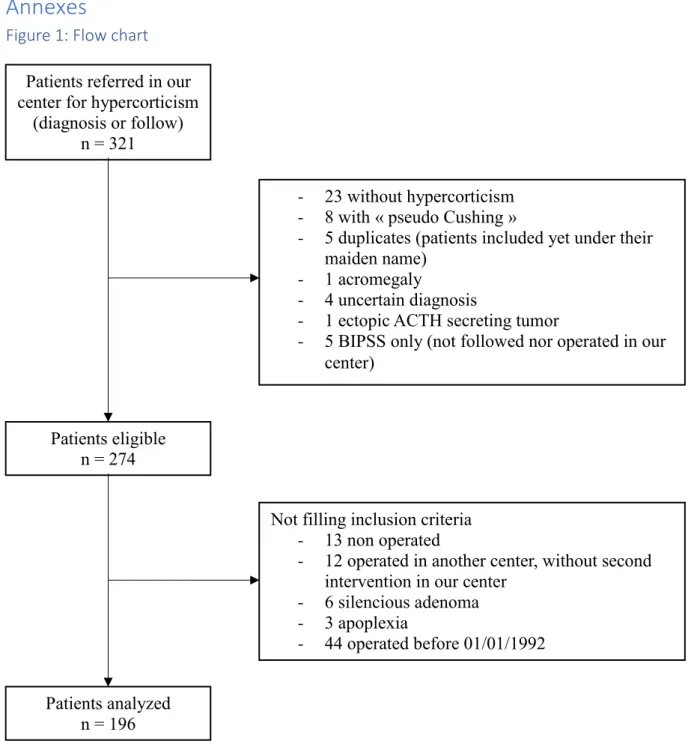
Patients population (Figure 1)The patients’ data were extracted from the medical records of patients referred to our center for established or putative CD, between 01/01/1992 and 09/31/2018. In agreement with
French legislation, an authorization of the National Information science and Liberties
Commission (CNIL) was obtained, through a delegation to our University Hospital Center,
since the database fulfilled all the conditions to be labeled “MR004”. Information letters were sent to patient to ensure they were not opposed to the use of their clinical data.
Exclusion criteria were: patients operated before 01/01/1992 (n=44), patients for whom
diagnosis of hypercorticism was finally ruled out (n=31) or remained uncertain (n=4), ectopic
ACTH secretion (n=1), patients who were not operated (n=13), patients operated in another
center (n=17, including patients referred only for BIPPS), patients with silent corticotroph
adenoma (n=6), discovery of the adenoma on a clinical presentation of pituitary apoplexy
(n=3). Patients who underwent a previous surgery in another center, but who were either not
in remission or with disease recurrence, and who underwent a second surgery in our center,
were included (n=5). Corticotroph macroadenomas were not excluded of the study as long as
they were not silent or not revealed by pituitary apoplexy. Altogether, 196 patients were
included (see results).
Preoperative Endocrine Examination
All patients presented clinical signs of Cushing’s syndrome (CS). Diagnosis of ACTH-dependent CS was based on elevation of free urinary cortisol, elevation of midnight salivary
of cortisol circadian pattern, and a normal or increased ACTH level. Pituitary origin was
assessed by CRH test, Desmopressin test, or 8mg Dexamethasone test, combined to MRI
adenoma findings.
Radiological procedure
All patients underwent pituitary MRI, without and with gadolinium injection, in T1 weighted
and T2 weighted sequences and sometimes dynamic sequences. All patients’ cases were presented in a multidisciplinary meeting including endocrinologists, neurosurgeons and a
neuroradiologist with expertise in pituitary imaging. For patients referred to our institution
with a MRI already performed in another institution, the quality of this MRI was assessed
during the multidisciplinary meeting and if this quality was judged insufficient, an additional
MRI was performed in our institution. The MRI was then defined as typical of an adenoma,
negative or inconclusive. Bilateral inferior petrosal sinus samplings (BIPSS) was performed
as previously described (21,22) in all 44 patients with negative MRI, and it was also
performed in the majority (34/44) of patients with inconclusive MRI, although 10 patients
with inconclusive MRI but typical tests of CD underwent pituitary surgery without BIPPS.
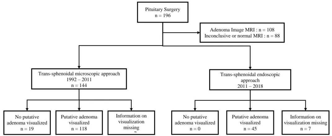
Surgical procedure (Figure 2 and 3)
All patients were operated by trans-sphenoidal approach, by two different expert surgeons
(JGP from 1992 to 2007 EG from 2007 to 2018). Until 2011, surgery relied on a sub-labial
approach guided by microscopy, but an extemporaneous measurement of ACTH in the
cavernous sinuses was performed according to Ludecke (23) as previously described (24):
briefly, after removal of the sellar floor, the neurosurgeon performed bilateral samplings in
the intercavernous sinus veins, and ACTH levels were measured extemporaneously, allowing
to calculate a per-operatory lateral gradient of ACTH in the cavernous sinuses. If no adenoma
half of the pituitary on the side of the dominant sinus, or the median part of the pituitary when
there was no dominant sinus. If an adenoma was identified, the surgeon performed resection
of the adenoma, including a small fragment of the surrounding adjacent normal pituitary.
At the end of year 2011, and for all patients operated later, the endoscopic technic was
introduced and used exclusively. With this approach, per-operatory measurement of ACTH in
the cavernous sinuses were not performed, which proved to have no consequences as an
adenoma was always identified at surgery.
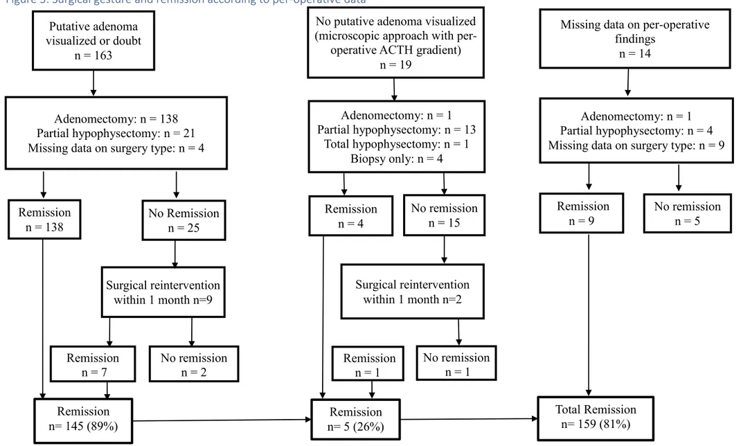
Eleven patients were submitted to an additional trans-sphenoidal surgery within a month
(Figure 2). These patients were characterized by a post-operative serum cortisol level above
remission levels (see below) and a surgical target based on pathological examination of the
first surgery. These early additional surgeries were hemi-hypophysectomy in case of a
previous adenomectomy, or a 2/3 hypophysectomy if a large exeresis had been first
performed. In two cases, total hypophysectomy was performed (Figure 2).
Histological examination
All operative tissues were sent for histological analysis. Tissues were fixed in formaldehyde
and then embedded in paraffine wax. Bloc was then sectioned into 3 µm sections and stained
with Hematoxylin Eosin Safran staining. Immunostaining then included chromogranin A and
cytokeratin AE1 and AE3 staining, to confirm the neuroendocrine nature of the tumor. Then
the corticotroph nature of pituitary adenoma was affirmed if either ACTH, beta endorphin or
MSH staining was positive. The staining is now performed automatically with instrument
Benchmark Ultra (Roche Diagnostics).
Definition of remission
Remission was defined as a post-operative serum cortisol level below 138 nmol/L (50 µg/L)
threshold were either re-operated within a month if adenoma tissue had been identified on the
pituitary fragments obtained during surgery (which was the case for 11 patients, see above
and figure 2) or reevaluated at 3 months post operatively : a normal free urinary cortisol was
then considered indicative of remission whereas an elevated free urinary cortisol was
considered indicative of persistence of CD.
Surgical complications and pituitary morbidity
Surgical complications such as mortality, meningitis, other infections, CSF leaks and
carotidian hemorrhage were collected from operative report and hospitalization report.
Patients were evaluated clinically and biologically at three post-operative months and the data
regarding pituitary deficiencies were collected from their medical report.
Somatotropic deficiency was defined as an IgF1 level below the normal range for age.
Thyrotropic insufficiency was defined as a FT4 below normal range. Gonadotropic
insufficiency was defined as total testosterone below normal range for men, or absence of
regular menstruations for women. For this reason, women taking an estrogen plus progestin
contraception, a progestin-only contraception or aged 52 years or more were not analyzed for
this criterion. Diabetes insipidus was defined as a polyuria with the need for a treatment with
Desmopressin.
Statistical analysis
Analysis were performed with the R Studio software (RStudio, 2016, 1.1.456 version)(26).
Fisher Freeman Halton test was used for the analysis of remission rates, the visualization rate
and the distribution of surgical procedure. Chi Square test was used for the analysis of
Results
196 patients were included in our study, 157 females and 39 males: 90 with microadenomas,
18 with macroadenomas, 44 with inconclusive MRI and 44 with negative MRI. The mean age
at operation was 41 years old.
Of note most of the 18 macroadenomas were “small macroadenomas”: the largest diameter was 18.3mm on average and between 10 - 15 mm; 16-20mm; 21-30mm and>30 mm in 9; 4; 3
and 2 patients, respectively.
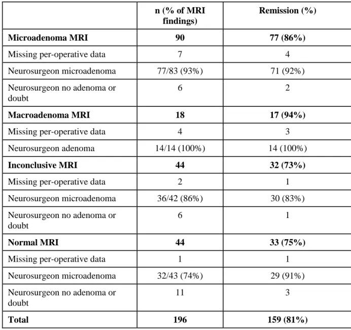
Remission rates
Results are presented in table 2. Post-operative remission rates were 85.5%, 94.4%, 72.7%
and 75.0% for microadenomas, macroadenomas, inconclusive MRI and negative MRI
respectively, and were not statistically different (p=0.09). Of note the only patient with a
macroadenoma who was not in remission had the largest macroadenoma, 42 mm.
Visualization of suspicious tissue
Results are presented in table 2 and supplemental table 1. Per-operative visualization rates
were high in all groups, although the 74.4% visualization rate of patients with a negative MRI
reached statistical difference from the 92.8%, 100%, 85.7% visualization rates of patients
with an MRI image typical of microadenomas, macroadenomas, or an inconclusive MRI
(p=0.03).
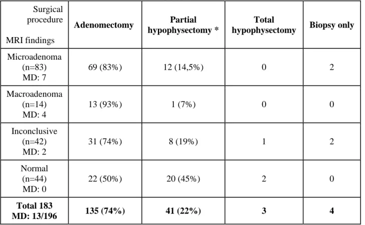
Distribution of surgical procedure
As described in the methods section, two surgical procedures were successively used (figure 1
and 2). Between 1992 to 2011, “classical” trans-sphenoidal microscopic technic was used. If no adenoma was identified, result of per-operatory lateral gradient of ACTH in cavernous
end of year 2011, trans-sphenoidal endoscopic technic was used exclusively and
per-operatory lateral gradient of ACTH in cavernous sinuses was not performed anymore.
The distribution of surgical procedures according to MRI is presented in table 3. The
adenomectomy rates are 83.1%, 92.9%, 73.8% and 50% for microadenoma MRI,
macroadenoma MRI, inconclusive MRI and negative MRI respectively (p<0.001). The
subtotal hypophysectomy rates are 14.5%, 7.1%, 19.0% and 45.5% for microadenoma MRI,
macroadenoma MRI, inconclusive MRI and negative MRI respectively. It must be stressed
that 97.6% of subtotal hypophysectomy were performed in patients operated before 2011 with
the microscopic technique.
Results of surgical exploration
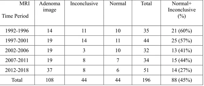
Table 1 compares the per-operative findings with pathological analysis.
For microadenomas MRI, 77 suspicious tissues were seen during operation: 69 were
histologically proven as corticotropic adenoma (89.6%). In 4 tissues, no adenoma was found,
another was proven as a prolactinoma and 2 pathological data were missing. Of the 4 samples
with no adenoma found at pathological examination, 2 / 4 patients were in remission.
For macroadenomas, 14 suspicious tissues were seen and all were confirmed as corticotroph
adenoma.
For inconclusive MRI, 36 suspicious tissues were seen during operation, but only 28 were
histologically proven as corticotroph adenomas (77.8%). In 5 tissues, no adenoma was found
(but 2 patients were in remission after surgical procedure). 1 tissue was histologically proven
as prolactinoma, 1 tissue was too damaged to confirm or exclude an adenoma (but patient was
in remission after surgery) and 1 histological data was missing.
For negative MRI, 32 suspicious tissues were seen during operation: 23 were histologically
were in remission after surgery. 2 tissues were too damaged to perform a precise pathological
analysis, but the 2 patients were in remission after surgery. 1 data was missing.
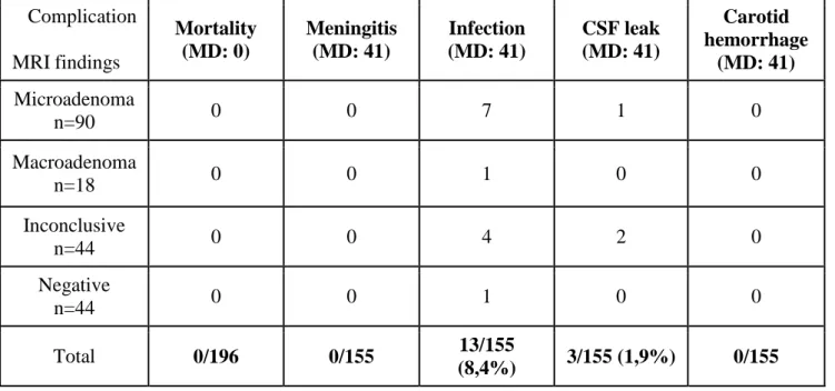
Non-endocrine complications of surgery
Peri operative morbidity was explored and results are presented in table 4. We can assess that
in our series, there was no nosocomial meningitis and no death related to surgery.
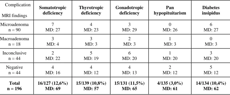
Endocrine complications of surgery
Endocrine complications at 3 months are presented in table 5. Somatotropic, thyrotropic,
gonadotropic deficiency occurred respectively in 12.6%, 10.8% and 11.5% of our patients.
Diabetes insipidus occurred in 10.4% of our patients.
Discussion
Cushing’s disease management is unequivocally based on TSS as the first-line treatment when MRI shows a clear image of adenoma. However, when MRI fails to detect a
microadenoma, the question of first line treatment is more debated, with some who favor TSS
even in case of negative MRI and others who recommend medical treatment in first intention,
in case of lack of expert surgeon, rather than proposing to send patients to an expert one (9).
In our series, rate of negative + inconclusive MRI decreases significantly from 60% to
27% throughout a 26 years period (p=0.015). If results may seem first very encouraging, they
however underline that, to date, MRI still misses 27% of adenomas. One may be tempted to
believe that with the advent of MRI 7 Teslas, negative MRI will be fewer and fewer. We
don’t agree with this assumption: we think that sometimes, MRI can be negative because adenoma signal is equivalent to healthy pituitary tissue and increasing resolution of MRI can’t
solve this problem. In their series, Yamada et al (20) show that in 18/183 patients (10%), MRI
resolution can also increase the number of false positive or detect litigious image. In
literature, the number of patient with MRI microadenoma, with no adenoma found in surgery
varies from 0 to 35% (16,20,27,28). In Feng et al (29), authors analyze concordance of
lateralization by MRI and surgical finding: concordance is 79%, meaning that in 21% of case,
MRI shows a false image of microadenoma.
We want to underline that even when MRI is inconclusive or negative, the surgeon
finds a pathological tissue in respectively 82% and 74.4% of cases. Our data corroborate those
still described in several series: the per-operative visualization rate for patient with negative
MRI ranges from 44% to 100% with a mean of 69% (15,16,27,28,30,31). It seems clear that
the eye of the neurosurgeon is better than MRI.
Since 2011, endoscopic trans-sphenoidal technic replaces microscopic technic in our center. It
is to note that since then, all adenomas have been seen in per operative examination, allowing
to perform only one hemi-hypophysectomy. If analyzed separately, the visualization rates for
inconclusive and negative MRI are 83 and 70% respectively for microscopic technic and
100% for both inconclusive and negative MRI with endoscopic technic.
We do not compare the performance of the two techniques to each other, because the
statistical power of the analysis would have not been not. However, data on per-operative
visualization, which is much higher with endoscopic technic, could let supposed that
performance would be better with endoscopic approach. A recent meta-analysis (32)
including data of 6695 patients, shows no difference for remission rate between the two
technics, except for macroadenoma, where performance of endoscopic technic was better.
Results for recurrence and mortality are identical for both technics. However, several recent
studies show a learning curve for endoscopic approach in pituitary surgery (33–35), with a
In our series, the remission rate for microadenoma is 86%, which is similar to results
of major series (> 50 patients) in the literature (12,13,15,28,36). The remission rate we report
for the macroadenomas is higher than in the literature: 94% of remission compared to
between 59 and 69% in other series (12,13,15,28,36), but it must be stressed that most of
these 18 macroadenomas were “small macroadenomas” with no cavernous sinus invasion.
Brichard et al (37) have a remission rate of 93% for macroadenomas after several TSS, for
similar reasons to ours.
For inconclusive MRI, the remission rate in our series is 73%. This MRI category is not
present in most published studies and is usually not analyzed separately from the negative
MRI category. Three series present results for inconclusive + negative MRI with remission
rates of 77.8% in Honegger et al (31), 69% in Rees et al (38), 67.8% in Bakiri et al (39) which
are comparable to our findings.
For negative MRI, the remission rate is 75%, which is in the same range than those reported
in literature, indicating remission rate between 57 and 71.4% (12,13,15,28,36).
These results are explained by a standardized care strategy: before 2011, surgeon’s gesture
was guided by a per-operative sampling of ACTH in intercavernous sinus veins; now,
endoscopic allow to maximize direct visualization of adenoma, without help of intercavernous
ACTH sampling.
In our study, the remission rate isn’t different between MRI types. Literature is controversial
on this subject: some studies show absence of statistically significative difference between
remission rates when analyzed in function of MRI type (19,40) while some others show a
difference (36,41).
We report no early death linked to operation in our series. For the other complications
(meningitis, infection, CSF leak and carotid hemorrhage), we report an occurrence of
the high rate of missing data (at least 20% for each category), our results are consistent with
those reported in literature with an occurrence rate of 0.4% to 12.5% for meningitis and 0.9%
to 27% for CSF leak (9).
For endocrine complications, our occurrence rate at 3 months is respectively 12.6%, 10.8%,
11.5%, 3% and 10.4% for somatotropic deficiency, thyrotropic deficiency, gonadotropic
deficiency, pan hypopituitarism and diabetes insipidus. Here again, high rate of missing data
must be taken in account. Our data are consistent with median of occurrence rate of other
studies published: we have an higher occurrence rate for somatotropic, gonadotropic
deficiency and diabetes insipidus (respectively 11.7, 8.6 and 4.9% reported in (9)) but an
lower occurrence rate for thyrotropic deficiency and pan hypopituitarism.
Mean and median of efficacy of available medical treatment are reviewed in (9). Mean
efficacy rate (ER) are variable between drugs: 64.3% for Ketoconazole, 71% for Metyrapone,
86.9% for Mitotane, 31.2% for Cabergoline, 14.6% to 26.2% for Pasireotide, 57.7% for
Mifepristone. Except for Mitotane, all drugs are less effective to achieve control of
hypercorticism than surgery, even, in our series, in patients with negative MRI (as a reminder,
75%). Moreover, Ketoconazole, Metyrapone and Mitotane expose to several complications:
liver failure in 18.7% of case with Ketoconazole, hirsutism in 36% of case with Metyrapone,
nausea/vomiting in 46.3% of case with Mitotane. Their costs vary between 10 000 and 30 000
euros/year in France while the cost of both performing BIPPS and trans-sphenoidal surgery by
an expert neurosurgeon is less than 20 000 euros. So that, the cost of treating medically a
single CD patient for 30 years with a normal/inconclusive MRI would allow to offer BIPPS
and trans-sphenoidal surgery to 15-45 patients.
New drugs are now emerging such as retinoic acid, EGFR, hsp90 and CDK inhibitors, but
also Osilidrostat or Levoketoconazole (42,43). Currently, no data is available about efficacy
Conclusion
Despite technological progress, MRI still miss 27.5% of pituitary microadenoma in our series,
even if rate of negative MRI decreases over the years.
The Surgeon’s eye seems to be more performant to detect some pituitary adenoma than MRI,
since surgeons visualize 82% of adenoma in patients with inconclusive MRI and 74.4% of
adenoma in patients with negative MRI. These visualization rates include results of both
microscopic and endoscopic approach, and if analyzed separately, the visualization rates in
endoscopic approach is 100% for inconclusive and negative MRI.
Remission rates are not statistically different whatever the MRI type. Our study supports the
previous results of neurosurgical series published in literature about data on remission in case
of negative MRI.
There is neither death nor meningitis in our series, as still reported in literature. 10 to 12% of
patients have at least one endocrine deficiency, but only 3% have pan hypopituitarism.
Because of its high efficacy rate, its low adverse effects and its lower cost, surgery should
systematically consider as a first option of treatment for CD. In case of lack of expert
neurosurgeon, we think that we shouldn’t consider medical treatment or adrenal surgery as
first option rather than refer patient to expert neurosurgeon.
Bibliography
1. Bourdeau I, Bard C, Forget H, Boulanger Y, Cohen H, Lacroix A. Cognitive function
and cerebral assessment in patients who have Cushing’s syndrome. Endocrinol Metab Clin North Am. 2005 Jun;34(2):357–369, ix.
2. Tiemensma J, Kokshoorn NE, Biermasz NR, Keijser B-JSA, Wassenaar MJE,
Middelkoop HAM, et al. Subtle cognitive impairments in patients with long-term cure of
Cushing’s disease. J Clin Endocrinol Metab. 2010 Jun;95(6):2699–714.
3. Starkman MN, Giordani B, Berent S, Schork MA, Schteingart DE. Elevated cortisol
levels in Cushing’s disease are associated with cognitive decrements. Psychosom Med. 2001 Dec;63(6):985–93.
4. Dekkers OM, Horváth-Puhó E, Jørgensen JOL, Cannegieter SC, Ehrenstein V,
Vandenbroucke JP, et al. Multisystem morbidity and mortality in Cushing’s syndrome: a cohort study. J Clin Endocrinol Metab. 2013 Jun;98(6):2277–84.
5. Pivonello R, Isidori AM, De Martino MC, Newell-Price J, Biller BMK, Colao A.
Complications of Cushing’s syndrome: state of the art. Lancet Diabetes Endocrinol. 2016;4(7):611–29.
6. Lacroix A, Feelders RA, Stratakis CA, Nieman LK. Cushing’s syndrome. Lancet Lond
Engl. 2015 Aug 29;386(9996):913–27.
7. Bolland MJ, Holdaway IM, Berkeley JE, Lim S, Dransfield WJ, Conaglen JV, et al.
Mortality and morbidity in Cushing’s syndrome in New Zealand. Clin Endocrinol (Oxf). 2011 Oct;75(4):436–42.
8. Ntali G, Asimakopoulou A, Siamatras T, Komninos J, Vassiliadi D, Tzanela M, et al.
Mortality in Cushing’s syndrome: systematic analysis of a large series with prolonged follow-up. Eur J Endocrinol. 2013 Nov;169(5):715–23.
9. Pivonello R, De Leo M, Cozzolino A, Colao A. The Treatment of Cushing’s Disease.
Endocr Rev. 2015 Aug 10;36(4):385–486.
10. Shimon I, Ram Z, Cohen ZR, Hadani M. Trans-sphenoidal Surgery for Cushing’s
Disease: Endocrinological Follow-up Monitoring of 82 Patients. Neurosurgery. 2002 Jul
1;51(1):57–62.
11. Hammer GD, Tyrrell JB, Lamborn KR, Applebury CB, Hannegan ET, Bell S, et al.
Trans-sphenoidal Microsurgery for Cushing’s Disease: Initial Outcome and Long-Term
Results. J Clin Endocrinol Metab. 2004 Dec 1;89(12):6348–57.
12. Hofmann BM, Hlavac M, Martinez R, Buchfelder M, Müller OA, Fahlbusch R.
Long-term results after microsurgery for Cushing disease: experience with 426 primary operations
over 35 years. J Neurosurg. 2008 Jan;108(1):9–18.
13. Lambert JK, Goldberg L, Fayngold S, Kostadinov J, Post KD, Geer EB. Predictors of
mortality and long-term outcomes in treated Cushing’s disease: a study of 346 patients. J Clin
Endocrinol Metab. 2013 Mar;98(3):1022–30.
14. Alexandraki KI, Kaltsas GA, Isidori AM, Storr HL, Afshar F, Sabin I, et al. Long-term
remission and recurrence rates in Cushing’s disease: predictive factors in a single-centre study. Eur J Endocrinol. 2013 Apr;168(4):639–48.
15. Dimopoulou C, Schopohl J, Rachinger W, Buchfelder M, Honegger J, Reincke M, et
al. Long-term remission and recurrence rates after first and second trans-sphenoidal surgery
for Cushing’s disease: care reality in the Munich Metropolitan Region. Eur J Endocrinol. 2014 Feb;170(2):283–92.
16. Ciric I, Zhao J-C, Du H, Findling JW, Molitch ME, Weiss RE, et al. Trans-sphenoidal
surgery for Cushing disease: experience with 136 patients. Neurosurgery. 2012
17. Tritos NA, Biller BMK, Swearingen B. Management of Cushing disease. Nat Rev
Endocrinol. 2011 May;7(5):279–89.
18. Semple PL, Vance ML, Findling J, Laws ER. Trans-sphenoidal Surgery for Cushing’s
Disease: Outcome in Patients with a Normal Magnetic Resonance Imaging Scan.
Neurosurgery. 2000 Mar 1;46(3):553–9.
19. Salenave S, Gatta B, Pecheur S, San-Galli F, Visot A, Lasjaunias P, et al. Pituitary
Magnetic Resonance Imaging Findings Do Not Influence Surgical Outcome in
Adrenocorticotropin-Secreting Microadenomas. J Clin Endocrinol Metab. 2004 Jul
1;89(7):3371–6.
20. Yamada S, Fukuhara N, Nishioka H, Takeshita A, Inoshita N, Ito J, et al. Surgical
Management and Outcomes in Patients with Cushing Disease with Negative Pituitary
Magnetic Resonance Imaging. World Neurosurg. 2012 Mar 1;77(3):525–32.
21. Lefournier V, Gatta B, Martinie M, Vasdev A, Tabarin A, Bessou P, et al. One
Transient Neurological Complication (Sixth Nerve Palsy) in 166 Consecutive Inferior
Petrosal Sinus Samplings for the Etiological Diagnosis of Cushing’s Syndrome. J Clin Endocrinol Metab. 1999 Sep 1;84(9):3401–6.
22. Lefournier V, Martinie M, Vasdev A, Bessou P, Passagia J-G, Labat-Moleur F, et al.
Accuracy of Bilateral Inferior Petrosal or Cavernous Sinuses Sampling in Predicting the
Lateralization of Cushing’s Disease Pituitary Microadenoma: Influence of Catheter Position and Anatomy of Venous Drainage. J Clin Endocrinol Metab. 2003 Jan 1;88(1):196–203.
23. Lüdecke DK. Intraoperative measurement of adrenocorticotropic hormone in
peripituitary blood in Cushing’s disease. Neurosurgery. 1989 Feb;24(2):201–5.
24. Passagia JG, Gay E, Chabre O, Martinie M, Labat-Moleur F, Bachelot I. [Role of
perioperative biological tests during the performance and follow-up of corticotroph adenoma
25. Nieman LK, Biller BMK, Findling JW, Murad MH, Newell-Price J, Savage MO, et al.
Treatment of Cushing’s Syndrome: An Endocrine Society Clinical Practice Guideline. J Clin Endocrinol Metab. 2015 Aug;100(8):2807–31.
26. RStudio Team. RStudio: Integrated Development for R. 2016;
27. Johnston PC, Kennedy L, Hamrahian AH, Sandouk Z, Bena J, Hatipoglu B, et al.
Surgical outcomes in patients with Cushing’s disease: the Cleveland clinic experience. Pituitary. 2017 Aug;20(4):430–40.
28. Cebula H, Baussart B, Villa C, Assié G, Boulin A, Foubert L, et al. Efficacy of
endoscopic endonasal trans-sphenoidal surgery for Cushing’s disease in 230 patients with
positive and negative MRI. Acta Neurochir (Wien). 2017;159(7):1227–36.
29. Feng M, Liu Z, Liu X, Bao X, Yao Y, Deng K, et al. Diagnosis and Outcomes of 341
Patients with Cushing’s Disease Following Transsphenoid Surgery: A Single-Center Experience. World Neurosurg. 2018 Jan 1;109:e75–80.
30. Chee GH, Mathias DB, James RA, Kendall‐ Taylor P. Trans-sphenoidal pituitary surgery in Cushing’s disease: can we predict outcome? Clin Endocrinol (Oxf). 2001 May 1;54(5):617–26.
31. Honegger J, Schmalisch K, Beuschlein F, Kaufmann S, Schnauder G, Naegele T, et al.
Contemporary microsurgical concept for the treatment of Cushing’s disease: endocrine outcome in 83 consecutive patients. Clin Endocrinol (Oxf). 2012 Apr;76(4):560–7.
32. Broersen LHA, Biermasz NR, van Furth WR, de Vries F, Verstegen MJT, Dekkers
OM, et al. Endoscopic vs. microscopic trans-sphenoidal surgery for Cushing’s disease: a
systematic review and meta-analysis. Pituitary. 2018 May 16;
33. Lofrese G, Vigo V, Rigante M, Grieco DL, Maresca M, Anile C, et al. Learning curve
of endoscopic pituitary surgery: Experience of a neurosurgery/ENT collaboration. J Clin
34. Qureshi T, Chaus F, Fogg L, Dasgupta M, Straus D, Byrne RW. Learning curve for
the trans-sphenoidal endoscopic endonasal approach to pituitary tumors. Br J Neurosurg. 2016
Nov 1;30(6):637–42.
35. Shikary T, Andaluz N, Meinzen-Derr J, Edwards C, Theodosopoulos P, Zimmer LA.
Operative Learning Curve After Transition to Endoscopic Trans-sphenoidal Pituitary Surgery.
World Neurosurg. 2017 Jun;102:608–12.
36. Chandler WF, Barkan AL, Hollon T, Sakharova A, Sack J, Brahma B, et al. Outcome
of Trans-sphenoidal Surgery for Cushing Disease: A Single-Center Experience Over 32
Years. Neurosurgery. 2016 Feb;78(2):216–23.
37. Brichard C, Costa E, Fomekong E, Maiter D, Raftopoulos C. Outcome of
Trans-sphenoidal Surgery for Cushing Disease: A Single-Center Experience over 20 Years. World
Neurosurg. 2018 Nov;119:e106–17.
38. Rees DA, Hanna FWF, Davies JS, Mills RG, Vafidis J, Scanlon MF. Long-term
follow-up results of trans-sphenoidal surgery for Cushing’s disease in a single centre using
strict criteria for remission. Clin Endocrinol (Oxf). 2002 Apr 1;56(4):541–51.
39. Bakiri F, Tatai S, Aouali R, Semrouni M, Derome P, Chitour F, et al. Treatment of
Cushing’s disease by trans-sphenoidal, pituitary microsurgery: prognosis factors and long-term follow-up. J Endocrinol Invest. 1996 Oct;19(9):572–80.
40. Sun Y, Sun Q, Fan C, Shen J, Zhao W, Guo Y, et al. Diagnosis and therapy for
Cushing’s disease with negative dynamic MRI finding: a single-centre experience. Clin Endocrinol (Oxf). 2012 Jun;76(6):868–76.
41. Rollin G, Ferreira NP, Czepielewski MA. Prospective evaluation of trans-sphenoidal
pituitary surgery in 108 patients with Cushing’s disease. Arq Bras Endocrinol Amp Metabol. 2007 Nov;51(8):1355–61.
42. Feelders RA, Newell-Price J, Pivonello R, Nieman LK, Hofland LJ, Lacroix A.
Advances in the medical treatment of Cushing’s syndrome. Lancet Diabetes Endocrinol. 2018 Jul 19;
43. Störmann S, Schopohl J. New and emerging drug therapies for Cushing’s disease. Expert Opin Pharmacother. 2018 Aug;19(11):1187–200.
Annexes
Figure 1: Flow chart Patients referred in our center for hypercorticism
(diagnosis or follow) n = 321
- 23 without hypercorticism - 8 with « pseudo Cushing »
- 5 duplicates (patients included yet under their maiden name)
- 1 acromegaly
- 4 uncertain diagnosis
- 1 ectopic ACTH secreting tumor
- 5 BIPSS only (not followed nor operated in our center)
Patients analyzed n = 196
Not filling inclusion criteria - 13 non operated
- 12 operated in another center, without second intervention in our center
- 6 silencious adenoma - 3 apoplexia
- 44 operated before 01/01/1992 Patients eligible
Figure 2: Surgical approach and per-operative visualization data
Pituitary Surgery n = 196
Trans-sphenoidal microscopic approach 1992 – 2011 n = 144 Trans-sphenoidal endoscopic approach 2011 – 2018 n = 52 No putative adenoma visualized n = 19 Putative adenoma visualized n = 118 No putative adenoma visualized n = 0 Putative adenoma visualized n = 45
Adenoma Image MRI : n = 108 Inconclusive or normal MRI : n = 88
Information on visualization missing n = 7 Information on visualization missing n = 7
Figure 3: Surgical gesture and remission according to per-operative data Putative adenoma visualized or doubt n = 163 Adenomectomy: n = 138 Partial hypophysectomy: n = 21 Missing data on surgery type: n = 4
No putative adenoma visualized (microscopic approach with
per-operative ACTH gradient) n = 19
Adenomectomy: n = 1 Partial hypophysectomy: n = 13
Total hypophysectomy: n = 1 Biopsy only: n = 4
Missing data on per-operative findings
n = 14
Adenomectomy: n = 1 Partial hypophysectomy: n = 4 Missing data on surgery type: n = 9
Remission n = 138 No Remission n = 25 Surgical reintervention within 1 month n=9 Remission n = 7 No remission n = 2 Remission n = 4 No remission n = 15 Surgical reintervention within 1 month n=2 Remission n = 1 No remission n = 1 Remission n = 9 No remission n = 5 Remission n= 145 (89%) n= 5 (26%) Remission Total Remission n= 159 (81%)
Table 1: Evolution of MRI performance with time MRI
Time Period
Adenoma image
Inconclusive Normal Total Normal+ Inconclusive (%) 1992-1996 14 11 10 35 21 (60%) 1997-2001 19 14 11 44 25 (57%) 2002-2006 19 3 10 32 13 (41%) 2007-2011 19 8 7 34 15 (44%) 2012-2018 37 8 6 51 14 (27%) Total 108 44 44 196 88 (45%)
Table 2: Relation between MRI findings and per-operative neurosurgeon’s finding and remission rates n (% of MRI findings) Remission (%) Microadenoma MRI 90 77 (86%)
Missing per-operative data 7 4
Neurosurgeon microadenoma 77/83 (93%) 71 (92%) Neurosurgeon no adenoma or
doubt
6 2
Macroadenoma MRI 18 17 (94%)
Missing per-operative data 4 3
Neurosurgeon adenoma 14/14 (100%) 14 (100%) Inconclusive MRI 44 32 (73%)
Missing per-operative data 2 1
Neurosurgeon microadenoma 36/42 (86%) 30 (83%) Neurosurgeon no adenoma or
doubt
6 1
Normal MRI 44 33 (75%)
Missing per-operative data 1 1
Neurosurgeon microadenoma 32/43 (74%) 29 (91%) Neurosurgeon no adenoma or doubt 11 3 Total 196 159 (81%) Statistics :
- Microadenoma VS Macroadenoma VS Inconclusive VS Normal: p=0.09 (Fisher Freeman Halton test)
- Negative VS Microadenoma: p=0.31 (Chi Square Test) - Inconclusive VS Microadenoma: p=0.21 (Chi Square Test)
Table 3: Distribution of surgical procedures according to MRI Surgical procedure MRI findings Adenomectomy Partial hypophysectomy * Total
hypophysectomy Biopsy only
Microadenoma (n=83) MD: 7 69 (83%) 12 (14,5%) 0 2 Macroadenoma (n=14) MD: 4 13 (93%) 1 (7%) 0 0 Inconclusive (n=42) MD: 2 31 (74%) 8 (19%) 1 2 Normal (n=44) MD: 0 22 (50%) 20 (45%) 2 0 Total 183 MD: 13/196 135 (74%) 41 (22%) 3 4
*Partial hypophysectomy includes hemi hypophysectomy (n = 17), subtotal hypophysectomy (n = 9) and median hypophysectomy (n = 15). For negative MRI, surgeon realized 6 hemi-hypophysectomy, 6 subtotal hypophysectomy and 8 median hypophysectomy. The gesture of hemi-hypophysectomy was guided by intraoperative measurement of ACTH, as described by Ludecke (24)
Table 4: Distribution of non-endocrine complications of surgery according to MRI Complication MRI findings Mortality (MD: 0) Meningitis (MD: 41) Infection (MD: 41) CSF leak (MD: 41) Carotid hemorrhage (MD: 41) Microadenoma n=90 0 0 7 1 0 Macroadenoma n=18 0 0 1 0 0 Inconclusive n=44 0 0 4 2 0 Negative n=44 0 0 1 0 0 Total 0/196 0/155 13/155 (8,4%) 3/155 (1,9%) 0/155
Table 5: Endocrine complications of surgery Complication MRI findings Somatotropic deficiency Thyrotropic deficiency Gonadotropic deficiency Pan hypopituitarism Diabetes insipidus Microadenoma n = 90 7 MD: 27 4 MD: 23 3 MD: 29 0 MD: 26 6 MD: 27 Macroadenoma n = 18 3 MD: 4 3 MD: 3 2 MD: 3 1 MD: 3 0 MD: 3 Inconclusive n = 44 2 MD: 22 5 MD: 19 6 MD: 20 1 MD: 20 3 MD: 20 Negative n = 44 4 MD: 16 4 MD: 12 4 MD: 13 2 MD: 12 5 MD: 12 Total n = 196 16/127 (12,6%) MD: 69 15/139 (10,8%) MD: 57 15/131 (11,5%) MD: 65 4/135 (3,0%) MD: 61 14/134 (10,4%) MD: 62
Supplemental Table 1 Microadenoma n = 83 MD: 7 Macroadenoma n = 14 MD: 4 Inconclusive n = 42 MD: 2 Negative n = 43 MD: 1 Total Putative adenoma visualized n = 77 (92,8%) n = 14 (100 %) n = 36 (85,7%) n = 32 (74,4%) 159 Corticotropic adenoma histologically proven 69 14 28 23 134 Remission 67 14 25 22 128 No adenoma found 5 5 6 16 Remission 3 2 4 9
Other histological type 1 1 2
Remission 0 1 1 Inconclusive histological exam 1 2 3 Remission 1 2 3 MD on histology 2 1 1 4 Remission 1 1 1 3 Putative adenoma visualized global remission rate 92% 100% 83% 87,5% 91% No adenoma visualized n = 4 n = 6 n = 9 19 Corticotropic adenoma histologically proven 2 2 Remission 2 2 No adenoma found 2 4 6 12 Remission 0 1 1 2
Other histological type 2 1 1 4
Remission 1 0 0 1
MD on histology 1 1
Remission 0 0
No adenoma found
global remission rate 25% 17% 33% 26%
Doubt on adenoma vision n = 2 n = 2 4 Corticotropic adenoma histologically proven 1 1 2 Remission 1 0 1 No adenoma found 1 1 Remission 0 0
Other histological type 1 1
Remission 0 0
Doubt on adenoma vision global remission rate
Remerciements
Aux membres du jury
Au Pr Emmanuel Gay, merci d’avoir accepté d’être président de ce jury. Vous ne vous en souvenez certainement pas, mais vous avez été mon premier « chef ». Au sein de votre service, en premier stage d’externat, j’ai découvert pour la première fois le monde hospitalier, fait mon premier « bip 120 », fait mon premier vrai examen neurologique, appris à m’habiller. J’ai encore le souvenir de vos « Oh là là » au bloc opératoire quand les hypophyses saignaient ! Je suis ressortie en me disant que peut être que la neurochirurgie pourrait être bien comme spécialité. Après, je me suis rappelée que j’aimais dormir et que je confondais ma droite et ma gauche, et j’ai trouvé plus sage de retourner rêver à l’endocrinologie. J’espère que vous prendrez plaisir à lire cette thèse, qui existe grâce à vos résultats (que ferait-on sans neurochirurgien ?)
Au Pr Olivier Chabre, merci de m’avoir encadrée tout au long de cette thèse et merci de continuer à me soutenir dans mon parcours professionnel. Je retiendrai de vous le médecin investi que vous êtes, le plaisir que vous prenez à enseigner, votre sens clinique, mais aussi le temps démesuré que vous passez à vous battre pour vos patients et votre disponibilité pour eux. Votre exigence envers les autres n’a d’égal que celle que vous avez envers vous-même et je vous remercie pour vos conseils. Votre rigueur nous pousse vers le meilleur. J’espère pouvoir avoir la chance de continuer à évoluer professionnellement à vos côtés.
Au Pr Pierre Yves Benhamou, merci de m’avoir fait faire mes premiers pas dans le service. D’abord en m’accueillant en P2 (même si c’était le hasard), puis en Master 1, puis en tant qu’interne et indirectement en Master 2. Merci pour votre oreille attentive (sans mauvais jeu de mot) pendant ces années d’internat, merci pour votre bienveillance constante. J’admire le calme et la patience dont vous faites preuve avec chacun, vous êtes un modèle d’humanité. Merci pour les encouragements.
A tous les deux, merci pour votre soutien. Je pense que vous faites partie des rares chefs à régulièrement féliciter leurs internes et leur dire lorsque ce qu’ils font est bien. J’apprécie énormément évoluer dans le confort de cette bienveillance. Merci encore.
Au Pr Jean Guy Passagia, merci d’avoir accepté d’être membre de ce jury. Car après tout, c’est aussi (et peut être d’abord !) grâce à vous que cette thèse existe. J’ai aussi eu le plaisir de vous côtoyer dans vos consultations en Neurochirurgie, où j’ai pu apprécier votre pédagogie envers les patients et votre façon de leur expliquer les choses et de les accompagner. J’ai le souvenir de vos cours d’anatomie, et je crois que je peux vous le dire, je ne me souviens plus tant du contenu de vos cours que de la façon passionnée dont vous les enseignez, même sur DVD ! J’avoue vous avoir un peu maudit sur le membre supérieur, je
n’aurai jamais imaginé qu’il puisse y avoir autant de muscles et d’insertions dont il faudrait apprendre les noms ! Je vous souhaite une belle fin de carrière.
Au Dr Virginie Lefournier, merci d’avoir accepté de faire partie de mon jury. Merci pour ton œil aiguisé et ta passion des images (« Mais regardez comme c’est beau », « Mais regardez-moi la qualité de cette IRM, c’est nul, ils ne sont même pas fichus de la faire dans le bon plan ! », « Ah non, là il est énorme »). Merci pour les petites tranches de rigolade en « Club hypophyse », même si après on se fait disputer par le Pr Chabre ^^. J’espère avoir le plaisir de continuer à te voir un jeudi sur deux.
Aux autres « chefs » m’ayant encadrée
Au Dr Sandrine Lablanche, merci pour ton soutien et ton aide durant ces 4 années, merci pour ton écoute. On m’a toujours dit que je ne serai pas une vraie interne de diabétologie tant que je n’aurai pas pleuré dans le bureau du Pr Benhamou… C’est finalement dans ton bureau que je serai venue chouiner un peu. Merci pour ton aide durant ce 6ème semestre pas facile sur le plan moral. Merci pour tes conseils sur mes choix professionnels. Merci pour ton encadrement pendant mon master 2. Et merci de me montrer tous les jours qu’on peut être une super chef, une super universitaire et une super maman. Tu me donnes l’espoir que mener une vie professionnelle et personnelle épanouissante sur tous les plans est possible.
Au Pr Anne Laure Borel, merci de faire partager ton savoir immense, tu m’épates, je ne sais pas comment tu as le temps d’être toujours au courant de toute la biblio. Je me rappelle encore ce moment où tu m’as dit « Justine, tu ne peux pas continuer à vivre sans PubMed plus longtemps ». Ces quelques minutes que tu as prises pour tout me montrer m’ont sans doute sauvé la vie pour ma thèse et mes autres présentations ! Tu es une véritable « warrior » et j’admire ta volonté à toute épreuve.
Au Dr Marie Muller, merci pour tes conseils sur la prise en charge des plaies de pied, merci pour m’avoir proposé un sujet de mémoire et m’avoir fait découvrir la recherche clinique prospective et ses aléas administratifs. Merci pour ton humour pince sans rire, et tes retours lorsque tu nous encadre. Merci d’être souvent un soutien pour les internes, tu es toujours la première à nous prendre le téléphone d’avis et à nous aider lorsque tu vois qu’on est en sous-effectif.
Au Dr Nelly Wion, merci de partager ton expérience avec nous, notamment sur les avis et de prendre le temps de venir voir chacun de tes patients en hospitalisation. Tu as une patience à toute épreuve avec les patients, qui sont parfois compliqués à gérer, notamment ceux de nutrition, face auxquels je me sens très démunie.
Au Dr Sandrine Coumes, merci de m’avoir fait poser ma première sonde nasogastrique ! Nous n’aurons pas beaucoup travaillé ensemble, mais c’est toujours un plaisir de te voir passer une tête souriante dans le bureau des internes.