Squatting test: a dynamic postural manoeuvre to study baroreflex
sensitivity
André J. Scheen · Jean-Christophe Philips
Division of Diabetes, Nutrition and Metabolic Disorders, Department of Medicine, CHU Liège, University of Liège, CHU Sart Tilman (B35), 4000 Liège 1, Belgium
Abstract
Introduction Squatting is an active posture test that can be used to assess baroreflex sensitivity. Indeed, the shift from squatting to standing imposes a major orthostatic stress leading to rapid and large changes in arterial blood pressure (BP) and heart rate (HR) allowing precise baroreflex assessment.
Material and methods BP and HR can be continuously and non-invasively monitored with a Finapres device. Results The standing to squatting transition is accompanied by rises in BP, pulse pressure and cardiac output, mainly due to increased venous return, and by a secondary reduction in HR. Conversely, the squatting to standing transition is associated with an immediate drop in BP and both reflex tachycardia and vasoconstriction. This mirror changes in BP and HR, mimicking those observed with the classical pharmacological approach using vasopressor/ vasodilating agents, allows the calculation of the so-called baroreflex gain.
Discussion The present review describes the haemodynamic changes occurring in normal subjects during the shifts from standing to squatting and from squatting to standing and discusses the underlying cardiovascular and autonomic mechanisms.
Conclusion This careful analysis in healthy individuals should help in the understanding of disturbances that may be observed in patients with autonomic dysfunction, such as in diabetic patients with cardiovascular autonomic neuropathy.
Keywords Baroreflex · Blood pressure · Finapres · Heart rate · Orthostatic hypotension · Squatting
INTRODUCTION
Although arterial blood pressure (BP) is regulated via the interaction of various local, humoral and neural factors, acute BP regulation during posture changes mainly involves baroreflexes. In response to baroreceptor activa-tion/deactivation, key determinants of BP, such as cardiac period (RR interval)/heart rate (HR) (via the sympathetic and parasympathetic nervous system) and vascular resistance (via the sympathetic nervous system), are modified to maintain BP homoeostasis [1]. In normal subjects, BP is tightly regulated and the variations are generally of limited amplitude and, even more typically, of short duration despite acute postural changes. However, alterations in baroreflex function with age may explain the higher risk of postural hypotension in elderly people [2]. Furthermore, several pathological entities may be associated with autonomic disorders and dysregulation of BP homoeostasis leading to orthostatic hypotension and syncope as most obvious adverse events [3, 4].
Non-invasive continuous monitoring of systolic and diastolic BP together with HR, using the servoplethysmo-manometry and the volume clamp technique at the finger level (Finapres®, Finometer®), has greatly facilitated the study of haemodynamic changes during postural tests in both clinical research and practice [5, 6]. Similar information can be obtained with non-invasive Finapres® BP recordings as with invasive intra-arterial pressure measurements (considered as the gold standard method) for the assessment of the continuous BP responses to orthostatic stress [5].
Various posture tests are classically used to assess haemodynamic orthostatic adaptation and autonomic function. The most popular posture test in clinical research is the passive head-up tilt table test at 70°. Alternatively, an active manoeuvre, the so-called squatting test, produces even a stronger acute orthostatic stress when the subject stands up [7]. Careful analysis of BP and HR changes during both transition states, from standing to squatting and from squatting to standing, respectively, provides interesting information as far as performance of baroreflexes and haemodynamic homoeostasis [6]. For instance, the analysis of HR and BP changes occurring
during a squatting test has been used by our group in patients with diabetes mellitus to assess orthostatic hypotension [7], cardiovascular autonomic neuropathy [7, 8] and pulsatile stress [8].
The present article is the first review paper written in English that systematically analyses the haemodynamic changes during standing-squatting-standing transitions in healthy subjects allowing the calculation of the so-called baroreflex gain. It aims at summarizing all the pertinent data published in the literature when using this active postural test to assess baroreflex sensitivity. This careful analysis of physiological changes during the transition from standing to squatting and from squatting to standing is a prerequisite before considering corresponding changes in subjects with autonomic dysfunction, such as diabetic patients with cardiovascular autonomic neuropathy [4].
CHANGES FROM STANDING TO SQUATTING
Sharpey-Schafer, in a study of normal subjects performed in 1956, found that squatting caused an increase in systemic arterial BP that was followed by bradycardia [9]. He suggested that squatting increased cardiac output (CO) by increasing venous return, and that the bradycardia was secondary to baroreceptor activity. In 1962, another study showed that squatting from the standing position increased arterial BP, CO and "central blood volume" [10]. All healthy subjects had an immediate increase in systolic BP, diastolic BP and pulse pressure (PP) when they squatted from the standing position. About four beats after squatting there was a bradycardia, which was interpreted as a baroreceptor response to the rise in systolic BP and PP. It has been demonstrated later on that lengthening of the R-R interval during squatting was abolished by atropine [11]. Squatting increased both CO and central blood volume [10]. The steady-state measurements of BP were all higher in the squatting than in the standing position, but were always less than the values obtained immediately after squatting (Fig. 1).
Prompt active postural manoeuvres induce an immediate arterial BP variation followed by a period of regulation. In the squatting manoeuvre, initial hypertension was initially explained by a rise of cardiac filling pressure due to "squeezing blood out of the veins of the legs", leading to an increase in stroke output by Frank-Starling
mechanism. The observations of an increase in central blood volume led to the idea of a rise of cardiac filling [10]. For a minor part, it was also explained by "kinking" of the femoral arteries.
However, no such consistent circulatory variations were detected when postural changes were realized in a water tank. Similarly, squatting in the lying position has no significant haemodynamic effects [10, 12]. These
observations demonstrate that squatting exerts its main action on the circulation not by kinking arteries and veins to the legs.
The haemodynamic changes induced by squatting are prompt rises in arterial BP and CO, as well [6, 10, 13]. Several reports have shown that squatting increases preload by augmentation of venous return, whereas systemic vascular resistance does not change by squatting [10, 14]. These changes will result in greater increase of systolic BP than of diastolic BP and thus augmentation of PP in squatting as compared to standing position. A study reported a significant increase in systolic/diastolic BP (+6.5/+3.7 mmHg) in healthy female and male adults [15]. Another study elucidated the pulsatile haemodynamic change induced by squatting and concluded that this change is induced by enhanced aortic wave reflection [16]. Although systolic BP (+8.2 mmHg), diastolic BP (+3 mmHg) and PP (+5.2 mmHg) during squatting were higher than those during standing, only the difference in systolic BP was statistically significant. Like in another study [13], we observed a 5-6 mmHg increase in PP in squatting as compared to standing position in normal subjects (Fig. 1). Interestingly, this increase was amplified in patients with long-lasting type 1 diabetes, a finding attributed to increased arterial stiffness [17].
Little data are available concerning the relation between the muscular pumping mechanism and the variation of superficial and deep venous pressure during normal action of the calf pump. A study determined the pressure values in three muscular compartments of the calf and in the deep and the superficial venous system and established correlation between muscular and venous pressure. When healthy young women were squatting, an increase of 414% in the deep posterior muscular compartment pressure, 734% in the superficial posterior compartment and 492% in the anterior tibial compartment was noticed. Thus, the significant boost in the pressure of all muscular compartments can partially explain the rise in the deep and superficial venous pressures and consequently the increased venous return [10]. The increased compartmental and venous pressures link back to the original idea of "squeezing blood out of the veins of the legs", which in the end looks quite close to the current thoughts of a muscle pump action leading to the initial sharper rise followed by persistently increased venous pressure.
Fig. 1 Haemodynamic changes during standing-squatting-standing in a normal subject. Changes in systolic
(SBP), diastolic (DBP) and mean blood pressure (MBP), pulse pressure (PP) and heart rate (HR) measured continuously with a Finapres® during a standing (0-60 s)— squatting (60-120 s)—standing (120-180 s) postural test in a normal subject. Please note the transient drop in BP with reflex tachycardia after standing up following squatting
The dynamics of haemodynamic and cardiac changes was investigated in healthy men by comparing non-inva-sively registered parameters of left ventricular performance in standing (control), at the onset of squatting ('prompt squat') and at 2 min of squatting [18]. Squatting produced decreases in HR, isovolumic contraction time, pre-ejection period and pulse transmission time from onset of depolarisation to the first heart sound, left ventricular ejection time and the ejection time index. These results of systolic time intervals are consistent with the bradycardia and increased ventricular filling induced by squatting. Major changes from control
measurements were found at the onset of squatting, showing the impact of prompt squat on left ventricular performance. The effects of changes in posture on left ventricular diameter and function were also studied by echocardiography in healthy children [19]. Squatting was accompanied by an increase in left ventricular cavity dimension, while HR fell slightly and calculated stroke index (+35%) and cardiac index (+33%) increased. Mean BP increased by 19%.
In summary, the transition from squatting to standing elicits muscle pumping and increased venous return, which leads to prompt increases in CO, systolic BP and PP, followed by reflex bradycardia.
CHANGES FROM SQUATTING TO STANDING
Squatting to standing involves volume changes and systemic pressure changes. Therefore, the hemodynamic results are due both to activation of volume sensors and pressure sensors. The immediate HR increase upon fast standing is mainly due to vagal withdrawal and then sympathetic activation.
A detailed description of normal haemodynamic changes during the transition from squatting to standing has been reported [6] (Fig. 1). Without exception, the first beat after changing from squatting to standing showed a decrease in systolic BP, diastolic BP and mean BP by 14.6, 10.6 and 13.9 mmHg, respectively. Mean BP reached a minimum of 57.8 mmHg after 7 s. Thereafter, the BP increased to a new level within about 15 s. Most subjects demonstrated a biphasic HR response. The maximum HR was reached after 11 s of standing. Propranolol, a beta-adrenergic blocking agent, markedly attenuated shortening of the R-R interval at standing from squatting [ 11 ]. In all experiments, the peaks in HR were distinctly delayed after the BP dips, suggesting that an arterial baroreflex could be implicated in the immediate HR increase. In the same experiments, the influence of posture on the rhythms in BP, HR and respiration was tested by means of spectral analysis [6, 20]. During each posture, the finger BP was recorded non-invasively together with cardiac intervals and respiratory movements. The power spectra obtained from 5-min samples showed that the respiratory components of cardiac interval and PP were reduced significantly in standing. Compared to squatting, a significant increase of total power in the medium frequency band (0.05-0.15 Hz) for cardiac interval, diastolic and mean BP could be detected.
To our knowledge, there is no detailed study reporting the changes in stroke volume and CO during the transition from squatting to standing. In a preliminary study, we combined measurements of stroke volume by bioimped-ance cardiography (Kardiocom ) with measurements of mean arterial BP and HR by the Finapres to derive CO and systemic vascular resistance [21]. We showed that the initial drops in stroke volume and systemic vascular resistance were more marked during the squat-stand transition, leading to a significantly greater initial drop in BP [7], compared to the reference tilt test, especially in diabetic patients with autonomic neuropathy. Over the first 30 s of a head-up tilt test, there is a steady parallel decline of thoracic blood volume and stroke volume; there is also an initial surge of CO followed by a steady decrease [22]. However, several differences may be pointed out between the active squatting to standing transition and the passive head-up tilt table manoeuvre, among which are the absence of kinking of the arteries and the almost absence of active muscular contraction during the tilt test. In contrast, the squatting test is accompanied by a strong muscular contraction leading to increased venous pumping during active stand up [23]. Furthermore, squatting may be described as a fast way of standing, contrasting with tilt that is a slow way of standing. Therefore, the homoeostatic mechanisms are the same, but with different quantity of the same time-dependent quality. A carefully performed study compared haemodynamic changes during active standing and following passive 60° tilt in young healthy subjects [24]. BP and HR were continuously recorded by a Finapres®, and simultaneous beat-to-beat stroke volume was obtained using an ultrasound Doppler technique. Active standing (but not from squatting position in that study) caused a transient but greater reduction of BP (-39 vs. -16 mmHg), and a higher increase of HR (+35 vs. +12 beats/min) than passive tilt during the first 30 s. There was a significantly larger increase in CO during active standing (+37 vs. 0%) and a more marked decrease in total peripheral resistance (-58 vs.-16%). A precipitous rise in intra-abdominal pressure (43 mmHg) could be observed upon rising only in active standing. This was interpreted as an indication of translocation of blood to the thorax. The greater BP reduction in the initial phase of active standing may reflect systemic vasodilatation caused by activation of cardiopulmonary baroreflexes; this is probably due to a rapid shift of blood from the splanchnic vessels in addition to the shift from muscular vessels associated with abdominal and calf muscle contraction [24]. At least similar, and probably even amplified, changes may be expected during the shift from squatting to standing.
An experimental study investigated if the cardiovascular adaptation observed following ten +75° head-up tilts in 70 min would improve the responses to the squatting-standing test [25]. After repetitive tilting, BP (diastolic more than systolic) was significantly increased during the first 30 s of post-squatting standing when compared with the same test performed before repetitive tilting. In contrast, there were no differences observed in the control group when comparing BP changes during the squat-stand transition before and after 70 min of rest. Thus, cardiovascular responses to the squatting-standing test can be improved with consecutive head-up tilts. This effect is predominantly due to an increase in diastolic BP, indicative of a change in vascular resistance. In summary, the squat-stand transition provokes an immediate ample reduction in BP, which most probably reflects a decrease in stroke volume and CO, and results in a rapid reflex tachycardia and peripheral vasoconstriction. Therefore, normal baroreflex homoeostasis leads to a return to baseline BP and HR within about 15-20 s.
CALCULATION OF THE BAROREFLEX GAIN
The arterial baroreflex is a key mechanism for BP homoeostasis and is physiologically relevant as an indicator of autonomic control. The sensitivity, or gain, of the arterial baroreflex is calculated routinely from the relation of tachycardie and/or bradycardic responses to decreases and/or increases in systemic arterial pressure [26, 27]. The classical assessment of baroreflex gain used a pharmacological approach with the infusion of angiotensin or phenylephrine to generate a vasopressor stimulus and of nitroprusside or amyl nitrite to induce vasodilation and BP fall [28, 29]. These pharmacological tools were designed to perturb the system, induce rapid and large HR responses and assess reflex gain. This "gold standard" approach is based on the fact that baroreceptor responses are greatest and most apparent with rapidly changing pressures, as opposed to stationary or minimally changing BP levels [30]. However, this procedure can only be performed in experimental conditions and is not used in clinical practice, leading to the increasing use of spontaneous concomitant changes in HR and BP [26, 27]. However, spontaneous short-term fluctuations in RR interval are not intimately and always linked to those in BP via the baroreflex, and thus simple observation of arterial BP and heart period alone may not reveal the extent of arterial baroreflex involvement [30, 31]. If baroreflex function is to be assessed with the fewest and safest assumptions, the input to the system should be driven externally to create large and apparent responses. A noninvasive method based on BP changes during the late phases of Valsalva manoeuvre was proposed. Indeed, phase IV of the Valsalva manoeuvre is normally characterized by BP overshoot with concomitant reflex bradycardia, allowing to calculate cardiac vagal baroreflex gain [32-35].
Cardiovagal barosensitivity can also be studied during early phase II of the Valsalva manoeuvre, which is characterized by a progressive fall in systolic BP associated with reflex tachycardia [36]. Of note, the Valsalva manoeuvre involves an increase in the CO2, and CO2 is known to affect the baroreceptor sensitivity.
Furthermore, the Valsalva manoeuvre is not an easy test to be performed and standardized, and results may be affected by various confounding factors [37].
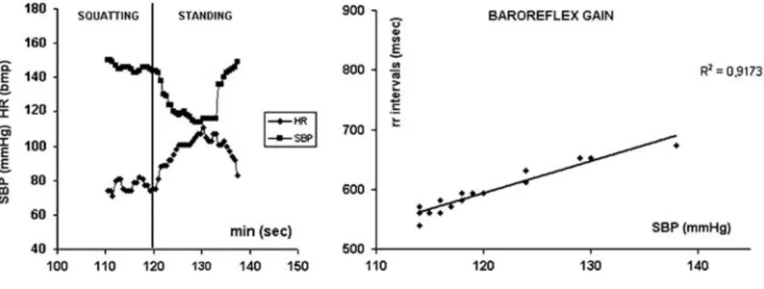
The squatting test may be used as an alternative dynamic procedure to assess the baroreflex gain [8, 38, 39]. During the transition from squatting to standing, there is an abrupt drop in BP associated with a reflex tachycardia, and these initial changes are followed by a rapid return to baseline values of both parameters (secondary BP increase and HR decrease) (Fig. 1). Propranolol markedly attenuated reflex tachycardia at standing from squatting [11]. The large mirror changes in HR and systolic BP during a squatting test allow to calculate a baroreflex gain; indeed, such haemodynamic changes are almost similar to those seen when using a vasodilating drug (hypotension with reflex tachycardia) and a vasoconstricting agent (hypertension associated with bradycardia) [26, 38]. The baroreflex gain was calculated by plotting the pulse intervals (R-R) against systolic BP during the transition phase from squatting to standing (Fig. 2). The slope represents the baroreflex sensitivity [39] and corresponds to changes in pulse intervals per millimetre change in arterial BP. The changes in systolic BP and HR are more abrupt and transient during the squat-stand manoeuvre than during the infusion of a vasodilating/vasopressor pharmacological agent. However, a Japanese study reported that indices of the squatting test show a significant correlation with baroreflex sensitivity assessed by the phenylephrine method in patients with diabetes [38].
Fig. 2 Calculation of the baroreflex gain during a squat-stand test. Mirror image of changes in heart rate (HR)
and systolic blood pressure (BP) during the transition from squatting to standing and illustration of the mode of calculation of the baroreflex gain by plotting RR intervals according to systolic BP
Fig. 3 Reproducibility of baroreflex gain (BRG) during a squat-stand test. Graphic representation according to
Bland and Altman [41] of repeated measures of baroreflex gain (BRG) calculated during a squat-stand test. According to this evaluation, 95% of between-test differences are expected to be less than two standard deviations. There does not appear to be any major relation between the between-test difference and the size of the mean BRG. Please note that there is a clear outlier in our series
A study that investigated the reproducibility of various pharmacological (phenylephrine infusion) and
physiological (Valsalva manoeuvre, spectral analysis) methods to assess baroreflex sensitivity in normal subjects reported quite heterogeneous results [40]. We studied the reproducibility of the measurement of baroreflex gain in 70 subjects who were submitted to two squat-stand tests (unpublished results). We used the recommended Bland and Altman's representation to assess repeatability of the measurement [41] and obtained acceptable results (Fig. ).
We also assessed the baroreflex sensitivity using such a squat-stand test in a large cohort of healthy subjects and we separated the population according to gender, body mass index and age. Baroreflex gain was similar in men and women, confirming previous observations obtained with the pharmacological reference test [42]. However, baroreflex gain was slightly but significantly decreased in overweight/obese subjects versus lean subjects and markedly decreased in elderly versus young individuals, also in agreement with data obtained from the pharmacological test using sequential nitroprusside-phenylephrine infusions [29] (Table 1).
Table 1 Baroreflex gain (BRG) calculated during a squatting test in healthy subjects divided according to gender, body mass index (BMI) and age
Subjects Gender (n, F/M) BMI (kg/m2) Age (years) BRG (ms mmHg-1) P
Female 78/0 22.3 ± 2.8 40 ± 12 3.64 ± 2.31 Male 0/77 25.1 ± 3.1 41 ± 12 3.42 ± 2.27 0.55 Lean 20/20 22.2 ± 1.6 50 ±6 4.03 ± 2.25 Overweigh t 20/20 28.6 ± 2.7 50 ±6 2.85 ± 2.23 0.03 Young 40/38 22.8 ± 2.7 31 ±6 3.63 ± 2.29 Old 22/22 24.7 ± 4.0 69 ±6 2.04 ± 2.24 0.0006
Finally, the changes in baroreflex gain measured during such a squatting test have been used to assess cardiac autonomic neuropathy in diabetic patients [8, 38]. As an example, we previously reported that the baroreflex gain progressively decreased according to the duration of the disease in a population of patients with type 1 diabetes, whereas such reduction was not significant in a control nondiabetic population matched for age [8]. In summary, the ample mirror changes in systolic BP and HR occurring during the initial squat-stand transition mimic those observed during a pharmacological test using vasodilator/vasopressor agents or during the Valsalva manoeuvre. By plotting R-R intervals according to systolic BP changes, a baroreflex gain can be calculated, which may be used to assess cardiovagal baroreflex sensitivity and autonomic function.
LIMITATIONS
There are some limitations of the use of the squat-stand test to calculate baroreflex sensitivity. First, many elderly or disabled individuals have much difficulties or are not capable of maintaining the squatting position, thus not allowing the correct assessment of haemodynamic changes during the squat-stand transition. Second, standardizing method for squatting has been requested, as various groups used different protocols [8, 11, 38]. Third, the calculation of baroreflex gain during a squatting test has not been validated in comparison to the classical pharmacological procedure in healthy volunteers [33, 34], even if a significant correlation between the barosensitivity indices measured by the two methods has been recently reported in diabetic patients [38]. CONCLUSION
Squatting may be used as a dynamic postural test capable of detecting baroreflex sensitivity by measuring continuously BP and HR during the transition from standing to squatting and even more from squatting to standing. Thus, squatting may be considered as an interesting posture in both experimental and clinical situations and a better understanding of the squatting-induced haemodynamic changes may be important in cardiovascular physiology and pathology. The knowledge of changes occurring in healthy subjects may help in analysing the abnormalities in patients with autonomic failure, especially diabetic patients with cardiovascular autonomic neuropathy.
Acknowledgments The authors thank Monique Marchand for her valuable help in the experimental measurements and in the illustrations of this paper.
Conflict of interest None declared.
References
1. Sunagawa K, Sato T, Kawada T (2001) Integrative sympathetic baroreflex regulation of arterial pressure. Ann N Y Acad Sci 940:314-323
2. Monahan KD (2007) Effect of aging on baroreflex function in humans. Am J Physiol Regul Integr Comp Physiol 293:R3-R12
3. Freeman R (2008) Clinical practice. Neurogenic orthostatic hypotension. N Engl J Med 358:615-624
4. Philips JC, Scheen, AT (2011) Squatting test: a posture to study and counteract cardiovascular abnormalities associated with autonomic dysfunction. Auton Neurosci doi:10.1016/j.autneu. 2011.03.001
5. Imholz BP, Wieling W, van Montfrans GA, Wesseling KH (1998) Fifteen years experience with finger arterial pressure monitoring: assessment of the technology. Cardiovasc Res 38:605-616
6. Rossberg F, Penaz J (1988) Initial cardiovascular response on change of posture from squatting to standing. Eur J Appl Physiol Occup Physiol 57:93-97
7. Scheen AJ, Juchmes J, Pochet T (1990) Noninvasive, beat-to-beat, investigation of the effects of posture on arterial blood pressure in diabetic neuropathy. Diab Metab 16:382-384
8. Philips JC, Marchand M, Scheen AJ (2009) Pulse pressure and cardiovascular autonomic neuropathy according to duration of type 1 diabetes. Diabetes Metab Res Rev 25:442-451
9. Sharpey-Schafer EP (1956) Effects of squatting on the normal and failing circulation. Br Med J 1:1072-1074
11. Marfella R, Giugliano D, Dimaro G, Acompora R, Giunta R, Donofrio F (1994) The squatting test—a useful tool to assess both parasympathetic and sympathetic involvement of the cardiovascular autonomic neuropathy in diabetes. Diabetes 43:607-612
12. Brotmacher L (1957) Haemodynamic effects of squatting during repose. Br Heart J 19:559-566
13. Rickards CA, Newman DG (2003) A comparative assessment of two techniques for investigating initial cardiovascular reflexes under acute orthostatic stress. Eur J Appl Physiol 90:449-457
14. Hanson P, Slane PR, Rueckert PA, Clark SV (1995) Squatting revisited: comparison of haemodynamic responses in normal individuals and heart transplantation recipients. Br Heart J 74:154-158
15. Kim KH, Cho JG, Lee KO et al (2005) Usefulness of physical maneuvers for prevention of vasovagal syncope. Circ J 69:1084-1088
16. Murakami T (2002) Squatting: the hemodynamic change is induced by enhanced aortic wave reflection. Am J Hypertens 15:986-988
17. Philips JC, Marchand M, Scheen AJ (2008) Squatting amplifies pulse pressure increase with disease duration in patients with type 1 diabetes. Diabetes Care 31:322-324
18. Lance VQ, Spodick DH (1977) Physiological responses to prompt and sustained squatting. Measurement by systolic time intervals. Br Heart J 39:559-562
19. Lewis BS, Lewis N, Gotsman MS (1980) Effect of standing and squatting on echocardiographic left ventricular function. Eur J Cardiol 11:405-412
20. Rossberg F, Penaz J (1992) Heart rate and arterial pressure variability in humans during different orthostatic load. Physiol Res 41:19-23
21. Scheen AJ, Marchand M, Lefèbvre PJ (1999) Cardiovascular monitoring during tilt and squatting tests in diabetic patients with autonomic neuropathy (Abstract). Diabetologia 42:A297
22. Smith JJ, Porth CM, Erickson M (1994) Hemodynamic response to the upright posture. J Clin Pharmacol 34:375-386
23. Alimi YS, Barthelemy P, Juhan C (1994) Venous pump of the calf—a study of venous and muscular pressures. J Vase Surg 20:728-735
24. Tanaka H, Sjoberg BJ, Thulesius O (1996) Cardiac output and blood pressure during active and passive standing. Clin Physiol 16:157-170
25. Berry NM, Rickards CA, Newman DG (2006) Squat-stand test response following 10 consecutive episodes of head-up tilt. Aviat Space Environ Med 77:1125-1130
26. La Rovere MT, Pinna GD, Raczak G (2008) Baroreflex sensitivity: measurement and clinical implications. Ann Noninvasive Electrocardiol 13:191-207
27. Parati G, Di Rienzo M, Mancia G (2000) How to measure baroreflex sensitivity: from the cardiovascular laboratory to daily life. J Hypertens 18:7-19
28. Pickering TG, Gribbin B, Sleight P (1972) Comparison of the reflex heart rate response to rising and falling arterial pressure in man. Cardiovasc Res 6:277-283
29. Rudas L, Crossman AA, Morillo CA et al (1999) Human sympathetic and vagal baroreflex responses to sequential nitroprus-side and phenylephrine. Am J Physiol 276:H1691-H1698
30. Lipman RD, Salisbury JK, Taylor JA (2003) Spontaneous indices are inconsistent with arterial baroreflex gain. Hypertension 42:481-487
31. Diaz T, Taylor JA (2006) Probing the arterial baroreflex: is there a 'spontaneous' baroreflex? Clin Auton Res 16:256-261
32. Palmero HA, Caeiro TF, Iosa DJ, Bas J (1981) Baroreceptor reflex sensitivity index derived from Phase 4 of the Valsalva maneuver. Hypertension 3:11-134—137
33. Goldstein DS, Horwitz D, Keiser HR (1982) Comparison of techniques for measuring baroreflex sensitivity in man. Circulation 66:432-439
34. Milic M, Sun P, Liu F et al (2009) A comparison of pharmacologic and spontaneous baroreflex methods in aging and hypertension. J Hypertens 27:1243-1251
35. Zollei E, Paprika D, Rudas L (2003) Measures of cardiovascular autonomic regulation derived from spontaneous methods and the Valsalva maneuver. Auton Neurosci 103:100-105
36. Schrezenmaier C, Singer W, Swift NM, Sletten D, Tanabe J, Low PA (2007) Adrenergic and vagal baroreflex sensitivity in autonomic failure. Arch Neurol 64:381-386
37. Looga R (2005) The Valsalva manoeuvre-cardiovascular effects and performance technique: a critical review. Respir Physiol Neurobiol 147:39-49
38. Nakagawa M, Shinohara T, Anan F et al (2008) New squatting test indices are useful for assessing baroreflex sensitivity in diabetes mellitus. Diabet Med 25:1309-1315
39. Zhang R, Claassen JA, Shibata S et al (2009) Arterial-cardiac baroreflex function: insights from repeated squat-stand maneuvers. Am J Physiol Regul Integr Comp Physiol 297:R116-R123
40. Lord SW, Clayton RH, Hall MC et al (1998) Reproducibility of three different methods of measuring baroreflex sensitivity in normal subjects. Clin Sci (Lond) 95:575-581
41. Bland JM, Altaian DG (1986) Statistical methods for assessing agreement between two methods of clinical measurement. Lancet 1:307-310
42. Tank J, Diedrich A, Szczech E, Luft FC, Jordan J (2005) Baroreflex regulation of heart rate and sympathetic vasomotor tone in women and men. Hypertension 45:1159-1164

![Fig. 3 Reproducibility of baroreflex gain (BRG) during a squat-stand test. Graphic representation according to Bland and Altman [41] of repeated measures of baroreflex gain (BRG) calculated during a squat-stand test](https://thumb-eu.123doks.com/thumbv2/123doknet/6115041.155591/6.892.113.653.217.506/reproducibility-baroreflex-graphic-representation-according-repeated-baroreflex-calculated.webp)
