0 FEBS 1994
Stability and structural analysis
of
or-amylase
from the antarctic psychrophile
Alteromonas haloplanctis A23
Georges FELLER', Franqoise PAYAN', Fabienne THEYS l , Minxie QIAN2, Richard HASER' and Charles GERDAY
Laboratory of Biochemistry, Institute of Chemistry B6, University of Lihge, Belgium
Laboratoire de Cristallographie et Cristallisation des Macromolecules Biologiques, Centre National de la Recherche Scientifique, FacultC de MCdecine Nord, Marseille, France
(Received February 14March 21, 1994) - EJB 94 0202/3
The a-amylase secreted by the antarctic bacterium Alteromonas haloplanctis displays 66%
amino acid sequence similarity with porcine pancreatic a-amylase. The psychrophilic a-amylase is however characterized by a sevenfold higher k,,, and k,,/K,,, values at 4°C and a lower conforma-
tional stability estimated as 10 kJ
.
mol-' with respect to the porcine enzyme. It is proposed that both properties arise from an increase in molecular flexibility required to compensate for the reduction of reaction rates at low temperatures. This is supported by the fast denaturation rates induced by temperature, urea or guanidinium chloride and by the shift towards low temperatures of the apparent optimal temperature of activity.When compared with the known three-dimensional structure of porcine pancreatic a-amylase, homology modelling of the psychrophilic a-amylase reveals several features which may be assumed to be responsible for a more flexible, heat-labile conformation: the lack of several surface salt bridges in the @/a)* domain, the reduction of the number of weakly polar interactions involving an aromatic side chain, a lower hydrophobicity associated with the increased flexibility index of amino acids forming the hydrophobic clusters and by substitutions of proline for alanine residues in loops connecting secondary structures. The weaker affinity of the enzyme for Ca2+ (Kd = 44 nM) and for C1F (Kd = 1.2 mM at 4°C) can result from single amino acid substitutions in the Ca2+-binding and CIF-binding sites and can also affect the compactness of a-amylase.
The physical basis of the forces driving the folding of a polypeptide chain and maintaining the final structure remains only understood in general terms. Accordingly, the relative contributions of the determinants of protein stability, mainly the various weak electrostatic interactions and the hydropho- bic effect, are still under debate (Creighton, 1991). Amongst the several approaches currently used, the analysis of pro- teins from extremophiles provides valuable insights on the molecular strategies adopted in response to environmental stress such as extremes of pH, pressure, salinity or temper- ature (Zuber, 1988 ; Jaenicke, 1991). Enzymes from thermo- philic microorganisms for example seem stabilized by strengthening one or a combination of noncovalent interac- tions making use of a small number of amino acid replace- ments (Fontana, 1991; Jaenicke, 1991).
By contrast, psychrophilic microorganisms persisting at low temperatures have received little attention. Enzymes from these organisms are characterized by marked heat labil- ity which has been commonly correlated with environmental conditions (Ushakov, 1964). However, enzymes from psy- chrophiles also display high specific activity which is the Correspondence to G. Feller, Laboratory of Biochemistry, Insti- tute of Chemistry B6, University of Lihge, B-4000 Likge, Belgium
Fax: +32 41563364.
Abbreviations. Np(Glcp),OCH,CH'+, 4-nitrophenyl-a-~-malto-
Enzymes. a-Amylase or 1,4-a-D-glUCan glucanohydrolase (EC heptaoside-4,6-O-ethylidene ; GdmC1, guanidinium chloride.
3.2.1.1).
main property for enzyme adaptation to catalysis at low envi- ronmental temperatures. Hochachka and Somero (1984) have argued that enzymes which can readily undergo conforma- tional changes during catalysis at low temperature are capa- ble of supplying most of the energy for activation events and can reduce the activation energy barrier of their chemical reactions. Thermal instability is regarded as the consequence of the highly flexible structure of cold-active enzymes. Psy- chrophilic enzymes that have evolved towards high confor- mational flexibility and catalytic efficiency therefore repre- sent appropriate candidates for protein-stability analysis. Cloning of genes from psychrophilic bacteria in Escherichia coli results in the expression of thermolabile recombinant
enzymes active at temperatures close to 0 "C demonstrating the intrinsic character of these properties (Feller et al., 1991; Davail et al., 1992; Rentier-Delrue et al., 1993). In most cases however, the analysis of psychrophilic protein se- quences has been impaired by insufficient isology with their mesophilic counterparts.
We have previously reported the characterization and the nucleotide sequence of the a-amylase secreted by a Gram- negative antarctic bacterium Alteromonas haloplanctis
(Feller et al., 1992). In this study, we report typical stability parameters of A. haloplanctis and porcine pancreatic a-amy- lases. A model of the heat labile a-amylase has been con- structed on the basis of the three-dimensional structure of the porcine enzyme recently solved at 0.21 -nm resolution (Qian et al., 1993).
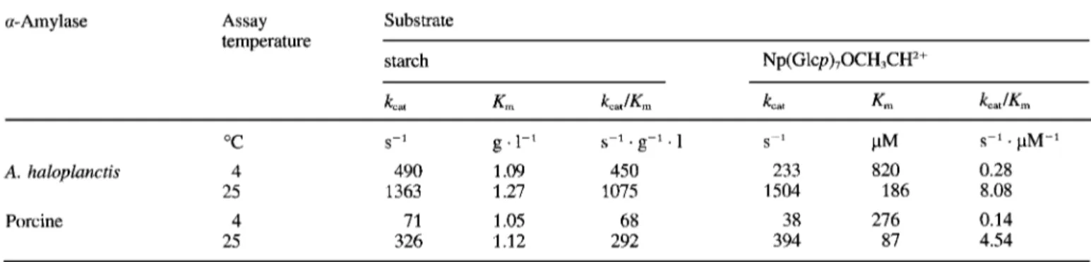
Table 1. Kinetic parameters for the amylolytic activity of A. haloplanctis and porcine pancreatic a-amylases. Linear reaction rates were recorded in 100 mM Hepes, 50 mM NaC1, 10 mM MgC12, pH 7.1. Assays using Np(Glcp)70CH3CH2+ as substrate also contained 23 U/ml a- glucosidase as coupling enzyme. Substrate concentrations were varied from 0.4-4 X K,. k,,, values for starch are calculated on the basis of
maltose released. Values are the mean of at least three determination sets. SE for k,,, and K , was S 5 % [ N ~ ( G ~ c ~ ) ~ O C H , C H ~ + ] and G8% (starch).
a-Amylase Assay Substrate
temperature
starch Np(Glcp),OCH,CH'+
k,, Km kcaJKm kcat Km kcatlK, "C 25 Porcine 4 25 A. haloplanctis 4 s-1
.
pM-1 S-' g .1-' s-'.
g-1 . 1 S-' PM 490 1.09 450 233 820 0.28 1363 1.27 1075 1504 186 8.08 71 1.05 68 38 276 0.14 326 1.12 292 394 87 4.54MATERIALS AND METHODS Sources
The heterotrophic aerobic strain A. haloplanctis A23 was isolated near the Dumont d'Urville antarctic station (60'40' S, 40"Ol'E). Porcine pancreatic a-amylase was from Sigma (A6255).
Purification of A. haloplanctis a-amylase
The antarctic strain was cultivated at 4°C for 3 days in 3.5 1 broth containing 10 g/l bactotryptone, 5 g/l yeast ex- tract, 20 g/l NaC1, 10 g/l sea salts, 20 g/l maltose, pH 7.6, vigorously aerated by air bubbling. After centrifugation at 11 000 g, the culture supernatant was adjusted to 0.02% NaN3, concentrated to 400 ml and diafiltrated against 50 mM Tris/HCl, 1 mM CaCl,, pH 7.5, using a Minitan tangential flow ultrafiltration unit (Millipore) fitted with PTGC mem- branes (10-kDa retention limit). The sample was loaded on a DEAE-agarose column (2.5 cmX40 cm) equilibrated in the above mentioned buffer and eluted with a NaCl linear gradient (500 ml each starting buffer, 0-0.8 M NaC1). Fractions containing amylolytic activity were concentrated to 10 ml and applied onto a Sephadex G-100 column (2.5 cmX 100 cm) eluted with 50 mM Tris/HCl, 1 mM CaCl,, pH 7.0, followed by gel filtration on an Ultrogel AcA 54 column (2.5 cmX 100 cm) eluted with the same buffer. For further experiments, the purified a-amylases were condi- tioned in the appropriate buffers by gel filtration on a PD-10 column (Pharmacia).
Enzyme assay
The standard assay was carried out at 25°C with the a-
amylase EPS kit (Boehringer) using 3.5 mM 4-nitrophenyl-
a-~-maltoheptaoside-4,6-O-ethylidene [Np(Glcp),0CH3CHZ+] as substrate and excess (23 U/ml) of a-glucosidase as cou- pling enzyme in 1 O O m M Hepes, 50mM NaCl, 1OmM MgCl,, pH 7.1. Activities towards the synthetic substrate were recorded in a thermostated Uvicon 860 spectrophotometer (Kontron) and values were calculated using an absorption co- efficient for 4-nitrophenol of 8980 M-'
.
cm-' at 405 nm (Raucher et al., 1985). The amylolytic activity was also deter- mined by the dinitrosalicylic acid method (Bernfeld, 1955) using 1% soluble starch (Sigma) as substrate in the above- mentioned buffer.Analytical procedures
The enzyme concentrations were determined spectropho- tometrically by using Aol" = 2.41 at 280 nm for the porcine a-amylase (Levitzki and Steer, 1974) and A''" = 1.90 at 280 nm for the bacterial a-amylase. The kinetic parameters k,,
and K , were determined by the initial velocity method using a nonlinear regression computer fit of the saturation curves.
Cl--free a-amylase was prepared by gel filtration on a PD- 10 column eluted with 25 mM Hepes/NaOH, pH 7.0. The dissociation constants for C1- were calculated from computer- fitted activation curves generated by NaCl titration in the above-mentioned reaction mixture except that the buffer was replaced by 25 mM Hepes/NaOH, pH 7.0. Apo a-amylase (Cl-, Ca2+-free) was prepared by overnight dialysis of the na- tive enzyme against 25mM Hepes/NaOH, 5 mM EGTA, pH 8.0. Activation kinetics obtained by calcium titration were performed in 25mM HepesBaOH, 50mM NaC1, 5 m M EGTA, 3.5 mM Np(Glcp)70CH,CH2+, 23 U/ml a-glucosidase, pH 8.0. The desired free Ca2+ concentration was set by addi- tion of 50 mM CaC1, according to a program developed by Robertson et al. (1982). The data points were computer fitted according to the Hill equation.
Guanidinium chloride (GdmCl) denaturation curves were recorded according to Pace et al. (1989). Enzymes (36 pg/ml) were subjected to increasing GdmCl concentrations in 30 mM Mops, pH 7.0. Following 4 h incubation at 25"C, the fluores- cence intensity of the samples was recorded using a Perkin Elmer LS 50 spectrofluorimeter at an excitation wavelength of 280 nm. Emission wavelengths were 340 nm and 347 nm for the porcine and A. haloplanctis a-amylases respectively.
Molecular modelling
The psychrophilic a-amylase model was constructed from the porcine pancreatic a-amylase coordinates using the pro- gram TURBO-FRODO (Roussel and Cambillau, 1990). Few insertions were operated and deletions mainly affected surface loops between Ap3 and Bfl, between Aa4 and A/35 and be- tween Ap8 and Aa8. Amino acid replacements were generated in the low energy conformation. Energy minimization and low-temperature molecular dynamics were carried out using X-PLOR (Briinger et al., 1987).
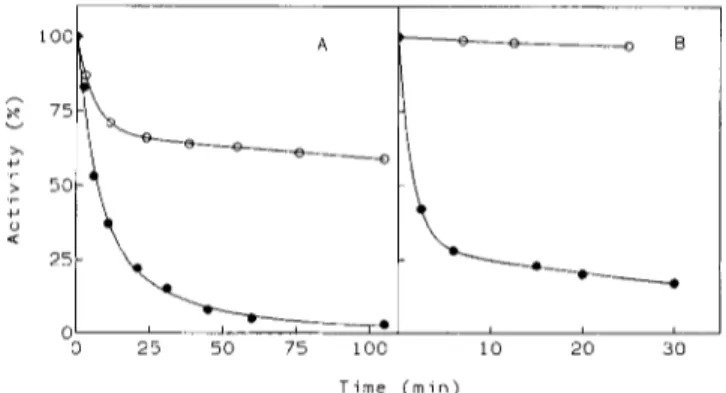
I -1 100- 75- v x +J -L 50- .r Y u a 15 30 45 60 75 0
I / ,
, , , . , , , , , ,, j
0 T e m p e r a t u r e ('C)Fig. 1. Effect of assay temperature on amylolytic activity. Specific activities of A. huloplunctis (0) and porcine pancreatic (0) a-amy- lases were recorded at increasing temperatures using 3.5 mM
Np(Glcp),OCH,CH*+ as substrate under standard assay conditions. Reaction rates are reported relative to the maximal activity recorded.
RESULTS
Thermodependence of enzyme activity
The kinetic parameters k,,, K , and the physiological effi-
ciency k,,lK, for the amylolytic activity of the bacterial and
the porcine a-amylases are given in Table 1. The k,,, of the
psychrophilic a-amylase towards the natural substrate at 4 "C is seven times the value displayed by the porcine enzyme. As a result of similar K , values for starch in both enzymes, the k,,,lK, ratio is also more favourable to A. haloplanctis a-amy-
lase. The same behaviour is recorded using the synthetic sub- strate Np(Glcp)70CH,CH2+, although the Michaelis constants of A. haloplanctis a-amylase are larger. The thermodependen-
cies of the amylolytic activity of both enzymes are shown in Fig. 1. The drastic shift of the apparent optimal temperature of activity (from 50°C for the porcine enzyme to 30°C for the bacterial enzyme) indlcates that thermally induced alterations of the catalytic mechanism occur at moderate temperatures in the case of the psychrophilic a-amylase.
a-Amylase stability
The thermal stability of the amylolytic activity of both a-
amylases is shown by the denaturation curves at 50°C (Fig. 2A). The half time of inactivation (tJ for the A. ha- loplanctis protein is approximately 20 times lower than for the porcine a-amylase. The difference in free energy of stabiliza- tion between both enzymes, estimated by the relation AAG =
2.3RTlog(t,,It,,), amounts to 8
kJ
.
mol-'. As shown in Fig. 2B, urea also induces a very fast inactivation of A. ha- loplanctis a-amylase. The GdmCl denaturation curves of both a-amylases monitored by intrinsic fluorescence are shown in Fig. 3. As the denaturant concentration increases, the fraction of native porcine enzyme molecules decreases according to a biphasic decay; the transition step can arise from the unfolding of different domains. The concentration for half denaturation [GdmC1],,2 is 1.4 M. In the case of A. haloplanctis a-amylase, GdmCl induces a fast denaturation of the protein structure with a [GdmCl],, value of 0.8 M. The curve only displays a minor deviation corresponding to the transition step of the porcine protein indicative of a less accentuated resistance of the bacterial a-amylase structure to GdmC1. The difference in conformational stability between the psychrophilic and the porcine a-amylases has been estimated by the relation AAG =T i m e ( m i n )
Fig.2. Thermal stability (A) and urea denaturation (B) of the amylolytic activity. (A) A. haloplunctis ( 0 ) and porcine pancreatic (0) a-amylases were incubated at 50°C in 50 mM Tris/HCl, 1 mM CaCl,, pH 7.0, and residual activities were recorded under standard assay conditions using 3.5 mM Np(Glcp),OCH,CHZ+ as substrate. (B)
A. huloplunctis and porcine pancreatic a-amylases (symbols as in A)
were incubated at 25°C in the above mentioned buffer including 3.2 M urea and residual activities were recorded under standard assay conditions.
"?.,
-
::I
v LL 4 0 'c 2 0 0 0 1 2 3 [ G d m C l ] ( M )Fie. 3. Guanidinium-chloride-induced unfolding
I
curves. The fol;ded fractionf, of A. haloplunctis ( 0 ) and porcine pancreatic (0) a-amylases is plotted for increasing GdmCl concentrations. Fluores- cence intensities of a-amylases (36 pg/ml) were recorded at 25°C in 30 mM Mops, pH 7.0. The fraction of protein in the folded state fp
was calculated using fF = y-y,,/yn-yo, where yN and yo are the fluo-
rescence intensities of the native state and the fully denatured state respectively; y is the fluorescence intensity at a given GdmCl concen- tration.
A([GdmCl],,2)m,, where ma is the average value of the slopes
from plots of [GdmCl] versus the free energy of unfolding, AG, = AG,,,-m[GdrnCl] (Pace et al., 1989). This difference in conformational stability amounts to 10
kJ
.
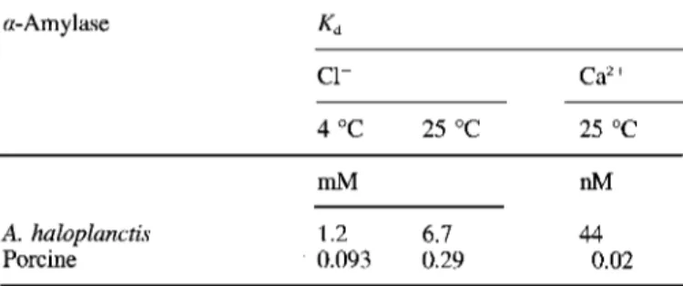
molF and is in reasonable agreement with the thermal inactivation estimate.Dissociation constants
Removal of either Ca2+ or C1- results in the reversible inactivation of A. haloplanctis a-amylase. This allows the de-
termination of the dissociation constants, Kdr by activation
ki-
netics following calcium or chloride titration. Fig. 4 shows the allosteric activation by these ions. The deduced Kd values are
compared in Table 2 with those previously determined for the pancreatic a-amylase. The binding affinity for both ions is
p C a 2 + N a C l ( m M )
Fig. 4. Activation of A. haloplanctis a-amylase by Caw and C1-.
(A) Calcium titration of the &'+-free enzyme in the presence of
5 mM EGTA and chloride; pCa2+ = -log[Ca2+]. (B) Chloride titra- tion of the Cl--free enzyme at 4°C (0) and 25°C (W).
Table 2. Dissociation constants of the Clb-amylase and the CaY amylase complexes. Kd data from A. haloplanctis a-amylase are de-
rived from activation kinetics experiments shown in Fig. 2. SE for Kd was S 5 % (Cl-) and 12% (Ca"). Kd data for porcine pancreatic a-
amylase are taken from Levitzki and Steer (1974).
a-Amylase Kd c1- Ca2+ 4 "C 25 "C 25 "C mM nM A. haloplanctis 1.2 6.7 44 Porcine 0.093 0.29 0.02
significantly lower in the psychophilic a-amylase as indicated by a 10-20-fold increase of the Kd value for C1- and a 2000- fold higher K,-value for Ca2+.
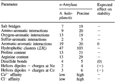
A. haloplanctis a-amylase structural model analysis
Amino acid sequences of A. huloplunctis and porcine pan- creatic a-amylases can be aligned with 53% positional iden- tity. This value increases to 66% when considering amino acid residues of the same type (Fig. 5). Conserved amino acids oc- cur essentially in the secondary structures of the porcine en- zyme and in regions bearing functional residues pertaining to the active site as well as to the ion-binding sites. This favoura- ble degree of identity allowed the building of a model of the three-dimensional structure of the psychrophilic a-amylase. The predicted molecular architecture follows the pattern of known a-amylase structures (Swift et al., 1991; Qian et al., 1993). The enzyme is made of a central barrel (domain A), a P-pleated domain B and a globular C-terminal domain C. Amino acids with their side chain directed towards the active site, amongst which Glu233, Asp300 and Asp197 are the main catalytic residues, are conserved suggesting a very close reaction mechanism in both enzymes. The structural model of A. huloplanctis a-amylase was analyzed in order to identify the weak interactions and the structural features potentially involved in the structure flexibility of the psychro- philic enzyme. The results are summarized in Table 3.
10 20 30 40 50
********TPTTFVHLFEWNWQDVAQECEQYLGPKGYAAVQVSPPNEHI**T*GS QYAPQTQSGRTSIVHFEWRWVDIALECERYLGPKGFGGVQVSPPNENVVVTNPS
A81 Aa 1 AB2a AB2b
60 70 80 90 100 110
O*WWTRYQPVSYELQSRGGNRAQFIDMVNRCSAAGVDIYVDTLINHM***AAGSG RPWWERYQPVSY KLCTRSGNENEFRDMVTRCNNVGVRIYVDAVI NHMCGSGAAAG
Aa2 A03 BB1 120 130 140 150 160 TGTAGNSFGN***KSFP*I*YSPQDFHES*C*T*INNSDYGNDRYRVQNCELVGL
TGTTCGSYQJGSWAVPY
S A W D F N D G K C K T A S ~ E S Y N D P Y Q V R D C Q L V ~ BC2 883 884 885 BB6 8137 B a l BB8 170 180 190 200 210 220 ADLDTASNYVQNTIAAYINDLQAIGVKGFRFDASKHVAASDIQSLMAK***VN** LDLALEKDYVRSMIADYLNKLIDIGVAGFRIDASKHMWPGDIKAVLDKLHNLNTN 8139 A a 3 A 0 4 A a 4 230 240 250 260 270 ****GS*PVVFQEVIDQGGEAVGASEYLSTGLVTEFKYSTELGNTFRNGSLA*** W F P A G S R P ~ V I D L G G E A I S S S E Y F G N G R V T E F K Y G A K L G T V V R K W S G E ~ AB5 A3105 A 0 6 A a 6 a 280 290 300 310 320 WLSNFGEGWGFMPSSSAVVFVDNHDNQRGHGGAGNVI*TFEDGRLYDLANVFMLA WGEGWGFMPSDMFVDNHDNQRGHGAGGSSILTFWDARLYKVAVGFMLAA3106b AB7 ACl7
340 350 360 370 380 YPYGYPKVMSSYDFHGDTDAGGP*N**V*PVHNNG*******NLE**CFASNWKC - HPYGFTRVMSSYRWARNFVNGEDVNDWIGPPNNNGVIKEVTINADTTC*GNDWVC A 0 8 390 400 410 420 430 EHRWSYIAGGVDFRNNTADNWAVTNWWDNTNNQISFGRGSSGHMAINKEDSTLTA EHRWREIRNMVWFRNV*VDGQPFANWWDNGSNQVAFGRGNRGFIVFNNDDWQ~ A a 8 CBl CC2 CB3 450 460 4 70 480 490 TVQTDMASGQYCNVLKGELSADAKSCSGEVITVNSDGTINLNIG**AWDAM*AIH TLQTGLPGGTYCDVISGW*GNSCTGIKVYVSSDGTAQFSISNSAEDPFIAIH CB4 CR5 CB6 CB7 CB8 CB9 CC10 KNAKLNTSSAS 453 AHA - AESKL****** 496 PPA
Fig. 5. Amino acid sequence alignment of A. haZoplanct?s and por- cine pancreatic a-amylases. Residues are numbered according to the porcine a-amylase sequence with no number for inserted residues and no number shift for deleted residues. The secondary structures of the porcine enzyme identified by Qian et al. (1993) are underlined; de- leted or inserted residues are indicated (*). 238 amino acids of A. haloplanctis a-amylase are conserved in the porcine enzyme. When considering residues of the same type (G = A = S, S = T, L = I =
V, F = W = Y, E = D = R = K, Q = N), 301 amino acid residues are conserved.
Disulfide bonds
A. haloplunctis a-amylase contains eight cysteine residues.
Sulfhydryl titration by 5,5'-dithiobis-(2-nitrobenzoic acid) of both the native and the denatured enzyme in 8 M urea indi- cates that there is no free thiol group. Since the positions of these cysteine residues correspond to residues forming disul- fide linkage in the porcine a-amylase, four conserved disulfide bonds are proposed for the bacterial enzyme (28-86, 141 -
160, 378-384,450-462).
DISCUSSION
The k,, and k,,lK,,, values of the a-amylase excreted by
A. huloplunctis are larger than the values determined for the porcine enzyme over a temperature range of 0°C to at least 25°C. This increase largely compensates for the reduction of the reaction rates occurring at low temperatures since both k,, and k,,lK, values for starch of the psychrophilic a-amylase at 4°C are higher than the corresponding values of the pancreatic
Table 3. Structural parameters potentially involved in the stability of a-amylase conformation. - , denotes an expected decrease of sta- bility; (), low or undefined effect. ZH,, sum of the hydrophobicity
values Hi from the PRIFT scale (Comette et al., 1987) for the 25 substitutions in the hydrophobic clusters of A. haloplunctis and por-
cine pancreatic a-amylases.
Parameter a - A m y 1 as e
A. halo- Porcine stability
planctis Expected effect on Salt bridges Amino-aromatic interactions Oxygen-aromatic interactions Sulfur-aromatic interactions Aromatic-aromatic interactions Hydrophobic clusters (ZH,) Proline content Arginine content Disulfide bonds
Helices dipoles - charges at Na Helices dipoles
+
charges at Ca Caz+ affinity C1- affinity 7 9 13 2 19 47 13 13 4 2 3 low lowenzyme at 25°C. Hochachka and Somero (1984) have pro- posed that this catalytic efficiency can be gained by an appro- priate folding flexibility. A loose conformation of the psychro- philic a-amylase is indeed suggested by the susceptibility of secondary structures to unfolding at moderate temperatures (Feller et al., 1992), by the fast denaturation rates induced by temperature, urea or GdmCl and by the shift of the apparent optimal temperature of activity. The high dissociation con- stants for Ca” and C1- possibly also reflect a less compact protein structure.
Ca2+-binding and C1--binding sites
Calcium coordination in porcine pancreatic a-amylase oc- curs through a pentagonal bipyramid involving the carboxyl oxygens of Asp167 and the carbonyl oxygen of His201 at the apices, three water molecules, of AsnlOO and the main- chain carbonyl of Arg158. The latter is replaced by Gln158 in
A. haloplanctis a-amylase whereas the other Ca2+ ligands are
conserved. As Argl58 in the porcine enzyme coordinates the Ca” ion via the main-chain carbonyl, there is apparently no drastic modification of the A. haloplanctis a-amylase binding site that would account for its 2000 times lower binding affin- ity. In the porcine enzyme however, Argl58 forms a salt bridge with Glu246 and, as a result of its charge resonance structure, also forms an amino-aromatic interaction with Trp203 (alanine in A. haloplanctis a-amylase). The three resi-
dues are conserved in all mammalian a-amylases. As these interactions are absent in the bacterial enzyme, Gln158 is less f i i y oriented in the Ca2+-binding site. The Ca” of a-amy- lases bridges a cleft between domains A and B forming the active site (Qian et al., 1993). If the high specific activity of the psychrophilic a-amylase is a consequence of a more flexi- ble active site, then the substitution Argl58+Gln is certainly of importance through its effect of weakening interdomain contacts around the catalytic cleft.
The chloride-binding site of the pancreatic a-amylase is composed by Arg195 (N@), Asn298 (N”) and the side-chain amine of Arg337 coordinating C1- in a bidentate mode. This chloride-binding site is conserved in all mammalian a-amy-
lases but differs in A. haloplanctis by a lysine residue instead of Arg337. As lysine can only provide an unidentate coordina- tion, this substitution can account for the 10-20-fold lower Cl--binding affinity of the psychrophilic a-amylase.
Calcium protects a-amylases against heat inactivation (Vihinen and MZntsAa, 1989) and we have found that chloride is required for optimal stability of A. haloplunctis a-amylase (Feller et al., 1992). It follows that the low Ca2+-binding and Cl--binding affinities are additional factors leading to a low stability. In thermophiles, stabilization by tight calcium bind- ing is well documented and involves either unusual high affin- ity or even extra Ca2’-binding sites (Teplyakov et al., 1990). No equivalent Cl--binding site has yet been described in ther- mophiles.
Conformational stability of the psychrophilic enzyme The various structural factors potentially promoting flexi- bility of A. haloplanctis a-amylase (Table 3 ) occur essentially in the domain A but also affect interactions between the three domains. When compared with the crystal structure of the pancreatic enzyme, the model of the psychrophilic a-amy- lase is characterized by the lack of 12 surface salt bridges. In most cases, this difference arises from the replacement of the basic residue of the pair by a glutamine residue or an aspara- gine residue. Weakly polar interactions were analyzed using the angles and interatomic distance parameters given by Bur- ley and Petsko (1988). As shown in Table 3, the number of amino-, oxygen-, and sulfur-aromatic interactions is lower in the bacterial protein. By contrast, the strong conservation of aromatic-aromatic interactions certainly reflects their essential function in the conformation of both enzymes. However, ther- mophilic enzymes and the thermostable a-amylase from Bacil-
lus lichenifomis are protected against irreversible thermoinac-
tivation through rigidification of the molecule by additional salt bridges (Perutz and Raidt, 1975; Tomazic and Klibanov, 1988) and weakly polar interactions (Teplyakov et al., 1990). Amongst the 84 residues forming the hydrophobic clusters of the porcine a-amylase (Raimbaud et al., 1989), 25 subsitu- tions were noted in the corresponding positions of A. ha-
loplanctis a-amylase. 80% of these substitutions are accompa-
nied by a sharp decrease of the hydrophobicity index using either the statistical PRTFT scale or the experimental scale of Aboderin (see Cornette et al., 1987 for normalized values). In addition, 72% of these substitutions increase the main-chain flexibility parameter derived by Karplus and Schultz (1985) from the crystallographic temperature factor of individual amino acids. Assuming that urea and GdmCl act mainly by weakening hydrophobic interactions, their strong denaturing effect on A. haloplanctis a-amylase also suggests a low hydro- phobicity of the core clusters. Conversely, several studies (Mozhaev et al., 1988), especially a study on the a-amylase of
Bacillus stearothermophilus (Brosnan et al., 1992) support a reinforcement of hydrophobic interactions in proteins from thermophiles.
The psychrophilic enzyme has the lowest proline content found in a-amylases. The sequence aligment (Fig. 5 ) shows that 9 proline residues are conserved in both enzymes whereas 12 proline residues are either deleted or substituted by small amino acids such as alanine. These substitutions/deletions mainly occur within loops or turns and favour the flexibility of the chain connecting adjacent secondary structures. Indeed, the thermostability of oligo-l,6-glucosidase from Bacillus
thermoglucosidasius has been correlated with the presence of
pyrrolidine ring severely restricts the available dihedral angles of the preceding residue and decreases the backbone entropy of unfolding. A. haloplanctis a-amylase also possesses a low arginine content. Since the charge resonance of the guanidi- nium group gives arginine the possibility to form more than one electrostatic bond (Mrabet et al., 1992), an increased argi- nine content has been correlated with heat stability in thermo- philic enzymes (Merkler et al., 1981).
The disulfide bond Cys70-CysllS is absent in A. ha-
loplanctis a-amylase. However, mesophilic a-amylases from Aspergillus oryzae (Swift et al., 1991) and Streptomyces limo-
sus (Long et al., 1987) are also devoid of this disulfide linkage which does not seem of crucial importance for stability. Nega- tively charged side chains at the N-terminal first turn of a
helices and positively charged side chains at the C-terminal last turn of a helices are considered as stabilizing factors (Shoemaker et al., 1987; Rentier-Delrue et al., 1993). A. ha- loplanctis a-amylase contains less favourable charge-dipole in-
teractions than the porcine enzyme, but unfavourable charge- dipole interactions are numerous in both a-amylases so that no definitive conclusion can be drawn.
In conclusion, the psychophilic enzyme alters several weak interactions in order to gain structural flexibility. Inter- estingly, enzymes from thermophiles reinforce the same type of interactions indicating that there is a continuum in the strat- egy of protein adaptation to temperature. One can reasonably assume that the lower conformational stability of A. ha-
loplanctis a-amylase (= 10 kJ
.
mol-’) is achieved by the net balance between exothermically formed weak bonds (electro- static, stabilized by a decrease of temperature) and endother- mically formed interactions (hydrophobic, destabilized at low temperature). The determination of the three-dimensional structure of A. haloplanctis a-amylase and the construction of site-directed mutants are now required for a detailed analysis of the adaptation parameters suggested by molecular model- ling.This study was supported by the Fonds de la Recherche Fonda- mentale et Collective research contract 2.452692 to C. G. We are also
grateful to the Institut Frangais de la Recherche Polaire for generous support and accomodation of research fellows at the antarctic base J-
S Dumont d’Urville and to N. Gerardin-Otthiers and R. Marchand for
expert technical assistance.
REFERENCES
Bemfeld, P. (1955) Amylases, a and /3, Methods Enzymol. 1, 149- 151.
Brosnan, M. P., Kelly, C. T. & Fogarty, W. M. (1992) Investigation
of the mecanisms of irreversible thermoinactivation of Bacillus
stearothennophilus a-amylase, Eur: J. Biochem. 203, 225-231.
Briinger, A. T., Kuriyan, J. & Karplus, M. (1987) Crystallographic R factor refinement by molecular dynamics, Science 235, 458-460. Burley, S. K. & Petsko, G. A. (1988) Weakly polar interactions in
proteins, Adv. Protein Chem. 39, 125-189.
Comette, J. L., Cease, K. B., Margalit, H., Spouge, J. L., Berzofsky, J.
A. & DeLisi, C. (1987) Hydrophobicity scales and computational techniques for detecting amphipathic structures in proteins, J. Mol. Biol. 195, 659-685.
Creighton, T. E. (1991) Stability of folded conformations, Curr: Opin.
Struct. Biol. I , 5 - 16.
Davail, S., Feller, G., Narinx, E. & Gerday, C. (1992) Sequence of the subtilisin-encoding gene from an antarctic psychrotroph Bacillus
TA41, Gene (Amst.) 119, 143-144.
Feller, G., Thiry, M. & Gerday, C. (1991) Nucleotide sequence of the lipase gene lip2 from the antarctic psychrotroph Moraxella TA144 and site-specific mutagenesis of the conserved serine and histidine residues, DNA Cell Biol. 10, 381 -388.
Feller, G., Lonhienne, T., Deroanne, C., Libioulle, C., Van Beeumen, J. & Gerday, C. (1992) Purification, characterization, and nucleo- tide sequence of the thermolabile a-amylase from the antarctic psychrotroph Alteromonas haloplanctis A23, J. Biol. Chem. 267,
5217-5221.
Fontana, A. (1991) How nature engineers protein (thermo)stability, in
Life under extreme conditions: biochemical adaptations (di Prisco,
G., ed.) pp. 89- 113, Springer Verlag, Berlin-Heidelberg. Hochachka, P. W. & Somero, G. N. (1984) Biochemical adaptations,
Princeton University Press, Princeton, NJ.
Jaenicke, R. (1991) Protein stability and molecular adaptations to ex- treme conditions, Eul: J. Biochem. 202, 715-728.
Karplus, P. A. & Schulz, G. E. (1985) Prediction of chain flexibility in proteins, Natunvissenschaften 72, 212-213.
Levitzki, A. & Steer, M. L. (1974) The allosteric activation of mam- malian a-amylase by chloride, Eur: J. Biochem. 41, 171-180.
Long, C. M., Virolle, M. J., Chang, S. Y., Chang, S. & Bibb, M. J. (1987) a-Amylase gene of Streptomyces limosus: nucleotide
sequence, expression motifs, and amino acid sequence homology to mammalian and invertabrate a-amylases, J. Bacteriol. 169,
5745 -5754.
Merkler, D. J., Farrington, G. K. & Wedler, F. C. (1981) Protein thermostability, lnt. J. Pept. Protein Res. 18, 430-442.
Mozhaev, V. V., Berezin, I. V. & Martinek, K. (1988) Structure-sta- bility relationship in proteins: fundamental tasks and strategy for the development of stabilized enzyme catalysts for biotechnology,
CRC Crit. Rev. Biochem. 23, 235-281.
Mrabet, N. T., Van den Broeck, A,, Van den Brande, I., Stanssens, P., Laroche, Y., Lambeir, A. M., Matthijssens, G., Jenkins, J., Chiadmi, M., Van Tilbeurgh, H., Rey, F., Janin, J., Quax, W. J., Lasters, I., De Mayer, M. & Wodak, S. J. (1992) Arginine residues as stabilizing elements in proteins, Biochemistry 31, 2239 - 2253. Pace, C. N., Shirley, B. A. & Thomson, J. A. (1989) Measuring the conformational stability of a protein, in Protein structure, a practi- cal approach (Creighton, T. E., ed.) pp. 311-330, IRL Press,
Oxford.
Perutz, M. F. & Raidt, H. (1975) Stereochemical basis of heat stability in bacterial ferredoxins and in haemoglobin A2, Nature 255,256-
259.
Qian, M., Haser, R. & Payan, F. (1993) Structure apd molecular model refinement of pig pancreatic a-amylase at 2.1 A resolution, J. Mol.
Biol. 231, 785-799.
Raimbaud, E., Buleon, A., Perez, S. & Henrissat, B. (1989) Hy- drophobic cluster analysis of the primary sequences of a-amylases,
lnt. J. Biol. Macromol. 11, 217-225.
Raucher, E., Neumann, U., Scaich, E., von Bulow, S. & Wahlefeld, A. W. (1985) Optimized conditions for determining activity con- centration of a-amylase in serum, with 1,4-a-D-4-nitrophenylmal- toheptaoside as substrate, Clin. Chem. 31, 14-19.
Rentier-Delrue, F., Mande, S. C., Moyens, S., Terpstra, P., Mainfroid, V., Goraj, K., Hol, W. G. J. & Martial, J. A. (1993) Cloning and overexpression of the triosephosphate isomerase genes from psy- chrophilic and thermophilic bacteria, J. Mol. Biol. 229, 85-93.
Robertson, S. P., Potter, J. D. & Rouslin, W. (1982) The Ca” and Mg2+ dependence of Caz+ uptake and respiratory function of por- cine heart mitochondria, J. Biol. Chem. 257, 1743-1748.
Roussel, A. & Cambillau, C. (1990) Turbo-Frodo, molecular model- ling package, in Silicon graphics geometry partners directory, p. 109, Silicon Graphics, Montain View, CA.
Shoemaker, K. R., Kim, P. S., York, E. J., Stewart, J. M. & Baldwin, R. L. (1987) Tests of the helix dipole model for the stabilisation of a-helices, Nature 326, 563-567.
Swift, H. J., Brady, L., Derewenda, Z. S., Dodson, E. J., Dodson, G. G., Turkenburg, J. P. & Wilkinson, A. J. (1991) Structure and molecular model refinement of Aspergillus oryzae (TAKA) a- amylase: an application of the simulated-annealing method, Acta
Cry~tallogl: B47, 535 -544.
Teplyakov, A. V., Kuranova, I. P., Harutyunyan, E. H., Vainshtein, B. K., Frommel, C., Hohne, W.,E. & Wilson, K. S. (1990) Crystal
structure of thermitase at 1.4A resolution, J. Mol. Biol. 214, 261 - 279.
Tomazic, S. J. & Klibanov, A. M. (1988) Why is one Bacillus a- amylase more resistant against irreversible thennoinactivation than UshAov, B. (1964) Themostability of cells and Proteins in Poikilo- Vihinen, M. & Miintsda, P. (1989) Microbial amylolytic enzymes,
in the loop regions of an extremely thermostable olig0-1,6-glucos- idase from Bacillus themzoglucosidasius KP1006, J. Biol. Chem.
Zuber, H. (1988) Temperature adaptation of lactate dehydrogenase. Structural, functional and genetic aspects, Biophys. Chem. 29, another? J. Biol. Chem. 263, 3092-3096. 266, 24287-24294.
therms, Physiol Rev. 44, 518-560
Cnt. Rev. Biochem. Mol. Biol. 24, 329-418.
Watanabe, K., Chishiro, K., Kitamura, K. & Suzuki, Y. (1991) Proline residues responsible for thermostability occur with high frequency
171-179.
Supplementary material. Stability and structural analysis of a-amylase from the antarctic psychrophile Alteromonas haloplanctis A23. Table S1. Salt bridges in A. haloplanctis a-amylase and porcine pancreatic a-amylase. Table S2. Aromatic- aromatic interactions in A. haloplanctis a-amylase and porcine pancreatic a-amylase. Table S3. Amino-, oxygen-, and sulfur- aromatic interactions in A. haloplanctis and porcine pancreatic a-amylases. Table S4. Proline residues in A. haloplanctis and porcine pancreatic a-amylases. Table S5. Amino acid substitutions in the hydrophobic clusters of A. haloplanctis a-amylase. This information is available, upon request, from the Editorial Office.