Asymmetric Cell Division Intersects with Cell Geometry: a
method to extrapolate and quantify geometrical parameters of
sensory organ precursors
Par Arturo Papaluca
Département de Pathologie et Biologie Cellulaire Faculté de Médecine
Mémoire présenté à la Faculté de Médecine en vue de l’obtention du grade de Maîtrise ès sciences (M.Sc) en Biologie Moléculaire
Option générale
Novembre, 2014
© Arturo Papaluca, 2014 Université de Montréal
Faculté des études supérieures et postdoctorales
Ce mémoire intitulé:
Asymmetric Cell Division Intersects with Cell Geometry: a method to extrapolate and quantify geometrical parameters of sensory organ precursors
Présentépar : Arturo Papaluca
A été évalué par un jury composé des personnes suivantes :
Jean-Claude Labbé, président-rapporteur Gregory Emery, directeur de recherche
Résumé
La division cellulaire asymétrique (DCA) consiste en une division pendant laquelle des déterminants cellulaires sont distribués préférentiellement dans une des deux cellules filles. Par l’action de ces déterminants, la DCA générera donc deux cellules filles différentes. Ainsi, la DCA est importante pour générer la diversité cellulaire et pour maintenir l’homéostasie de certaines cellules souches. Pour induire une répartition asymétrique des déterminants cellulaires, le positionnement du fuseau mitotique doit être très bien contrôlé. Fréquemment ceci génère deux cellules filles de tailles différentes, car le fuseau mitotique n’est pas centré pendant la mitose, ce qui induit un positionnement asymétrique du sillon de clivage.
Bien qu’un complexe impliquant des GTPases hétérotrimériques et des protéines liant les microtubules au cortex ait été impliqué directement dans le positionnement du fuseau mitotique, le mécanisme exact induisant le positionnement asymétrique du fuseau durant la DCA n'est pas encore compris. Des études récentes suggèrent qu’une régulation asymétrique du cytosquelette d’actine pourrait être responsable de ce positionnement asymétrique du faisceau mitotique. Donc, nous émettons l'hypothèse que des contractions asymétriques d’actine pendant la division cellulaire pourraient déplacer le fuseau mitotique et le sillon de clivage pour créer une asymétrie cellulaire. Nos résultats préliminaires ont démontré que le blebbing cortical, qui est une indication de tension corticale et de contraction, se produit préférentiellement dans la moitié antérieure de cellule précurseur d’organes sensoriels (SOP) pendant le stage de télophase.
Nos données soutiennent l'idée que les petites GTPases de la famille Rho pourraient être impliqués dans la régulation du fuseau mitotique et ainsi contrôler la DCA des SOP. Les paramètres expérimentaux développés pour cette thèse, pour
étudier la régulation de l’orientation et le positionnement du fuseau mitotique, ouvrirons de nouvelles avenues pour contrôler ce processus, ce qui pourrait être utile pour freiner la progression de cellules cancéreuses. Les résultats préliminaires de ce projet proposeront une manière dont les petites GTPases de la famille Rho peuvent être impliqués dans le contrôle de la division cellulaire asymétrique in vivo dans les SOP. Les modèles théoriques qui sont expliqués dans cette étude pourront servir à améliorer les méthodes quantitatives de biologie cellulaire de la DCA.
Mots-clés : Précurseurs d’organe sensoriel (SOP), mécanisme du fuseau mitotique, cellule sort déterminants, petites GTPases, blebbing cortical.
Abstract
Asymmetric cell division (ACD) consists in a cellular division during which specific cell fate determinants are distributed preferentially in one daughter cell, which then differentiate from its sibling. Hence, ACD is important to generate cell diversity and is used to regulate stem cells homeostasis. For proper asymmetric distribution of cell fate determinants, the positioning of the mitotic spindle has to be tightly controlled. Frequently, this induces a cell size asymmetry, since the spindle is then not centered during mitosis, leading to an asymmetric positioning of the cleavage furrow.
Although small small GTPases have been shown to act directly on the spindle, the exact mechanism controlling spindle positioning during ACD is not understood. Recent studies suggest that an independent, yet uncharacterized pathway is involved in spindle positioning, which is likely to involve an asymmetric regulation of the actin cytoskeleton. Indeed, actin enables spindle anchoring to the cortex. Hence we hypothesize that asymmetric actin contractions during cytokinesis might displace the mitotic spindle and the cleavage furrow, leading to cell size asymmetry. Interestingly, from our preliminary results we observed that cortical blebbing, which is a read-out of cortical tension/contraction, preferentially occurs on the anterior side of the dividing sensory organ precursor (SOP) cells at telophase.
Our preliminary data support the idea that Rho small GTPases might be implicated in regulation of the mitotic spindle hence controlling asymmetric cell division of SOP cells. The experimental settings developed for this thesis, for studying regulation of the mitotic spindle orientation and positioning will serve as proof of concept of how geneticist and biochemist experts could design ways to control such process by different means in cancerous cells. The preliminary results
from this project open novel insights on how the Rho small GTPases might be implicated in controlling asymmetric cell division hence their dynamics in vivo of such process during SOP development. Furthermore, the assays and the theoretical model developed in this study can be used as background that could serve to design improved quantitative experimental methods for cell biology synchronizing sub-networks of ACD mechanism.
Keywords: Sensory organ precursors (SOP), mitotic spindle mechanism, cell fate determinants, small GTPases, cortical blebbing.
Contents:
Résumé --- i
Abstract --- iii
Contents: --- v
Index of tables: --- viii
Index of figures: --- ix
Acknowledgements --- xii
1. Introduction --- 1
1.1. Preamble : Subject Situation --- 2
1.2. Asymmetric Cell Division --- 3
1.3. Drosophila melanogaster as a model system --- 6
1.3.1. Asymmetric cell division in Drosophila melanogaster --- 8
1.3.2. Strength of Sensory Organ Precursors to study asymmetric cell division --- 9
1.4. Molecular regulators of asymmetric cell division in sensory organ precursors --- 11
1.4.1. Polarization --- 11
1.4.2. Segregation --- 12
1.4.3. Spindle Orientation --- 13
1.4.4. Mitosis and Cytokinesis --- 14
1.5. The small GTPases family of proteins --- 16
1.5.1. The small GTPases function as molecular switches --- 16
1.5.2. The small GTPases function in Drosophila melanogaster --- 18
1.5.2.1. The Rho smallGTPases activity --- 18
1.5.2.1.1. General roles of Rac1 and Cdc42 on the actin cytoskeleton --- 19
1.5.3. Cdc42, Rac1 and their relation with Par proteins --- 22
1.6. Blebs: Possible implication with the mitotic spindle --- 25
1.7. Geometry of asymmetric cell division --- 29
1.9. Specific aims --- 30
1.9.1. Determining geometrical parameters of SOP asymmetric division: --- 30
1.9.2. Determining the sub-cellular activation of Rho small GTPases during ACD: - 31 1.9.3. Assessing the effect of perturbing the actin cytoskeleton on SOP division: --- 31
2. Materials and Methods --- 32
2.1. Drosophila fly stocks and genetic crosses --- 33
2.1.1. Fly Stocks: --- 33
2.1.1.1. Definitions: --- 34
2.1.2. Genetic crosses performed at 25°C: --- 35
2.2. Procedure to dissect Drosophila pupae --- 36
2.2.1. Required materials: --- 36
2.2.2. Procedure: --- 37
2.2.3. Pupae mounting: --- 37
2.3. Microscopy, image acquisition and processing--- 40
2.4. Time-lapse imaging and quantification --- 40
2.5. Assembling a procedure to extract geometrical parameters from SOP cells --- 41
2.5.1. Detailed procedure to quantify spindle positioning --- 43
2.6. Crescent formation-expansion --- 52
2.7. Blebs quantification --- 53
3. Results --- 54
3.1. Extracting geometrical parameters --- 55
3.1.1. Using DNA as reference for the positioning of the mitotic spindle --- 55
3.1.2. Using Aurora for the positioning of the mitotic spindle --- 59
3.1.3. Comparing quantification of the mitotic spindle in SOP cells with DNA and Aurora as references --- 59
3.1.4. Setting a threshold for the mitotic spindle positioning --- 64
3.2. Crescent formation-expansion --- 65
3.2.1. Setting a threshold for crescent expansion (width) --- 69
3.3.1. Quantifying blebs with Pon-GFP, Moesin-GFP and Lgl3A-GFP --- 72
4. Discussion and Conclusion --- 75
4.1. Mitotic spindle orientation, small GTPases and blebbing --- 76
4.2. Geometry of the Sensory Organ Precursor --- 80
4.3. Perspectives and future approach --- 81
5. Bibliography --- 83
6. Appendix --- i
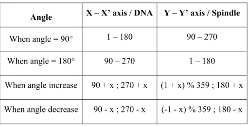
6.1. Table of angle combinations --- ii
Index of tables:
Table 1: The following table enlists the genetic crosses we performed in this study, indicating the female and male fly stocks used and the experimental purpose. ... 35 Table 2: Combination of values for corresponding DNA and Spindle positions... 48 Table 3: Angle measurements with DNA and Aurora as references. ... 63 Table 4: Table of P-values for bleb counts for corresponding stages during asymmetric cell division cycle ... 74
Index of figures:
Figure 1: Discovery of Asymmetric cell division with the Ascidian embryo. ... 5
Figure 2 : Drosophila melanogaster as a model system. ... 8
Figure 3: Model for asymmetric cell division in Drosophila melanogaster sensory organ precursor (SOP). ... 10
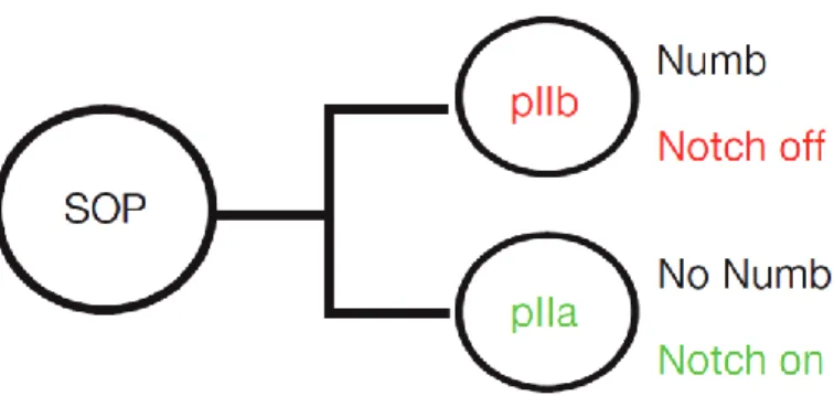
Figure 4: Numb-Notch activation during asymmetric cell division of SOP cells. ... 11
Figure 5: Proper spindle orientation leads to proper segregation of cell-fate determinants. ... 15
Figure 6: The GDP-GTP cycle of small GTPases. ... 17
Figure 7: Bleb life cycle. The bleb expansion-retraction cycle can be subdivided into three phases: bleb initiation (nucleation), expansion and retraction. ... 28
Figure 8: Step by step dissection procedure showed in images for live cell imaging of SOP cells. ... 39
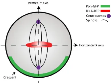
Figure 9: Cartoon depicting horizontal and vertical axis of polarity. ... 42
Figure 10: Image J layout with angle tool ... 44
Figure 11: Modelization of an SOP cell as a circle... 45
Figure 12: Angle measurement comparisons in SOP cells modelized as circles. ... 47
Figure 13: Compass plots showing Spindle / DNA positions. ... 51
Figure 14: Modelization of crescent formation-expansion. ... 52
Figure 15: Blebbing quantification. A blebbing SOP cell with quantification circle. ... 53
Figure 16: Quantification of geometrical parameters of wild type SOP cells ... 57
Figure 17: Mitotic spindle positioning during a time-course. ... 58
Figure 18: Quantification of geometrical parameters of wild type SOP cells with Aurora-GFP. ... 61
Figure 19: Mitotic spindle comparison using DNA and Aurora. ... 62
Figure 20: Probability histogram of Gaussian-distributed of angle measurements of the mitotic spindle of 100 random wild type SOP cells at the onset of metaphase. ... 65
Figure 21: Quantification of Pon-GFP crescent expansion. ... 67 Figure 22: Quantification of Pon-RFP crescent expansion. ... 68 Figure 23: Quantification of Pon-GFP and RFP crescent expansion... 69 Figure 24: Probability histogram of Gaussian-distributed of angle measurements of the crescent expansion (width) of 100 random wild type SOP cells at the onset of metaphase. ... 70 Figure 25: Blebbing SOP cell. ... 72 Figure 26: Bleb quantification during the asymmetric cell division cycle. ... 74
Dedicated to my Mother and Father for everything!
To those who are longing to change and ready to change to a meaningful and successful life of abundance and happiness.
Acknowledgements
A lot can change in 6 years, a plethora of events and a variation of all of it. Then there are some things that do not change at all like love, madness, adventure and friendship. This goes to my beloved family, for undying love and support. Papá, Mamá, Fer, Uly, Marti and the Papaluca Dog squad! You all are unique in your own special way and I love you just like that! Abuela y Tía for love and prayers. I am extremely grateful to have such a distinctive family. My brothers in music Sir Pop, Harrison and Vikingo for your patience, support, riff worship and keeping Ballad of the Owl perpetual (Wise guys, GoodFellas). My dear friend and colleague Mohan Malleshaiah for being an amazing friend! Eternal thanks to the Father of Creation and the Mother of Nature for amaze me every day and putting me in the right path.
The work of this thesis would not have been possible without support and strength from God and my own patience and dedication. I am very grateful for the training I received in high-resolution microscopy. I would like to thank the members of the lab, express my gratitude to the members of my thesis committee at Institute for Research in Immunology and Cancer (IRIC) and CHU Saint-Justine for accepting to review my work, and IRIC - Bioimaging Facility for allowed me to perform all my microscopy work.
As always,
May the Beer in your glasses never end! May the Rock N’ Roll in your lives always be loud!
May Eternal Peace never leave your soul! Cheers!
1.1. Preamble : Subject Situation
The tight control of cellular self-renewal, cell fate diversity and daughter cells differentiation is orchestrated by extrinsic and intrinsic asymmetric cell divisions1,2. Understanding this mechanism of generation of distinct cell fates is pivotal in specific areas of research such as drugs development and cancer medical therapy. Several research works have reported a strong connection between polarity proteins and small GTPases, showing that activation of these proteins requires physical interactions with constitutively active small GTPases3,4. The small GTPases family of proteins have been the focus of cancer research since the discovery of the isoform H-Ras p21 mutant in human tumour cells 5. This discovery highlighted the importance of the implication of proteins from the small GTPases family in different types of diseases and cellular processes. Therefore, they represent important targets for symmetric and asymmetric cell division pathway activation and control, since they can regulate a wide variety of related functions6,7 serving as excellent candidates to shed more light into the regulation of asymmetric cell division.
Previous work done by Cabernard et al.8 demonstrated a spindle-independent mechanism for cleavage furrow positioning in Drosophila
melanogaster neuroblasts. They identified that furrow specific proteins are localized
at the basal cortex at anaphase onset and can induce furrow displacement in the total absence of the mitotic spindle. The authors showed a very interesting mechanism for asymmetric cell division leading to the hypothesis that another regulatory pathway possibly involving small GTPases might act through the actin cytoskeleton. Such mechanism raises several questions: (i) Whether Rho small GTPases and their connection to polarity proteins can regulate mitotic spindle orientation? (ii) If they regulate the stability of the cleavage furrow’s position? (iii) Whether they influence
the balance of polar forces and tension release that define asymmetric division? (iv) How spindle positioning can be quantified in order to statistically differentiate between wild type and abnormal conditions? To start answering such questions, we used Drosophila melanogaster sensory organ precursors (SOP) as a model system since it allows the use of genetic tools and advanced in vivo 4D time-lapse microscopy techniques. The aims of this master thesis are (i) To determine geometrical parameters of SOP division, (ii) Determining the sub-cellular activation of Rho small GTPases during asymmetric cell division (ACD) and (iii) Assess the effect of perturbing the actin cytoskeleton on SOP division.
Using high-resolution 4D confocal microscopy techniques, we developed a simple method that uses available geometrical parameters to assess perturbations of ACD in SOPs due to abnormal Rho small GTPase activity, which allows us to determine spindle positioning over time regarding to other axes of polarity. Our approach will help to better understand the mechanism of mitotic spindle positioning and how it can be regulated by influential polarity proteins and individual Rho small GTPases during ACD. Moreover, our method could be implemented for other investigations to extract parameters in order to differentiate important observations in the asymmetrically dividing cells of the sensory organ precursors; such as mitotic spindle positioning, bleb formation and polarity crescent formation-expansion at metaphase, to mention a few.
1.2.
Asymmetric Cell Division
The process of asymmetric cell division was first described a century ago by American biologist Edwin Conklin. Using ascidian embryos, he observed that during early division, an area of yellow cytoplasm was always co-segregating with cells that will eventually differentiate from the others and become muscle cells9
(Figure 1). This observation opened the field of study of asymmetric cell division until today. The process of asymmetric cell division has been fascinating scientists for more than a century, leading research in the field using various model organisms such as the worm Caenorhabditis elegans, the fruit fly Drosophila melanogaster and mammalian systems like Mus musculus1,2. Cellular diversity is generated by the processes of symmetric and asymmetric cell divisions. Following symmetric cell divisions, daughter cells can acquire different fates depending on the cellular environment. Hence, this type of cell diversity is known to be extrinsic. Cellular self-renewal is also orchestrated by the asymmetric distribution of different cell fate determinants occurring in several steps processes recognized as intrinsic asymmetric cell divisions1. Although asymmetric cell division has fascinated scientists for over a century, a thorough understanding of the underlying mechanisms has only recently emerged. Much of this increased knowledge has come from studies in Drosophila melanogaster and Caenorhabditis elegans that have led to the identification of conserved cellular principles and molecular players that govern asymmetric cell division.
Intrinsic asymmetric cell division occurs in simple step processes. First, after –mother symmetry breaking, the mother cell becomes polarized. Second, cell-fate determinants are segregated towards both distinct poles of the mother cell. Third, the mitotic spindle is aligned so in turns the cleavage furrow results in the proper inheritance of cell-fate determinants to the daughter cells. Fourth, during mitosis following by cytokinesis, different fates for the daughter cells are established. As a result of these crucial steps, the generation of two daughter cells born at the same time, are not identical2,10. Therefore, asymmetric cell division is pivotal for generating cell diversity. In this thesis we focus on intrinsic asymmetric cell division using the well known biological model system Drosophila
Figure 1: Discovery of Asymmetric cell division with the Ascidian embryo. Depicted is Edwin Conklin’s original drawings of a one-cell stage (left) and eight cell stage (right) embryo (A) Yellow pigment representing the crescent (cr) of mesodermal substance (marked by black circles) co-segregating with muscle cells of the tadpole. (B) Schematic representation of Edwin Conklin’s observations pinpointing the asymmetric segregation and localization of the yellow pigment forming muscle cells (Adapted from 9,11).
1.3. Drosophila melanogaster as a model system
The fruit fly Drosophila melanogaster serves as one of the most studied biological systems and is a splendid model for studies towards understanding cellular processes and development of multi-cellular organisms. Used in physiology and genetics studies, Drosophila melanogaster contributes to the development of a broad variety of genetics and microscopy tools which have been carefully designed and optimized to study any specific gene function in this wonderful organism 12, 13. For the purpose of this master’s thesis, live imaging is a crucial tool in order to understand in vivo processes occurring throughout the entire cell cycle. Using the fruit fly Drosophila melanogaster as a model, we are able to fulfill such a requirement since it allows in vivo tracking of each step of the dynamic process of development at both tissue and cellular levels14, 15, 16.
The approach taken in this research project consists in following asymmetric cell divisions linked exclusively to the proper and tight alignment of the mitotic spindle. Drosophila melanogaster provided us with the working model of choice, the Sensory Organ Precursors. These particular cells display a wide range of asymmetric morphologies such as daughter cells size and asymmetric division variants like cell fate determinants, which helped the development of this project. Moreover, this multi-cellular organism allows the in vivo study of developmental processes like the cell cycle, actin cytoskeleton organization, cellular trafficking, memory systems, metabolic regulation, signalling processes, chromosome recombination and receptor behaviour occurring during development 17,18.
Figure 2 : Drosophila melanogaster as a model system. (A) The Drosophila life cycle. The transition from an embryo to a first instar larva is called hatching. The transitions between larval instars are molts. The process that converts a third instar larva to a pupa is pupariation. Emergence of the adult from the pupal case is called eclosion (Adapted from Genetics: From Genes to Genomes Book 19). (B, C, D, E) Examples of live imaging using diverse Drosophila cell lineages (B) Sensory Organ Precursor cell at anaphase onset during asymmetric cell division. Cell fate determinants (green) co-segregate with anterior PIIB daughter cell. DNA (red) (C) Egg chamber with migrating border cells cluster (green) (D) An adult fly expressing GFP-actin in bristles and socket cells (Guild Lab, University of Pennsylvania) (E) Regulation of cell fate within neuroblast cell lineages (Doe Lab, Institute of Neuroscience, University of Oregon). Images in B and C were acquired at the IRIC Bio-imaging facility, Emery Lab.
1.3.1. Asymmetric cell division in Drosophila melanogaster
Asymmetric cell division of somatic cells was first described in Drosophila
melanogaster by Rhyu et al. 20. They characterized the function of asymmetrically distributed cell fate determinant Numb. Rhyu et al. observed that during mitosis, the fate determinant protein Numb was always segregating towards one of the two daughter cells 20. They observed that Numb localized on one side of the cell forming a crescent during early metaphase. This observation became a characteristic behaviour of cell fate determinants which also led the identification of others. Furthermore, it was shown that Numb is implicated in the regulation of external sensory organs 21. Partial or total loss of Numb leads to abnormal development of external sensory organs supporting the importance of proper cell fate determinants inheritance amongst daughter cells during asymmetric cell division.
1.3.2. Strength of Sensory Organ Precursors to study asymmetric cell division
Asymmetric cell division of Sensory Organ Precursor (SOP) cells occurs along the anterior-posterior axis of the fly notum. Single SOP cells “PI” are able to generate two daughter cells of different sizes and fates. The anterior “PIIB” cell gives rise to neurons and sheath cells and the posterior “PIIA” cell gives rise to socket and hair cells11,22 (Figure 3). More specifically, this cell fate differentiation comes from complex signaling cues between PIIA and PIIB. This mechanism of differentiation requires the ligand Delta in PIIB and the receptor protein Notch in PIIA cells. This is one of the mechanism responsible for different cell fate distribution at the moment of division22,23. Directional signalling between PIIA and PIIB is in part established through the asymmetric distribution of Numb and Neuralized (Neur). Numb and it’s anchor protein Partner of Numb (Pon) act as cell fate determinant markers during asymmetric cell division being unequally localized
in SOP cells24. Numb is inherited by the anterior PIIB cell where Notch signal is shut down, and is absent in the PIIA cell where Notch signal is active (Figure 4). Numb and its partner Sanpodo play a role in establishing Notch signaling at cytokinesis onset20,25. Numb regulate Notch trafficking and establishes directional signaling during cytokinesis25. In neuroblasts and SOP cell lineages, Numb’s localization at the pole is facilitated by its anchor protein Pon2. These two proteins have been very instrumental for live cell imaging studies of asymmetric cell division in Drosophila melanogaster.
Figure 3: Model for asymmetric cell division in Drosophila melanogaster sensory organ precursor (SOP). All SOP cells divide along the anterior-posterior axis of the pupa. Cell fate determinants (green) are segregated into the smaller anterior daughter cell (PIIB), making it different from its posterior sibling (PIIA). PIIB gives rise to neurons and sheath cells, whereas PIIA gives rise to socket and hair cells.
Figure 4: Numb-Notch activation during asymmetric cell division of SOP cells. (Adapted from Couturier et al.25)
1.4.
Molecular regulators of asymmetric cell
division in sensory organ precursors
1.4.1. PolarizationThe Par protein complex has a conserved function in establishing proper cell polarity during asymmetric cell division in C. elegans and Drosophila
melanogaster10. This occurs by a series of phosphorylation events, which has been proposed to also take place in SOP cells. At mitosis, activation of the mitotic kinase Aurora-A promotes a phosphorylation cascade. When phoshorylated, Aurora-A in turns phosphorylates and thus activates a aPKC’s regulatory subunit Par6. During interphase, Lgl gets phosphorylated leading to its release from the cell cortex. It is then released from aPKC, which contributes to the dissociation of the Par6/ aPKC /Lgl complex. Next, Baz is recruited to form the Par6/aPKC/Baz complex, allowing aPKC to phosphorylate Numb leading to its localization at the anterior pole the cell cortex hence being inherited by PIIB26. This mechanism reveals how Numb is
localizing asymmetrically and demonstrates how cell polarity can be linked to the cell cycle. Moreover, loss of the Par polarity complex at the cortex abrogates the mitotic spindle positioning during anaphase, resulting in the formation of daughter cells of equal sizes27,28. Therefore, both the Par complex and cell shape changes contributes to the regulation of the orientation and position of the mitotic spindle demonstrating the importance of the Par complex.
This cell polarity model can be summarized in four simple steps. (i) During mitosis, the Par proteins along with cell fate determinants set up a polarity axis. (ii) This axis is used for mitotic spindle positioning and for asymmetric localization of cell fate determinants at the cell poles. (iii) During the transition from anaphase to telophase, this tightly controlled orientation, positions the mitotic spindle ensuring proper asymmetric localization of cell fate determinants (iv) At two cell stage, cell fate determinants are inherited by only one daughter cell2,24,29. Our study focuses on the possible mechanisms that regulate the orientation and position of the mitotic spindle as the driving force for asymmetric cell division in SOP cells.
1.4.2. Segregation
Several mechanisms of unequal protein segregation have been proposed to occur through a phosphorylation cascade26,30. One of them occurs in the Drosophila neuroblast cell lineage, Partition defective (Par) proteins Par6, Baz and aPKC form a complex and localize at the apical pole guiding the localization of the cell fate determinants Prospero (Pros), Numb, his anchor protein Pon and the adaptor protein Miranda (Mira) to the basal pole. This tight localization ensures proper segregation into the basal daughter cell2,11,30. Next, the Par complex phosphorylates the cytoskeletal protein Lethal (2) giant larvae (Lgl) recruiting cell fate determinants to the cortex. aPKC phosphorylates Lgl leading the release from the cortex and the actin cytoskeleton, prohibiting the localization of cell fate determinants to the apical
pole and excluding Numb and Neuralized from the posterior pole. This phosphorylation event restricts Lgl activity and Miranda localization to the basal pole of the cell30. Despite this precise phosphorylation mechanism, an over expression of a non-phosphorylatable version of Lgl (Lgl3A) is sufficient to disrupt cell fate determinants destiny.
1.4.3. Spindle Orientation
During cell fate diversity generation, the mitotic spindle plays a pivotal role, orchestrating the mechanisms for unequal segregation of cell fate determinants, influencing the proper inheritance by the two daughter cells and assuring appropriate cell size. The mitotic spindle is a conserved cell division structure from yeast to humans28. This machinery features two spindle poles from which emanate three classes of microtubules from their minus-ends, (i) kinetochores, that are attached to chromosomes, (ii) interpolar microtubules, that are structured in an antiparallel fashion in the middle of the spindle poles and (iii) astral microtubules, that diverge towards the cell cortex from the spindle poles and use their plus-ends to attach the spindle to the cell cortex28,31. The tight coordination of these structures orchestrates cell division, serving as a pulling force for chromosome segregation.
Mitotic spindle positioning depends on subtle interactions between astral microtubules and the cell cortex32. The dynein-dynactin complex is the main player responsible for mitotic spindle alignment along the anterior-posterior axis of the pI cell in C. elegans embryos33. The action of the dynein-dynactin complex is a conserved spindle alignment and pulling force mechanism across species28. Dynein associates with the dynactin complex which foster dynein to its cargo proteins allowing the complex moving the spindle towards the cortex34,35. In D.
melanogaster, several proteins are needed for polarity and spindle position during
inscuteable (Pins), Locomotion defects (Loco), Mushroom body defective (Mud) and Gαi forming the Mud-Pins-Loco-Gαi complex at the anterior side of neuroblasts and SOP cells10,36. Par proteins Par6, Baz and aPKC and smallGTPase Cdc42 associate with the Mud-Pins-Loco-Gαi complex through the dynein-dynactin complex allowing proper orientation and positioning of the mitotic spindle during asymmetric cell division28. The control of mitotic spindle orientation and positioning in developmental systems is based on the full coordination of the previously mentioned mechanism and the activity of cortical blebbing.
1.4.4. Mitosis and Cytokinesis
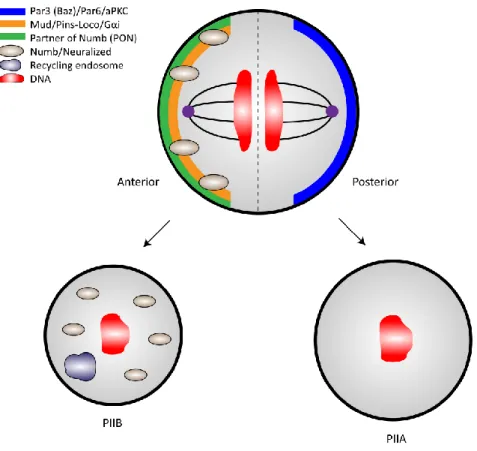
During mitosis, following by cytokinesis, several components are pivotal for proper cell-fate inheritance by the two daughter cells. Time-lapse quantitative experiments demonstrated that Pon-GFP is recruited to the cortex of the PI cell upon progress into mitosis becoming enriched on the anterior pole of the SOP11. Proteins like actin and Myosin II are required for –anterior pole- enrichment of Numb and its anchor protein Pon, suggesting a mechanism that drives asymmetric segregation of cell-fate10. In turns, Pon and Numb are inherited only by the PIIB daughter cell. In SOP cells, polarity proteins Par6, Baz and aPKC interact with each other forming the Par complex which localizes to the posterior pole cortex. The posterior localization of the Par complex along with the anterior localization of Numb, Pon and Neuralized and spindle proteins Mud, Pins, Loco and Gαi, establish the axis of polarity, essential for spindle orientation and asymmetric protein localization during mitosis. Finally, at cytokinesis, cell-fate determinants are inherited respectively by only one daughter cell (Figure 5).
Figure 5: Proper spindle orientation leads to proper segregation of cell-fate determinants. First: spindle orientation controls the axis of cell division and determine cell-fate determinants segregation in an asymmetric fashion. Second: position of the spindle within the dividing cell determine the relative size of the two daughter cells.
1.5. The small GTPases family of proteins
Developmental processes in multi-cellular organisms are controlled by specific proteins, which are part of a wide variety of complex signaling networks. Amongst those regulatory proteins is the Ras family of guanosine triphosphates (small GTPases). The small GTPases family is constituted of the Ras, Rho, Rab, Ran and Arf subfamilies. Each of these subfamilies is found in different functional branches across species37. These proteins are of special interest because they regulate intracellular signal transduction pathways in response to external and internal stimuli. They act as molecular binary switches that are either turned on or off depending on the cell’s needs (Figure 6). Small GTPases are known to be involved in various coordinated processes such as cell polarity 3,38, polarized growth 39, collective cell migration 40, vesicle trafficking 41,42, actin and septin organization and development 43, cell cycle regulation and cell survival 44. This conserved family of proteins has been well studied in humans, budding yeast Saccharomyces
cerevisiae, the fruit fly Drosophila melanogaster and the worm Caenorhabditis elegans44,45. Thus, small GTPases serve as an excellent working mechanism for the development of complex signaling processes established at both levels of functional and structural levels 46.
1.5.1. The smallGTPases function as molecular switches
The small GTPases family of proteins features a GDP-GTP cycle mechanism which is similar among small GTPases subfamilies such as Ras, Rho, Rab, Ran and Arf. The small GTPases cycle of activation and inactivation is controlled by GEFs (Guanine Exchange Factors) that stimulate the exchange of GDP into a GTP. The inactivation is controlled by GAPs (small GTPase activating proteins) that promote hydrolysis from GTP into GDP. The mechanism of small
GTPases activation and inactivation relies on specific membrane receptors, which sense extracellular signals, leading to the initiation of complex signal transduction pathways. This particular mechanism leads to the recruitment of a specific GEF for the activation of a small GTPase through binding to GTP (GTP-bound state). These GEF proteins can act specifically on one small GTPase or on several of them. This active signal is controlled when GTP gets hydrolyzed ending in a GDP-bound state. Hence, cellular behaviours can be determined by single or multi small GTPase specificity 47.
Figure 6: The GDP-GTP cycle of small GTPases. These proteins are in their active state when bound to a GTP molecule and are inactive when bound to a GDP molecule. Small GTPases activation is controlled by GEF (Guanosine Exchange Factors) that stimulates the exchange of GDP into a GTP and inactivation is controlled by GAP (small GTPase Activating Proteins) that hydrolyse the GTP into GDP.
1.5.2. The small GTPases function in Drosophila melanogaster
In the past years, the fruit fly Drosophila melanogaster has risen as a wonderful genetic system for the study of small GTPase proteins in developmental and molecular processes. We are interested in better understanding how the particular family of Rho small GTPases is implicated in the actin cytoskeleton, cell polarity and asymmetric cell division.
1.5.2.1. The Rho small GTPases activity
The Rho subfamily of small GTPases was found to be evolutionary conserved across species37,48. This particular subfamily of proteins is in charge of maintaining the appropriate cell morphology and coordinates migratory movements, which are essential for homeostasis and dynamic processes 49. The principal members of this family are Rho, Rac and Cdc42. These proteins function as molecular binary switches changing from a GTP-bound active state to a GDP-bound inactive state, depending on intra or extra-cellular signals (Section 1.5.1). Rho small GTPases are also regulated by third class of regulatory protein called Guanine Nucleotide Dissociation Inhibitors (GDIs). These regulators not only prevent the GDP ↔ GTP exchange cycle, but also maintain proteins in their GDP inactive state and prevent their localization at the membrane 6,7,50.
The fruit fly Drosophila melanogaster undergoes several morphological changes during development 19. Therefore this subfamily of proteins participates actively in many processes including regulation of the actin cytoskeleton, cell growth, cell fates, cell survival and differentiation, axonal guidance, cell-cell interaction and cell proliferation, which implicates control of the cell cycle 7. These are fundamental processes that are essential for development in higher organisms including Drosophila. The small GTPases Rac1 and Cdc42 are the most
investigated since they are involved in regulating many cellular functions through protein-protein interaction dynamics. This diverse regulation happens via a number of effector molecules which have been well characterized in structure and functions7,51. Moreover work done by members of the Emery lab, demonstrated that Rac1 activity and polarization during collective cell migration is regulated by members of the Rab small GTPases family52. The actin cytoskeleton organization plays an important role in determining cell polarization and proper distribution of cell fate determinants. This section describes such processes and the involvement of small GTPases Rac1 and Cdc42 in the generation of several cell lineages and specific functions in fruit flies.
1.5.2.1.1. General roles of Rac1 and Cdc42 on the actin cytoskeleton
The small GTPase Rac1 is able to control through a series of complex signaling pathways, some of the most important processes of cell morphology. The principal roles of Rac1 include regulation of the actin cytoskeleton, epithelial morphogenesis and axon growth and guidance 53. This particular small GTPase has two homologs, Rac2 and Mtl, having overlapping roles in the control of Drosophila development 54. To this date, not much information is known about Rac1 being involved in regulating asymmetric cell division in SOP cells. Our focus on Rac1 relies particularly on evidence suggesting it has one of the principal roles in regulating the actin cytoskeleton. Rac1 is present in almost all eukaryotic systems, conserved from yeast to humans 55. Such conservation suggests that basic mechanisms involved in cell morphology were conserved during evolution. These mechanisms have designated the finest tasks in development and maintenance of the actin cytoskeleton. In Drosophila, mammalian systems and other organisms, rearrangement of the actin cytoskeleton is necessary for cell shape changes driving
cell movements and migration. These dynamic changes are responsible for cell migration in higher organisms48,56.
Cell migration features a series of subsequent dynamic actions, including lamellipodia extension, formation of focal adhesions and contractions, all requiring tight control of the actin cytoskeleton and it’s downstream effectors57. These include WAVE/Scar, Sra1, PAK and Plexin-B1 amongst the most notorious effectors involved in actin cytoskeleton regulation 7. Rac1 is able to induce localized actin branches formation, which leads the generation of polarized morphological changes known as protrusions. These protrusions, in cooperation with other mechanisms help the cell to control the direction of migration 58. Also, FRET biosensors data revealed that Rac1 localizes at the leading edge of these protrusions in vivo, suggesting a strong influence on remodelling of the actin cytoskeleton for cell movement59.
The multiple roles of Rac1, Cdc42 and other small GTPases became evident when extensive studies began implementing ectopic expression of constitutively active and dominant negative mutant of these proteins 60. For small GTPases, a constitutively active mutant is when they are unable to hydrolyze GTP and a dominant negative mutation is when they are unable to remove GDP 61. The most common constitutively active mutants are found in the P-loop, when the catalytic Glycine (G) residue at position 12 is exchanged by Valine (V) (G12V) and in the catalytic residue, when Glutamine (Q) at position 61 is exchanged by Leucine (L) (Q61L) resulting in a GTP-lock state 44,61,62. As well, the most notorious dominant negative mutants are based on the Ras S17N, founded as Serine (S) at position 17 is exchanged by Asparagine (N) (S17N) and Aspartic Acid (D) at position 119 is exchanged by Asparagine (D119N)63,64. These single nucleotides substitution lead to a GDP-lock state unable to interact with downstream effectors. Such mutations
have been found in cancerous cells when small GTPases functions have been completely abrogated5.
Recently, a few examples of altered Rac1 and Cdc42 small GTPase’s function have been reported in lymphocytes development, differentiation, activation and migration. It has been demonstrated that Rac1 has a pivotal role in B cell development, where loss of Rac1 blocks migration processes. This leads to an arrest in B cell development in the spleen, showing that Rac1 is required during the earlier stage of transitional B cells in mammalian systems65. Rac1 and Cdc42 coordinate actin polymerization and hence cell motility in vivo. This dynamic coordination is completely loss when known dominant negative and constitutively active versions of Rac1 and Cdc42 are expressed66. Data regarding actin cytoskeleton regulation also suggest a coordinated task between Rac1 and Cdc4267. This spatiotemporal coordination between Rac1 and Cdc42 has been observed using FRET biosensors, such as when activation between these small GTPases overlap, it results in a protrusion-retraction cycle68. Moreover, the use of a dominant negative version of Rac1 leads to memory deregulation acting through remodelling of the actin cytoskeleton69. All these results confirm the implication of Rac1 and Cdc42 in coordinating the actin cytoskeleton in higher organisms.
Cdc42 is known as the master regulator of cell polarity70, is a highly conserved small GTPase essential for establishment and maintenance of cell homeostasis from yeast to humans37. It acts as a molecular binary switch modulating a wide range of signalling processes. Mutant versions of Cdc42 show defects in the organization of actin cytoskeleton and septins, which have pivotal roles during progression of the cell cycle. This main regulator is known to be involved in processes like actin patch polarization38 and controlling the formation of actin bundles containing filopodia at the cellular periphery. Furthermore, Cdc42 regulates the pheromone response pathway71, actin cable nucleation and septin organization,
which are implicated in the maintenance of cell morphology72. Cdc42 functions at the plasma membrane, localized at specific domains, and coordinating polarized organization of the actin cytoskeleton during cell migration. In the next section I will describe the link between Cdc42 and polarity proteins and their involvement in the regulation of asymmetric cell division in Drosophila neuroblast cell lineages and the worm Caenorhabditis elegans. These are the major reasons why we chose the small GTPases Rac1 and Cdc42 and to study their involvement in the regulation of the mitotic spindle during asymmetric cell division. They are very efficient at inducing different phenotypes through coordination of actin cytoskeleton dynamics. Moreover, they can induce malignant cells forming tumours in humans.
1.5.3. Cdc42, Rac1 and their relation with Par proteins
As described above, the small GTPases Rac1 and Cdc42 play key signaling roles in regulating cell polarity and the actin cytoskeleton. The two smallGTPases share 70% sequence conservation and identity among them and with human homolog Rac1 and Cdc4273,74. This suggests that non-conserved sites might be defining different specificities and thus specific functions75. Since there is a strong link between Cdc42 and Par proteins as well as sequence similarity between Cdc42 and Rac1, we wondered whether Rac1 could contribute to the regulation of polarity cues. The first link between Rho smallGTPases, polarity proteins and asymmetric cell division came from research done in Caenorhabditis elegans and Drosophila
melanogaster. In these eukaryotes, the unequal distribution of polarity proteins and
cell fate determinants coordinates local differences regarding the actin cytoskeleton and actomyosin meshwork10.
The Par proteins were first indentified in a genetic screen using the embryos of the worm C. elegans. It encodes six different proteins all required for proper asymmetric cell division76. After fertilization, C. elegans embryos divide
asymmetrically along the anterior-posterior axis of the cell. Par3 and Par6 proteins segregate to the anterior pole, whilst Par1 with Par2 segregate to the posterior pole. This coordinated localization of Par proteins leads to an actomyosin meshwork restricted to the anterior pole which promotes contractility while the posterior pole remains non-contractile77,78. The small GTPase Cdc42 provides the link and regulates the actomyosin complex with Par proteins through a series of protein-protein interactions77,79.
Similar to Drosophila sensory organ precursors, the neuroblast cell lineages divide asymmetrically, but along the apical–basal polarity plane following a similar protein segregation mechanism in both SOPs and C. elegans. During asymmetric cell division of neuroblasts, the polarity proteins Bazooka (Baz, Par3 homolog in
Drosophila), aPKC and Par6 segregate to the apical pole. Alignment of the mitotic
spindle along the apical–basal axis is controlled by Scribble (Scrib), Discs large (Dlg) and Lethal giant larvae (Lgl)2. Cell fate determinants Prospero, Brat and Numb localize at the basal pole and hence segregate into the basal daughter cell2,11. The basal localization of cell fate determinants depends on the asymmetric actomyosin contraction at the apical pole of neuroblasts80. This regulation of actomyosin occurs by apical restriction of Myosin II (Squash (Sqh) in Drosophila) by Lgl. This mechanism takes place when Lgl gets phosphorylated by aPkc, which restricts Lgl at the apical pole30. Par6 and aPkc establish polarity by localizing on the apical pole of the daughter cell. It has been reported that apical localization of Par6 requires physical interaction with Cdc42, which acts downstream of Baz to establish polarity81. Dominant negative and constitutively active versions of Cdc42 are able to dislocate such epithelial polarity3, demonstrating the role of a small GTPase in the regulation of epithelial polarity during asymmetric cell division of
Despite the sequence similarities between Drosophila small GTPases Rac1 and Cdc42, in vitro binding assays between Drosophila Par6 and Rac1 does not show a physical interaction, with neither wild type, dominant negative nor constitutively active versions of Rac13. However, a physical interaction between
Drosophila Par6 and Rac1 has been detected in a yeast two-hybrid assay screen82. Also, a physical interaction between mammalian Rac1 and Par6 has been detected, suggesting a possible role in coordinating polarity of asymmetric cell division in mammals83. These overlapping results led us to investigate protein-protein interactions between Par proteins and the small GTPases Rac1 and Cdc42.
Cdc42 and Rac1 interact with proteins that feature a short conserved motif named CRIB (Cdc42/Rac1 interacting binding) 84. Remarkably, Par6 possesses a semi-CRIB motif and an adjacent PDZ domain required for its biological regulation and interaction with Cdc42 and Rac14,85. The crystal structure of a complex between Cdc42 and Par6 inducing a conformational change upon direct binding has been reported. As well for Rac14 but with lower affinity due to two overlapping residues in sequence with Cdc4286. To this point, published data suggests connection between Rac1 and polarity cues. However, neither physical, genetic nor molecular evidence link Rac1 to the control of the mitotic spindle and hence regulation of asymmetric cell division. Therefore we are aiming to unveil whether or not there exists a pathway that involves this small GTPase.
1.6.
Blebs: Possible implication with the mitotic
spindle
In living cells cortical tension is the combined result of the physical properties of the membrane and the cortical network87. Cellular tension release relies on a very efficient cortical mechanism inducing cortex deformation around the cell surface. Such mechanism in living cells is known as the process of membrane blebbing88,89. Blebs have been well studied during cell division in mammalian systems and regularly observed from anaphase to cytokinesis88. They also play a role in cell migration and tissue morphogenesis90. Although bleb studies have reported stabilization of cell shape during cytokinesis91, the role in asymmetric cell division still remains unclear. In non-motile cells, several membrane-associated proteins have been shown to coordinate the sequential recruitment-expansion cycle of blebs. Such proteins include actin cytoskeleton membrane linker proteins, actin bundling proteins and contractile proteins88. Some commons proteins involved in expansion are the, small GTPase RhoA, ROCK, Myosin II and Src. The most notorious proteins involved in retraction include, Ezrin, Actin, Moesin and Myosin II88,92.
Research on blebbing have shown that during asymmetric cell division of C.
elegans Q neuroblasts cells, unbalanced contraction at the anterior pole directly
through Myosin II action generates daughter cells of different sizes. This suggests a Myosin II driven mechanism on the anterior-posterior poles of a dividing cell helping to regulate the size and fate of the daughter cells93. In Drosophila neuroblasts Myosin II regulates asymmetric cell division by excluding cell fate determinants from the apical pole. From prophase to metaphase, Myosin II restricts these cell fate determinants from the apical pole. From anaphase to telophase, Myosin II concentrates at the cleavage furrow of the contractile ring to promote
cytokinesis and complete the proper distribution of cell fate determinants from the neuroblast to its daughter cells94. Also Myosin is required for proper orientation and position of the mitotic spindle in Drosophila neuroblasts95. Myosin helps orient the mitotic spindle by 90° alignment along the apical-basal axis and by localizing the adaptor protein Miranda at the basal pole. This suggests a mechanism where a higher actin contraction at the basal pole of neuroblasts is required for proper mitotic spindle positioning during asymmetric cell division. The interaction of the mitotic spindle with the cell cortex is one of the principal regulators of the spindle alignment. It is thought that actin contraction forming blebs can orchestrate the orientation of the mitotic spindle. Therefore, unbalance of this process could randomize the proper orientation. These proposed mechanisms in Drosophila and C.
elegans are important in order to better understand unbalanced contraction
differences and the role of blebs around the cell cortex. These studies led us to question how asymmetric actin contractions of SOP cells occur during cytokinesis, and whether they play a role in the positioning of the mitotic spindle.
Figure 7: Bleb life cycle. The bleb expansion-retraction cycle can be subdivided into three phases: bleb initiation (nucleation), expansion and retraction. (A) Bleb initiation can result from a local detachment of the cortex from the membrane (left model) or from a local rupture of the cortex (right model). (B) Hydrostatic pressure in the cytoplasm (Pint) then drives membrane expansion by propelling cytoplasmic fluid through the remaining cortex (left model) or through the cortex hole (right model). Concomitantly, the membrane can detach further from the cortex, increasing the diameter of the bleb at the base (dashed line). (C) As bleb expansion slows down, a new actin cortex reforms under the bleb membrane. (D) Recruitment of myosin to the new cortex is followed by bleb retraction. Pext, extracellular hydrostatic pressure (Adapted from Charras and Paluch 88).
1.7.
Geometry of asymmetric cell division
Accurate asymmetric cell division requires precise coordination of the mitotic spindle. Complex signaling pathways respond to cortical tension generated by both, internal and external environmental changes to control the mitotic spindle28. The orientation and position of the mitotic spindle in SOP cells determines the relative size of the PIIA and PIIB daughter cells, as well as determines the proper distribution of cell fate determinants96. The mechanism that controls asymmetric cell division of SOP cells copes with subtle geometrical changes in the orientation of the mitotic spindle, and understanding the geometry that leads to proper asymmetric cell division is a difficult task.
Cell geometry plays an important role in controlling the cell cycle in different animal cells97. Research on C. elegans and different Drosophila
melanogaster cell lineages including neuroblasts, SOP cells, intestinal stem cells98, epidermoblasts99, and many others, feature specific cell components like cortical cell polarity proteins and polarized cortical pulling forces28 that can monitor cell geometry. These specific components are used to control the mitotic spindle and cell fate determinants inheritance28. It is thought that geometry sensing mechanisms control decisions ensuring proper cell division100 in living organisms. These observations converged into the key idea that the majority of signaling cues plays an important role in determining the cell geometry, which controls the mitotic spindle, cell fate determinant localization, crescent formation and inheritance, chromosome segregation and daughter cells of different sizes. These geometrical parameters become interesting to determine and quantify the mitotic spindle orientation and other parameters during asymmetric cell division of SOP cells. One of the principal aims of this research work relies on the proper acquisition and quantification of such geometrical parameters.
1.8.
Hypothesis
We are interested to investigate whether the small GTPases control the positioning of the mitotic spindle during ACD in SOP cells. I want to establish a method to visualize SOP cells at different stages of the ACD cycle and quantify various parameters of mitotic spindle along the course of ACD. Therefore, we hypothesize that “Asymmetric actin contractions during cytokinesis might displace the mitotic spindle and the cleavage furrow, leading to cell size asymmetry”. In order to test this hypothesis, I propose, first to implement different quantification methods to assess the subtle dynamics of a normal ACD. After, this will serve to detect subtle movements of the mitotic spindle after perturbing the asymmetric cell division mechanism of SOP cells. Simultaneously, this study will shed more light regarding the sub-cellular activation of certain proteins like small GTPases, which might be involved in regulating mitotic spindle dynamics in SOP cells.
1.9.
Specific aims
1.9.1. Determining geometrical parameters of SOP asymmetric division: By using Drosophila lines expressing markers for the asymmetrically distributed protein Pon (Pon-GFP and Pon-RFP), for DNA (Histone-RFP) and for centrosome (Aurora-GFP), I will determine the exact geometry of ACD of SOP cells. Using 4D confocal microscopy, I will perform tridimensional, multichannel, high-speed acquisition of dividing SOP cells. Since the DNA, as well as the centrosomes and Partner of Numb do not overlap, I can image these proteins simultaneously. From these experiments, I will determine several parameters regarding the geometry of SOP division, such as the exact positioning of the spindle
and the cleavage furrow, the crescent formation-expansion and diameters of the two daughter cells. This will allow us to construct a precise mathematical model of SOP division and to measure subtle alteration of ACD as described in aim#3.
1.9.2. Determining the sub-cellular activation of Rho small GTPases during ACD:
From previous work done on collective cell migration, the laboratory has acquired and developed tools like Rac1-FRET and Cdc42-FRET, to determine where Rho small GTPases are active in vivo. Here, we will take advantage of such FRET biosensors, in order to determine where Rho small GTPases are activated during SOP division.
1.9.3. Assessing the effect of perturbing the actin cytoskeleton on SOP division:
Here, I will perturb the actin cytoskeleton by different means to determine its role in positioning the mitotic spindle. Initially, I will focus on Rho small GTPases. I will express DN and CA forms to determine their effect on the geometry of SOP division. As certain experiments are out of the scope for this master thesis, future work could be done. Taking advantage of photoactivatable versions of Rho small GTPases and FRET probes, the dynamics of Rho small GTPases can be further explored.
The experiments of this project consisted of four important steps: basic genetic crossing of Drosophila virgin females with young males, efficient dissection of flies at pupal stage, high definition four-dimensional (4D) confocal microscopy and the image analysis. We built custom homemade image analysis and quantification tools. Together, these procedures led to efficient analyses of the changes in asymmetric cell division of SOP cells.
2.1.
Drosophila fly stocks and genetic crosses
The Drosophila fly stocks were maintained at 18°C and 25°C. All genetic crosses were performed at 25°C. The stocks used in this study are listed below along with the relevant reference (the laboratory and Bloomington (BL) stock number). Fly crosses were performed before image acquisition through confocal microscopy.
2.1.1. Fly Stocks:
w; Neuralized Gal4 / Tm6b, Tb (G019)
w; UAS H2A::RFP, UAS Pon.LD::GFP, Neuralized Gal4 / Tm6b, Tb on III (This study)
w; Neuralized Gal4, UAS H2A::RFP / Tm6b (This study)
y[1] w[*]; P{w[+mC]=UAS-Rac1.T17N}1 on III (BL6292) (O017) y[1] w[*]; P{w[+mC]=UAS-Cdc42.T17N}3 on II (BL6288) (O054) w[1118]; P{w[+mC]=UAS-RhoL.T25N}AM / Cyo on II (BL4849) (O073) y[1] w[*]; P{w[+mC]=UAS-Rac1-FRET} / Cyo ; MKRS / Tm6b (From Ramel et al. 52)
w[1118]; P[mW, UAS Pon.LD::RFP], Neuralized Gal4, P[mW, UAS Aurora::GFP] / Tm6b, Tb on III (396) (From Schweisguth Lab Gomes et
al. 101)
w[1118]; P[mW, pNeuralized, H2B::RFP] on I (619) (From Schweisguth Lab published by Gomes et al. 101)
w[1118]; P{w[+mC]=UAS-Moesin.GFP} on III (O046)
w[1118]; P{w[+mC]=UAS-Lgl3A.GFP}/ Tm3 on III (O578) (From Wirtz-Peitz et al. 26)
The small GTPase fly lines and the UAS-Rac1-FRET probe were balanced on the II and III chromosomes using the double balancer line:
w; If / Cyo ; MKRS / Tm6b, Tb (T033)
2.1.1.1. Definitions:
Neuralized Gal4: Tissue Gal4 driver used for specific gene expression in the sensory organ precursor cells.
Partner of Numb (Pon): Cell fate determinant marker used to follow asymmetric cell divisions of SOP cells. Tagged with a green fluorescence protein (GFP) and a red fluorescence protein (RFP).
Histone 2 A and Histone 2 B: DNA marker tagged to a red fluorescence protein.
Aurora A (AurA): Centrosomes marker tagged to a green fluorescence protein (GFP).
Moesin (Moe): Actin binding moesin used for cortex marker tagged to a green fluorescence protein (GFP).
Lgl3A: Lethal (2) Giant Larvae, triple alanine mutant, prevents the release of Lgl from the cortex during ACD. This allows the visualization of blebs formation until division. Tagged to a green fluorescence protein (GFP).
If: Irregular facets, dominant mutation that results in small white eyes with fused ommatidia. Marker used to follow the second chromosome.
CyO: Curly of Oster, which wings are curled at the end. Marker used to follow the second chromosome.
MKRS: Minute-Karmoisin-Rosy-Stubble, which flies that, features short bristles. Marker used to follow the third chromosome.
Tm6b: Tubby, marker used to follow the third chromosome and to differentiate crosses at pupal stage prior to dissection.
2.1.2. Genetic crosses performed at 25°C:
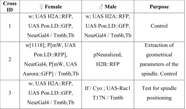
Table 1: The following table enlists the genetic crosses we performed in this study, indicating the female and male fly stocks used and the experimental purpose. All crosses were performed at 25°C.
Cross
ID ♀ Female ♂ Male Purpose
1 w; UAS H2A::RFP, UAS Pon.LD::GFP, NeurGal4 / Tm6b,Tb w; UAS H2A::RFP, UAS Pon.LD::GFP, NeurGal4 / Tm6b,Tb Control 2 w[1118]; P[mW, UAS Pon.LD::RFP], NeurGal4, P[mW, UAS Aurora::GFP] / Tm6b,Tb pNeuralized, H2B::RFP Extraction of geometrical parameters of the spindle. Control 3 w; UAS H2A::RFP, UAS Pon.LD::GFP, NeurGal4 / Tm6b,Tb If / Cyo ; UAS-Rac1 T17N / Tm6b
Test for spindle positioning
4 w; UAS H2A::RFP, UAS Pon.LD::GFP, NeurGal4 / Tm6b,Tb UAS-Cdc42 T17N / Cyo ; MKRS / Tm6b
Test for spindle positioning 5 w; UAS H2A::RFP, UAS Pon.LD::GFP, NeurGal4 / Tm6b,Tb UAS-RhoL T25N / Cyo ; MKRS / Tm6b
Test for spindle positioning 6 w; NeurGal4 / Tm6b,Tb UAS-Rac1-FRET/ Cyo ; MKRS / Tm6b,Tb FRET 7 w; NeurGal4, UAS
H2A::RFP / Tm6b UAS Moesin GFP Blebs quantification 8 w; NeurGal4, UAS
H2A::RFP / Tm6b
UAS Lgl3A GFP /
Tm3 Blebs quantification
2.2.
Procedure to dissect Drosophila pupae
2.2.1. Required materials:
Zeiss Stereo Discovery V8 microscope (Carl Zeiss, Oberkochen, Germany) Custom microscope plastic slide (feature a small canal where pupae are
placed)
Micro cover glass (No.1.5 mm 22 x 40 mm) (VWR, Radnor, United States) Dissection forceps (size 5 or 5.5) and scissors
Thin paint brush
5 cc syringe for oil distribution Rubber glue
Confocal microscope with digital camera and image acquisition software
2.2.2. Procedure:
Set up the fly crosses or place flies from a stock you wish to image in several fresh vials at 25°C.
SOP cells generally begin to proliferate on the pupae thorax at fifteen hours after the onset of pupariation, we therefore select pupae at 0 hour after pupae formation, which can be recognized by their white color.
Incubate the pupae at 25°C for 15 hours.
Collect pupae and place them into a rubber petri dish with the ventral side down. Grasp the edge of the operculum (the circular hatch on the anterior dorsal tip of the pupae case) with special forceps and carefully cut slowly with scissors. (Figure 8 A)
Gently lift, remove, and discard the operculum, revealing the head along with the notum of the pupa.
After the cut, use the forceps to begin tearing along the side of the pupal case. Lift the midsection of the pupae case from the torn side and bring it over to the opposite side. Pupal case can be removed completely or partially. (Figure 8 B)
2.2.3. Pupae mounting:
Isolated pupae have to be placed on the center of the custom plastic slide dorsal side up with head facing the anterior side. (Figure 8 C, D)
Using a 5 cc syringe filled with halocarbon oil, apply a thin uniform layer in the middle of the micro cover glass. An oil overload can cause asphyxia to flies preventing proper asymmetric division of SOP cells.
Place a small drop of water (1 μl) on the sides of a 22 x 40 mm micro cover glass and place it on the above preparation such that the small halocarbon
oil contacts the surface you want to image, full notum in this case). Compress gently to form a complete seal and flat contact surface between the micro cover glass and pupae cuticle.
Sample can then be imaged on an inverted or upright confocal microscope fitted with laser scanning or spinning disk, as well two-photon confocal abilities. (Figure 8 E)
Being careful enough might allow adult flies to be recovered after several days.
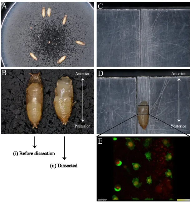
Figure 8: Step by step dissection procedure showed in images for live cell imaging of SOP cells. (A) Few pupae placed in rubber petri dish after being incubated for 15 hours at 25°C ready for dissection. (B) A pair of pupae before (i) and after dissection (ii). Head is toward anterior and abdomen is toward posterior (C) Empty custom made plastic slide featuring pupa fitting groove (D) Slide featuring a pupa placed on groove ready for live imaging (E) An array of few SOP cells on the notum of a fly pupa. Asymmetric segregation of cell fate determinants can be