Science Arts & Métiers (SAM)
is an open access repository that collects the work of Arts et Métiers Institute of Technology researchers and makes it freely available over the web where possible.
This is an author-deposited version published in: https://sam.ensam.eu Handle ID: .http://hdl.handle.net/10985/11815
To cite this version :
Sébastien JÉGOU, Laurent BARRALLIER - Determination of residual stresses within submicroscopic and coherent precipitates - 2012
Any correspondence concerning this service should be sent to the repository Administrator : archiveouverte@ensam.eu
Determination of residual stresses within
sub-microscopic and coherent precipitates
S. Jégou, L. Barrallier
MecaSurf Laboratory, Arts et Métiers ParisTech, 2 cours des Arts et Métiers, 13617 Aix-en-Provence, France
When phase transformations occur during thermochemical processes such as nitriding, residual stresses find origins through the volume change accompanying the surface microstructure evolution. The macroscopic stresses are a mixture of stresses at the phase scale. However diffraction analysis for micro-stress determination can highly depend on the volume fraction, size and nature of phases. Correlations between macro- and micro-stresses are thus limited. This work aims to determine residual stresses in sub-microscopic and coherent precipitates in order to better understand multi-scale stress development during thermo-chemical surface treatment such as in case of gaseous nitriding.
Nitriding of steels (Fe-3wt.%Cr-0.35wt.%C or Fe-1wt.%Cr-0.2wt.%Al-0.35wt.%C) involves a complex microstructural gradient both in time and depth including diffusion of nitrogen and precipitation of sub-microscopic and coherent nitrides MN (M=Cr, Al…) but also coarsening, dissolution and re-precipitation of carbides accompanied by transverse diffusion of carbon [1]. Residual stresses have been characterized using layer removal techniques and laboratory X-ray diffraction analysis [2]. However residual stresses of nitrides have never been analysed yet essentially due to their size (~ 20 nm) associated with their coherency with the ferritic matrix. Moreover uncertainties about plastic straining during nitriding still exist [3,4,5]. As a consequence, macro- and micro-stress analyses cannot be coupled to estimate residual stresses within nano-scale nitrides.
These restrictions can be overcome by performing high-energy synchrotron diffraction in transmission geometry in order to maximize the gauge volume and nano-scale precipitate scattering. In that respect, we carried out diffraction analysis at beamline P07, using a photon energies of hν = 100 keV. The beam size was set to 1 mm width and 0.025 mm height in order to perform analysis with a 25 µm depth step. A flat panel detector with a CsI-scintillator having a size of 0.4 x 0.4 m2 (2048 x 2048 pixels) was used at distances of 1700 and 2100 mm from the sample. Figure 1 shows an example of a scattering image from the near surface (depth = 75 µm) of a nitrided Fe-3wt.%CR-0.35wt.%C steel grade. The most intense peaks come from the scattering of the ferritic matrix. As expected, secondary rings are observed that correspond to the scattering of the precipitation, either cementite Fe3C or nitride MN. However, more precise identification of
peaks has to be done in order to choose the best scattering direction aiming strain analysis. Assuming that the stress component perpendicular to the beam is null, which is often valid for surface stress analysis, the determination of the in-plane biaxial stress should then be possible within each kind of phase identified.
Moreover in order to have an idea of the influence of the divergence of the beam diffracted on the position of the scattering peaks, and so the longitudinal resolution, two scans were carried out along the principal axes (horizontal and vertical axes) using a point detector. The scattering beam was set to 1.5 x 0.1 mm and 5 x 0.15 mm for horizontal and vertical scans respectively. In case of the horizontal analysis, an analyser crystal (Si1-XGeX gradient crystal) having an angular acceptance of
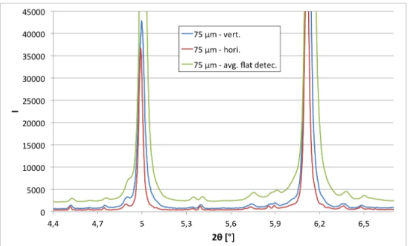
about 25 arcsec and a reflectivity of about 90 % was also available. Figure 2 shows a comparison of these scans for a depth of 75 µm below the free surface. The averaging of rings intensities from the flat detector is also given for the corresponding depth. Clearly the error on the peak position is decreased by controlling the size of the scattering radiation. These scans also show that the half width of diffraction peaks is decreased by using an analyser crystal. Therefore better resolved strain analysis should be expected from point detector scans along the main directions in the present work.
Strain analysis within nano-scale precipitates appears being possible through high-energy synchrotron diffraction in transmission mode. One can actually take advantage of the higher gauge volume (mainly limited by the depth step needed in the present work) that can be used as compared to the reflection mode. However preliminary results suggest that the scattering conditions have to be optimized in order to increase the resolution of such strain analysis. For this purpose, we are planning a new study of strain analysis on similar system using some optimizations of the scattering beam such as using a collimator between the sample and the detector that can be made from two slits and/or rotating the sample by 90° around the incident beam in order to be able to use the analyser crystal only available for horizontal scan at P07 beamline.
Figure 1: Scattering data imaging from high-energy synchrotron diffraction of a nitride Fe-3wt.%Cr-0.35wt.%C steel grade at a depth of 75 µm below the free surface. Main rings correspond to the scattering of the ferritic matrix.
Figure 2: Horizontal and vertical diffraction analyses of a nitride Fe-3wt.%Cr-0.35wt.%C steel grade at a depth of 75 µm below the free surface. Use of a point detector and an analyser crystal in case of the horizontal scan. The averaging of the rings intensity from the flat detector analysis at the corresponding depth is given for comparison.
References
[1] S. Jegou, L. Barrallier, R. Kubler and M.A.J. Somers, J. Heat Treatm. Mat. 66, 135 (2011). [2] S. Jegou, L. Barrallier, R. Kubler, Act Mat. 58, 2666 (2010).
[3] P. Buchhagen, T. Bell, Comp. Mat. Sci. 7, 228 (1996)
[4] L. Barrallier, R. Soto, J.M. Sprauel, A. Charai, Met. Mat. Trans. A 28, 851 (1997) [5] W. Daves, F.D. Fischer, Mat. Sci. For. 163-165, 713 (1998)