HAL Id: tel-01137592
https://tel.archives-ouvertes.fr/tel-01137592
Submitted on 31 Mar 2015HAL is a multi-disciplinary open access archive for the deposit and dissemination of sci-entific research documents, whether they are pub-lished or not. The documents may come from teaching and research institutions in France or abroad, or from public or private research centers.
L’archive ouverte pluridisciplinaire HAL, est destinée au dépôt et à la diffusion de documents scientifiques de niveau recherche, publiés ou non, émanant des établissements d’enseignement et de recherche français ou étrangers, des laboratoires publics ou privés.
Jaime Gonzalo Cordova
To cite this version:
Jaime Gonzalo Cordova. Role of CTGF and TNF on fibrosis in muscular dystrophy. Genomics [q-bio.GN]. Université Pierre et Marie Curie - Paris VI; Universidad católica de Chile, 2014. English. �NNT : 2014PA066546�. �tel-01137592�
Pontificia Universidad Católica de Chile Université Pierre et Marie Curie
ROLE OF CTGF AND TNF ON FIBROSIS IN MUSCULAR DYSTROPHY
By:
JAIME GONZALO CÓRDOVA STEGER
Pontificia Universidad Católica de Chile Université Pierre et Marie Curie
ROLE OF CTGF AND TNF ON FIBROSIS IN MUSCULAR DYSTROPHY
Tesis entregada a la Pontificia Universidad Católica de Chile en cumplimiento parcial de los requisitos para optar al grado de Doctor en Ciencias con mención en Biología Celular y Molecular.
Por:
JAIME GONZALO CÓRDOVA STEGER
Tutor: Dr. Enrique Brandan Co-tutor: Dr. Pascal Bigey
Pontificia Universidad Católica de Chile Université Pierre et Marie Curie
ED 387: iViv: Interdisciplinaire pour le vivant
ROLE OF CTGF AND TNF ON FIBROSIS IN MUSCULAR DYSTROPHY Thèse de doctorat de Biologie Cellulaire et Moléculaire
Par :
JAIME GONZALO CÓRDOVA STEGER
Dirigée par : Dr. Enrique Brandan et Dr. Pascal Bigey Devant un jury composé de : Dr. Antoine Kirchler Dr. Germain Trugnan Dr. Martín Montecino
Dr. Claudio Cabello
To my beloved parents, Marta y Jaime, and to my dear Pachi.
ACKNOWLEDGEMENTS
I am sincerely grateful to:
My advisors, Dr. Enrique Brandan and Dr. Pascal Bigey, for the continuous support and guidance throughout my research project, for the stimulating discussions and their valuable suggestions that helped me to design and perform this work. Also, their effort in proof reading the drafts, are greatly appreciated.
Members of the evaluation committee: Dr. Germain Trugnan, Dr. Antoine Kirchler, Dr. Martín Montecino and Dr. Claudio Cabello; who reviewed this thesis and contributed to improve the manuscript with their insightful comments and corrections.
I also thanks to for the pre-doctoral fellowships from: Beca VRI de doctorado 2007, PUC; Beca de doctorado 2008 and Beca de apoyo a la realización de tesis doctorado 2009-2010, CONICYT; Beca de pasantía MECESUP 2009 and 2011; Beca Chile cotutela de doctorado 2010.
This work was supported by grants from the Basal Center of Excellence in Science in Technology (CONICYT-PFB12/2007) FONDECYT 1110426 and Fundación Chilena para Biología Celular Proyecto MF-100 to Dr. Enrique Brandan.
INDEX Abbreviations VI Abstract VIII Resumen X Résumé XII 1. Introduction 1
1.1 Duchenne muscular dystrophy 2
1.2 Muscle damage and repair 5
1.3 Pro-fibrotic and pro-inflammatory cytokines in DMD 8
1.4 Muscle dystrophy therapies 13
1.5 Hypothesis and Objectives 16
2. Chapter I 18
2.1 Abstract 20
2.2 Introduction 20
2.3 Materials and Methods 23
2.4 Results 26
2.6 References 33
2.7 Figures and Tables 40
3. Chapter II 48
3.1 Abstract 50
3.2 Introduction 50
3.3 Materials and Methods 54
3.4 Results 56
3.5 Discussion 59
3.6 References 62
3.7 Figures 69
4. Discussion 77
4.1 CTGF expression and transcriptional regulation 78
4.2 Tumor Necrosis Factor 82
4.3 Combined therapies for the treatment of DMD 84
5. Conclusions 86
FIGURES AND TABLES INDEX 1. Chapter I. Figure 1 40 2. Chapter I. Figure 2 41 3. Chapter I. Figure 3 42 4. Chapter I. Figure 4 43 5. Chapter I. Figure 5 44 6. Chapter I. Figure 6 45 7. Chapter I. Table 1 46
8. Chapter I. Supplementary Figure 1 47
9. Chapter II. Figure 1 69
10. Chapter II. Figure 2 70
11. Chapter II. Figure 3 71
12. Chapter II. Figure 4 72
13. Chapter II. Figure 5 73
14. Chapter II. Figure 6 74
15. Chapter II. Figure 7 75
ABBREVIATIONS
ATRA: All-trans retinoic acid
CTGF: Connective Tissue Growth Factor DISC: Death Inducing Signaling Complex DD: Death Domain
DMD: Duchenne Muscular Dystrophy
DGAC: Dystrophin Glycoproteins-Associated Complex ECM: Extracellular Matrix
ET: Electrotransfer
GRMD: Golden Retriever Muscular Dystrophy HFMD: Hypertrophic Feline Muscular Dystrophy HGF: Hepatocyte Growth Factor
LRP: Low Density Lipoproteins-Related Receptor MAPK: Mitogen-Activated Protein Kinase
RAR: Retinoic Acid Receptor RXR: Retinoic X Receptor SBE: SMAD Binding Element
SMA: Smooth Muscle Actin
SMAD: Small Mothers Against Decapentaplegic TA: tibialis anterior
TFBS: Transcription Factor Binding Sites TGF: Transforming Growth Factor type β TGFBR: TGF-β Receptor
TNF: Tumor Necrosis Factor TNFR: TNF Receptor
UTR: Untranslated Region
VEGF: Vascular Endothelial Growth Factor wt: wild-type
ABSTRACT
The Duchenne Muscular Dystrophy (DMD) is an X-linked disease characterized by progressive and accumulative damage in the muscle due to the absence of the dystrophin protein. Fibrosis, the excessive accumulation of extracellular matrix (ECM) proteins, is also present in the muscle of DMD patients and several animal models (such as the mdx mice), due to continuous inflammation in the tissue produced by contraction-relaxation cycles. Among the factors that induce fibrosis are Transforming Growth Factor type β (TGF-β) and Connective Tissue Growth Factor (CTGF), the latter being a target of the TGF-β/Small Mothers Against Decapentaplegic (SMAD) signaling pathway and is the responsible for the profibrotic effects of TGF-β and are augmented in fibrosis tissues. Little is known about the regulation of the expression of CTGF mediated by TGF-β in muscle cells. In here, we described a novel SMAD Binding Element (SBE) located in the 5’ UTR region of the CTGF gene important for the TGF-β mediated expression of CTGF in myoblasts. In addition, our results suggest that additional transcription factor binding sites (TFBS) present in the 5’ UTR of the CTGF gene are important for this expression.
On the other hand, the Tumor Necrosis Factor (TNF) is a potent inflammatory cytokine that is present in DMD muscles and is responsible for muscle necrosis and inflammatory cell
infiltration in several tissues. In this study, we show that the increased expression of the soluble TNF Receptor I (smTNFRI) by electrotransfer (ET) in the tibialis anterior (TA) muscle attenuates inflammation, damage and fibrosis in the skeletal muscle of the mdx mice. In addition, we found an increase in isolated muscle strength in the mdx mice. Therefore, we propose that ET could be used as an efficient anti-TNF therapy for treating muscle dystrophies.
RESUMEN
La Distrofia Muscualar de Duchenne (DMD) es una enfermedad ligada al X, que se caracteriza por daño progresivo y acumulativo en el músculo debido a la ausencia de la proteína distrofina. La fibrosis, que es la excesiva acumulación de proteínas de la matriz extracelular (ECM), también está presente en el músculo de pacientes de DMD y varios modelos animales (como el ratón mdx), debido a la inflamación continua en el tejido producida por repetidos ciclos de contracción y relajación. Entre los factores que indicen fibrosis se encuentran el Factor de Crecimiento Transformante tipo β (TGF-β) y el Factor de Crecimiento de Tejido Conectivo (CTGF), este último es blanco de la señalización mediada por TGF-β/Small Mothers Against Decapentaplegic (SMAD) y es responsable de los efectos profibróticos de TGF-β y está aumentado en tejidos fibróticos. Poco se sabe de la regulación de la expresión de CTGF mediada por TGF-β en células musculares. Es esta tesis, describimos un nuevo elemento de unión a SMAD (SBE) localizado en la región 5’ UTR del gen de CTGF, que es importante para la expresión de CTGF mediada por TGF-β en mioblastos. Adicionalmente, nuestros resultados sugieren que sitios de unión de factores de transcripción (TFBS) adicional, presentes en la región 5’ UTR del gen de CTGF son importantes para su expresión.
Por otra parte, el Factor de Necrosis Tumoral (TNF) es una potente citoquina inflamatoria que está presente en músculos de pacientes con DMD y que es responsable de la necrosis en el músculo y de la infiltración de células inflamatorias en distintos tejidos. En esta tesis, encontramos que el aumento en la expresión del receptor soluble de TNFR I (smTNFRI) mediante la técnica de electrotransferencia (ET) en el músculo tibialis anterior (TA) atenúa la inflamación, el daño y la fibrosis en el músculo esquelético del ratón mdx. Adicionalmente, encontramos un aumento significativo en la fuerza isométrica en músculo aislado del ratón mdx. Por este motivo, proponemos que la ET, podría ser usada como una terapia eficiente contra TNF para el tratamiento de distrofias musculares.
RÉSUMÉ
La dystrophie musculaire de Duchenne (DMD) est une maladie liée à l'X caractérisée par la détérioration progressive et cumulative des muscles en raison de l'absence de la protéine dystrophine. Le muscle des patients atteints de DMD ainsi que plusieurs modèles animaux de la maladie (comme le souris mdx), présentent également une fibrose, accumulation excessive de protéines de la matrice extracellulaire, à cause de l'inflammation continue dans le tissu produit par des cycles de contraction et de relaxation. Parmi les facteurs qui induisent la fibrose, il y a le Facteur de Croissance de Transformation de type β (TGF-β) et le Facteur de Croissance du Tissu Conjonctif (CTGF). Ce dernier est une cible de la voie de signalisation médiée par TGF-β/Small Mothers Against Decapentaplegic (SMAD) et est responsable des effets profibrotiques de TGF-β, d’où son augmentation dans les tissus de la fibrose. La régulation de l'expression de CTGF médiée par TGF-β dans les cellules musculaires est peu connue. Dans ce travail, nous avons décrit un nouvel élément de liaison SMAD (SBE) situé dans la région 5’UTR du gène de CTGF, important pour l'expression de CTGF médiée par le TGF-β dans des myoblastes. De plus, nos résultats suggèrent que les sites de liaison supplémentaires du facteur de transcription (TFBS) présents dans le 5’UTR du gène de CTGF sont importants pour cette expression.
Par ailleurs, le Facteur de Nécrose Tumorale (TNF) est une cytokine inflammatoire puissante qui est présente dans les muscles atteints de DMD et est responsable de la nécrose du muscle et de l'infiltration de cellules inflammatoires dans plusieurs tissus. Dans cette étude, nous montrons que l'augmentation de l’expression du récepteur soluble de TNF I (smTNFRI) par électrotransfert (ET) dans le muscle tibialis anterior (TA) de la souris atténue l'inflammation, les dommages et la fibrose dans le muscle squelettique des souris mdx. En outre, nous avons constaté une augmentation de la force musculaire dans le muscle isolé chez la souris mdx. Par conséquent, nous proposons l'ET comme thérapie efficace anti-TNF pour le traitement de dystrophies musculaires.
1. INTRODUCTION
The pathophysiological fibrosis is characterized by an excessive accumulation of extracellular matrix components (ECM), produced by a cascade of events occurring after a tissue injury and resulting in permanent scars formation.
Fibrosis could alter tissue function and causes chronic diseases in several organs and tissues such as kidney, liver, lung, muscle, etc (Wynn, 2008). Despite the wide range of tissues susceptible to fibrosis, fibrotic tissues share common features, such as cell degeneration, inflammatory cell infiltration, chronic inflammation and proliferation of fibroblast-like cells (Serrano et al., 2011). This imbalance is also supported by the production of growth factors, proteolytic enzymes, angiogenic factors and fibrogenic cytokines, which together disturb the microenvironment of the damaged tissue and stimulate the deposition of connective tissue elements that progressively reshape, destroy and replace the normal tissue architecture. However, the identity of some cellular factors involved in the fibrogenic pathways is still unknown. Therefore, the improvement of our understanding of those factors as well as the mechanisms involved in the fibrogenic process is crucial for the development of new and more powerful strategies to the treatment of fibrosis-related diseases.
1.1 Duchenne muscular dystrophy
The skeletal muscle fibrosis is frequently associated with a clinical and molecular heterogeneous group of diseases known as muscular dystrophies. Phenotypically, these diseases are characterized by inflammation and weakening of the muscle tissue, which compromises patient mobility (Durbeej and Campbell, 2002, Amato and Griggs, 2011).
Muscular dystrophies are a diverse group of genetic muscular diseases, being Duchenne Muscular Distrophy (DMD) the most severe (Shieh, 2013). DMD is an X-linked disease that affects between one in 3600 to 6000 live male births (Bushby et al., 2010). Patients with this condition gradually show muscle weakness, they require the use of wheelchair in their teens and they present orthopedic and respiratory complications that lead to death between the second and third decade of life (Bushby et al., 2010). Cardiac involvement is also common, 25% of DMD patients show evidence of preclinical cardiomyopathies before their sixth year of age and by the time they reach age of 30, almost 100% of the patients have developed some kind of heart disease (McNally, 2007).
At the molecular level, DMD is characterized by a severe reduction or the absence of the protein dystrophin (Koenig et al., 1987, Kunkel et al., 1987). The dystrophin gene is about 2500 Mb, conformed by 86 exons and located in Xp21 (Muntoni et al., 2003). The dystrophin protein is 427 kDa in its complete form (there are also shorter isoforms) is expressed in skeletal, cardiac muscle and brain, predominantly. Specifically in the muscle, dystrophin is expressed under the plasma membrane of muscle fibers (sarcolemma) and it is anchored to the actin cytoskeleton and to several transmembrane glycoproteins (dystrophin glycoproteins-associated complex or DGAC) through β-dystroglycan (Blake et al., 2002). Since DGAC also binds to ECM proteins,
dystrophin act as a bridge that anchors the muscle fiber cytoskeleton to the extracellular space, which brings stability to the muscles fiber during cycles of contraction and relaxation (Ervasti and Sonnemann, 2008). The absence of dystrophin, might cause the rupture of the sarcolemma of the muscle fiber during contraction (Allen and Whitehead, 2011). Even though dystrophin act as a scaffolding protein, it seems that this is not its only function, because it could play a role in intracellular signaling through its association with DGAC (Batchelor and Winder, 2006). At a cellular level, muscle tissue of DMD patients show evidence of degeneration, regeneration, myofiber atrophy, fatty accumulation and necrosis of muscle fibers, inflammation and fibrosis (Spencer and Tidball, 2001, Alvarez et al., 2002, Desguerre et al., 2009a, Desguerre et al., 2009b, Serrano and Munoz-Canoves, 2010, Zhou and Lu, 2010, Villalta et al., 2011). Even in the first biopsies performed by Duchenne and reported in 1868 was described a “hyperplasia of the interstitial connective tissue with production of fibrotic tissue, as the main anatomical lesion of the muscles in the pseudohypertrophic paralysis” (Tyler, 2003). At early stages of the disease, necrotic muscle fibers rise and therefore areas of muscle regeneration emerge. Later, after repeated cycles of degeneration a decreased ability of muscle regeneration take place together with a chronic inflammatory process and a significant increase of fibrosis (Emery, 2002). Excessive ECM proteins in fibrotic conditions also affects the interaction between the sarcolemma and the basal lamina (Ervasti, 2007). Several ECM molecules that are augmented in the dystrophic muscles of mice and humans have been identified as collagen I and III, and fibronectin (Morrison et al., 2000, Wynn, 2008).
There are several animal models for DMD as the dog, GRMD (golden retriever muscular dystrophy), the cat, HFMD (hypertrophic feline muscular dystrophy), and the mouse, mdx (x-linked muscular dystrophy). Neither of the aforementioned models express dystrophin, but the
dog is the model that resembles most to the disease in humans (Blake et al., 2002). However, the most studied animal model for DMD is the mdx mouse. This mouse is considered a valid genetic model of the disease, since it has a point mutation in exon 23 of dystrophin gene that produces a stop codon which cause the absence of the protein in the sarcolemma of muscle fibers (Sicinski et al., 1989). Although mdx mice are normal at birth, skeletal muscles undergo an extensive process of degeneration between 3-5 postnatal weeks. This acute degeneration phase results in an extensive regeneration process with a progressive fibrosis. However, for unknown reasons in older animals the muscle regeneration process fails and the mice become extremely weak and die earlier than the wild-type (wt) animals (Tanabe et al., 1986, Pastoret and Sebille, 1995, Caceres et al., 2000).
Interestingly, mdx mouse histopathology is similar to that observed in DMD patients. Moreover, these animals show a progressive atrophy and loss of muscle mass. Due to continuous cycles of muscle degeneration and regeneration, mdx mice exhibit an increased variability of muscle fiber size, nuclei located in a central position and an increased presence of myofibroblasts and inflammatory type of cells (Bulfield et al., 1984, Briguet et al., 2004).
The fibrotic phenotype in mdx mice is less severe than in DMD patients, mainly in the muscles of lower extremities as the tibialis anterior, an effect caused by captivity. However, pathological features of mdx diaphragm are more similar to that of DMD limb muscles due to the constant movement of this muscle in the breathing process (Stedman et al., 1991, Connolly et al., 2001). The muscle damage in the limbs of mdx mice can be accelerated through exercise protocols to emulate the DMD phenotype in humans (De Luca et al., 2005, Morales et al., 2013b, Cabrera et al., 2014).
The excess of ECM components produces muscle dysfunction and contributes to the lethal phenotype of this disease (Desguerre et al., 2009b). Although the use of cellular and gene therapies that restore dystrophin expression might deliver a cure for DMD, so far there are no effective therapies against this disease. Recent studies have shown that decreasing muscle fibrosis could represent an effective therapy for DMD (Morales et al., 2013b, Acuna et al., 2014, Cabrera et al., 2014). Reducing fibrosis not only improves muscle function but increases the process of muscle regeneration (Cohn et al., 2007, Turgeman et al., 2008) and improves cell transplant to restore potential dystrophin in the muscle fiber (Cordier et al., 2000, Gargioli et al., 2008, Morales et al., 2013b, Cabrera et al., 2014) suggesting that antifibrotic therapies combined with cell therapy would have a significant potential in the treating of this disease.
Therefore understanding the cellular and molecular mechanisms involved in the development fibrosis associated with dystrophin deficiency is critical to the development of therapies antifibrotic for DMD.
1.2 Muscle damage and repair
Regenerative capacity of skeletal muscle depends on a population of cells located under the basal lamina of muscle fibers, called satellite cells. In normal conditions, satellite cells are mitotically inactive, but in response to stimuli such as stress induced by trauma, they become activated and start to proliferate and differentiate to form new fibers or merging to pre-existing fibers (Seale and Rudnicki, 2000, Hawke and Garry, 2001, Chen and Goldhamer, 2003).
After acute injury, normal muscle repair starts by removing damaged or dead fibers through inflammatory cells such as macrophages, and then replacing or repairing the injured tissue through satellite cells (Mauro, 1961). In chronic fibrosis cases, including DMD, newly produced muscle fibers are susceptible to degeneration because they carry a molecular defect that leads to repeated cycles of degeneration and regeneration of the muscle fibers and allows the establishment a chronic inflammatory process (Porter et al., 2002). In the muscle of DMD patients, this chronic injury leads not only to ECM deposition, but also to a decreased nutrition of the muscle fibers (Klingler et al., 2012) and a depletion of the muscle’s satellite cells (Charge and Rudnicki, 2004). Yet, cellular and molecular mechanisms that underlie the inflammatory process and lead to fibrosis onset and development are still unknown. Therefore, the identification of those factors that link both processes is key to the development of new therapeutic strategies.
Macrophages are the main type of inflammatory cells found after muscle injury. In the dystrophic muscle, macrophages remove dead cells and also modulate the regeneration process (Tidball, 2005). Current evidence suggests that nature, duration and intensity of the inflammatory response in damaged muscle critically influences normal muscle repair, while in the dystrophy muscle, promotes the formation of fibrotic tissue, particularly during disease progression (Tidball, 2005). Interfering the transient inflammatory response induced by an acute injury can affect removal of cell debris and therefore the formation of new muscle fibers. However, interfering chronic inflammation in muscular dystrophies has a beneficial effect since it decreases degeneration and blocks fibrosis progression, thus improving regeneration (Tidball, 2005). Indeed, several studies have demonstrated that anti-inflammatory agents, acting on cytokines (such as TNF and its cellular receptors) and proteins of the pro-inflammatory
pathways (such as NF-κB), slow the progression of dystrophy in mdx mice (Pelosi et al., 2007, Peterson and Guttridge, 2008, Radley et al., 2008, Cabrera et al., 2014).
After muscle injury, a heterogeneous population of macrophages playing opposite roles have been identified (Arnold et al., 2007). At early stages after a muscle injury, a population of macrophages (M1, positive ED-1 (CD68)), producing high levels of pro-inflammatory cytokines such as TNF and IL-1β, are found in association to monocytes recruitment and removal of necrotic material (Arnold et al., 2007). Later, during advanced stages of the regeneration process, a population of anti-inflammatory macrophages (M2C, positive ED-2 (CD163)) are found in abundance when tissue repair is carrying on (Arnold et al., 2007). Thus, pro-inflammatory macrophages might increase myogenic cell proliferation, whereas anti-inflammatory macrophages could stimulate differentiation, in vitro (Arnold et al., 2007). There is an interesting work that uses a co-injection of macrophages and human myoblasts in immunodeficient mice. They show that injecting pro-inflammatory macrophages together with human myoblasts, enhances the proliferation state of the myoblasts as well as their migration and, more importantly, these pro-inflammatory macrophages can switch to an anti-inflammatory state in vivo, and then start to stimulate differentiation and muscle regeneration (Bencze et al., 2012).
1.3 Pro-fibrotic and pro-inflammatory cytokines in DMD
Most pro-fibrogenic factors are produced by inflammatory cells infiltrated into the tissue, mesenchymal cells, fibroblasts and also by specific cells in the tissue, facilitating the fibrogenic paracrine effects and, therefore, perpetuating an inflammation-driven fibrosis. The activation of fibroblasts and the expression of ECM components are stimulated by pro-fibrotic cytokines like Transforming Growth Factor type β (TGF-β) (Wynn, 2008).
TGF-β is a potent pro-fibrotic cytokine that contributes to the pathogenesis of several fibrotic disorders (Branton and Kopp, 1999), including muscular dystrophies (Bernasconi et al., 1999). To date, there are three isoforms described for TGF-β (TGF-β1, TGF-β2 and TGF-β3) and all of them are synthetized as precursor proteins (Zhou et al., 2006). The canonical TGF-β signaling pathway is the following: TGF-β binds to the TGF-β receptor type II (TGFBRII), which forms a complex with TGF-β receptor type I (TGFBRI) and causes the phosphorylation and activation of TGFBRI, this complex phosphorylates SMAD2/3, which, in turn binds SMAD4 (Massague, 1998). In the nucleus, the SMAD proteins recognize the sequence called SMAD Binding Element (SBE), first described as 5’-GTCTAGAC-3’ (Zawel et al., 1998). Later, it was shown that SMAD complex recognizes the sequence 5’-GTCT-3’ or its complement 5’-AGAC-3’, although the optimal binding sequence is thought to be 5-CAGAC-3’ and, more importantly, the affinity observed of SMAD for this sequence was shown too low to be effective alone in vivo (Shi et al., 1998). The short length of the SBE (calculations show that it should be present once every 1024 bp in the genome), the low specifity (SMAD1, SMAD3 and SMAD4 can bind to the SBE) and the low affinity binding of SMAD proteins, suggest that additional components should be required for a specific, high-affinity binding of
SMAD-containing complexes to target genes (Massague and Wotton, 2000, Massague et al., 2005). On the other hand, it also has been shown that TGF-β can signal through several additional pathways, like the p38 mitogen-activated protein kinase (MAPK), activated ERK, c-abl, JNK, among others (Shi and Massague, 2003). These signaling pathways appear to be modifying the expression of genes in a selective manner. For example, FAK, JNK and TAK1 are required for the differentiation of fibroblasts to a fibrotic phenotype known as myofibroblasts, whose main characteristic is its high capacity to synthesize ECM proteins and have contractile activity due to expression of alpha smooth muscle actin (α-SMA) (Vaughan et al., 2000, Hinz, 2007).
TGF-β is present in the muscles of patients with several congenital dystrophies, including DMD, and in the mdx diaphragm (Bernasconi et al., 1995, Bernasconi et al., 1999, Zhou et al., 2006). Moreover, the direct in vivo injection of recombinant TGF-β in the muscle stimulates the expression of TGF-β in muscle cells in an autocrine fashion and induces the formation of connective tissue in the area of injection (Zhu et al., 2007, Brandan et al., 2008). Interestingly, it has been found that TGF-β induces the expression of Connective Tissue Growth Factor (CTGF/CCN2) in fibroblasts (Igarashi et al., 1993) and, more importantly, it has been shown that the pro-fibrotic effects of TGF-β are CTGF-dependent (Grotendorst, 1997, Leask and Abraham, 2004).
CTGF is a cysteine-rich modular protein belonging to the Cyr61/CTGF/NOV (CCN) family of growth factors (Perbal, 2004). CTGF is involved in a number of biological processes including differentiation, proliferation (Yosimichi et al., 2001, Grotendorst et al., 2004), adhesion (Ball et al., 2003), migration (Gao and Brigstock, 2006), apoptosis (Hishikawa et al., 1999a, Hishikawa et al., 1999b), ECM production (Frazier et al., 1996), chondrogenesis (Ivkovic et al., 2003) and angiogenesis (Babic et al., 1999). One of the main features of CTGF
is to be the main inducer of ECM production and therefore of the fibrotic process in a large variety of fibrotic diseases (Brigstock, 1999, Leask and Abraham, 2006). Based on studies of expression of CTGF during development, it has been suggested that this factor plays a role in the formation of cartilage, bone, teeth and maturation of nerve cells (Leask and Abraham, 2003). Consistent with this, mice deficient in CTGF die at birth due to alterations in specific production of bone matrix, chondrocyte proliferation and ossification of the ribs (Ivkovic et al., 2003). Although CTGF is not expressed under normal conditions in most adult tissues, expression of this protein can be induced by TGF-β, hepatocyte growth factor (HGF), vascular endothelial growth factor (VEGF), angiotensin II, glucocorticoid, endothelin-1 and hypoxia, among other stimuli (Leask and Abraham, 2006, Leask, 2009).
To date, a specific high-affinity receptor for CTGF has not been identified, but it is known that some of its functions, such as adhesion and migration, requires the presence of integrins and heparan sulfate proteoglycans (Babic et al., 1999, Ball et al., 2003, Gao et al., 2004). CTGF also interacts with low density lipoproteins-related receptor (LRP-1) through which it would be internalized and degraded via endosomes (Segarini et al., 2001). We have also shown that Decorin interacts with CTGF and inhibits its pro-fibrotic activity (Vial et al., 2011, Brandan and Gutierrez, 2013).
CTGF levels correlate with the degree and severity of fibrosis in many fibrotic tissues. Some of them include skin disorders such as systemic sclerosis and keloids (Igarashi et al., 1993), atherosclerotic lesions (Oemar et al., 1997), pulmonary fibrosis (Lasky et al., 1998), renal fibrosis (Ito et al., 1998), chronic pancreatitis (di Mola et al., 1999) and liver fibrosis (Paradis et al., 1999). In addition, CTGF is increased in the muscle tissue of patients with different dystrophies, including DMD (Sun et al., 2008), and in the mdx mice (Cabello-Verrugio et al.,
2012a, Morales et al., 2013b). Additionally, we found that the exogenous increase of CTGF in the muscle of wild type mice led to a decrease in muscle strength and an increase in the expression of ECM proteins (Morales et al., 2011). Also, we have previously shown that TGF-β induces CTGF mRNA and protein expression, and also that CTGF itself reduces differentiation markers in myoblasts, like desmin and MyoD along with an increase in FN accumulation (Vial et al., 2008). Furthermore, in another previous work, we showed that reducing CTGF expression or blocking CTGF function in mdx mice, slowed down the progression of the dystrophic phenotype, seen as an increase in muscle strength, a reduction in the deposition of ECM proteins and, more important, led to a better response to muscle stem cell therapy in treated mdx mice (Morales et al., 2013b). These findings confirm that CTGF is an attractive target for antifibrotic therapy, so it is essential to understand how its expression is regulated, particularly in muscle cells.
The first attempt to study the regulation of CTGF by TGF-β, was done by Grotendorst et al. where they identified a TGF-β response element, using a 900 bp fragment of the CTGF promoter controlling the expression of the luciferine gene in human skin fibroblasts (Grotendorst et al., 1996). When we tested this promoter in myoblasts cells in response to TGF-β, we found a weak induction of luciferase, but that it was surprisingly low compared to our observation of the mRNA induction by Northern blot analysis (Vial et al., 2008). One explanation for this difference is that in myoblasts, there are additional transcription factor binding sites (TFBS) that are required for the induction of CTGF by TGF-β. A TGF-β response element was described to control the TGF-β mediated expression of CTGF in fibroblasts (Grotendorst et al., 1996) and a SBE (Holmes et al., 2001), however, the full 5’ UTR region was not included in these studies. Also, several other transcription factors have been described to
contribute to the TGF-β mediated expression of CTGF: SP1 in scleroderma fibroblasts (Holmes et al., 2003), MAPKs and PKC in mesangial cells (Chen et al., 2002) and fibroblasts (Leask et al., 2003); and AP-1 in keloid fibroblasts (Xia et al., 2007) and nucleus pulposus (Tran et al., 2010).
Regarding inflammation, Tumor necrosis factor (TNF) is a potent inflammatory cytokine that increases when myofibers are damaged, it is expressed in myoblasts and myotubes (Collins and Grounds, 2001) and it is also increased in the plasma levels of DMD patients (Porreca et al., 1999). TNF is mainly produced by macrophages, and also by a variety of other tissues including lymphoid cells, mast cells, endothelial cells, fibroblasts and neuronal tissue (Wajant et al., 2003). TNF is produced mainly as a transmembrane proteins which is cleaved by the metalloprotease TNF-converting enzyme, then, TNF acts as a homotrimer that binds to the TNF Receptors to exert its effects (Grell, 1995, Wajant et al., 2003). The TNF receptor I (TNFRI), which is ubiquitously expressed, or the TNF receptor II, which is mostly inducible and present in endothelia and hematopoietic cells (Tracey et al., 2008). More importantly, TNFRI has been recognized as the main TNFR responsible for the initiation of the inflammatory response (Loetscher et al., 1993, van der Poll et al., 1996). In addition, soluble versions of the TNF receptors (sTNFR) occurs naturally and might have a role in the modulation of the TNF inflammatory response (Engelmann et al., 1990, Nophar et al., 1990, Seckinger et al., 1990). Also, the levels of sTNFRs in serum increase with several pathological conditions (Aderka et al., 1991, Cope et al., 1992, Diez-Ruiz et al., 1995, Torre-Amione et al., 1996, Thevenon et al., 2010).
We have previously shown that by expressing a chimeric protein composed of the sTNFRI receptor coupled with the Fc fragment of IgG1 by electrotransfer (ET) in skeletal muscle, is
effective for the treatment of rheumatoid arthritis in a mouse model (Bloquel et al., 2004) and uveitis in a rat model (Bloquel et al., 2006). Therefore, these data suggests that the expression of sTNFRI by ET might be used as an efficient anti-TNF therapy for treating muscle dystrophies.
1.4 Muscle dystrophy therapies
A study performed on twenty five DMD patients, showed that among several pathological features, including myofibrillar atrophy, necrosis and replacement by fatty tissue, only fibrosis observed in biopsies from early stages of disease correlates to poor muscle strength and results in age-related progressive muscle weakening (Emery, 2002). This finding supports the idea that fibrosis directly contributes to progressive muscle dysfunction that leads to dead in DMD patients.
Treatment of DMD patients involves corticosteroids (prednisone) administration, which partially improves muscular strength and extends the ability to walk in the early years, but eventually produces undesirable side effects (Angelini, 2007). Until now, there are not effective treatments to combat fibrosis in DMD.
Among the therapies assayed for DMD treatment, there are gene therapies including to reverse gene mutations that prevent the proper expression of dystrophin, the use of overexpression vectors, as well as attempts at exon-skipping (Lu et al., 2011, Adkin et al., 2012) to generate functional dystrophin protein isoforms. Furthermore, there have been cellular therapies using different cells with myogenic potential, several reports shows that cells derived from bone marrow have been used (Mafi et al., 2011), satellite cells (Cerletti et al., 2008, Sacco
et al., 2008), pericytes (Peault et al., 2007), muscle-derived stem cells (Qu-Petersen et al., 2002) and mesangioblasts (Sampaolesi et al., 2006). However, the effectiveness of these approaches is still a problem. Furthermore, the amount of target tissue for these interventions and an excess in connective tissue and ECM impose a physical barrier, which could impair the efficacies of these therapies (Muir and Chamberlain, 2009). In this context, several issues have been observed in cell therapies for DMD. First, the injected cells are distributed locally, which means that a patient must perform multiple injections to treat a complete muscle (Huard et al., 1992). Second, an immune response against injected satellite cells has been found even in the case of coincidence between the major histocompatibility complexes, therefore new therapies should consider an inhibition of the inflammatory response that eventually caused the death of most satellite cells during the first 72 hours after the injection (Fan et al., 1996, Guerette et al., 1997). On the other hand, the efficiency of transplanted cells can be increased by using cells with myogenic potential overexpressing metalloproteinase MMP-9 (Gargioli et al., 2008), belonging to a proteolytic family of enzymes that have multiple ECM components as substrates (Woessner, 1991). This suggests that cells that have the capacity to degrade the ECM become more migratory and have better distribution in the dystrophic muscle.
Antagonism to TGF-β signaling by a variety of strategies have been shown to inhibit fibrosis and enhance muscle regeneration in several experimental models. However, no agent has shown the ability to reduce fibrosis once formed. For example, direct immunomodulation of TGF-β inhibited the accumulation of connective tissue (Isaka et al., 2000, Denton et al., 2007) and progression of fibrosis in the diaphragm of mdx mice, but increased significantly inflammation (Andreetta et al., 2006). These data suggest a strong relationship between fibrosis and inflammation, so therapies that inhibits inflammation and decrease fibrosis could improve
cell-based therapies in DMD. We have shown before that decreasing fibrosis has a beneficial effect in cell based therapies (Morales et al., 2013b, Cabrera et al., 2014).
Several anti-TNF therapies are in use for the treatment of rheumatoid arthritis using a recombinant versions of sTNFRII or blocking antibodies (Thalayasingam and Isaacs, 2011) and also, a pegylated form of sTNFRI has been used with good results reducing renal fibrosis (Therrien et al., 2012). Two of those therapies have been used in relation to muscle dystrophy. Enbrel® (etanercept), a chimeric protein of the STNFRII with the Fc fragment of human IgG, has been used to successfully protect dystrophic muscle from inflammatory damage in the mdx mice (Nemoto et al., 2011) and Remicade® (infliximab), an anti-TNF antibody, has also been used and shown to reduce muscle fiber necrosis in dystrophic mice (Grounds and Torrisi, 2004). In addition, a modification of the infliximab antibody, cV1q, has been used to reduce damage and necrosis in muscles of wt and mdx mice (Radley et al., 2008, Piers et al., 2011). All these therapies rely on the production of purified proteins, which can be expensive and the successful treatment require repetitive injections. Gene therapy offers many advantages compared to recombinant protein therapies, such as low cost, good quantity and long-term production of the required protein with a single procedure and little secondary effects. We have previously shown that by expressing a chimeric protein composed of the sTNFRI receptor coupled with the Fc fragment of IgG1 by electrotransfer (ET) in skeletal muscle, has been proven effective for the treatment of rheumatoid arthritis in a mouse model (Bloquel et al., 2004) and uveitis in a rat model (Bloquel et al., 2006). Therefore, these data shows that the expression of sTNFRI by ET might be used as an efficient anti-TNF therapy for treating muscle dystrophies.
At this point, we have described a very complex scenario showing that fibrotic and pro-inflammatory cytokines are present in the muscles of DMD patients and in the mdx mice, which
can be inducing fibrosis. As CTGF has been shown as the growth factor responsible for many of the pro-fibrotic effects of TGF-β, it appear as an attractive target for therapy design in DMD and to study its expression in the skeletal muscle. On the other hand, by using the TA muscle as a biofactory for producing a blocking agent for TNF, might be a novel and efficient way to reduce inflammation and fibrosis in the skeletal muscle of the mdx mice and inflammation-related pathologies.
1.5 Hypothesis and Objectives.
Hypothesis 1:
Novel transcription factor binding sites control TGF-β mediated CTGF expression in C2C12 myoblasts.
General Objective 1:
To study the transcriptional regulation of CTGF in C2C12 myoblasts. Specific Objectives 1:
To identify the regions of the CTGF promoter that are relevant for the TGF-β mediated expression of CTGF in C2C12 myoblasts by the use of deletion mutants of a vector carrying 5kb of the murine CTGF promoter.
To analyze TFBS that participate in the transcriptional regulation of CTGF mediated by TGF-β in C2C12 myoblasts by site-directed mutagenesis.
Hypothesis 2:
Using the TA muscle for producing sTNFRI will block inflammation and reduce damage and fibrosis in the dystrophic muscle.
General Objective 2:
To evaluate the role of TNF in the onset and progression of fibrosis in the dystrophic muscle of the mdx mice.
Specific Objective 2:
To evaluate the blockage of TNF by the expression of soluble TNF receptor I in the TA muscle of the mdx mice and its relationship with the onset and progression of fibrosis.
2. CHAPTER I
The results presented in the next section were obtained to accomplish the Objective 1, previously described.
The manuscript presented below, was submitted in August 2014 to Journal of Cellular Biochemistry (Manuscript ID: JCB-14-0445).
A novel SMAD binding element in the 5’ UTR of Connective Tissue Growth Factor Gene controls its expression in myoblasts in response to Transforming Growth Factor β
Running Title: Transcriptional regulation of CTGF in myoblasts
Gonzalo Córdova1, 2, Alice Rochard2, Daniel Scherman2, Pascal Bigey2* and Enrique
Brandan1*
1 Laboratorio de Diferenciación Celular y Patología, Centro de Regulación Celular y Patología
(CRCP), Departamento de Biología Celular y Molecular, Pontificia Universidad Católica de Chile, Santiago, Chile.
2 Unité de Technologie Chimique et Biologique pour la Santé, CNRS, UMR8258, Paris,
F-75006 France; INSERM U1022; Université Paris Descartes; ENSCP Chimie-ParisTech. * To whom correspondence should be addressed: Enrique Brandan. Catholic University of Chile, Santiago, Chile. Email: [email protected]. Pascal Bigey. Université Paris Descartes, Paris, France. Email: [email protected].
Abstract
Fibrotic disorders are characterized by an increase in extracellular matrix protein expression and deposition, being Duchene Muscular Dystrophy one of them. Among the factors that induce fibrosis are Transforming Growth Factor type β (TGF-β) and Connective Tissue Growth Factor (CTGF), the latter being a target of the TGF-β/SMAD signaling pathway and is the responsible for the profibrotic effects of TGF-β. CTGF and TGF-both cytokines are increased in tissues affected by fibrosis but little is known about the regulation of the expression of CTGF mediated by TGF-β in muscle cells. In here, we described a novel SMAD Binding Element (SBE) located in the 5’ UTR region of the CTGF gene important for the TGF-β mediated expression of CTGF in myoblasts. In addition, our results suggest that additional transcription factor binding sites (TFBS) present in the 5’ UTR of the CTGF gene are important for this expression.
Keywords: Fibrosis, CTGF/CCN2, skeletal muscle, Duchenne Muscular Dystrophy, TGF-beta, SMAD.
Introduction
The main feature of fibrotic disorders is the increased expression and accumulation of extracellular matrix (ECM) proteins, like fibronectin and collagen. These disorders are found in several tissues, like the kidney (Ito et al. 1998), liver (Paradis et al. 2002), lung (Lasky et al. 1998) and heart (Lang et al. 2008). In Duchenne Muscular Dystrophy (DMD), an X-linked recessive disease, characterized by a severe and progressive muscle loss, fibrosis is also observed (Blake et al. 2002). Fibrosis is the result of chronic inflammatory reactions induced by tissue injury, among other factors (Wynn 2008). In the muscle of DMD patients, this chronic
injury leads not only to ECM deposition, but also to a decreased nutrition of the muscle fibers (Klingler et al. 2012) and a depletion of the muscle’s satellite cells (Charge et al. 2004). Fibrosis is also observed in DMD animal models including the mdx mice (Bulfield et al. 1984, Stedman et al. 1991, Caceres et al. 2000, Passerini et al. 2002).
Among the factors that contribute to fibrosis, one of the most important is transforming growth factor type β (TGF-β), which augmented expression has been described in the muscles of patients with several congenital dystrophies, including DMD, and in the mdx diaphragm (Bernasconi et al. 1995, Bernasconi et al. 1999, Zhou et al. 2006). The canonical TGF-β signaling pathway is the following: TGF-β binds to the TGF-β receptor type II (TGFBRII), which forms a complex with TGF-β receptor type I (TGFBRI) and causes the phosphorylation and activation of TGFBRI, this complex phosphorylates SMAD2/3, which, in turn binds SMAD4 (Massague 1998). In the nucleus, the SMAD proteins recognize the sequence called SMAD Binding Element (SBE), first described as 5’-GTCTAGAC-3’ (Zawel et al. 1998). Later, it was shown that SMAD complex recognize the sequence GTCT-3’ or its complement 5’-AGAC-3’, although the optimal binding sequence is thought to be 5-CAGAC-3’ and, more importantly, the affinity observed of SMAD for this sequence was shown too low to be effective
in vivo (Shi et al. 1998). The short length of the SBE (calculations show that is should be present
once every 1024 bp in the genome), the low specifity (SMAD1, SMAD3 and SMAD4 can bind to the SBE) and the low affinity binding of SMAD proteins, suggest that additional components should be required for a specific, high-affinity binding of SMAD-containing complexes to target genes (Massague et al. 2000, Massague et al. 2005). It has been found that TGF-β induces the expression of connective tissue growth factor (CTGF/CCN2) in fibroblasts (Igarashi et al. 1993)
and, more important, the pro-fibrotic effects of TGF-β are CTGF-dependent (Grotendorst 1997, Leask et al. 2004).
CTGF is a member of the CCN family of proteins. CTGF is a secreted protein involved in many physiological processes, including adhesion, angiogenesis, migration, tissue repair and bone formation (reviewed in (Leask et al. 2006)). In pathological conditions, it has been described to participate in cancer progression (reviewed in (Chu et al. 2008, Dhar et al. 2010)), it’s been proposed to have a central role in fibrosis in several tissues (reviewed in (Lipson et al. 2012, Leask 2013)) and, importantly, to be required for the onset of fibrosis in vivo (Li et al. 2006, Liu et al. 2011).
CTGF is increased in the muscle tissue of patients with different dystrophies, including DMD (Sun et al. 2008), and mdx (Cabello-Verrugio et al. 2012, Morales et al. 2013). Additionally, we found that the exogenous increase of CTGF in the muscle of wild type mice led to a decrease in muscle strength and an increase in the expression of ECM proteins (Morales et al. 2011). We have previously shown that TGF-β induces CTGF mRNA and protein expression, and also that CTGF itself reduces differentiation markers in myoblasts, like desmin and MyoD along with an increase in fibronectin accumulation (Vial et al. 2008). Furthermore, in another previous work we showed that reducing CTGF expression or blocking CTGF function in mdx mice, slowed down the progression of the dystrophic phenotype, seen as an increase on muscle strength, a reduction in the deposition of ECM proteins and, more important, led to a better response to muscle stem cell therapy in treated mdx mice (Morales et al. 2013). These findings confirm that CTGF is a very interesting target for antifibrotic therapy, so it is essential to understand how its expression is regulated, particularly in muscle cells.
A TGF-β response element was described to control the TGF-β mediated expression of CTGF in fibroblasts (Grotendorst et al. 1996) and a SBE (Holmes et al. 2001), however, the full 5’ UTR region was not included in these studies. Also, several other transcription factors have been described to contribute to the TGF-β mediated expression of CTGF: SP1 in scleroderma fibroblasts (Holmes et al. 2003), MAPKs and PKC in mesangial cells (Chen et al. 2002) and fibroblasts (Leask et al. 2003); and AP-1 in keloid fibroblasts (Xia et al. 2007) and nucleus pulposus (Tran et al. 2010).
In this work, we describe a novel SBE located in the 5’ UTR region of the murine CTGF gene that regulates the expression of CTGF induced by TGF-β in C2C12 myoblast cell line.
Materials and Methods Cell culture
C2C12 mouse myoblast cells were acquired from the American Type Culture Collection and were grown in DMEM culture medium (Life Technologies) with 10% Fetal Bovine Serum (FBS, HyClone) and Penicillin-Streptomycin (Life technologies) in a culture chamber at 37°C, 5% CO2 and controlled humidity. This cell line correspond to a subcloning made by Blau et al
(Blau et al. 1985) from the myoblast cell line produced by Yaffe et al (Yaffe et al. 1977). These myoblasts have the capability of differentiate and fuse, forming contractile myotubes in differentiation conditions as described (Larrain et al. 1997).
Animals
C57BL/6JRj animals (Charles River) were kept in temperature and humidity controlled facility, and had free access to water and food until they were used for study at 8 weeks of age. All protocols were conducted in strict accordance and with the formal approval of the Animal Ethics
Committee of the Pontificia Universidad Católica de Chile and following the Paris Descartes Ethics Committee recommendations.
Transfections and luciferase reporter assay
C2C12 cells were plated on 24-well plates 24 hours prior to the transfection procedure, until 60-70% confluence was reached. Plates were rinsed with PBS and medium was replaced with Opti-MEM (Life Technologies). Later, cells were incubated with the different plasmid constructions, Lipofectamine and PLUS Reagent in Opti-MEM according to the manufacturer protocol (Life Technologies) for 4 hours. At that point, FBS was added to reach a final concentration of 10% and cells were cultured for 3 hours. The cells were then serum-starved for 12-14 hours and 10 ng/mL TGF-β (R&D systems) or vehicle was then added to the culture and incubated for further 24 hours and cells were lysed and assayed with Dual-Luciferase Reporter Assay System according to manufacturer instructions (Promega). pRL-SV40 (Promega) plasmid was used as internal transfection control and pBluescript II (Agilent) plasmid was used to normalized the amount of DNA transfected in each well. Light emission of luciferase and renilla was measured with Mithras LB 940 Multimode Microplate Reader (Berthold).
CTGF promoter cloning and plasmid construction
To clone the promoter of CTGF, we used was the BAC RP24-346F6 (Access number BH044826, from Children's Hospital Oakland Research Institute, CHORI) as template, which is part of a genomic library constructed from the spleen and brain of C57BL6/J mice. Using Pfu polymerase (Fermentas) and a standard PCR protocol, we cloned a 5091 bp fragment (ranging from -4872 to +219 of the CTGF gene) into the pGL3 vector (Promega) that includes the full 5’ UTR region of the CTGF gene and it was fully sequenced in both strands with primer walking procedure, Genbank accession number KF905227. In addition, all the deletion mutants were
constructed using PCR in the same way as the full plasmid and sequenced. The pCTGF-0.9 vector was kindly donated by A. Leask (Abraham et al. 2000).
Site-directed mutagenesis
Site-directed mutagenesis was performed with QuikChange II Site-Directed Mutagenesis Kit (Stratagene) according to manufacturer protocol. Primer used for mutations were designed with QuikChange Primer Design online software (Stratagene). For SBE mutation, 5'-CCG CCT GGA GCG TCC AAA AAC CAA CCT CCG C-3' and 5'-GCG GAG GTT GGT TTT TGG ACG CTC CAG GCG G-3' primers were used, bases used for mutations are underlined and correct mutations were confirmed by sequencing.
Electrotransfer procedure and in vivo luciferase activity
C57BL6/J animals of 8 weeks of age were anesthetized by intraperitoneal injection of 0.3 ml of a mix of ketamine (100 mg/kg, Chlorkétam, Vétoquinol, Paris, France) and xylazine (10 mg/kg, Rompun, Bayer Santé, Puteaux, France) in 0.9% NaCl sterile solution. Hind legs were shaved and 30 µg of plasmid diluted in 40 µL of saline, or saline alone, were injected into tibialis
anterior muscle of both legs. Then, the muscle was coated with conductive gel (Eko-gel,
Eurocamina, Italy) to ensure electrical contact and two homemade stainless steel external plate electrodes were placed about 5mm apart at each side of the leg. Eight transcutaneous pulses of 200 V/cm and 20 ms were then applied at a frequency of 4 Hz with a square pulse electroporator (Sphergen, Evry, France). To measure luciferase activity in vivo after electrotransfer, mice were anesthetized and 10 mg/mL luciferine solution (Synchem) in sterile saline was injected intraperitonealy. Optical imaging was performed as described elsewere (Bloquel et al. 2006). Briefly, luminescence was detected using a cooled GaAs intensified charge-coupled device (ICCD) camera (Photon-Imager; Biospace, Paris, France). Distance from the lens to the mouse
was of 30 cm. Operating temperature was set at -25°C. Duration of luminescence acquisition was 120s and was initiated 3 minutes after injection of the substrate.
In silico analysis of the CTGF promoter
The promoter sequence of CTGF was analyzed using the MatInspector tool (Quandt et al. 1995, Cartharius et al. 2005) and Transfac® vertebrate database version 7.0.
Results
CTGF promoter expression and analysis in myoblasts and skeletal muscle
The first attempt to study the regulation of CTGF by TGF-β, was done by Grotendorst et al. where they identified a TGF-β response element, using a 900 bp fragment of the CTGF promoter controlling the expression of the luciferase gene in human skin fibroblasts (Grotendorst et al. 1996). When we tested this promoter (in here called pCTGF-0.9) in myoblasts cells in response to TGF-β, we found a weak induction of luciferase, this induction was surprisingly low compared to our observation of the important TGF-β mediated induction of CTGF mRNA seen in the same cell line by Northern blot analysis (Vial et al. 2008). One explanation for this difference is that in myoblasts, there are additional transcription factor binding sites (TFBS) that are required for the induction of CTGF by TGF-β present in more distal regions of the CTGF promoter or in the 5’ UTR of the CTGF gene. To test this hypothesis, we cloned a 5091 bp fragment of the murine CTGF promoter, ranging from -4972 to +219 of the CTGF gene, in the pGL3 vector and we conducted luciferase assays in myoblasts to test its response to TGF-β. We found that the vector carrying the larger fragment of the CTGF promoter (pmCTGF-5.1) shows an increased response to TGF-β than the vector carrying the shorter fragment (pCTGF-0.9)
(Figure 1). This result suggests that there are other TFBS that might have a role in the TGFβ-mediated expression of CTGF in myoblast cell line.
Next, we wanted to know if the pmCTGF-5.1 construct could be expressed in skeletal muscle. For that purpose, we electroporated the pmCTGF-5.1 plasmid in tibialis anterior muscle of C57BL/6JRj mice and we measured the expression of luciferase in vivo. We found that the pmCTGF-5.1 plasmid shows an increased expression in the muscle than the empty vector (pGL3) from day 1 to day 7 post electrotransfer (ET) (Figure 2A and 2B). The control situations, saline injection and injection of plasmids without electrical pulses, showed no detectable luminescence (data not shown). These results suggest that the CTGF gene could be transcriptionally activated in mature muscle fibers.
CTGF promoter in silico analysis
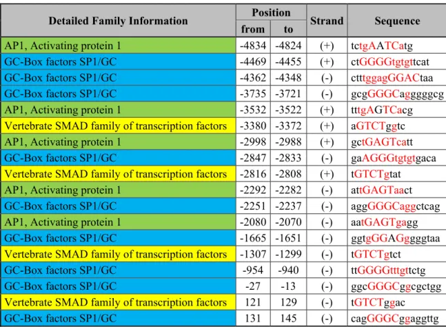
In order to elucidate which TFBS could be responsible for the TGF-β mediated expression of CTGF in myoblasts, we used the MatInspector tool in order to identify the TFBS present in the CTGF promoter with special focus on those related to TGFβ. The analysis, showed 1250 putative TFBS, including several sites related to β. The TFBS of importance for TGF-β/SMAD are summarized in Table 1: eight AP-1 sites were found, together with ten SP1 sites and four SBEs. A TATA-box (-38 to -32) was also recognized in the CTGF gene.
The 5’ UTR region of the CTGF gene bears elements of transcriptional regulation in response to TGF-β
To analyze which TFBS are responsible for the expression of CTGF in response to TGF-β, we constructed deletion mutants by PCR as shown in Figure 3 and we conducted reporter assays on
myoblasts. As shown in Figure 4, all the deletion mutants showed a significant decrease in the activation mediated by TGF-β and no additional decrease was found between the deletion mutants, suggesting that the region comprised between -4872 and -4578 of the CTGF promoter region carries regulatory elements that could be controlling the TGF-β mediated expression of CTGF in myoblasts. This region has several putative TFBS that could account for the decrease in transcriptional activation of the CTGF promoter when deleted and we chose to test the AP-1 site (tctgAATCatg) located in -4834 to -4824 (Table 1), because it has been shown that AP-1 transcription factors could act synergically with SMAD3 to promote gene expression (Verrecchia et al. 2001, Sundqvist et al. 2013, Bai et al. 2014). The mutation of the AP-1 site showed no decrease of the reporter gene expression (Supplementary Figure 1A and 1B) suggesting that the AP-1 site is not involved in the TGF-β mediated expression of CTGF. Further analyses are required in order to comprehend how this region regulates the expression of the CTGF gene in myoblasts.
The bioinformatical analysis also showed the presence of a SBE in the 5’ UTR region of the CTGF promoter (Table 1). Therefore, we first decided to construct a deletion mutant of pmCTGF-5.1 that lacks most of the 5’ UTR region of the CTGF gene (Figure 5A). The deletion of the 5’ UTR region of the CTGF gene showed a significant reduction on the TGF-β mediated CTGF expression (Figure 5B), suggesting that there are relevant TFBS that are responsible for the induction of the CTGF gene expression by TGF-β.
A SBE in the 5’UTR of the CTGF gene is important for the induction of CTGF by TGF-β In the 5’ UTR of the CTGF gene, there is a SBE (Table 1, 121 to 129) that could be important for the induction of CTGF by TGF-β in myoblasts. To test this hypothesis, we mutated the SBE
changing the important nucleotides in the sequence (Figure 6A) and performed reporter assays of the constructs. As seen in Figure 6B, there is a significant reduction in the expression of the reporter gene when the SBE is mutated, suggesting that this sequence is important for the transcriptional control of CTGF expression by TGF-β in myoblasts. It is important to notice that the mutation on the SBE reduced the expression of the reporter gene expression by 32% (Figure 6B), whereas the deletion of the 5’ UTR reduced the expression by 67% (Figure 5B). This suggests that there are additional TFBS that are important in the TGF-β mediated expression of CTGF. Further experiments will be required in order to fully understand the mechanisms involved in this process in myoblasts.
Discussion
CTGF plays a central role in the onset and maintenance of fibrosis in the skeletal muscle. Exogenous increase of CTGF in the muscle of wild type mice can induce augmented extracellular matrix components and decrease isometric force in the muscle, all features of dystrophic pathologies (Morales et al. 2011). Interestingly, returning of CTGF expression to normal levels, led to a reversion of the fibrotic phenotype (Morales et al. 2011), showing that therapies against fibrotic pathologies could be successful even when the disease is already present. Also, reducing CTGF levels slowed down the progression of muscular dystrophy in the mdx mice and led to an increase in cell therapy (Morales et al. 2013).
There are compiling evidence showing that the muscle fiber might be an important source for CTGF production in the dystrophic context. Usually, CTGF is not expressed in the normal state of the muscle but CTGF levels increase importantly when damage and inflammation are present, and under pathological conditions (Cabello-Verrugio et al. 2012). TGF-β is increased in the
muscles of DMD patients (Bernasconi et al. 1995) and those of several dystrophic mice (Onofre-Oliveira et al. 2012). Moreover, CTGF also contributes to an increase in TGF-β binding to its receptors and an increase in TGF-β signaling (Abreu et al. 2002). It has also been shown that CTGF and TGF-β act cooperatively to elicit a fibrotic tissue response (Wang et al. 2011). In the mdx mice, TGF-β expression seems to be originated in areas populated by inflammatory cells and regenerating fibers (Zhou et al. 2006). This correlates with the fact that CTGF is expressed in the endomysium and regenerating fibers of human dystrophic patients (Sun et al. 2008) and, as we showed in here, that CTGF promoter can be activated in the skeletal muscle of mice. However, the induction of CTGF mediated by TGF-β in muscle cells has not been extensively studied. We have previously shown that TGF-β can induce the expression of CTGF in myoblast and C2C12-derived myotubes (Vial et al. 2008), this has been also found in rat L6-derived myotubes (Maeda et al. 2005), so it is of particular interest to characterize the regulation of the expression of CTGF in myoblasts, myotubes and skeletal muscle. Further analyses will be required in order to fully comprehend how CTGF expression is regulated in muscle cells. In this paper, we found a novel SMAD Binding Element in the 5’ UTR of the CTGF gene that is important for the TGF-β mediated expression of CTGF in myoblasts. In addition, we showed that the 5’ UTR might have additional TFBS important for CTGF expression in myoblasts. The full 5’ UTR region was not included in previous studies regarding TGF-β mediated CTGF expression (Chen et al. 2002, Holmes et al. 2003, Leask et al. 2003, Xia et al. 2007, Tran et al. 2010) and our bioinformatical analysis shows the presence of several TFBS that could be acting together with the SBE described in this paper for the TGF-β mediated CTGF expression in myoblasts. It is known that SBEs alone are not strong enough to confer TGF-β inducibility, due to SMADs low binding affinity to this site (Massague et al. 2005), this might be the reason why
we couldn’t immunoprecipitate SMAD3 in the CTGF promoter when we analyzed the novel SBE located in the 5’ UTR (data not shown). Due to the possible interaction of SMAD3 with other factors and the formation of a bigger transcriptional complex, the SMAD3 protein might have not been exposed within the complex to allow the recognition by the antibody during the ChIP procedure. Within the 5’ UTR region that confers TGF-β induction to the reporter gene, we found several TFBS that could be acting together with the SBE in the 5’ UTR of CTGF gene. Among these TFBSs, a SP1 site which is located in very close proximity to the SBE (131 to 145) and could be evaluated for the TGF-β induction of CTGF, as the SP1 factors are reported to be acting together with SMADs proteins to enhance transcription (Botella et al. 2009, Lu et al. 2010, Fausther et al. 2012).
Our data also indicates that an upstream region (-4872 and -4578) of the CTGF promoter is involved in TGF-β mediated expression of CTGF and that the AP-1 site located in this region would not be involved with this induction. Between the TFBSs found in this region, there are two TCF/LEF (-4810 to -4794 and -4721 to -4705) sites that could be implicated in the expression of CTGF. Interestingly, several experimental evidences show a cross-talk between Wnt and TGF-β signaling, and Wnt pathway has been proposed as a novel therapeutical target for fibrotic disorders (reviewed in (Cisternas et al. 2014)).
The GTGTCAAGGGGTC element described first as a TGF-β response element (Grotendorst et al. 1996) and later named BCE-1 (Chen et al. 2002), was recognized as a RXR heterodimer and Nuclear receptor subfamily 2 factors binding site in our bioinformatical analysis (160 to -148). This site was recognized by Retinoic Acid Receptor/Retinoid X Receptor (RAR/RXR) heterodimers and was important for All-trans retinoic acid (ATRA) mediated expression of CTGF in fibroblasts (Fadloun et al. 2008). In addition, ATRA therapy Induces myositis in
leukemia patients (van Der Vliet et al. 2000, Pecker et al. 2014), suggesting that ATRA might have a role in inflammation in the muscle. On the other hand, in activated and hence, fibrogenic hepatic stellate cells that participates in hepatic fibrosis, the levels of RAR and RXR were diminished, together with lower concentration of RXR/RAR activators (Ohata et al. 1997) and, in another study, several agonists of RAR and RXR produced a decrease in the expression of fibrotic proteins (Hellemans et al. 2004). ATRA was also found to reduce the expression of TGF-β and CTGF in scleroderma fibroblasts (Xiao et al. 2011). Moreover, ATRA has been reported to reduce TGF-β expression and signaling in lung fibrosis (Song et al. 2013) and mesangial cells (Han et al. 2014). Taken together, this evidence shows that the effect of retinoic acid in fibrosis is not yet clear (reviewed in (Zhou et al. 2012)) but it could be an interesting approach to explore its effect in CTGF and TGF-β expression and signaling in muscular fibrosis.
Recently, it has been shown a fibrotic effect of CTGF in the absence of TGF-β signaling in liver fibrosis (Sakai et al. 2014). This evidence shows the relevance of CTGF in fibrotic disorders and that, other signaling pathways, besides TGF-β/SMAD signaling, are involved in CTGF expression. Therefore, the study of the precise regulation of CTGF expression can be helpful to understand the mechanisms of the onset and progression of fibrosis in different tissues.
Acknowledgments
This study was supported by research grants from CARE PFB12/2007, FONDECYT 1110426, Fundación Chilena para Biología Celular Proyecto MF-100. Beca VRI de doctorado 2007, PUC. Beca de doctorado 2008 and Beca de apoyo a la realización de tesis doctorado 2009-2010,
CONICYT. Beca de pasantía MECESUP 2009 and 2011. Beca Chile cotutela de doctorado 2010.
We are grateful to the Animal Housing Facility (in vivo experiments) of the Centre de Recherche Pharmaceutique de Paris (Paris Descartes University).
Disclosure
The authors declare they have no competing interests as defined by molecular medicine, or other interests that might be perceived to influence the results and discussion reported in this paper.
References
Abraham, D. J., X. Shiwen, C. M. Black, S. Sa, Y. Xu and A. Leask (2000). Tumor necrosis factor alpha suppresses the induction of connective tissue growth factor by transforming growth factor-beta in normal and scleroderma fibroblasts. J Biol Chem 275(20): 15220-15225.
Abreu, J. G., N. I. Ketpura, B. Reversade and E. M. De Robertis (2002). Connective-tissue growth factor (CTGF) modulates cell signalling by BMP and TGF-beta. Nat Cell Biol 4(8): 599-604.
Bai, G., T. D. Hock, N. Logsdon, Y. Zhou and V. J. Thannickal (2014). A far-upstream AP-1/Smad binding box regulates human NOX4 promoter activation by transforming growth factor-beta. Gene 540(1): 62-67.
Bernasconi, P., C. Di Blasi, M. Mora, L. Morandi, S. Galbiati, P. Confalonieri, F. Cornelio and R. Mantegazza (1999). Transforming growth factor-beta1 and fibrosis in congenital muscular dystrophies. Neuromuscul Disord 9(1): 28-33.
Bernasconi, P., E. Torchiana, P. Confalonieri, R. Brugnoni, R. Barresi, M. Mora, F. Cornelio, L. Morandi and R. Mantegazza (1995). Expression of transforming growth factor-beta 1 in dystrophic patient muscles correlates with fibrosis. Pathogenetic role of a fibrogenic cytokine. J Clin Invest 96(2): 1137-1144.
Blake, D. J., A. Weir, S. E. Newey and K. E. Davies (2002). Function and genetics of dystrophin and dystrophin-related proteins in muscle. Physiol Rev 82(2): 291-329.
Blau, H. M., G. K. Pavlath, E. C. Hardeman, C. P. Chiu, L. Silberstein, S. G. Webster, S. C. Miller and C. Webster (1985). Plasticity of the differentiated state. Science 230(4727): 758-766.