HAL Id: tel-01356238
https://tel.archives-ouvertes.fr/tel-01356238
Submitted on 25 Aug 2016HAL is a multi-disciplinary open access archive for the deposit and dissemination of sci-entific research documents, whether they are pub-lished or not. The documents may come from teaching and research institutions in France or abroad, or from public or private research centers.
L’archive ouverte pluridisciplinaire HAL, est destinée au dépôt et à la diffusion de documents scientifiques de niveau recherche, publiés ou non, émanant des établissements d’enseignement et de recherche français ou étrangers, des laboratoires publics ou privés.
synapse formation : towards potential therapies for
intellectual disability
Mariana Ramos
To cite this version:
Mariana Ramos. Unraveling the impact of IL1RAPL1 mutations on synapse formation : towards potential therapies for intellectual disability. Neurons and Cognition [q-bio.NC]. Université Sorbonne Paris Cité, 2015. English. �NNT : 2015USPCB058�. �tel-01356238�
THESE
Ecole doctorale BioSPC
Spécialité Neurosciences
Présentée par
Mariana Ramos-Brossier
Pour obtenir le grade de
DOCTEUR DE L’UNIVERSITE PARIS DESCARTES
Unraveling the impact of IL1RAPL1 mutations on synapse
formation: towards potential
therapies for intellectual
disability
Sous la direction du Dr. Pierre Billuart
Soutenue le 9 octobre 2015, devant le jury composé de :
Dr. Barbara Bardoni
Rapportrice
Dr. Cyrille Vaillend
Rapporteur
Dr. Patricia Gaspar
Examinatrice
Dr. Yann Humeau
Membre invité
REMERCIEMENTS
Je remercie avant tout Pierre Billuart pour sa grande disponibilité et pour sa direction tout au long de cette thèse. Je lui suis reconnaissante de m’avoir fait confiance et m’avoir donné une grande liberté. Merci pour ces 4 années et tout ce qu’elles m’ont apporté scientifiquement et humainement.
Je remercie les docteurs Barbara Bardoni, Cyrille Vaillend et Patricia Gaspar pour le temps consacré à l’évaluation et à la critique de ce travail.
Un grand merci à tous les collaborateurs qui ont enrichi ce travail:
Yann Humeau à Bordeaux, pour ses commentaires et sa collaboration tout au long de ce travail de thèse, et pour m’avoir initié à l’électrophysiologie. Je remercie aussi les membres de son équipe, Xander, Etienne, Christelle, Marylin, Elizabeth pour leur aide précieuse et leur bonne humeur.
Ringrazio Carlo Sala e Caterina Montani a Milano per la loro collaborazione, che e stata une esperienza molto gratificante.
Hamid Meziane et Yann Hérault à Illkich, pour les études de comportement. Jozef Gecz and Anna Hacket in Australia, and Franck Kooy and Bart Loeys in Belgium, for their invaluable contribution to the genetics studies.
Un grand merci à l’Ecole des Neurosciences de Paris, non seulement pour le financement, mais aussi pour leur soutien logistique dès mon arrivé à Paris, et pour m’avoir offert l’opportunité d’appartenir à ce grand réseau. Merci particulièrement à Jean Antoine Girault, Yvette Henin, Andre Sobel, Laura et Deborah. Je remercie également la Fondation EDF, qui m’a permis de travailler une année de plus pour terminer mes travaux en cours.
Je remercie l’Université Paris Descartes, et à Michelle Pitte pour l’aide administrative.
Merci à tous les membres de l’Institut Cochin qui sourient (ou pas) dans les couloirs, et à Agnès Fouet pour sa bienveillance. Merci aux plateformes de l’Institut Cochin sans lesquelles ce travail n’aurait pas pu avoir lieu, spécialement la plateforme de microscopie (Béatrice, Thomas et Pierre) pour leur aide et expertise précieuse, ainsi qu’à l’animalerie.
Un énorme merci aux membres de l’équipe BiBi:
Jamel Chelly pour m’avoir accueilli dans son équipe et pour ses conseils précieux. Olivier pour son conseil scientifique; Nicolas pour ses critiques toujours constructives et son aide précieuse; Chloé pour sa bonne humeur et son exemple de travail ; Yoann a pesar de que nuestra relación tuvo un inicio tormentoso, gracias por tu ayuda siempre.
Ainsi qu’à à tous les membres présents et passés du laboratoire : Thierry, Nadia, Laurence, Karine, Juliette, Giuseppe, Adelyn, Léa, Audrey, Loïc, Julia, Miriam, Christine, Adrienne, Devina, Justine, Matthieu, Laure Elise, Anne, Benjamin, Pang, France, Madison, Cécile, Remi, Bénédicte, Marine, Mélanie, Claire, Hamida, Sylvain…
Je voudrais remercier particulièrement les membres de la plateforme 5004 pour leur amitié et les bonbons partagés au rythme de radio latina: Rodrick, pour faire monter la température dans le bureau; Julie pour toutes les discussions et pour être toujours à l’écoute; Flora pour sa franchise et son énergie; Julien pour partager mes peines de neurones; et Laetitia pour sa grande gentillesse, repose en paix.
Pendant cette thèse j’ai eu l’opportunité d’encadrer Geoffroy Goujon, que je remercie pour son aide technique et son optimisme. C’était une grande expérience pour moi, et je lui souhaite beaucoup de réussite pour la suite.
Merci à Maryse pour son efficacité imbattable et sa bonne humeur, et à Josiane, Nathalie, Mireille et Annick pour l’animation dans les couloirs du 5ème.
Merci à Julie Dam, Ralph Jokers, Denis Hervé, Salah El Mestikawy, Diana Sakae et Bruno Ragazzon pour leur aide et conseils techniques.
Gracias a Magda Giordano y Ranulfo Romo por sus consejos, y por apoyarme y alentarme a venir a Francia.
Merci à toutes les personnes qui ont réaffirmé mon engagement envers la vulgarisation scientifique, et qui m’ont permis d’apprendre et de m’améliorer. A tous ceux qui ont subi les présentations, résumés, posters et explications, et qui ont survécu pour me dire ce qui n’était pas clair.
À mes parents, ma famille et à mes amis, au loin mais près au même temps, et à ceux qui m’ont entouré, motivé et inspiré depuis mon arrivée en France. A Ximena por estar ahí siempre, y en todos lados. A Benjas, Lalo y Nica por tantas risas. A Yavé, Maja, Ana, Gerardo, Inseparable, Oscar, Mag, Romu, Lily, Oriane, Benjamin, Olivia, à tous les Brossier et les Percoco. Merci aux salseros, joueurs et aux polyglottes pour les bons moments partagés. A Elisa que no deja de hacerme sentir orgullosa.
A Stefen, qui a cumulé 58,630,231 points-thèse grâce à son soutien constant et inconditionnel, indispensable à la réalisation de cette thèse. Merci d’être monté dans ce train et parcourir avec moi autant de kilomètres.
ABSTRACT
Preserving the integrity of neuronal synapses is important for the development and maintenance of cognitive capacities. Mutations on a growing number of genes coding for synaptic proteins are associated with intellectual disability (ID), a neurodevelopmental disease characterized by deficits in adaptive and intellectual functions. The present work is dedicated to the study of one of those genes, IL1RAPL1, and the role of its encoding protein in synapse formation and function. IL1RAPL1 is a trans-membrane protein that is localized at excitatory synapses, where it interacts with the postsynaptic proteins PSD-95, RhoGAP2 and Mcf2l. Moreover, the extracellular domain of IL1RAPL1 interacts trans-synaptically with the presynaptic phosphatase PTPδ.
We studied the functional consequences of two novel mutations identified in ID patients affecting this IL1RAPL1 domain. Those mutations lead either to a decrease of the protein expression or of its interaction with PTPδ, affecting in both cases the IL1RAPL1-mediated excitatory synapse formation. In the absence of IL1RAPL1, the number or function of excitatory synapses is perturbed, leading to an imbalance of excitatory and inhibitory synaptic transmissions in specific brain circuits. In particular, we showed that this imbalance in the lateral amygdala results in associative memory deficits in mice lacking Il1rapl1. Altogether, the results included in this work show that IL1RAPL1/PTPδ interaction is essential for synapse formation and suggest that the cognitive deficits in ID patients with mutations on IL1RAPL1 result from the imbalance of the excitatory and inhibitory transmission. These observations open therapeutic perspectives aiming to reestablish this balance in the affected neuronal circuits.
Key-words: intellectual disability, synapses, IL1RAPL1, excitatory/inhibitory balance.
RESUME
L’intégrité des synapses neuronales est primordiale pour le développement et le maintien des capacités cognitives. Des mutations dans des gènes codant pour des protéines synaptiques ont été trouvées chez des patients atteints de déficience intellectuelle (DI), qui est une maladie neurodéveloppementale ayant des conséquences sur les fonctions intellectuelles et adaptatives. Ce travail de thèse porte sur l’étude de l’un de ces gènes, IL1RAPL1, dont les mutations sont responsables d’une forme non-syndromique de DI liée au chromosome X, et sur le rôle de la protéine IL1RAPL1 dans la formation et le fonctionnement des synapses. IL1RAPL1 est une protéine trans-membranaire qui est localisée dans les synapses excitatrices où elle interagit avec les protéines post-synaptiques PSD-95, RhoGAP2 et Mcf2l. De plus, IL1RAPL1 interagit en trans- avec une protéine phosphatase présynaptique, PTPδ, via son domaine extracellulaire.
Nous avons étudié les conséquences fonctionnelles de deux nouvelles mutations qui affectent le domaine extracellulaire d’IL1RAPL1 chez des patients présentant une DI. Ces mutations conduisent soit à une diminution de l’expression de la protéine, soit à une réduction de l’interaction avec PTPδ affectant ainsi la capacité d’IL1RAPL1 à induire la formation de synapses excitatrices. En absence d’IL1RAPL1, le nombre ou la fonction des synapses excitatrices est diminué, ce qui mène à un déséquilibre entre les transmissions synaptiques excitatrice et inhibitrice dans des régions spécifiques du cerveau. Dans le cas particulier de l’amygdale latérale, nous avons montré que ce déséquilibre conduit à des défauts de mémoire associative chez la souris déficiente en Il1rapl1.
L’ensemble des résultats qui font partie de ce travail montre que l’interaction IL1RAPL1/PTPδ est essentielle pour la formation des synapses et suggère que les déficits cognitifs des patients avec une mutation dans il1rapl1 proviennent du déséquilibre de la balance excitation/ inhibition. Ces observations ouvrent des perspectives thérapeutiques visant à rétablir cette balance dans les réseaux neuronaux affectés.
TABLE OF CONTENTS
ABBREVIATIONS ... 1
INTRODUCTION ... 3
1. Synapses – targets for intellectual disability ... 3
1.1. Synaptic functions are impaired in intellectual disability ... 6
1.1.1. Regulation of cytoskeleton dynamics ... 7
1.1.2. Presynaptic vesicle cycling and exocytosis ... 10
1.1.3. Organization of postsynaptic protein complexes ... 12
1.1.4. Trans-synaptic signaling ... 14
2. IL1RAPL1, a synaptic protein implicated in non-syndromic ID... 15
2.1. Identification of IL1RAPL1 as a gene related to ID ... 15
2.2. IL1RAPL1 structure and expression ... 17
2.3. Molecular partners of IL1RAPL1 and effects on neuronal physiology ... 19
2.3.1. NCS-1 and calcium-regulated exocytosis ... 20
2.3.2. PSD-95 and excitatory postsynaptic organization ... 22
2.3.3. PTPδ and synaptogenesis ... 24
2.3.4. Regulators of Rho GTPases and neuronal morphology ... 31
2.3.4.1. RhoGAP2 and dendritic spine formation ... 32
2.3.4.2. Mcf2l and stabilization of glutamatergic synapses... 33
2.3.4.3. Neurite branching ... 34
2.3.5. Other potential IL1RAPL1 interacting proteins ... 35
2.4. Phenotypic characterization of Il1rapl1 KO mouse ... 36
2.5. IL1RAPL1 is a member of IL1 receptor family ... 38
2.5.1. IL1β signaling in the brain and regulation of synaptic function ... 38
2.5.2. Regulation of cognitive functions by IL1R members ... 43
TABLE OF CONTENTS
2.5.3. IL1 receptor family members involved in synaptogenesis ... 43
2.5.4. IL1β-induced signaling in neurons and astrocytes ... 44
RESULTS ... 48
1. Novel IL1RAPL1 mutations associated with intellectual disability impair synaptogenesis. ... 49
2. Target-specific vulnerability of excitatory synapses leads to deficits in associative memory in a model of intellectual disorder. ... 67
3. Unpublished work ... 85
3.1. Behavioral and cellular deficits in Il1rapl1 KO mouse are rescued by α5IA treatment ... 85
3.1.1. Context ... 85
3.1.2. Material and methods ... 88
3.1.3. Results ... 90
DISCUSSION AND CONCLUSIONS... 94
1. Importance of IL1RAPL1 - PTPδ interactions for synapse formation ... 94
2. Role of IL1RAPL1 in synapse formation and in E/I balance ... 100
3. Towards a treatment for restoring E/I balance and improve cognitive deficits in ID mouse models ... 104
APENDIX ... 108
LIST OF FIGURES
Figure 1. Neurons are polarized cells. ... 3
Figure 2. Chemical synapses are excitatory or inhibitory. ... 4
Figure 3. Regulation of small GTPases activity by GEFs and GAPs. ... 8
Figure 4. Regulators of small GTPases associated with ID. ... 10
Figure 5. Presynaptic proteins associated with ID. ... 11
Figure 6. Postsynaptic organization is affected in ID. ... 13
Figure 7. Cell adhesion is affected in ID. ... 15
Figure 8. Gene and protein organization of human IL1RAPL1. ... 18
Figure 9. IL1RAPL1 is present in excitatory but not inhibitory synapses. ... 19
Figure 10. IL1RAPL1 regulates N-type Ca2+ current through NCS-1. ... 21
Figure 11. IL1RAPL1 interacts with PSD-95 and regulates its targeting to excitatory postsynapses. ... 24
Figure 12. Pre and postsynaptic differentiation is mediated by different IL1RAPL1 protein domains. ... 25
Figure 13. PTPδ structure and trans-synaptic interaction with the extracellular domain of IL1RAPL1. ... 27
Figure 14. Mapping of the IL1RAPL1/PTPδ interaction. ... 29
Figure 15. IL1RAPL1 interacts with two regulators of small GTPases activity through its TIR domain, RhoGAP2 and Mcf2l. ... 32
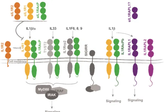
Figure 16. Interleukin 1 receptor family of proteins regulate cytokines signaling. ... 39
Figure 17. IL1β induces different cellular signaling in different brain cell types. ... 45
Figure 18. Crystal structure of Il1rapl1 and Ptpδ interaction. ... 51
Figure 19. Excitatory/inhibitory balance is perturbed in the absence of Il1rapl1. ... 68
Figure 20. Effect of α5IA on contextual memory in wild-type and Il1rapl1 KO mice.. 90
Figure 21. Gabra5 mRNA relative abundance in wild-type and Il1rapl1 KO hippocampal neurons in culture. ... 91
LIST OF FIGURES
Figure 22. c-Fos mRNA relative abundance in hippocampal neurons after bicuculline or α5IA treatment ... 92 Figure 23. Postsynaptic changes after α5IA treatment ... 93 Figure 24. Presynapses are not altered after α5IA treatment ... 93 Figure 25. Synaptogenesis period in rat hippocampus and in hippocampal neurons in culture. ... 95 Figure 26. Cerebellar circuits are affected during development in the absence of Il1rapl1. ... 102 Figure 27. Possible mechanism for α5IA-induced improvement in hippocampal-dependent tasks in Il1rapl1 KO mice ... 106 Table 1. Reported mutations on IL1RAPL1 in ID patients and their consequences for protein function ... 108
1
ABBREVIATIONS
α5IA 3-(5-methylisoxazol-3-yl)-6-[(1-methyl-1, 2, 3-triazol-4-yl) methyloxy]-1, 2,
4-triazolo [3, 4-a] phthalazine
AHC adrenal hypoplasia congenital,
AMPA α-amino-3-hydroxy-5-methylisoxazole-4-propionic acid
ASD autism spectrum disorder
BRET bioluminescence resonance energy transfer
CAMs cell adhesion molecules
CNV copy number variant
DCN deep cerebellar nuclei
E/I excitation/inhibition
ECD extracellular domain
G granule cells
GABA γ-aminobutyric acid
GAP GTPase-activating proteins
GDP guanosine diphosphate
GEF guanine nucleotide exchange factors
GKD glycerol kinase deficiency
GTP guanosine triphosphate
ID intellectual disability
IEG immediate early gene
Ig immunoglobulin
IL1R1 interleukin 1 receptor
IL1RAcP interleukin 1 receptor accessory protein
IL1RAcPb interleukin 1 receptor accessory protein isoform b
IL1RAPL1 interleukin 1 receptor accessory protein like 1
IL1β interleukin 1 beta
In interneurons
IQ intelligence quotient
JNK c-Jun N-terminal kinase
KO Knockout
LA lateral amygdala
LTD long-term depression
2
MAPK Mitogen-activated protein kinases
MD muscular dystrophy
meA/B mini-exon A/B
mEPSCs miniature excitatory postsynaptic currents
NCS-1 neuronal calcium sensor-1
NFκB nuclear factor κB
NMDA N-methyl-D aspartate
NPCs neural precursor cells
NT neurotransmitter
N-VGCC N-type voltage-gated calcium channels
P post-natal
PC Purkinje cells
PSD postsynaptic density
PSD-95 postsynaptic density protein 95
PTPδ protein tyrosine phosphatase δ
SAPK stress-activated protein kinases
SE startle epilepsy
SEM standard error of the mean
sIPSC spontaneous inhibitory post-synaptic currents
SV synaptic vesicle
VGlut1 vesicular glutamate transporter 1
WS West syndrome
3
INTRODUCTION
1. Synapses – targets for intellectual disability
Synapses are the elementary units in the formation of the neural circuits that regulates all the functions of the nervous system. These specialized structures mediate the contact and communication between nerve cells, and in mammalian brains we can count several billions of synapses (~1015 of synapses in the human brain). Although
electrical synapses occur in every brain region, the most common mechanism for signaling between neurons is the neurotransmitter-releasing chemical synapse. Throughoutthis text I will talk about chemical synapses, that are formed by the axon of a presynaptic neuron, and either the dendrites or the cell body of the target postsynaptic neuron (Figure 1).
Neurons are polarized cells extending neurites, dendrites and axon, away from their cell body in order to contact other nerve cells.
Figure 1. Neurons are polarized cells. An hippocampal neuron in culture was transfected with a plasmid carrying GFP fluorescent protein for visualization of neurites (dendrites and axons) and dendritic spines. The inset in the right shows a dendrite and dendritic spines. Synapses are mainly formed between axons and dendrites, as shown in the left inset. Scale bar 10 μm.
4 The morphology of neurites affects synaptic signaling, integration, and connectivity, and their diversity reflects the complexity and specificity of neural circuits. Neurites formation begins shortly after neurons complete their migration during brain development, and synapse formation, or synaptogenesis in the human brain occurs between gestational age week 20 until the adolescence (Tau & Peterson 2010). Scaffolding cells and molecular gradients are important in the assembly of synaptic connections, which will be constantly refined and modified throughout life. Synapses consist of two asymmetrically components separated by the synaptic cleft (about 20 nm): a presynaptic and a postsynaptic specialization that differ in their chemical, structural and functional characteristics. Even if large evidence of the participation of non-neuronal (glial) brain cells on synapse physiology, like microglia and astrocytes, I will only focus on the neuronal pre and postsynaptic partners.
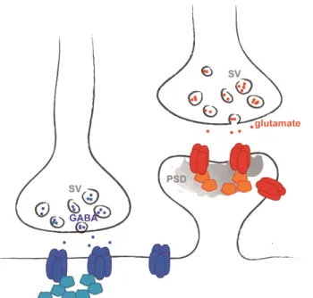
Two major classes of chemical synaptic transmission are present in the central nervous system, excitatory and inhibitory (Figure 2). The differences arise from the type of neurotransmitter released (glutamate or γ-aminobutyric acid (GABA), respectively) and
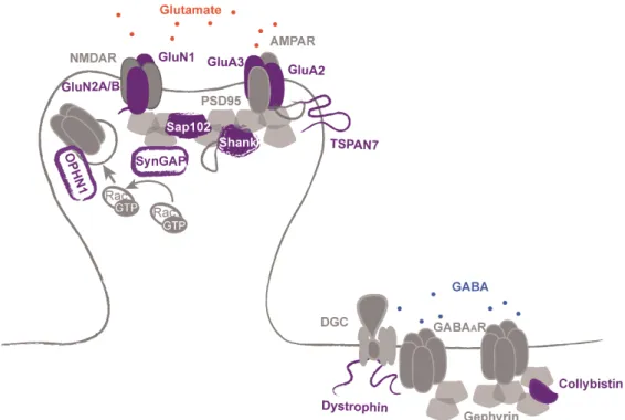
Figure 2. Chemical synapses are excitatory or inhibitory. Excitatory glutamatergic (red) are housed on dendritic spines, and inhibitory GABAergic (blue) synapses on dendritic shafts or neuronal soma. Each type of synapse is characterized by different neurotransmitter (NT), receptors (dark blue and red) and scaffolding proteins (light blue and orange). SV: synaptic vesicle. PSD: postsynaptic density.
5 from the molecular components (receptors, scaffolding proteins, effectors) that are present in them. The total number of synapses formed and the ratio of excitatory/inhibitory synaptic inputs that a neuron receives determine its excitability. Most excitatory synapses are housed on dendritic spines, small actin-rich protrusions extending from dendrites, and inhibitory synapses are formed on the dendritic shaft and on the cell soma. Excitatory synapses are characterized by a morphological and functional specialization of the postsynaptic membrane called the postsynaptic density (PSD), which is usually located at the tip of the dendritic spine. The PSD contains the glutamate receptors, as well as a large diversity of associated signaling and structural molecules.
Synapses have a high degree of molecular complexity, with elements acting as neurotransmitter receptors, adhesion molecules, signaling effectors, scaffolds, kinases and phosphatases, translation factors, cytoskeleton, ion channels, modification enzymes… Comparative studies between different species have suggested that the evolutionary diversification of the synapse components underlies the complexity in signal processing and behavior (Emes et al. 2008; Bayés et al. 2012).
Communication at chemical synapses involves the release of neurotransmitters (NT) from the presynaptic terminals in response to electrical impulses (action potentials), diffusion of the NT across the synaptic clefts, and NT binding to postsynaptic receptors. The postsynaptic compartment converts these chemical signals back into action potentials, allowing their propagation. The presynaptic terminals contain synaptic vesicles (SV) filled with neurotransmitters and a dense matrix of cytoskeleton and scaffolding proteins, the active zone, that contains all the machinery to release SV into the synaptic cleft. Diverse cell-adhesion molecules hold pre and postsynaptic specializations together through trans-synaptic interactions. On the postsynaptic specialization, glutamate and GABA receptors interact with the neurotransmitter and transduce its binding into electrical excitation or inhibition of the postsynaptic cell. These receptors form large signaling complexes, which send downstream signals into the postsynaptic cell and mediate feedback regulation of synaptic transmission. Dynamic regulation of function and localization of glutamate and GABA receptors mediate many forms of postsynaptic plasticity, including long-term potentiation (LTP) and long-term depression (LTD) of synaptic strength (two processes considered as representing the cellular basis of learning and memory), as well as the coupling of
6 synaptic activity to regulation of gene expression (Vithlani et al. 2011; Wang et al. 2012).
Given the crucial role of synapses on brain function, it’s not surprising that disruption in key synaptic components lead to brain diseases. These synaptic diseases, or synaptopathies, have received particular interest in recent years (Grant 2012).
Since large scale screening of genetic information is available, the understanding of synapse pathology is increasing by systematically examining mutations in genes coding for all proteins in the synapse. Using this approach, it has been estimated that mutations on genes coding for synaptic proteins may play a role in around 133 brain diseases, including neurodegenerative diseases, motor disorders, epilepsy and cognitive disorders such as intellectual disability (ID) (Bayés et al. 2011).
To date, a large amount of genes encoding for synaptic proteins were associated with intellectual disability, the principal interest of the present work. Throughout this manuscript, I will focus on one of these genes, interleukin 1 receptor accessory protein-like 1 (IL1RAPL1) and address the consequences of mutations on this gene on synaptic formation and function.
1.1. Synaptic functions are impaired in intellectual disability
Intellectual disability (ID) is a common neurodevelopmental disorder characterized by an intelligence quotient (IQ) of 70 or below, and by impairments in adaptive behaviors including conceptual, social and practical areas (American Psychiatric Association 2000). This disorder was formerly known as mental retardation, but the term was changed in October 2010, when Rosa’s law was signed in the United States as an effort to change the stigma among ID patients. The prevalence of ID is between 1 and 3% and is present in every social class and culture. ID was formerly classified in the basis of the IQ (from mild to profound severity) or into syndromic and non-syndromic depending on the presence of other clinical or morphological features. Recently, the DSM-5 rather set the severity in function of the adaptive behavior. Moreover, the classification of non-syndromic ID is debated because even if it has been traditionally defined by the presence of intellectual disability as the sole clinical feature, it is difficult to rule out the presence of more subtle neurological anomalies and psychiatric disorders in these patients.
7 ID can be caused by environmental and/or genetic factors. However, for up to 60% of cases there is no identifiable cause. Environmental exposure to certain teratogens, viruses or radiation can cause ID, as can severe head trauma or injury causing lack of oxygen to the brain. Genetic causes accounts for 25–50% of ID cases, although this number increases with ID severity. Among the genetic causes, chromosome abnormalities, large deletions and pathogenic copy number variants (CNV) have been found to be associated with ID in a large number of studies, and have contributed to the discovery of many ID-associated genes. Down syndrome, caused by human trisomy 21, and Fragile X syndrome due to mutations in the FMR1 gene, are the most frequent genetic form of ID (Ropers 2010). A significant number of ID genes code for synaptic proteins, and it is noteworthy that many of them converge into common cellular pathways, allowing to determine the specific pathways perturbed in ID.
ID is considered as part of broad spectrum of neurodevelopmental disorders, including autism spectrum disorders (ASD). Clinically and genetically, these disorders are difficult to separate clearly, and the majority of ASD and ID genes share common cellular pathways. In this section I will describe briefly some of the synaptic features affected by genes whose mutations were described in patients with ID, associated or not with ASD or with other syndromes. These synapse-related ID proteins are involved in different cellular pathways but I will focus on those ID-related proteins involved in cytoskeleton dynamics, presynaptic vesicle cycling and exocytosis, organization of postsynaptic complexes and trans-synaptic signaling. This lets apart important processes underlying synaptic physiology like the regulation of transcription, protein synthesis and degradation that are also regulated by ID genes (Vaillend et al. 2008; Humeau et al. 2009; van Bokhoven 2011; Verpelli et al. 2013; Kroon et al. 2013; Srivastava & Schwartz 2014; Maurin et al. 2014; Volk et al. 2014).
1.1.1. Regulation of cytoskeleton dynamics
In mature neurons, the dendritic spines, small actin-rich protrusions extending from dendrites, house most excitatory synapses. Dendritic spines undergo marked changes in shape and number during development and in response to environmental stimuli. Changes in the number and morphology of spines and neurites are observed in several
8 neurodevelopmental diseases, including Rett syndrome, Fragile X syndrome, and ID (Xu et al. 2014; Castets et al. 2005; Khelfaoui et al. 2007).
Dynamic rearrangements of actin filaments, the predominant cytoskeletal element in dendritic spines (12% of total PSD proteins), regulates the formation and reorganization of dendritic spines (Matus 2000; Sheng & Kim 2011). Small GTPases are important regulators of the actin cytoskeleton that play essential roles in the development and remodeling of dendritic spines. These proteins account for the 8% of total PSD proteins and their activity is sensitive to synaptic transmission and can regulate activity-induced maturation of the synapse. This superfamily of proteins comprises several subfamilies like RhoA. The members of RhoA small GTPases subgroup include Ras homolog gene family member A (RhoA), Ras-related C3 botulinum toxin substrate (Rac1) and cell division cycle 42 (Cdc42). In neurons, Rac1, Ras and Cdc42 activity promote the formation, growth and maintenance of spines, whereas RhoA induces spine retraction and loss.
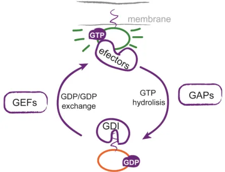
Figure 3. Regulation of small GTPases activity by GEFs and GAPs. In their GDP-bound state, small GTPases are maintained in an inactive form by GDIs and are located in the cytoplasm. GEFs catalyze the exchange of GDP for GTP and release small GTPases from the GDI complex. Active small GTPases translocate to the membrane, where they interact with their effectors. Small GTPases return to the inactive state by GAP-mediated GTP hydrolysis.
9 Spatial and temporal regulation of Rho GTPases activity is achieved by guanine nucleotide exchange factors (GEFs) and GTPase-activating proteins (GAPs) that activate and inhibit Rho GTPases activity respectively (Figure 3). In the inactive GDP-bound form, RhoA is locked in the cytosol by guanine dissociation inhibitors (GDIs). RhoGEFs catalyze the exchange of GDP for GTP to activate RhoA, by releasing it from the RhoA-GDI complex. Activated RhoA translocates to plasma membrane where it interacts with different effectors to transduce the signal. RhoA activation is turned off by RhoGAPs that induce the hydrolysis of GTP to GDP (Sasaki & Takai 1998). Other subfamilies of small GTPases, like Ras, Arf and Rab, similarly regulated by specific GAPs and GEFS, are also linked to cytoskeleton dynamics in neurons.
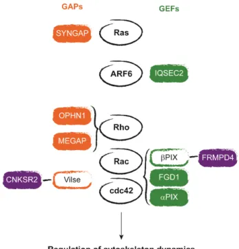
Mutations in regulators and effectors of the Rho GTPases have been found to underlay various forms of ID (syndromic written in green, and non-syndromic in blue) and are shown in Figure 4 (Newey et al. 2005; Ba et al. 2013). For example, the RhoGAPs oligophrenin 1 (OPHN1) and MEGAP (SRGAP3), the Rac and Cd42GEF αPIX (ARHGEF6) and FGD1 (FGD1), as well as the RhoGTPases effector p21-activating kinase 3 PAK3 (PAK3), are involved in regulating changes in spine morphology (Billuart et al. 1998; Endris et al. 2002; Lebel et al. 2002; Bienvenu et al. 2000; Boda et al. 2004). Spine morphogenesis is also regulated by CNKSR2/CNK2 (CNKSR2) that interacts principally with the Rac and Cdc42 GAP ARHGAP33 (also known as Vilse) but also with βPIX and the scaffolding protein PSD-95 (Tarpey et al. 2009; Houge et al. 2012; Lim et al. 2014; Hu et al. 2015). Preso (FRMPD4) is another PSD-95-interacting ID protein (Hu et al. 2015) that also associates with actin filaments and βPIX, and is a positive regulator of spine density (Lee et al. 2008). A member of the Ras superfamily, the small GTPase ADP-ribosylation factor 6 (ARF6) is also known to impact actin dynamics in the brain, and is regulated by the ID-related protein IQSEC2/BRAG2 (IQSEC2) (Shoubridge et al. 2010; Raemaekers et al. 2012). SYNGAP (SYNGAP) is a RasGAP that regulates spine maturation (Hamdan, Gauthier, et al. 2009; Hamdan, Daoud, et al. 2011; Clement et al. 2012). These examples strongly link ID with defects of actin dynamics that underlies defects in spine formation and morphology.
10
Figure 4. Regulators of small GTPases associated with ID. Proteins activating small GTPases (GAPs) are shown in orange, and GEFs are represented in green. Empty rectangles indicates proteins not associated with ID. Protein represented in violet interact with GAPs or GEFs and with the scaffolding protein PSD-95 found in excitatory postsynapses.
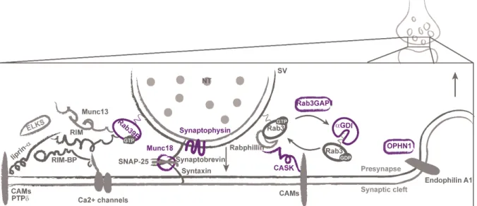
1.1.2. Presynaptic vesicle cycling and exocytosis
Neurotransmitters are stored in synaptic vesicles at presynaptic terminals. Several presynaptic proteins involved in the regulation of synaptic vesicle release, including vesicle docking, priming, fusion, endocytosis and recycling are found to be defective in syndromic (written in green) and non-syndromic (in blue) ID (Figure 5). Synaptic vesicle exocytosis is restricted to a small section of the presynaptic membrane called the active zone. This zone is composed of evolutionarily conserved proteins (the core is composed of RIM, Munc13, RIM-BP, liprin-α, and ELKS proteins) and performs the principal functions of neurotransmitter release: docks and primes synaptic vesicles, recruits calcium (Ca2+) channels to the docked and primed vesicles, tethers the
vesicles and Ca2+ channels to synaptic cell-adhesion molecules, and mediates
presynaptic plasticity (Südhof 2012). Because Ca2+ entry governs neurotransmitter
release, regulation of the Ca2+ channel is an important control point for synaptic
transmission on the presynapses. Upon stimulation, the synaptic vesicles are translocated to the active zone, anchored to the presynaptic membrane and made ready for fusion. In response to Ca2+ influx, docked vesicles undergo exocytosis and
11 release neurotransmitters into the synaptic cleft, where neurotransmitter can bind to their receptors in the postsynaptic element. The synaptic vesicle fusion machinery for exocytosis is mediated by the SNARE (soluble N-ethymalemide sensitive factor attachment protein receptor) complex composed of three SNARE proteins (syntaxin, SNAP-25, and synaptobrevin) and Munc18 (STXBP1), that was found mutated in patients with severe ID (Saitsu et al. 2008; Hamdan, Piton, et al. 2009). Synaptophysin (SYP) is also an ID protein found in synaptic vesicles that interacts with synaptobrevin, an essential component of SNARE complex (Tarpey et al. 2009; Gordon & Cousin 2013).
The synaptic vesicles are covered with Rab proteins, principally Rab3a, a subgroup of the Ras superfamily, which regulates vesicle trafficking and neurotransmitter release. Rab39B (Rab39B) and some proteins regulating this signaling pathway have been associated with ID: αGDI (GDI1), Rab3GAP1 (Rab3GAP1) and the calcium/calmodulin-dependent serine protein kinase CASK (CASK) (Giannandrea et al. 2010; D’Adamo et al. 1998; Aligianis et al. 2005; Hackett et al. 2010). Both, αGDI and Rab3GAP1 causes Rab3a to remain inactive by maintaining it in its GDP-bound form. CASK is a member of the membrane associated guanylate kinase (MAGUK) family of scaffolding proteins that interacts with rabphilin3a, an effector of Rab3a, which stabilizes Rab3a in its active state on the vesicle (Zhang et al. 2001).
Figure 5. Presynaptic proteins associated with ID. Protein members of the active zone and SNAREs are shown in the left. The ID presynaptic proteins involved in synaptic vesicles (SV) exocytosis and endocytosis (right) are shown in violet. CAMs: cell-adhesion molecules.
12 Loss of oligophrenin 1 signaling at presynapses has been shown to impair synaptic vesicle cycling at hippocampal synapses by forming a complex with endophilin A1, a protein implicated in several stages of synaptic vesicle endocytosis (Khelfaoui et al. 2009; Nakano-Kobayashi et al. 2009).
1.1.3. Organization of postsynaptic protein complexes
Disruption of signaling pathways in excitatory glutamatergic or inhibitory GABAergic synapses contributes to the cognitive impairment and behavioral anomalies in ID. However, most of the studies of the synaptic role of ID-associated proteins have been focused on excitatory synapses.
The integrity and composition of postsynaptic density (PSD) protein complexes is crucial for proper excitatory synaptic function. Two main types of ionotropic glutamate receptors are found in glutamatergic synapses: the N-methyl-D aspartate (NMDA) that transmits signals, and the α-amino-3-hydroxy-5-methylisoxazole-4-propionic acid (AMPA) receptors that triggers long-term changes in synaptic transmission. Both receptors are composed of different subunits that determine the signaling properties of the receptor, like channel conductance, signaling, localization, and interaction partners. Synaptic strength, or plasticity, is determined by the number of AMPA receptors that are inserted in the postsynaptic membrane, a process regulated by multiple mechanisms (phosphorylation of receptor subunits, for example). Mutations in genes encoding subunits glutamate receptors have been linked to syndromic(written in green) and non-syndromic (in blue) ID (Figure 6). Examples of this are the AMPA receptor subunit GluA2 (GRIA2) and GluA3 (GRIA3), and the NMDA receptor subunits GluN1 (GRIN1), GluN2A (GRIN2A), and GluN2B (GRIN2B) (Tzschach et al. 2010; Gécz et al. 1999; Wu et al. 2007; Hamdan, Gauthier, et al. 2011; Reutlinger et al. 2010; Endele et al. 2010).
Among the ID–associated proteins that regulates AMPA receptor trafficking and stabilization to the membrane (and thus excitatory synaptic function) are SynGAP (SYNGAP1), the MAGUK protein SAP102 (DLG3), oligophrenin 1 and TSPAN7 (TM4SF2) (Kim et al. 2005; Tarpey et al. 2009; Elias et al. 2008; Nadif Kasri et al. 2009; Zemni et al. 2000; Abidi et al. 2002; Bassani et al. 2012). Homer and Shank proteins are among the most abundant scaffolding proteins in the PSD, and form a network
13 structure serving as an assembly platform for other PSD proteins. SHANK2 and
SHANK3 were found mutated in ID patients (Berkel et al. 2010; Durand et al. 2007).
Figure 6. Postsynaptic organization is affected in ID. ID-related proteins involved in excitatory (left) or inhibitory (right) postsynaptic organization and neurotransmitter receptor stabilization to the membrane are shown in violet.
Unlike excitatory ones, inhibitory synapses are mostly formed on the dendritic shaft or in the cell soma. In those synapses, there are two classes of GABA receptors: ionotropic GABAA and metabotropic GABAB, but most of the actions of GABA are
mediated via GABAA receptors. GABAA receptors are anchored postsynaptically by
gephyrin, a scaffolding protein that interacts with the cytoskeleton and with multiple signaling molecules. Modification of gephyrin clustering properties enable structural and functional regulation of inhibitory neurotransmission (Tyagarajan & Fritschy 2014). Mutations in ARHGEF9, coding for collybistin, a Cdc42 GEF essential for the clustering gephyrin, have been associated with ID (Marco et al. 2008; Lesca et al. 2011; Tyagarajan & Fritschy 2014). Dystrophin (DMD) is part of the dystroglycan complex (DGC), a large, membrane-spanning protein complex that links the cytoskeleton to the extracellular matrix. At inhibitory synapses this complex stabilizes GABAA receptors clusters (Perronnet & Vaillend 2010).
14 1.1.4. Trans-synaptic signaling
Cell-adhesion molecules (CAMs) play critical roles in brain development by ensuring proper synapse formation bridging the pre- and postsynaptic compartments, as well as during the maturation and maintenance of synapses (Yogev & Shen 2014). These proteins represent 7% of the PSD proteins (Sheng & Kim 2011). It has been suggested that CAMs might have overlapping functions or act together at synaptic sites, as no single pair of synaptic adhesion molecules seems to be sufficient to accomplish all aspects of synaptic development. Mutations in several CAMs are associated with syndromic (written in green) and non-syndromic (in blue)ID (Figure 7). The majority of CAMs at synaptic clefts are members of the cadherin, immunoglobulin and integrin families, as well as neurexins and neuroligins. CAMs provide anchors for scaffolding proteins like members of the MAGUK family, and several SH3 and multiple ankyrin repeat domain proteins (Shanks) (Südhof 2008).
Presynaptic neurexins and postsynaptic neuroligins are Ca2+-dependent cell adhesion
molecules that participate in the formation of both excitatory and inhibitory synapses. Neurexins encode two major isoforms, α (long) and β (short), differing in their extracellular domains. Trans-synaptic binding of neurexins to neuroligins is mediated by the sixth LNS (laminin, neurexin, sex-hormone-binding globulin) domain of α-neurexin, and the single LNS-domain of β-neurexin. Through their cytoplasmic tail, neuroligins bind to intracellular class-I PDZ-domains such as those contained in PSD-95, a postsynaptic MAGUK protein, whereas neurexins contain a binding site for class-II PDZ- domains that binds to the PDZ-domain of CASK and related proteins. Thanks to gene promoter diversity and complex alternative splicing, neurexins can be found in thousands of different isoforms, which contributes to the diversity and specificity of synapses in the nervous system (Südhof 2008).
The genes encoding neurexin 1 (NRXN1) and neuroligins 3 and 4 (NLGN3 and
NLGN4) were associated with ID, as well as CASPR2 (CNTNAP2) a neurexin-related
protein that contains additional extracellular domains not found in α-neurexins (Jamain et al. 2003; Laumonnier et al. 2004; Zweier et al. 2009). DGC complex (see below) was shown to bind to neurexins at inhibitory synapses (Reissner et al. 2014). Similarly to neurexin/neuroligins system, IL1RAPL1 (IL1RAPL1) associates trans-synaptically with the receptor tyrosine phosphatase PTPδ, and binds to PSD-95. These interactions will be discussed in detail later on.
15
Figure 7. Cell adhesion is affected in ID. Cell adhesion molecules or their interacting proteins that are associated with ID are shown in violet.
CASK binds to the cytoplasmic tails of the presynaptic cell adhesion molecules neurexin 1 and KIRREL3/NEPH2 (KIRREL3), a member of immunoglobulin superfamily (Hata et al. 1996; Bhalla et al. 2008). These observations suggest that CASK may participate in the translation of extracellular interactions of cell-surface proteins into an intracellular response.
2. IL1RAPL1, a synaptic protein implicated in non-syndromic ID
Among the ID related genes coding for synaptic proteins, particular importance will be given to IL1RAPL1 that is the main subject of this thesis work. This gene is located in X chromosome so is called an X-linked gene. An increasing number of functions and molecular partners for this protein have been found, and since its first description in 1999, investigating the physiopathology of IL1RAPL1 has been one of the interests of our research team.
2.1. Identification of IL1RAPL1 as a gene related to ID
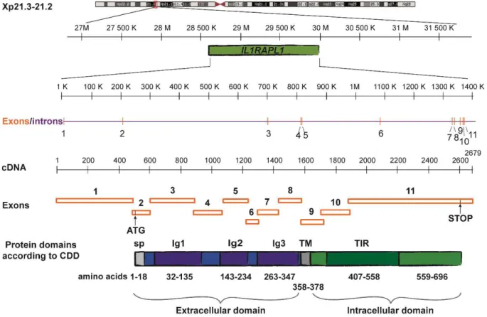
The interleukin 1 receptor accessory protein-like (IL1RAPL) gene was described as an ID gene by Carrie and collaborators in 1999. The Xp22.1–21.3 locus on the X chromosome was first identified by linkage analysis of large families, and IL1RAPL gene was identified through the detection of two inherited overlapping micro deletions
16 in this region associated exclusively with non-syndromic ID (Carrié et al. 1999).
IL1RAPL gene is composed of 11 exons of which 10 are coding. The two IL1RAPL
mutations characterized by Carrié and collaborators were a nonsense mutation (c.1377C>A, Y459X) and deletion of exons 3-5. Shortly after, Jin and collaborators reported the deletion of IL1RAPL exons 9 to 11 as responsible of the ID in a patient that also presented muscular dystrophy, glycerol kinase deficiency, and adrenal hypoplasia (Jin et al. 2000). Those pathologies are explained by the deletion of GK and DAX-1 genes and the last exon of dystrophin gene, in addition to the deletion found in IL1RAPL. In that study, a gene homologous to IL1RAPL in Xq22 was identified. Following this observation, the protein product of the first described gene was named IL1RAPL1 and the homologous one IL1RAPL2, but they are also known as TIGIRR-2 (three immunoglobulin domain-containing IL1 receptor-related) or IL1R8, and TIGIRR-1 or ILTIGIRR-1-R9, respectively (Born et al. 2000; Sana et al. 2000).
Since then, several mutations on IL1RAPL1 gene were reported, and they are listed in Table 1 (Appendix). Most of them are large deletions including several exons (Carrié et al. 1999; Nawara et al. 2008; Piton et al. 2008; Whibley et al. 2010; Behnecke et al. 2011; Mikhail et al. 2011; Franek et al. 2011; Youngs et al. 2012; Barone et al. 2013; Mignon-Ravix et al. 2014; Tucker et al. 2013; Redin et al. 2014) or nonsense mutations leading to truncated proteins (Kozak et al. 1993; Carrié et al. 1999; Tabolacci et al. 2006). However, there are some reports of duplications of X chromosome regions encompassing several genes including IL1RAPL1 in patients presenting ID or developmental delay and autistic features ((Honda et al. 2010; Utine et al. 2014), J. Lauer and F. Kooy personal communication). Surprisingly, most deletions involve one or more of the first 7 exons, and some authors suggested that this is probably due to the particular recombination potential of this region (Leprêtre et al. 2003; Tabolacci et al. 2006). This suggestion is supported by the reports of two patients with chromosome inversions with the breakpoint in IL1RAPL1 intron 2 (Bhat et al. 2008) and exon 6 (Leprêtre et al. 2003).
In agreement with the X chromosome-linked recessive transmission, patients with mutations in IL1RAPL1 gene are males, whereas females are identified as carriers in these families. However, some of these carriers show less severe phenotypes, like learning impairment or mild ID (Tabolacci et al. 2006; Bhat et al. 2008; Leprêtre et al. 2003; Piton et al. 2008). This may be due to the X chromosome inactivation that some
17 studies assessed in patient’s fibroblasts, but no clear correlation between X chromosome inactivation in those cells and patients’ phenotype has been established (Tabolacci et al. 2006; Nawara et al. 2008; Behnecke et al. 2011; Franek et al. 2011). Most of the mutations on this gene are associated with non-syndromic ID (Allen-Brady et al. 2011) but some studies have also associated IL1RAPL1 with mild dysmorphic features (Leprêtre et al. 2003), autism spectrum disorder (ASD) (Piton et al. 2008; Bhat et al. 2008; Butler et al. 2015), or startle epilepsy (Dinopoulos et al. 2014). As mentioned before, when large deletions in IL1RAPL1 region include contiguous genes, ID can be associated with muscular dystrophy, metabolic and hormonal diseases such as glycerol kinase deficiency and adrenal hypoplasia (Jin et al. 2000; Sasaki et al. 2003; Zhang et al. 2004).
2.2. IL1RAPL1 structure and expression
Two different transcripts were described for IL1RAPL1 gene, one of ~9.5 kb and another of ~6.5 kb (Carrié et al. 1999; Born et al. 2000). The longer transcript is present in both adult and fetal tissue, whereas the short is only present in adult brain. The larger transcript produces a 696 amino acid protein, whereas the shorter transcript may result from alternative splicing and should lead to the first 259 amino acids identical to larger isoform differing only in the last 2 amino acids. No experimental evidence has been provided for the existence of the shorter protein.
IL1RAPL1 codes for a transmembrane protein sharing structural domains with
members of interleukin 1 (IL1) receptor family (Carrié et al. 1999; Jin et al. 2000). For this reason it was named after its similarity with human interleukin 1 accessory protein (IL1RAcP), and it was speculated that interleukin 1 signaling could have a role in the pathophysiology of ID.
As represented in Figure 8, IL1RAPL1 includes a signal peptide with a predicted cleavage site between Ser18 and Leu19, an extracellular domain (357 amino acids), composed of three immunoglobulin-like (Ig-like) domains (Schreuder et al. 1997); a short transmembrane domain (21 amino acids), and an intracellular domain (318 amino acids) composed of a TIR (Toll/IL1 receptor (Rock et al. 1998)) domain and a 138 amino acid C-terminal tail. The extracellular domain of this protein is N-glycosylated at sites Asn63, 122, 138, 213, 264 and 331 (Yamagata, Yoshida, et al.
18 2015). Some proteins interacting with intra- and extra-cellular domains of IL1RAPL1 have been identified. Their characterization and function is mentioned below.
Figure 8. Gene and protein organization of human IL1RAPL1. CDD: NCBI's conserved domain database (Marchler-Bauer et al. 2014). Modified from Pavlowsky 2009.
In the mouse, Il1rapl1 transcript is expressed in the brain, principally in the hippocampus, the olfactory bulb and mammillary bodies (Carrié et al. 1999; Houbaert et al. 2013). Il1rapl1 transcript is expressed in neurons at 18 days in vitro (DIV) as well as in astrocytes derived from primary cultures (unpublished observations). As shown in Figure 9, overexpression of IL1RAPL1 showed that this protein is located in excitatory synapses, since it co-localizes with VGlut1 and PSD-95, excitatory pre and postsynaptic markers, respectively. In the other hand, IL1RAPL1 does not co-localize with inhibitory synapses markers, like VGat and gephyrin.
In the next section I will address the known roles of the ID-associated synaptic protein IL1RAPL1.
19
Figure 9. IL1RAPL1 is present in excitatory but not inhibitory synapses. Overexpressed IL1RAPL1 co-localizes with excitatory (PSD-95 and VGlut) but not inhibitory (Gephyrin and VGat) post and presynaptic markers. From Pavlowsky and collaborators (2010), and unpublished observations. Scale bar 10 μm.
2.3. Molecular partners of IL1RAPL1 and effects on neuronal physiology
Since its homology to interleukin 1 receptor family, it could be expected that IL1RAPL1 participates in interleukin 1 signaling. Until now, there is no direct evidence to support this cellular function (see hereafter) but it was suggested instead that IL1RAPL1 and its homologous IL1RAPL2 could have completely different signaling capacities in the nervous system.
A crystallographic study suggested that the TIR domain of human IL1RAPL1 forms dimers (Khan et al. 2004). Evidence of this could be observed in cells overexpressing IL1RAPL1 (Bahi et al. 2003), but until now the biological role of this dimerization is not clear. Evidence of IL1RAPL1 localization in presynaptic compartment has been provided in the zebrafish, where overexpressed Il1rapl1b is located in axons terminals of olfactory sensory neurons and co-localizes with the presynaptic protein synaptobrevin (Yoshida & Mishina 2008). The overexpression or down regulation of
il1rapl1b in olfactory sensory neurons increases or decreases respectively presynaptic
vesicle accumulation (Yoshida & Mishina 2008), a process in which the C-terminal domain of Il1rapl1b is required.
In mammals, most of the studies have addressed the postsynaptic role of IL1RAPL1, and even if there is some evidence of its role as a presynaptic protein (Bahi et al. 2003), this was observed in a cell line and remains to be confirmed in neurons. We have observed less or no IL1RAPL1 staining in the axons of mouse cultured neurons and that was also reported by others (Yoshida et al. 2011), supporting why most of the studies consider IL1RAPL1 as a postsynaptic protein.
20 Several studies have addressed the question of the interactions of IL1RAPL1 with other proteins (Bahi et al. 2003; Pavlowsky, Gianfelice, et al. 2010; Valnegri et al. 2011; Yoshida et al. 2011; Hayashi et al. 2013). Until now only the role of some of these IL1RAPL1 partners is known, and others remain to be validated and elucidated. Below I will address each of the known IL1RAPL1 partners, and how these interactions regulate several aspects of synapse and neuron physiology.
2.3.1. NCS-1 and calcium-regulated exocytosis
As mentioned above, the C-terminal domain of zebrafish Il1rapl1b is required for synaptic vesicle accumulation (Yoshida & Mishina 2008). This ~140 amino acid C-terminal of IL1RAPL1 is unique to this protein, which may confer some specific signaling characteristics that could be explained by interactions with partners through this tail. Bahi and collaborators used the IL1RAPL1 intracellular domain as a bait in a yeast two-hybrid screen and found the neuronal calcium sensor-1 (NCS-1) as an interacting protein (Bahi et al. 2003). In that study, they also identified the interacting region of IL1RAPL1 (amino acids 549-644), which spans the last 20 amino acids of the TIR domain and the half of the C-terminal specific domain, and defined the conserved Leu606 amino acid as critical for binding. On the other hand, NCS-1 interacts with IL1RAPL1 via the amino acids 174–190 on its C-terminal domain. This interaction was confirmed in vitro by GST pull down assays and in vivo by co-immunoprecipitation assays in HeLa cells, and was shown to be calcium-independent. Moreover, the same study showed that in IL1RAPL1-overexpressing PC12 cells the secretion of growth hormone induced by ATP is reduced. A mutation on NCS-1 gene (Arg102Gln) was found in patient with ASD, but it does not affect its binding to IL1RAPL1 (Piton et al. 2008; Handley et al. 2010).
Altogether, this data suggests that IL1RAPL1, through interaction with NCS-1, is a negative regulator of exocytosis, which could in part explain the increase of synaptic vesicles stained by synaptobrevin after Il1rapl1b overexpression (Yoshida & Mishina 2008). The mechanism of exocytosis regulation by NCS-1/IL1RAPL1 interaction was elucidated by Gambino and collaborators. By overexpressing IL1RAPL1 and down regulating NCS-1 in PC12 cells, they showed that IL1RAPL1, through NCS-1, silences the activity of N-type voltage-gated calcium channels (N-VGCC). The down regulation
21 of the activity of this channel by IL1RAPL1 inhibits secretion of growth hormone in PC12 cells, as shown in Figure 10 (Gambino et al. 2007).
Figure 10. IL1RAPL1 regulates N-type Ca2+ current through NCS-1. Left: Overexpression of IL1RAPL1
abolishes N-type Ca2+ current in PC12 cells, which is not observed when NCS-1 is down regulated in
IL1RAPL1-overexpressing cells (IL1RAPL1 + siNCS-1). Black traces indicate Ca2+ currents under
control conditions (ctrl), and red traces indicate Ca2+ currents in the presence of 1 μM ω-conotoxin
(GVIA), a specific antagonist of N-type voltage gated Ca2+ channels (N-VGCC). Right: The
IL1RAPL1-NCS-1 interaction at presynapses could regulate synaptic vesicle (SV) exocytosis by inhibiting N-VGCC, as observed for growth hormone secretion in PC12 cells by Gambino and collaborators in 2007. NT: neurotransmitter.
NCS-1, found from yeast to humans, is a member of a family of calcium-binding proteins called neuronal calcium sensors (NCS), which include neurocalcin, visinin-like protein (VILIP), hippocalcin, recoverin, guanylate cyclase activating protein (GCAP), and K+ channel interacting protein (KChIP). These sensors are mainly expressed in the nervous system where they are involved in a number of calcium signaling pathways. In particular, NCS-1 is widely expressed in the brain, and there is evidence of its role in pre and postsynaptic compartments (Jinno et al. 2002; Martone et al. 1999; Jo et al. 2008).
In addition to the impact on secretion, the down regulation of N-VGCC by IL1RAPL1 and NCS-1 inhibits NGF-induced neurite elongation in PC12 cells. The negative regulation of NCS-1 in neurite outgrowth has been observed in different cell lines and
22 in neurons from Drosophila melanogaster (Chen et al. 2001; Hui et al. 2006). The roles of the NCS-1 fly homologue, Frequenin, in secretion and in neurite elongation are independent: Chronic expression of an interfering Frequenin C-terminal peptide affects both neuromuscular axon terminal branching, or structural complexity, and neurotransmitter release, while acute application of the same peptide produces a reduction of neurotransmitter release without an effect on axon morphology (Romero-Pozuelo et al. 2007).
At the presynaptic terminal, NCS-1 has been shown to regulate voltage gated calcium channels, including P/Q-, L- and N-type, as formerly mentioned. Those calcium channels group to the presynaptic membrane a large signaling complex containing SNARE proteins (see Figure 5), Ca2+-binding proteins (including NCS-1),
calcium-dependent kinases and scaffolding proteins. For example, presynaptic P/Q type CaV2 channels, whose Ca2+-dependent inactivation is regulated by its interaction with
NCS-1 in superior cervical ganglion neurons, provide a rapid and spatially limited Ca2+ entry
that initiates synaptic transmission (Yan et al. 2014). Through interactions with SNARE proteins and scaffolding proteins, these Ca2+ channels bring docked synaptic vesicles
close to the source of Ca2+ entry, allowing them to respond efficiently to the Ca2+
increase. Through specific interactions with Ca2+-binding proteins and kinases like
CaMKII, the activity of Ca2+ channels can be regulated to mediate forms of synaptic
plasticity. The regulation of voltage gated Ca2+ channels (in particular N-type) by
IL1RAPL1 through NCS-1 suggests a role for IL1RAPL1 in presynaptic function. Up to now, there is no direct evidence of the function of this protein in synaptic vesicle exocytosis in neurons.
2.3.2. PSD-95 and excitatory postsynaptic organization
The first evidence of the presence of IL1RAPL1 at the synapse was provided by a study by Pavlowsky and collaborators where, using subcellular fractionation, endogenous IL1RAPL1 protein was found enriched in postsynaptic density (PSD) fractions (Pavlowsky, Gianfelice, et al. 2010). By overexpressing this protein in mouse neurons in culture they observed that IL1RAPL1 co-localizes with excitatory pre (VGlut1) and post (PSD-95 and Shank) synaptic markers (Figure 9). Moreover, using the entire C-terminal domain of IL1RAPL1 (amino acids 390 – 696) as a bait for a two hybrid yeast system, Pavlowsky and collaborators found a direct interaction with PSD-95, SAP-97 and PSD-93. These are all members of the PSD-95 like subfamily of the
23 MAGUK family of scaffolding proteins (Cho et al. 1992), but only the interaction with PSD-95 was further characterized.
PSD-95 is the prototypical member of MAGUK family of proteins and constitutes a signaling hub that promotes formation and maturation of dendritic spines (El-Husseini et al. 2000). MAGUKs share a common organization with three N-terminal PSD-95/Dlg/ZO-1 (PDZ) domains, a Src-homology 3 (SH3) domain, and a C-terminal guanylate kinase (GK) domain catalytically inactive. PSD-MAGUKs are required for synaptic targeting of different glutamate receptors and their role is finely regulated during development (Elias et al. 2006).
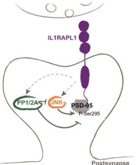
PSD-95 interacts via its first two PDZ domains (PDZ1 and PDZ2) with a non-canonical PDZ binding domain within the last 8 amino acids of the C- terminal tail of IL1RAPL1 (Pavlowsky, Gianfelice, et al. 2010). IL1RAPL1 overexpression in mouse or rat hippocampal neurons largely increases PSD-95 cluster number. This increase is not observed when IL1RAPL1 lacking the last 8 amino acids (Δ8) is overexpressed, showing that interaction with PSD-95 is necessary for the IL1RAPL1-induced increase of PSD-95 clusters (Pavlowsky, Gianfelice, et al. 2010).
Besides PSD-95, IL1RAPL1 overexpression also increases VGlut1 staining and dendritic spine number, suggesting a global increase in excitatory synapse number. This was confirmed by increases miniature excitatory postsynaptic currents (mEPSC) in neurons overexpressing IL1RAPL1. In addition, VGlut1 staining and dendritic spine number are also increased in neurons overexpressing Δ8 IL1RAPL1 mutant, suggesting that these are events independent of IL1RAPL1/PSD-95 interaction (Pavlowsky, Gianfelice, et al. 2010). Accordingly, hippocampal neurons from Il1rapl1 knockout (KO) mouse show ~25% less PSD-95 clusters number compared with wild-type neurons, and this was congruent with a decrease of miniature excitatory postsynaptic currents (mEPSCs) frequency.
IL1RAPL1 overexpression induces a significant increase of PSD-95 phosphorylation on Ser295 (Figure 11). This increase is dependent on IL1RAPL1 interaction with this scaffolding protein because Δ8 IL1RAPL1 mutant is not able to increase PSD-95 phosphorylation (Pavlowsky, Gianfelice, et al. 2010). According to the fact that phosphorylation on Ser295 by c-Jun N-terminal kinase (JNK) is important to target PSD-95 to synapses (Kim et al. 2007; Thomas et al. 2008), the increase/decrease of
24 PSD-95 at the synapse observed after overexpressing or knocking down Il1rapl1, respectively, may result from the changes in PSD-95 and JNK phosphorylation. Indeed, phosphorylation of PSD-95 and JNK is reduced in Il1rapl1 KO cortical neurons. The mechanism of phosphorylation regulation could involve the PP1/2A phosphatases, as their pharmacological inhibition partially rescued the phosphorylation deficits in
Il1rapl1 KO neurons (Pavlowsky, Gianfelice, et al. 2010).
Figure 11. IL1RAPL1 interacts with PSD-95 and regulates its targeting to excitatory postsynapses. In addition to interacting with PSD-95, IL1RAPL1 regulates the phosphorylation at Ser295 of this scaffold protein. This process is regulated by direct or indirect activation of JNK and/or PP1/2A.
2.3.3. PTPδ and synaptogenesis
Synapse formation is initiated at contact sites between axon terminals and dendrites (see Figure 1). There, pre and postsynaptic adhesion molecules form trans-synaptic complexes to induce pre and postsynaptic differentiation (Figure 7).
In mouse hippocampal and cortical neurons in culture, overexpression of IL1RAPL1 induces excitatory presynaptic differentiation and dendritic spine formation (Pavlowsky, Gianfelice, et al. 2010; Valnegri et al. 2011; Yoshida et al. 2011). Interestingly, although IL1RAPL1 extracellular domain is sufficient for inducing this presynaptic differentiation, both extracellular and intracellular TIR domains are required for postsynaptic changes (Figure 12) (Valnegri et al. 2011; Yoshida et al. 2011).
25
Figure 12. Pre and postsynaptic differentiation is mediated by different IL1RAPL1 protein domains. Pre (Synaptophysin, up) and post (PSD-95, down) synaptic markers are increased in mouse hippocampal mature neurons transfected with IL1RAPL1 (WT). In the absence of the C-terminal domain (ΔC), only synaptophysin increase is observed. On the other hand, in the absence of the two first Ig-like domains (ΔN), neither pre nor postsynaptic markers are increased.
These observations suggest a differential role of each IL1RAPL1 extra and intracellular domains in synaptic differentiation. These observations were also the starting point for the exploration aiming at finding partners of IL1RAPL1 extracellular domain, which may exert its synaptogenic activity by interacting with presynaptic proteins.
Using affinity chromatography, two research groups identified simultaneously protein tyrosine phosphatase δ (PTPδ) as a binding partner of IL1RAPL1 extracellular domain (Valnegri et al. 2011; Yoshida et al. 2011).
PTPδ is a member of the 2A subfamily of receptor-like protein tyrosine phosphatases (Tonks 2006). This family of proteins is composed of three members in vertebrates, leukocyte common antigen-related (LAR, which gives the name of the family), PTPσ, and PTPδ, that share overall 66% of amino acid identity (Pulido, Serra-Pagès, et al. 1995). mRNAs encoding the three family proteins show overlapping and differential distribution patterns in mouse brain (Kwon et al. 2010). In human tissue, PTPδ mRNA is predominantly detected in brain and, to a lesser extent, in heart, placenta, and kidney (Pulido, Serra-Pagès, et al. 1995). In the mouse brain, PTPδ mRNA is found in
26 hippocampus, in the reticular thalamic area, and is more present in the cortical layer IV of cortex compared with other layers (Kwon et al. 2010).
Some de novo deletions, duplications and single nucleotide polymorphisms (SNPs) in the 5’UTR of PTPRD gene are associated with attention-deficit hyperactivity disorder (ADHD) (Elia et al. 2010), autism spectrum disorder (ASD) (Pinto et al. 2010), bipolar disorder (Malhotra et al. 2011), and restless syndrome (Schormair et al. 2008; Elia et al. 2010; Yang et al. 2011). Recently, Choucair and collaborators identified an inherited homozygous deletion with breakpoints in PTPRD gene in a patient with ID, growth retardation, hearing loss, trigonocephaly and scaphocephaly (malformation of the skull) (Choucair et al. 2015).
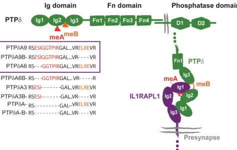
As the other members of the LAR family, PTPδ contains extracellular typical cell adhesion immunoglobulin-like (Ig) and fibronectin III (FN) domains modified by alternative splicing, which mediate diverse extracellular protein interactions (Figure 13). The intracellular domain involves two intracellular protein tyrosine phosphatase (PTP) domains: a membrane-proximal D1 domain with robust catalytic activity and a membrane-distal D2 domain with residual or no catalytic activity (Takahashi & Craig 2013). Up to three endoprotease cleavage sites are located 81-87 amino acids N-terminal from the transmembrane domain, constitutively generating an extracellular subunit that remains non covalently bound to the phosphatase domain subunit (Pulido, Krueger, et al. 1995). However, the functional significance of this modification is not clear.
Ptprd deficient mice are semi lethal, since they have difficulties in taking food, but they
have a normal brain morphology (Uetani et al. 2000). They exhibit impaired hippocampal-dependent learning and, although the hippocampus is histologically normal, the hippocampal LTP is increased. These data support the important role of PTPδ on hippocampal function, but unlike Il1rapl1 KO mice (see later), the LTP does not correlate positively with the impaired learning ability in the absence of PTPδ (Uetani et al. 2000).
PTPδ extracellular domain promotes neurite growth in chicken neurons (Wang & Bixby 1999) and acts as an attractant for growing axons (Sun et al. 2000), but no PTPδ interacting proteins were yet associated with these effects. PTPδ was first shown to have homophilic interactions (Wang & Bixby 1999), but besides IL1RAPL1, postsynaptic binding partners in trans-synaptic complexes identified so far are
netrin-27 G ligand-3 (NGL-3) (Kwon et al. 2010), Slit and Trk-like family member 1-6 (Slitrk1-6) (Takahashi et al. 2012; Yim et al. 2013; Yamagata, Sato, et al. 2015), and interleukin 1 receptor accessory protein (IL1RAcP) (Yoshida et al. 2012).
Figure 13. PTPδ structure and trans-synaptic interaction with the extracellular domain of IL1RAPL1. Up: Structure and domain of PTPδ. The isoforms generated by meA and meB splicing are shown, and those contained in the violet rectangle correspond to the ones interacting with IL1RAPL1. Right: Schematic representation of the interaction between IL1RAPL1 extra cellular domain and PTPδ Ig domain. Modified form Yoshida et al., 2011 and Yamagata, Yoshida et al., 2015.
Like other members of LAR family and their postsynaptic partners, IL1RAPL1/PTPδ interaction has three general functions in synaptic organization:
The first one is to mediate cell–cell adhesion at synapses. Wang and Bixby first described PTPδ as an adhesion molecule, and trans-synaptic adhesion mediated by PTPδ is highly selective, diverse and organized. This is clearly supported by two recent studies that show the crystal structures of PTPδ interacting with three of its partners, IL1RAPL1, IL1RAcP and Slitrk2 (Yamagata, Yoshida, et al. 2015; Yamagata, Sato, et al. 2015).
The diversity of trans-synaptic partners lead to a specific regulation of inhibitory and excitatory synaptogenesis. For example, interaction of PTPδ with IL1RAPL1 or Slitrk2 function selectively in excitatory synaptic organization, whereas interaction with Slitrk3 in inhibitory synaptic organization (Takahashi et al. 2012; Yim et al. 2013). However, despite its synaptogenic activity, Slitrk2 has not been found enriched in PSD fraction samples (Yim et al. 2013). As discussed further, PTPδ-IL1RAcP interaction induces