Ascididemin and meridine stabilise G-quadruplexes and inhibit telomerase
in vitro
Lionel Guittata,1, Anne De Ciana,1, Frédéric Rosub, Valérie Gabelicac, Edwin De Pauwc, Evelyne Delfourned,
Jean-Louis Mergnya,
a
Laboratoire de Biophysique, Muséum National d'Histoire Naturelle, INSERM UR 565, CNRS UMR 5153, 43 rue Cuvier, 75231 Paris cedex 05, France
b
Laboratoire de Chimie Physique Biologique, Université de Liège, Institut de Chimie, Bat. B6c, B-4000 Liège, Belgique c
Laboratoire de Spectrométrie de Masse, Université de Liège, Institut de Chimie, Bat. B6c, B-4000 Liège, Belgique dLaboratoire SPCMIB, Université Paul Sabatier, UMR CNRS 5068, 118 route de Narbonne, 31062 Toulouse cedex 4, France
1 These two authors contributed equally to this work.
Abstract
Ascididemin and Meridine are two marine compounds with pyridoacridine skeletons known to exhibit interesting antitumour activities. These molecules have been reported to behave like DNA intercalators. In this study, dialysis competition assay and mass spectrometry experiments were used to determine the affinity of ascididemin and meridine for DNA structures among duplexes, triplexes, quadruplexes and single-strands. Our data confirm that ascididemin and meridine interact with DNA but also recognize triplex and quadruplex structures. These molecules exhibit a significant preference for quadruplexes over duplexes or single-strands. Meridine is a stronger quadruplex ligand and therefore a stronger telomerase inhibitor than ascididemin (IC50=ll
and >80 µM, respectively in a standard TRAP assay).
Keywords: Telomeres; Telomerase inhibitor; G-quadruplex; G-Quartet; DNA ligand
1. Introduction
Researchers have long been investigating natural compounds as new anticancer drugs. In this way, different original metabolites with interesting cytotoxic properties have been reported from marine source. Several marine-derived metabolites have reached clinical trials as antitumour agents. Among marine alkaloids, the pyridoacridines seem to be the largest group, with most of the compounds being isolated from sponges and runicates (for a review, see [1]). In 1991, Schmitz et al. [2] described the isolation and structure of meridine (Fig. 1A, left) from the ascidian Amphicarpa meridiana. Ascididemin (Fig. 1A, right) was isolated in 1988 by Kobayashi et al. [3] from a tunicate Didemnum sp. Ascididemin and meridine differ by the attachment of the benzene ring, the position of a nitrogen atom and a hydroxyl substituent in meridine. Different mechanisms of action for these polycyclic aromatic compounds have been proposed. Ascididemin is a conventional Topo II poison, but it is unlikely that topoisomerases are the main cellular targets of these molecules. Ascididemin is also capable of reductive DNA cleavage mediated by reactive oxygen species [4]. The binding mode of these derivatives to double-stranded DNA has been studied. Ascididemin intercalates between base pairs with a preference for GC-rich sequences [5].
Besides duplexes, DNA is prone to structural polymorphism and a number of alternative DNA structures have been described to date [6,7]. DNA conformation may differ from a regular double-helix and may involve the association of more than two strands, leading to the formation of triplexes and quadruplexes. Alternative DNA structures offer significant differences in terms of electrostatics, shape and rigidity compared to single- or double-stranded DNA. Therefore, specific recognition of unusual DNA structures such as triplexes and quadruplexes by small molecules such as pyridoacridines should be possible.
Fig. 1. (A) Formula of ascididemin (left), meridine (right) and RHPS4 (center) [27,54,66,67]. (B) A G-quartet (left) and a schematic drawing of an intramolecular antiparallel G-quadruplexes involving 3 quartets.
G-quadruplexes are a family of secondary DNA structures formed in the presence of monovalent cations that consist of four-stranded structures stabilised by quartets (Fig. 1B) [8,9]. There is a renewed interest for G-quadruplex structures due to their putative biological regulatory function. Telomeres protect chromosomal ends from fusion events and provide a mean for complete replication of the chromosome. Telomere repeats are added by a specialized enzyme, telomerase, which is overex-pressed in most tumour cells. In contrast, normal
fibroblasts exert a very low level of telomerase previously thought to lack telomerase activity [10]. The 3'-terminal region of the G-rich strand of human telomeres is single-stranded and may adopt a G-quadruplex conformation [11,12]. This structure has been shown to directly inhibit telomerase elongation in vitro [13]. Therefore, ligands that selectively bind to G-quadruplex structures may interfere with telomere structure and telomere elongation and replication of cancer cells [14,15]. Several reviews concerning telomerase inhibitors in general or quadruplex-based telomerase inhibitors have been published in the last few years [16-23]. The recent discovery of natural compounds such as cryptolepine [24] and telomestatin [25,26] or of synthetic pentacyclic acridines (such as RHPS4)[27] that interact with G-quartets led us to test the binding of ascididemin and meridine to G-quadruplexes.
2. Materials and methods 2.1. Compounds
Ascididemin and meridine were synthesized according to published procedures [28,29]. Stock solutions (1 mM for meridine, 2 mM for ascididemin) were prepared in DMSO and kept at -20 °C. Their formula are shown in Fig. 1A.
2.2. Oligonucleotides
Oligodeoxynucleotide probes were synthesized by Eurogentec (Belgium) on the 1-µmol scale. Purity was checked by gel electrophoresis. All concentrations were expressed in strand molarity using a nearest-neighbour approximation for the absorption coefficients of the unfolded species [30]. All polynucleotides were ordered from Amersham-Pharmacia. F21MB is a doubly labelled 21-base-long oligomer that mimics the human telomeric guanine-rich strand with FAM at 5' end and DABCYL at 3' end respectively. It was synthesized and purified by Eurogentec (Belgium).
2.3. Dialysis experiments
The initial dialysis protocol was defined by Ren and Chaires [31-33]. We have adapted this test to accommodate a different set of nucleic acid structures [24,34]. Briefly, a buffer consisting of 15 mM sodium cacodylate (pH 6.5), 10 mM MgCl2, and 185 mM NaCl was used for all experiments. All structures used in these experiments
were stable at room temperature under the chosen experimental conditions. 400 mL of the dialysate solution containing 1 µM ligand were used for each competition dialysis assay. A volume of 200 µL at 75 µM monomeric unit (nucleotide, base pair, base triplet or quartet) of each of the nucleic acids samples was pipeted into a separate Dialyzer unit (Pierce). A panel of 19 different nucleic acids structures were used. They have been described in details elsewhere [24]. All 19 dialysis units were then placed in the beaker containing the dialysate solution. The beaker was covered with Parafilm and wrapped in foil, and its contents were allowed to equilibrate with continuous stirring at room temperature (20-22 °C) overnight. At the end of the equilibration period, DNA samples were carefully removed to microfuge tubes, and treated with 1% SDS. The ligand concentration in each sample was determined by absorbance.
2.4. Fluorescence studies
Fluorescence can be used to probe the secondary structure of oligodeoxynucleotides mimicking repeats of the guanine-rich strand of vertebrate telomeres, provided a FAM molecule (fluorescent tag) and a Dabcyl (quencher) are attached to the 5' and 3' ends of the oligonucleotide, respectively [35]. Fluorescence measurements with the F21MB oligonucleotide (0.2 µM) were initially performed on a Spex Fluoromax 3 instrument with 600 µL of solution in a 0.1 M lithium chloride, 10 mM sodium cacodylate pH 7.2 buffer [36]. In the experiments presented here, a real time PCR apparatus (MX3000P, Stratagene) was used, allowing the simultaneous recording of independent 96 samples. Each sample was studied in a 10 mM lithium cacodylate pH 7.2 buffer containing 50 mM sodium chloride + 50 mM lithium chloride or 5 mM potassium chloride + 95 mM lithium chloride (buffer conditions labelled "Na+" and "K+", respectively). LiCl is added in order to approach physiological ionic strength
without stabilising the quadruplex (lithium is a monocation which does not interact with G-quartets). The melting of the G-quadruplex was monitored in the presence and or in absence of the dye by measuring the fluorescence of fluorescein [36]. Several concentrations of each compound (1, 3, 5 or 10 µM) were tested, alone or in the presence of a double-stranded competitor (a self-complementary, 26 bp-long oligodeoxynucleotide "ds26", sequence 5' d-CAATCGGATCGAATTCGATCCGATTG 3'). This competitor, which is susceptible to "trap" a drug, will lower the stabilisation of the F21MB quadruplex. In the absence of a quadruplex-interacting drug, this competitor has no effect on the melting temperature of the F21MB quadruplex. All experiments were performed at least in duplicate, with a volume of 25 µL for each sample. Emission of fluorescein was normalized between 0 and 1, and the T1/2 was defined as the temperature for which the normalized emission is 0.5.
2.5. Mass spectrometry
Oligodeoxynucleotides d-CGTAAATTTACG ("Dk33", M = 3644.45 Da), d-CGCGAATTCGCG ("Dk66", M = 3646.44 Da), CGCGGGCCCGCG ("Dk100", M = 3678.40 Da), TGGGGT (M= 1863.26 Da),
d-GGGGTTTTGGGG (M = 3788.50 Da), d-(GGGTTA)3GGG (M = 6653.35 Da) were purchased from
Eurogentec (Angleur, Belgium) and used without further purification. Duplex and quadruplex solutions were prepared according to previous reports [34]. Ammonium acetate was chosen as the electrolyte for its
compatibility with electrospray mass spectrometry. Experiments were performed on an LCQ mass spectrometer (Finnigan, San Jose, CA, USA) operated in the negative ion mode with a needle voltage of -2.6 kV. The experimental conditions were optimized to avoid denatura-tion of the duplex or quadruplex species. Full scan MS spectra were recorded in the m/z range [1000-2000], and 50 scans were summed for each spectrum. As the interaction of these compounds with DNA is relatively weak, spectra of 2:1 (10 µM drug, 5 µM structure) of all possible structure (duplex or quadruplex)+drug combinations were recorded. Methanol (15%) was added to the samples just before injection to obtain a stable electrospray signal. The rate of sample infusion into the mass spectrometer was 4 µL/min. As previously discussed, the relative intensities (obtained from a sum of 50 spectra) of the free and bound DNA in the mass spectra are assumed to be proportional to the relative abundances of these species in solution. The amount of bound ligand expressed in monomeric unit (base pair or quartet in the DNA target) is determined by dividing the total amount of bound ligand by the number of monomeric units in the DNA targets.
2.6. TRAP assay
The TRAP reaction was performed in a 20 mM Tris-HCl pH 8.3 buffer containing 63 mM KC1, 1.5 mM MgCl2,
1 mM EGTA, 0.005% Tween 20, 0.1 mg/mL BSA, 50 µM dTTP, dGTP and dATP, 5 µM dCTP, and the oligonucleotides TS AATCCGTCGAGCAGAGTT-3') (0.4 µM), ACX
(5'-GCGCGGCTTACCCTTACCCTTACCCTAACC-3') (0.4 µM), NT (5'-ATCGCTTCTCGGCCTTTT-3') (0.4 µM) and TSNT (5'-ATTCCGTCGAGCAGAGTTAAAAGG-CCGAGAAGCGAT-3') (20 amoles), 2 units of Taq polymerase, 0.02 mCi/mL dCTP32 (3000 Ci/mmol) and 200 ng of A431 CHAPS extracts. Various
concentrations (0.5-100 µM) of ascididemin or meridine were tested, using freshly prepared aqueous dilutions of the compounds. As the stock solutions were prepared in DMSO, up to 5% DMSO was present (i.e., for 50 µM meridine or 100 µM ascididemin); we checked that 5% DMSO alone had no effect on telomerase activity (data not shown) After telomerase elongation for 15 min at 30 °C, 30 cycles of PCR were performed (94 °C 30 s, 50 °C 30 s and 72 °C for 90 s). Telomerase extension products were then analysed on a denaturing 8%
polyacrylamide, 7 M urea, 1 X Tris Borate EDTA (TBE) vertical gel.
3. Results
3.1. Equilibrium dialysis
To evaluate the selectivity of ascididemin and meridine for different DNA structures, we performed a
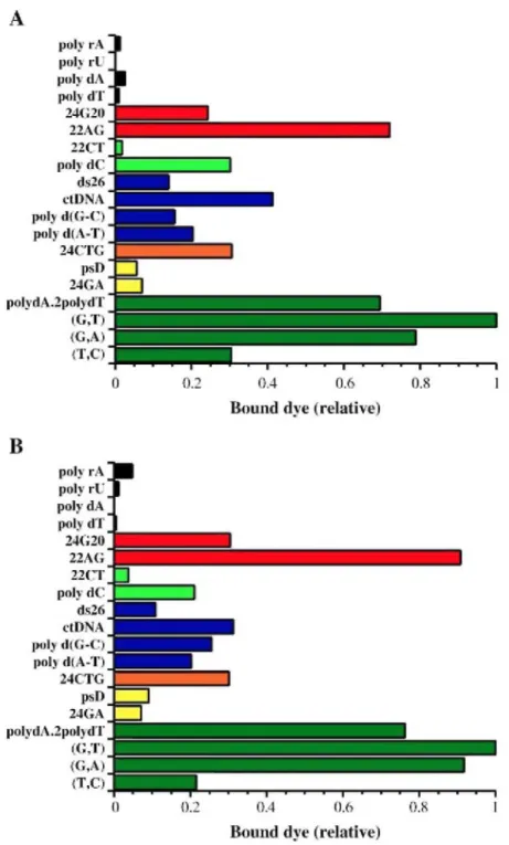
competitive dialysis experiment using 19 nucleic acids structures (described in Materials and methods) against a common ligand solution. More products accumulate in the dialysis tube containing the structural form with the highest ligand binding affinity [31,32]. It is possible to correlate the amount of the bound dye to a given structure with the affinity of the dye for that sample. As shown in Fig. 2A, ascididemin interacts preferentially with triplex and quad ruplex structures, whereas it shows a very weak affinity for single-strands and a moderate affinity for duplexes. Meridine (Fig. 2B) also interacts preferentially with triplex and quadruplex structures (especially with the human telomeric quadruplex, 22AG) as compared to single-strands and duplexes. The binding profile of the two compounds relative to higher-order DNA structures was quite unexpected and prompted us to confirm this interaction using different techniques.
3.2. Mass spectrometry
The ability of mass spectrometry to investigate drug-DNA interactions has been reviewed recently [37]. The binding stoichiometry, the relative binding affinities and the binding constants for DNA double-helices of various sequences may be determined and DNA complexes with intercalators and minor groove binders have been studied [38]. DNA quadruplex structures may also be investigated by electrospray mass spectrometry [39]. Fig. 3 presents the results obtained for two different quadruplexes, i.e., dTG4T, a tetramolecular quadruplex, and Telo, corresponding to 3.5 repeats of the human telomeric motif. These sequences are reminiscent of tetraplexes 24G20 and 22AG used in the equilibrium dialysis assays, respectively. Formation of stable complexes between ascididemin (or meridine) and each quadruplex is demonstrated by the presence of peaks corresponding to 1:1 or 2:1 drug-quadruplex complexes. The ratios of the intensities of the peaks of the complexes and the free
quadruplex obtained with meridine were more important (compare panel B with A, and panel D with C) suggesting that meridine has a slightly higher affinity for quadruplexes than ascididemin, in agreement with equilibrium dialysis.
Fig. 2. Equilibrium dialysis. All measurements were performed in a 185 mM NaCl, 10 mM MgCl2, 15 mM Na-cacodylate buffer (pH 6.5). The nucleic acid names are given on the left and all structures are described in Materials and methods. Triplexes are shown in green. Unusual parallel stranded duplexes, trinucleotide repeats and regular duplexes are shown in yellow, orange and blue, respectively. i-DNA are shown in light green and quadruplexes in red. Single-stranded polynucleotides are shown in black. (A) Ascididemin. (B) Meridine.
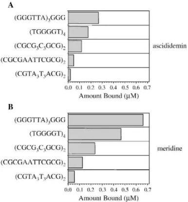
The affinities of ascididemin and meridine for various DNA forms were then analysed by measuring their interactions with 12 base pairs duplexes with different GC content, [34]. Fig. 4 shows the quantitation of the results for 5 different structures (2 quadruplexes and 3 duplexes) for ascididemin (Fig. 4A) and meridine (Fig. 4B). Overall, binding of meridine to all 5 DNA structures was stronger compared to ascididemin (compare X-axis values on Fig. 4A and B). An apparent preference for quadruplexes over duplexes is also suggested by the
higher amount bound per motif. Again, a preference for the human telomeric sequence over the d[TGGGGT]4
tetramolecular quadruplex is found. Within duplexes, binding was stronger to GC-rich over AT-rich sequences, as expected for DNA intercalators.
3.3. Fluorescence melting experiments
Dialysis and mass spectrometry experiments suggest that ascididemin and meridine interact with quadruplexes. Another method was implemented to confirm the interaction of these dyes with quadruplexes. We monitored by fluorescence the melting temperature of a fluorescent-tagged G-quadruplex (F21MB:
5'-Fluorescein-GGGTTAGGGT-TAGGGTTAGGG-3'-DABCYL representing the human telomeric motif) in the presence of various concentrations (1-10 µM) of ascididemin (Fig. 5A, C, E) and meridine (Fig. 5B D F). In sodium, there is little stabilisation of the G-quadruplex with ascididemin (+1.2 °C at the highest concentration: 10 µM) and a weak stabilisation (∆T1/2 = +3.6 °C at 10 µM compound) with meridine.
In the next series of experiments, sodium was replaced with potassium (panels C and D). Unsurprisingly, such a change led to a large increase of the melting temperature of the quadruplex alone as potassium is preferred over sodium by quadruplexes in general [8] and by the human intramolecular telomeric quadruplex in particular [35,40]. In order to stay in the same range of melting temperature for the quadruplex alone in sodium and in potassium, we chose to decrease the concentration of potassium to 5 mM and add 95 mM LiCl to keep a constant ionic strength. Under these experimental conditions, T1/2 was 54.9 °C (i.e., close to the T1/2 of 53.2 °C found in
50 mM NaCl + 50 mM LiCl). More unexpected was the observation that the stabilisation induced by
ascididemin and meridine is more pronounced in potassium than in sodium. At 10 µM, ascididemin and meridine stabilised the quadruplex by 5.7 and 11.4 °C, respectively. Lower concentrations (1, 3 or 5 µM) led to a less pronounced effect for both compounds. For example, ∆T1/2 at 1 µM were +0.9 and +1.4 °C for 1 µM ascididemin
and meridine, respectively, and +2.9 and +7.1 °C for 5 µM ascididemin and meridine, respectively. In the concentration range tested (1-10 µM), meridine was always more potent than ascididemin towards quadruplex stabilisation.
Fig. 3. Mass Spectrometry spectra. ESI-MS spectra of mixtures of 10 µM ascididemin (A, C) or meridine (B, D) with the parallel tetramolecular quadruplex d(TG4T)4 (panels A and B) or the human intramolecular quadruplex (panels C and D). Each structure was tested at a concentration of 5 µM with 10 µM ligand. Spectra were recorded using the LCQ instrument.
Fig. 4. Quantitation of ligand bound by Mass Spectrometry analysis. Concentration of bound ligand (per motif) deduced from the ESI-MS spectra of mixtures of 10 µM ascididemin (panel A) or meridine (panel B) with five different structures (5 µM): the human intramolecular quadruplex (GGGTTA)3GGG, the parallel tetramolecular quadruplex d[TG4T]4 and three self-complementary duplexes.
Finally, the stabilisation observed in potassium at 10 µM compound was challenged by the addition of a double-stranded competitor (Fig. 5E-F). Addition of a large excess of ds26, a self-complementary 26 base-long oligonucleotide of mixed base content had little effect on the ∆T1/2 obtained with meridine: the stabilisation was
+8 °C in the presence of 30 µM ds26 (which corresponds to 390 µM in base pairs, or a 600-fold molar excess in base pairs over base quartets). This value is relatively close to the +11.4 °C stabilisation observed in the absence of a competitor, showing that meridine is weakly trapped by the presence of double-stranded DNA. For ascididemin, which is a weaker stabiliser (+5.7 °C at 10 µM), 30 µM ds26 led to a slightly more pronounced deleterious effect (∆T1/2 +2.0 °C). These results are in agreement with the equilibrium dialysis and mass
spectrometry measurements which indicate a preferential interaction of ascididemin and meridine with the quadruplex-forming oligonucleotides.
3.4. Telomerase inhibition
Telomerase inhibition efficiency was measured by a TRAP assay. The non-folded, single-stranded form of the primer TS is required for optimal telomerase activity and quadruplex formation has been shown to directly inhibit telomerase elongation in vitro. A growing number of small molecules have been discovered to inhibit telomerase activity by inducing and/or stabilising G-quartet structure. Therefore, we performed here a TRAP assay with increasing concentrations (ranging 1 to 50 or 100 µM) of the compounds. Ascididemin inhibits telomerase with an IC50 of 87 µM (Fig. 6A). In agreement with its higher affinity for quadruplexes, meridine is
clearly more potent: partial disappearance of the TRAP ladder is already found at 5 µM compound, and an IC50
of 11 µM may be determined (Fig. 6B). The presence of an internal control (ITAS) discriminates inhibition of Taq polymerase during PCR amplification and telomerase elongation. A weak inhibition of the ITAS is detected with meridine at 50 µM (Fig. 6B, rightmost lane). Its IC50 against telomerase (11 µM) is significantly lower,
demonstrating that telomerase inhibition is not the result of an artefactual PCR inhibition. Nevertheless, this IC50
against telomerase is relatively modest when compared with the best G4-based telomerase inhibitors described and reflects a relatively weak interaction with G-quadruplexes.
Fig. 5. Stabilisation of human telomeric quadruplex by ascididemin and meridine. The thermal denaturation temperature of F21MB (followed by a variation of the fluorescence emission signal at 516nm)in 50 mMNaCl+50 mM LiCl (panels A and B) or 5 mMKCl+95 mM LiCl (panels C-F) is recorded alone (solid line) or in the presence of 1, 3, 5 or 10 µM of ascididemin (left: panels A, C and E) or meridine (right: panel B, D and F). Inverted triangles: 1 µM drug; squares: 3 µM; crosses: 5 µM; diamonds: 10 µM. Stabilisation is more
pronounced with meridine and when Na+ is replaced by K+. Panels E and F: the stabilisation induced by 10 µM ascididemin or meridine (diamonds) is partially maintained in the presence of 3 µM (dotted line); 10 µM (dashes) or 30 µM double-stranded competitor (large dashes) as compared to the control (solid line: no compound added).
Fig. 6. Telomerase inhibition by ascididemin and meridine in a TRAP assay. Increasing concentrations of compounds were added in the TRAP mixture in the presence of an internal control (ITAS). TRAP activity was determined with 200 ng of a CHAPS extract of A431 cell line. IC50 of 11 µM and 87 µM were determined for meridine and ascididemin, respectively.
4. Discussion
Marine organisms provide a valuable source for natural products and the biological activities of marine alkaloids have been studied widely. In this paper, we have used several methods to study the interaction of two alkaloids, ascididemin and meridine, with unusual DNA structures. These compounds bind to G-quadruplexes, as demonstrated by dialysis, mass spectrometry and fluorescence melting studies. However, their binding affinities are relatively modest (especially ascididemin) and these compounds also bind to triplexes. This is a recurrent observation for a number of quadruplex ligands: it has been relatively difficult to dissociate triplex and
quadruplex affinity. Most of the quadruplex ligands exhibit similar and sometimes higher affinity for antiparallel triplexes [24,41]. Another potential problem, when in vivo applications are contemplated, comes from the fact that very few quadruplex ligands distinguish between the various classes of G-quadruplexes (intra- or inter-molecular, parallel or antiparallel). Ascididemin and meridine are interesting exception to this rule, as they significantly prefer the human telomeric intramolecular quadruplex over the parallel one (see Fig. 2).
Meridine stabilises the F21MB quadruplex by 5 °C at 1 µM in potassium whereas the best G4 ligands stabilise by 20 °C or more. These molecules cannot be considered as potent telomerase inhibitors (IC50=11-87 µM) as the best inhibitors described so far have an IC50 of 50 nM or lower. A surprising observation was the systematic
higher stabilisation observed when sodium is replaced by potassium. This holds true both for ascididemin (+3.4 instead of +1.2 °C) and meridine (+11.2 instead of+6.0 °C) at 10 µM, and is also valid at other dye
concentrations. These data indicate that these dyes prefer to bind to the fluorescent F21MB oligonucleotide in potassium rather than in sodium. As recent data demonstrated that the crystal [12] and solution structure [42,43] of the human telomeric quadruplex in potassium may be different from the structure in sodium [11], one may propose that these two ligands have a preference for parallel over antiparallel quadruplexes. This preference for the K+ over the Na+ form seems to be a general rule for most of the ligands tested so far (L. Guittat and A. De
Cian, unpublished observations).
The number of G4 ligands has grown rapidly over a few years: a range of molecules has been shown to inhibit telomerase through binding to its substrate [14,26,27,36,44-59]. On the other hand, few natural products have been reported as G-quadruplex-mediated telomerase inhibitors, although one, telomestatin, is exceptionally potent with an IC50 of 5 nM against telomerase [25]. Even in view of the variability of the TRAP assay (in our hands, telomestatin has an IC50 of around 100 nM under our standard conditions), this molecule is, to our knowledge, one of the best small molecule telomerase inhibitor described so far. Telomestatin stabilises quadruplexes [26,60,61], interacts with the telomeric overhang [62] and is active against leukemia cells [63,64].
These observations prompted us to search for other natural compounds that inhibit telomerase. General features of molecules that bind to G4 tetraplexes include a large flat aromatic surface, as ascididemin and meridine and are likely to adopt the terminal stacking mode. Despite their modest affinity for quadruplexes, ascididemin and meridine are promising as they bind specifically to higher order nucleic acid structures, as demonstrated by a variety of techniques (equilibrium dialysis, electrospray mass spectrometry, UV-vis spectroscopy and
fluorescence melting assays). Due to technical constraints, buffer conditions may be different for each method. For example, NH4+ is the monocation of choice for ESI-MS. Despite these problems, all methods indicate that
these two compounds bind to quadruplexes, although to a different extent. The most potent G4 stabiliser (meridine) is also the most potent telomerase inhibitor. This is in agreement with what was previously observed with dibenzophenanthro-lines [36] or triazines [65]. This should stimulate the interest in the modification of these marine alkaloids in order to provide ligands with higher affinity and inhibitory potential. In particular, the five aromatic cycles of meridine are arranged in a similar way as another synthetic pentacyclic acridine, 3,11-difluoro-6,8,13-trimethyl-8H-quino[4,3,2-kl]acridinium methosulfate (RHPS4). In order to understand the better affinity of meridine as compared to ascididemin, the interaction energy of these compounds with the
intramolecular antiparallel G-quadruplex structure (A(G3T2A)3G3 in Na+ [11] PDB code 143D) was calculated
with the amber99 force field after a simulated annealing (SA) docking. Final energy minimization of the complex was performed using conjugate gradient (Polak-Riebiere) until a RMS gradient of 0.05 kcal/(A mol) was reached. The 1:1 end-stacked complexes have binding energies of -49 kcal/mol (ascididemin) and -53.1 kcal/mol (meridine). Molecular modeling therefore confirms that meridine is a better G4-ligand than
ascididemin. However, its calculated interaction energy remains modest compared to RHPS4 (-98.2 kcal/mol; data not shown). This positively-charged compound interacts with the human telomeric quadruplex, inhibits telomerase at submicromolar levels (IC50 of 0.33 µM), induces growth inhibitory effects in human tumour cell
lines as well as cell cycle alterations, apoptosis and telomere dysfunction [27,66]. This compound may also interact with the parallel-stranded DNA quadruplex d[TTAGGGT]4 by stacking at the ends of the G-quadruplex
[67]. The partial positive charge on position 13-N of the acridine ring of RHPS4 appears to act as a "pseudo" potassium ion and is positioned above the centre of the G-tetrad in the region of high negative charge density [67]. This charge, which is absent in meridine, could explain, at least in part, the large difference in affinity and telomerase inhibition found between meridine and RHPS4 (IC50 = 11 and 0.33 µM, respectively). Although
meridine is currently inferior to the reference natural compound telo-mestatin (both in terms of quadruplex stabilisation and telomerase inhibition), efforts will now be aimed at synthesizing meridine analogs bearing one positive charge or more in order to increase affinity for quadruplexes.
Acknowledgements
We thank L. Lacroix, S. Amrane (MNHN, Paris, France) Patrick Mailliet and Eliane Mandine (Aventis Pharma, Vitry France) Jean-François Riou (Université de Reims) and Madhu Chauhan (University of Cape Town) for helpful discussions. V. Gabelica is a postdoctoral research fellow of the FNRS (Fonds National de la Recherche Scientifique, Belgium). This work was supported by an ARC grant (no. 3365) (to J-L.M.).
References
[1] E. Delfourne, J. Bastide, Marine pyridoacridine alkaloids and synthetic analogues as antitumour agents, Med. Res. Rev. 23 (2003) 234-252.
[2] F.J. Schmitz, F.S. De Guzman, M.B. Hossain, D. van der Helm, Cytotoxic aromatic alkaloid from the ascidian Amphicarpa meridiana and Leptoclinides sp.: Meridine and 11-hydroxyascididemin, J. Org. Chem. 56 (1991) 804-808.
[3] J. Kobayashi, J. Cheng, H. Nakamura, Y. Ohizumi, Ascididemin, a novel pentacyclic aromatic alkaloid with potent antileukemic activity from the okinawan tunicate Didemnum sp., Tetrahedron Lett. 29 (1988) 1177-1180.
[4] S.S. Matsumoto, M.H. Sidford, J.A. Holden, L.R. Barrows, B.R. Copp, Mechanism of action studies of cytotoxic marine alkaloids: ascididemin exhibits thiol-dependant oxidative DNA cleavage, Tetrahedron Lett. 41 (2000) 1667-1670.
[5] I. Bonnard, N. Bontemps, S. Lahmi, B. Banaigs, G. Combaut, C. Francisco, P. Colson, C. Houssier, M. Waring, C. Bailly, Binding to DNA and cytotoxic evaluation of ascididemin, the major alkaloid from the Mediterranean ascidian Cystodytes dellechiajei, Anti-Cancer Drug Des. 10 (1995) 333-346.
[6] A. Rich, DNA comes in many forms, Gene 135 (1993) 99-109.
[7] M. Mills, L. Lacroix, P. Arimondo, J.L. Leroy, J.C. Francois, H.H. Klump, J.L. Mergny, Unusual DNA conformations: implications for telomeres, Curr. Med. Chem., Anti-Cancer Agents 2 (2002) 627-644.
[8] J.R. Williamson, G-quartet structures in telomeric DNA, Annu. Rev. Biophys. Biomol. Struct. 23 (1994) 703-730.
[9] J.T Davis, G-quartets 40 years later: from 5'-GMP to molecular biology and supramolecular chemistry, Angew. Chem., Int. Ed. 43 (2004) 668-698.
[10] K. Masutomi, E.Y. Yu, S. Khurts, I. BenPorath, J.L. Currier, GB. Metz, M.W. Brooks, S. Kaneko, S. Murakami, J.A. DeCaprio, RA. Weinberg, SA. Stewart, W.C. Hahn, Telomerase maintains telomere structure in normal human cells, Cell 114 (2003) 241-253. [11] Y Wang, D.J. Patel, Solution structure of the human telomeric repeat d[AG3(T2AG3)3] G-Tetraplex, Structure 1 (1993) 263-282. [12] G.N. Parkinson, M.P.H. Lee, S. Neidle, Crystal structure of parallel quadruplexes from human telomeric DNA, Nature 417 (2002) 876-880.
[13] A.M. Zahler, J.R. Williamson, T.R. Cech, D.M. Prescott, Inhibition of telomerase by G-quartet DNA structures, Nature 350 (1991) 718-720.
[14] D. Sun, B. Thompson, B.E. Cathers, M. Salazar, S.M. Kerwin, J.O. Trent, T.C. Jenkins, S. Neidle, L.H. Hurley, Inhibition of human telomerase by a G-quadruplex-interactive compound, J. Med. Chem. 40 (1997) 2113-2116.
[15] J.L. Mergny, C. Helene, G-quadruplex DNA: a target for drug design, Nat. Med. 4 (1998) 1366-1367.
[16] J.L. Mergny, J.F Riou, P. Mailliet, M.P Teulade-Fichou, E. Gilson, Natural and pharmacological regulation of telomerase, Nucleic Acids Res. 30 (2002) 839-865.
[17] G. Saretzki, Telomerase inhibition as cancer therapy, Cancer Lett. 194 (2003) 209-219.
[18] E.M. Rezler, D.J. Bearss, L.H. Hurley, Telomere inhibition and telomere disruption as processes for drug targeting, Annu. Rev. Pharmacol. Toxicol. 43 (2003) 359-379.
[19] J. Cuesta, M. Read, S. Neidle, The design of G-quadruplex ligands as telomerase inhibitors, Mini Rev. Med. Chem. 3 (2003) 11-21. [20] L.K. White, W.E. Wright, J.W. Shay, Telomerase inhibitors, Trends Biotech. 19(2001) 114-120.
[21] P.T. Rowley, Development of telomerase inhibitors, Expert Opin. Ther. Pat. 11 (2001) 1815-1823. [22] S.M. Kerwin, G-quadruplex DNA as a target for drug design, Curr. Pharm. Des. 6 (2000) 441-471.
[23] L. Guittat, P. Alberti, D. Gomez, A. de Cian, G. Pennarun, T. Lemarteleur, C. Belmokhtar, R. Paterski, H. Morjani, C. Trentesaux, E. Mandine, F. Boussin, P. Mailliet, L. Lacroix, J.-F. Riou, J.-L. Mergny, Targeting human telomerase for cancer therapeutics,
Cytotechnomogy 45 (2004) 75-90.
[24] L. Guittat, P. Alberti, F Rosu, S. Van Miert, E. Thetiot, L. Pieters, V. Gabelica, E. De Pauw, A. Ottaviani, J.F. Riou, J.-L. Mergny, Interactions of cryptolepine and neocryptolepine with unusual DNA structures, Biochimie 85 (2003) 535-547.
[25] K. Shin-ya, K. Wierzba, K. Matsuo, T. Ohtani, Y. Yamada, K. Furihata, Y. Hayakawa, H. Seto, Telomestatin, a novel telomerase inhibitor from Streptomyces anulatus, J. Am. Chem. Soc. 123 (2001) 1262-1263.
[26] MY. Kim, H. Vankayalapati, K. Shin-Ya, K. Wierzba, L.H. Hurley, Telomestatin, a potent telomerase inhibitor that interacts quite specifically with the human telomeric intramolecular G-quadruplex, J. Am. Chem. Soc. 124 (2002) 2098-2099.
[27] S. Gowan, R. Heald, M. Stevens, L. Kelland, Potent inhibition of telomerase by small-molecule pentacyclic acridines capable of interacting with G-quadruplexes, Mol. Pharmacol. 60 (2001) 981-988.
[28] F. Bracher, Total synthesis of the pentacyclic alkaloid ascididemin, Heterocycles 29 (1989) 2093-2095.
[29] N. Bontemps, E. Delfourne, J. Bastide, C. Francisco, F. Bracher, Total synthesis of the marine pentacyclic alkaloid meridine, Tetrahedron 53 (1997) 1743-1750.
[30] C.R. Cantor, M.M. Warshaw, H. Shapiro, Oligonucleotide interactions. 3. Circular dichroism studies of the conformation of deoxy-oligonucleotides, Biopolymers 9 (1970) 1059-1077.
[31] J.S. Ren, J.B. Chaires, Sequence and structural selectivity of nucleic acid binding ligands, Biochemistry 38 (1999) 16067-16075. [32] J.S. Ren, J.B. Chaires, Rapid screening of structurally selective ligand binding to nucleic acids, in: J.B. Chaires, M.J. Waring (Eds.), Drug Nucleic Acid Interaction, Academic Press Inc., 525 B Street, Suite 1900, San Diego, CA 92101-4495, USA, 2001, pp. 99-108. [33] J.B. Chaires, J. Ren, D. Hamelberg, A. Kumar, V. Pandya, D.W. Boykin, W.D. Wilson, Structural selectivity of aromatic diamidines, J. Med. Chem. 47 (2004) 5729-5742.
[34] F. Rosu, E.D. Pauw, L. Guittat, P. Alberti, L. Lacroix, P. Mailliet, J.-F. Riou, J.-L. Mergny, Selective interaction of ethidium derivatives with quadruplexes, Biochemistry 42 (2003) 10361-10371.
[35] J.L. Mergny, J.C. Maurizot, Fluorescence resonance energy transfer as a probe for G-quartet formation by a telomeric repeat, ChemBioChem 2 (2001) 124-132.
[36] J.L. Mergny, L. Lacroix, M.P. Teulade-Fichou, C. Hounsou, L. Guittat, M. Hoarau, PB. Arimondo, J.P. Vigneron, J.M. Lehn, J.F. Riou, T. Garestier, C. Helene, Telomerase inhibitors based on quadruplex ligands selected by a fluorescent assay, Proc. Natl. Acad. Sci. U. S. A. 98 (2001)3062-3067.
[37] J. Beck, M.L. Colgrave, S.F. Ralph, M.M. Sheil, Electrospray ionization mass spectrometry of oligonucleotide complexes with drugs, metals, and proteins, Mass Spectrom. Rev. 20 (2001) 61-87.
[38] V. Gabelica, E. DePauw, F. Rosu, Interaction between antitumor drugs and a double-stranded oligonucleotide studied by electrospray ionization mass spectrometry, J. Mass Spectrom. 34 (1999) 1328-1337.
[39] F. Rosu, V. Gabelica, C. Houssier, P. Colson, E. De Pauw, Triplex and quadruplex DNA structures studied by electrospray mass spectrometry, Rapid Commun. Mass Spectrom. 16 (2002) 1729-1736.
[40] J.L. Mergny, A.T. Phan, L. Lacroix, Following G-quartet formation by UV-spectroscopy, FEBS Lett. 435 (1998) 74-78.
[41] J.S. Ren, J.B. Chaires, Preferential binding of 3,3'-diethyloxadicarbo-cyanine to triplex DNA, J. Am. Chem. Soc. 122 (2000) 424-425. [42] A.T. Phan, D.J. Patel, Two-repeat human telomeric d(TAGGGT-TAGGGT) sequence forms interconverting parallel and antiparallel G-quadruplexes in solution: distinct topologies, thermodynamic properties, and folding/unfolding kinetics, J. Am. Chem. Soc. 125 (2003) 15021-15027.
[43] P. Balagurumoorthy, S.K. Brahmachari, Structure and stability of human telomeric sequence, J. Biol. Chem. 269 (1994) 21858-21869. [44] R.T. Wheelhouse, D. Sun, H. Han, F.X. Han, L.H. Hurley, Cationic porphyrins as telomerase inhibitors: the interaction of terra (N-methyl-4-pyridyl) porphyrin with quadruplex DNA, J. Am. Chem. Soc. 120 (1998) 3261-3262.
[45] O.Y. Fedoroff, M. Salazar, H. Han, V.V. Chemeris, S.M. Kerwin, L.H. Hurley, NMR-based model of a telomerase inhibiting compound bound to G-quadruplex DNA, Biochemistry 37 (1998) 12367-12374.
[46] P.J. Perry, M.A. Read, R.T. Davies, S.M. Gowan, A.P. Reszka, A.A. Wood, L.R. Kell, S. Neidle, 2,7-disubstituted amidofluorenone derivatives as inhibitors of human telomerase, J. Med. Chem. 42 (1999) 2679-2684.
[47] R.J. Harrison, S.M. Gowan, L.R. Keil, S. Neidle, Human telomerase inhibition by substituted acridine derivatives, Bioorg. Med. Chem. Lett. 9 (1999) 2463-2468.
[48] S. Neidle, R.J. Harrison, A.P. Reszka, M.A. Read, Structure-activity relationships among guanine-quadruplex telomerase inhibitors, Pharmacol. Ther. 85 (2000) 133-139.
[49] V. Caprio, B. Guyen, Y. Opoku-Boahen, J. Mann, S.M. Gowan, L.M. Kelland, M.A. Read, S. Neidle, A novel inhibitor of human telomerase derived from 10 H-indolo[3,2-b]quinoline, Bioorg. Med. Chem. Lett. 10 (2000) 2063-2066.
[50] F. Koeppel, J.F. Riou, A. Laoui, P. Mailliet, P.B. Arimondo, D. Labit, O. Petigenet, C. Helene, J.L. Mergny, Ethidium derivatives bind to G-quartets, inhibit telomerase and act as fluorescent probes for quadruplexes, Nucleic Acids Res. 29 (2001) 1087-1096.
[51] S. Missailidis, J. Stanslas, C. Modi, M.J. Ellis, R.A. Robins RA, C.A. Laughton, M.F. Stevens, Antitumor polycyclic acridines. Part 12. Physical and biological properties of 8,13-diethyl-6-methylquino [4,3,2-kl]acridinium iodide: a lead compound in anticancer drug design, Oncol. Res. 13 (2002) 175-189.
[52] P. Alberti, P. Schmidt, C.H. Nguyen, M. Hoarau, D. Grierson, J.L. Mergny, Benzoindoloquinolines interact with DNA quadruplexes and inhibit telomerase, Bioorg. Med. Chem. Lett. 12 (2002) 1071-7074.
[53] D.F. Shi, R.T. Wheelhouse, D.Y. Sun, L.H. Hurley, Quadruplex-interactive agents as telomerase inhibitors: synthesis of porphyrins and structure-activity relationship for the inhibition of telomerase, J. Med. Chem. 44 (2001) 4509-4523.
[54] R.A. Heald, C. Modi, J.C. Cookson, I. Hutchinson, C.A. Laughton, S.M. Gowan, L.R. Kell, M.F.G Stevens, Antitumor polycyclic acridines. 8. Synthesis and telomerase-inhibitory activity of methylated pentacyclic acridinium salts, J. Med. Chem. 45 (2002) 590-597. [55] S.M. Gowan, J.R. Harrison, L. Patterson, M. Valenti, M.A. Read, S. Neidle, L.R. Kelland, A G-quadruplex-interactive potent small-molecule inhibitor of telomerase exhibiting in vitro and in vivo antitumor activity, Mol. Pharmacol. 61 (2002) 1154-1162.
[56] L. Rossetti, M. Franceschin, A. Bianco, G. Ortaggi, M. Savino, Perylene diimides with different side chains are selective in inducing different G-quadruplex DNA structures and in inhibiting telomerase, Bioorg. Med. Chem. Lett. 12 (2002) 2527-2533.
[57] M. Read, R.J. Harrison, B. Romagnoli, F.A. Tanious, S.H. Gowan, A.P. Reszka, W.D. Wilson, L.R. Kell, S. Neidle, Structure-based design of selective and potent G quadruplex-mediated telomerase inhibitors, Proc. Natl. Acad. Sci. U. S. A. 98 (2001) 4844-4849. [58] A. Maraval, S. Franco, C. Vialas, G. Pratviel, M.A. Blasco, B. Meunier, Porphyrin-aminoquinoline conjugates as telomerase inhibitors, Org. Biomol. Chem. 1 (2003) 921-927.
[59] R.J. Harrison, J. Cuesta, G Chessari, M.A. Read, S.K. Basra, A.P. Reszka, J. Morrell, S.M. Gowan, CM. Incles, F.A. Tanious, W.D. Wilson, L.R. Kell, S. Neidle, Trisubstituted acridine derivatives as potent and selective telomerase inhibitors, J. Med. Chem. 46 (2003) 4463-4476.
[60] F. Rosu, V. Gabelica, K. Shin-ya, E. DePauw, Telomestatin induced stabilization of the human telomeric DNA quadruplex monitored by electrospray mass spectrometry, Chem. Commun. 34 (2003) 2702-2703.
[61] T. Lemarteleur, D. Gomez, R. Paterski, E. Mandine, P. Mailliet, J.-F. Riou, Stabilization of the c-myc gene promoter quadruplex by specific ligands inhibitors of telomerase, Biochem. Biophys. Res. Commun. 323 (2004) 802-808.
[62] D. Gomez, R. Paterski, T. Lemarteleur, K. Shinya, J.L. Mergny, J.F. Riou, Interaction of telomestatin with the telomeric single-strand overhang, J. Biol. Chem. 279 (2004) 41487-41494.
[63] T. Tauchi, K. Shinya, G. Sashida, M. Sumi, A. Nakajima, T. Shimamoto, J.H. Ohyashiki, K. Ohyashiki, Activity of a novel G-quadruplex-interactive telomerase inhibitor, telomestatin (SOT-095), against human leukemia cells: involvement of ATM-dependent DNA damage response pathways, Oncogene 22 (2003) 5338-5347.
[64] A. Nakajima, T. Tauchi, G. Sashida, M. Sumi, K. Abe, K. Yamamoto, J.H. Ohyashiki, K. Ohyashiki, Telomerase inhibition enhances apoptosis in human acute leukemia cells: possibility of antitelomerase therapy, Leukemia 17 (2003) 560-567.
[65] J.F. Riou, L. Guittat, E. Renou, P. Mailliet, A. Laoui, O. Petigenet, C. Helene, J.L. Mergny, Cell senescence and telomere shortening induced by a new series of specific quadruplex DNA ligands., Proc. Natl. Acad. Sci. U. S. A. 99 (2002) 2672-2677.
[66] C. Leonetti, S. Amodei, C. DAngelo, A. Rizzo, B. Benassi, A. Antonelli, R. Elli, M.F.G. Stevens, M. DIncalci, G. Zupi, A. Biroccio, Biological activity of the G-quadruplex ligand RHPS4 (3,11-difluoro-6,8,13-trimethyl-8H-quino[4,3,2-kl]acridinium methosulfate) is associated with telomere capping alteration, Mol. Pharmacol. 66 (2004) 1138-1146.
[67] E. Gavathiotis, R.A. Heald, M.F.G. Stevens, M.S. Searle, Drug recognition and stabilisation of the parallel-stranded DNA quadruplex d(TTAGGGT)(4) containing the human telomeric repeat, J. Mol. Biol. 334(2003)25-36.
![Fig. 1. (A) Formula of ascididemin (left), meridine (right) and RHPS4 (center) [27,54,66,67]](https://thumb-eu.123doks.com/thumbv2/123doknet/5832334.141205/2.918.147.610.164.822/fig-formula-ascididemin-left-meridine-right-rhps-center.webp)