Tunnel/Pouch versus Coronally Advanced Flap Combined with a Connective Tissue Graft for the Treatment of Maxillary Gingival Recessions: Four-Year Follow-Up of a Randomized Controlled Trial.
Texte intégral
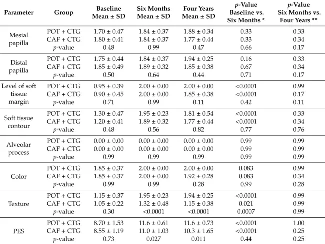
Figure
Documents relatifs
pensant faire découvrir aux élèves de nouvelles cultures alors qu’en fait, il accentue malheureusement les généralisations. L’élève en question peut ainsi
Maintenance of infliximab treatment in ankylosing spondylitis: Results of a one-year randomized controlled trial comparing systematic versus on-demand treatment.. Maxime
Stable coral reef habitats over geo- logical time may have promoted fish biodiversity by acting as (i) refugia, which preserved species from extinction because of habitat loss ( 4 ,
Cette thèse présente une Gestion basée objectif pour des Processus Métier Flexibles (GoPMF), avec les concepts sous-jacents, les processus de
Les paupières du jour se ferment J’entends
Puis le PE dit « Point to a tinsel » (par exemple) et les élèves doivent montrer avec leur doigt la flashcard qui correspond (ici : la guirlande de Noël).. Jeu : le jeu de Kim
Dans les deux pays, les principales mesures favorisant le retrait d’activité des travailleurs âgés sont en place depuis les années 1960 (préretraites FNE en France,
Participant Vedolizumab dose (mg) MedDRA PT As classi fi ed in text Medically signi fi cant observations at time of death Duration on study (days) Death during the TEAE reporting period
