Third-Order Nonlinear Optical Study
on
SubIimated/Langmuir-Blodgett Thin Films of
Lanthanide Porphyrin Phthalocyanine
Dimer/Heterodimer and Symmetric Trimer
Systems
by
Time-Resolved Non-Degenerate Four-Wave
Mixing
by
Mr. PAN, Ligang
Thesis Submitted in Partial Fullfillment of the Requirements
for
the Degree of Doctor of Science (Ph.D.)
Departement de physique, Faculte des sciences,Universite de Sherbrooke, Sherbrooke, Quebec, Canada.
October 1996
Members of Jury: Invited Member:
—Professor SergeJandl —ProfessorMarguerite-Marie Denariez-Roberge — Professor Denis Moris
Professor Daniel Houde (Director of the Thesis)
— Professor Laurent G. Caron (Directer of the Thesis )
1*1
National Library of Canada Acquisitions and Bibliographic Services 395 Wellington Street Ottawa ON K1AON4 Canada Bibliotheque nationale du Canada Acquisitions et services bibtiographiques 395, rue Wellington Ottawa ON K1AON4 CanadaYour file Votre reference Our file Notre r6f6rence
The author has granted a
non-exclusive licence allowing the National Library of Canada to reproduce, loan, distribute or sell copies of this thesis m microform,
paper or electronic formats.
The author retains ownership of the copyright m this thesis. Neither the thesis nor substantial extracts from it may be printed or otherwise
reproduced without the author's
permission.
L'auteur a accorde une licence non exclusive permettant a la
Bibliotheque nationale du Canada de
reproduire, prefer, distribuer ou vendre des copies de cette these sous
la forme de microfiche/fihn, de
reproduction sur papier ou sur format electronique.
L'auteur conserve la propriete du droit d'auteur qui protege cette these.
Ni la these ni des extraits substantiels de celle-ci ne doivent etre imprimes
ou autrement reproduits sans son
autonsation.
TOjftit^r
M$^m
^mmyw^^
x ^m^w, a^Nrn
To My Wife SUN Zhaoxia,
Our Sons Yingzhou & Xiongfei,
and Our Parents and Grand-Parents!
A ma femme SUN Zhaoxia,
nos enfants Yingzhou & Xiongfei,
et nos parents et grand-parents!
Zu meiner Frau SUN Zhaoxia,
unseren Kindem Yingzhou & Xionfei,
und unseren Eltem und Gropeltem!
^<7)^^
.b^LO^AMSS^,
t)kLk60M^, m <):/?^& ^ z,
-b/^L kb<r>W^/^t,
hk L ^60.bL?^S^b(f^&^(;
& L ^ (f ^ t!
K Moefl Focno^ce Cyn UaomHa,
Ham™ /leTHM Iteuoy H mnoH$bi,
Ham™ PO^HJIHM,
Resume
Ce travail porte sur 1'etude par la technique de melange a quatre ondes resolu dans Ie temps ("time-resolved four-wave mbdng" ou "TRFWM") de couches minces de differents materiaux organiques d'interet pour 1'optique non-Uneaire. n s'agit de couches sublimees du dimere neodymium porphyrme phthalocyanme Pc Nd Pc*, de 1'heterodimere cerium porphyrine phtfaalocyanine Pc2~Ce TPP2', des trimeres cerium
porphyrine phthalocyanine TPP Ce Pc Ce TPP et neodymium porphyrine
phthalocyanine Pc2'NdfflTPP Nd Pc , ainsi que de couches Langmuir-Blodgett du dimere mixte cobalt porphyrme phthalocyanine CoPC22+4 /H^PcTS4'.
Cette these fait etat de plusieurs premieres: premiere etudepar TRFWM de couches organiques sublimees, premiere observation et investigation de la contribution des mecanismes de diffusion d I'attenuation du reseau de dif fraction transitoire genere par TRFWM, premiere observation et investigation d'un effet photorefractif dans les
couches minces de multimeres organiques, et premiere etude et depistage d'un nouveau phenomene de mutation de mode de vibration.
Les valeurs absolues de la susceptibilite opdque non-lmeaire de troisieme-ordre ^<3)
des couches ont pu etre determinees grace a une etude comparee avec une lame de verre de quartz servant de reference.
Ce travail montre que la technique de TRFWM est non seulement utile mats surtout tres sensible et fort puissante pour 1'etude des procedes optiques non-lineaires dynamiques de troisieme ordre dans les materiaux organiques. EUe pennet entre autre de mesurer des mecanismes mobservables par absorption transitoire ou par d'autres methodes d'optique non-lineaire.
Les etudes des systemes dimere/heterodimere et trimere/heterodimere de terres rares porphyrine phthalocyanine monti-ent enfin que ces materiaux sont tres interessants pour leurs proprietes optiques non-lmeaires. Ce sont de nouvelles families de materiaux organiques tres prometteurs pour les etudes et les applications de I'effet photorefractif. Ils sont polyvalents et ont un grand potentiel comme materiaux optiques
Abstract
Time-resolved four-wave mixing (TRFWM) studies have been earned out on
sublimated fihns of the neodymium phthalocyanine dimer Pc Nd Pc , the cerium
porphyrin phthalocyanine sandwich mixed heterodimer Pc2'CeIVTPP2\ the cerium porphyrin phthalocyanine symmetric trimer TPP2~CemPc2~CemTPP2~, the neodymium porphyrin phthalocyanine trimer Pc2~Nd TPP2~Nd Pc2', and Langmuir-Blodgett fihns of the cobalt porphyrin phthalocyanine mixed dimer
CoPC^z^/H^PcTS4-.
In this work, we have presented a number of novel results: the first non-degenerate time-resolved four-wave mixing (NDTRFWM) experiment on a sublimated film, the first observation and identification of the diffusion contribution to the degrating process, the first observation and identification of photoreft active effect in an organic multimer thin film, and the first observation and identification of a correlated phonon mode shifting phenomenon.
The absolute values of the fhird-order nonlinear optical susceptibility ^ of the samples have been determined by comparing the third-order nonLuiear optical responses of the samples with those of a slide of reference fused quartz under tfae same experimental conditions.
It is shown that TRFWM is a very useful, very sensitive and very powerful tool to investigate the dynamics of the third-order nonlinear optical processes m a medium, especially those that can not be detected by transient absoiption and other means of probing nonlinear optical properties.
Through this work, it can be seen that lanthanide porphyrm phfhalocyamne dimer/ heterodimer trimer/heterotrimer systems could be a new family of organic materials
most promising for the investigation and the applications of photorefractive effect
m
^7:^4S^Kft-^^^^-^^-fc»^X^Pc2-NdmPc', ^^^^^^=-l
frPc2-CeIVTPP2-, tPh^^t^%ftTPP2-CeffiPc2-CefflTPP2-,t^f^t^%-^ Pc2-NdfflTPP2-NdfflPc2-^fM, ^4^*^»:£^r.XftCoPC^4'/H,PcTS4-^J Langmuir-Blodgett^MMf^ ?J-1^ ^T^^ ® &5,^(TRFWM)^ ^.
^X^ 7 lt—?9:?«)S±Wttl!tWfB|pT^H-a;g)@%^. ifii
kbttsl^^^6<)N-l'?l^^^Eg?A%»(TRFWM)^^^^^^f^%^^^^^
*, ifr-^.feSTrtfe^ig^-wasi^tiE. ^^, ^^a%^?f-%^,T^ft.-^
M + ^ ^-^^asTit^K^^^js^M-^tg^^^^^^^sea.
3li±fc^^N^^^^ft"F^^^&^^^-^*^^^S?N^ti7fc*^
SL, ^ilH^ti^si^t^fTfe^^^^A.
^^i£^7N-N'q~^^0t%^(TRFWM)^^^^^+5.yr^a'|i^:^^
^,6^-^tt^. ttXf. A^^S^AX^, A^^?t^?^¥^%
^7fcTtX^-ffet^'l±7fe^^^^a^^l6««S{^^*i±a*^> XAiS^.
A+j^^A, ^^"^'tk^=-^^/^^K^ ^iW^X-f^^
^t6A^ffl^w%^%?lf$efcS»^Ki-^STfi()®:&^a^ft*f^^ssi. ^ st,
^^MA^siS^t^^li, A-ftAt^^Mfe^^Mt^aiiTt:^
ft.
Acknowledgement
First of all, I would like to thank my two directors. Professor Laurent G. Caron, former Chairman of Canadian Association of Physics, and Professor Daniel Houde. It is Professor Laurent G. Caron who gave me the opportunity to study and work in I'Universe de Sherbrooke. Without his kindness and discerning eyes, I would have had no chance and no scope to exercise my abilities. It is Professor Daniel Houde who set up the femtosecond laser laboratory and proposed a research project which, although very difficult, proved to be very interesting, meaningful, and worth investigating in time and efforts. In his lab, I spent five imforgettable and fmitful years learning a great deal from him and accumulating the experiences that allowed me to mature as a scientific researcher. I would like to add that he is one oftfae best teachers I have ever seen. Without his attentive and enlightening guidance, tfais work would have been impossible.
Second I would like to thank Eh-. Thu-Hua Tran-Thi, her group in CEA, Centre d'Etudes Atomiques de Saclay, France, and her collaborators Professor Andre De Cian, Professor Driss Chabach, Professor Raymond Weiss. She and her group made an
important contribution to this work. During my visiting in CEA, I benefited a lot from
her kind arrangements and enthusiastic help.
I would like to thank Professor Marcel Aubin, who gave me my first French professional course in physics. With his warm and gentle help, I passed a drfficult turning point in my life.
I want to thank Professor Serge Jandl, Chamnan of Dq)artment of Physics, and his wife Carla Jandl. The former gave me a course in physics, the latter gave me a course in French. From their teachings, I gained a lot
I would like to thank Dr. Paul Grenier, who worked witfa Professor. Daniel Houde
to establish the femtosecond laser lab and helped me to quickly get access to all the facilities.
I wish to thank Dr. Rashmilkant Sudiwala, a specialist in Langmuir-Blodgett techniques. By means of his kind help, I got into the LB world and was able to complete an important part of this work.
I would like to thank Professor Andre-Marie Tremblay, Professor Alain Caille, and Professor Andre Lemieux, for their kind help to me, and thank the Department of Physics and C.R.P.S. ( Centre de Recherche en Physique du Solide ) of the Department of Physics for the financial support.
Many thanks to Professor Le Dao and his group for their collaboration in making the sublimated fihns.
Furthermore, I would like to thank Professor Jean Beerens and Mr. Jacques Corbin of the Department of Physics of Sherbrooke University, for their help m measuring the thickness of the samples, and thank Professor Jacques Beauvais, Ie directeur de Salle Blanche and Mr. Michel Paquette, du Departement de Genie Electrique de FUniversUe de Sherbrooke, for their help in measuring the refractive index of the samples.
I also thank Dr. Jean Fran^ois Lipskier, Dr. Thierry Foumier and Dr. Isabelle Salabert. Their photo-chemical study on the lanthanide porphyrm phthalocyanme dimer/heterodimer trimer/heterotrimer by transient absorption was of great importance to this work.
Thanks also are given to the Medical Research Council of Canada and the Centre of Excellence in Molecular and Interfacial Dynamics, CIDA and France-Quebec, for their support of this work.
Finally, I would like to thank Dr. Claude Pepin, Ms. Stephanie Marengo and all the members of the groupe CRM ( Conseil de Recherche Medicale ) de recherches en sciences dos radiations du Departement de Medecine Nucleaire et de Radiobiologie, Faculte de Medecine, Umversite de Sherbrooke. Thanks to all the known or unknown persons who have contributed to this research work.
I.
II.
III.
VII.
VIII.
DC.
X.Resume
Abstract
?
Acknowledgement
Table of Contents
List of Figures
List of Tables
Table of Contents
• 1. • • 11.m.
IV
VI. Xl. XV111.XI. ListofAbreviations xix.
Introduction 1.
Chapter 1
General Consideration 5.
1.1. Ultrafast Time-Resolved Spectroscopy 5 .
1.1.1. Time-Resolved Spectroscopy 7.
1.1.2. Pump-Probe Technique 8.1.1.3. White-Continuum Generation 12.
1.1.4. Basic Research Opportunities 12.
1.1.5. Transient Grating Method 13.
1.1.5.1. Formation of the Transient Grating 13.
1.1.5.2. Detection of the Decay of the Transient Grating 1 4. 1.1.5.3. Comparison of the Transient Grating Method
with the Transient Absorption Technique 15.
1.2. Experimental Materials Used 1 8.
1.2.1. Nonlinear Optical Materials 18.
1.2.2. Porphyrin and Phthalocyanine 20.
Phthalocyanine Dimeric/Heterodimeric Trimeric/
Heterotrimeric Prototype Model System 22.
1.3. Two Important Experimental Phenomena
Encountered 29.
1.3.1. Electron Transfer 29.
1.3.2. Photorefractivity 32.
1.4. Outline of Our Research Work 35.
Chapter 2
Theoretical and Experimental Backgrounds of
Nonlinear Optics 36.
2.1. Basis for Macroscopic Electromagnetic
Theory ofNonlinear Optics 36.
2.2. Macroscopic Expressions for Nonlinear Optics 39.
2.3. Jth-Order Nonlinear Optical Interaction
Processes in a Medium 44.
2.4. Third-Order Nonlinear Optical Effects:
Four-Wave Interactions 47.
2.5. Nonlinear Optical Experiment Requirements 48.
2.6. Four-Wave Interaction (FWI) and Four-Wave
Mixing (FWM) 49.
2.7. Transient Response of a Medium in
Four-Wave Mixing 51.
2.7.1 Classical Field Theory View 51.
2.7.2. Coupled Phonons 56.
2.7.3. Population Relaxation 57.
Chapter 3
3.1. Experimental Set-up 5 8 .
3.2. Femtosecond Laser Generator and Monitor 61.
3.3. Femtosecond Laser Amplifier 63.
3.4. Four-Wave Mixing System 65.
3.4.1. Spatial Arrangement of Four-Wave Mixing Beams 66.
3.4.2. Synchronization of the Two Pump Pulses 67.
3.5. Four-Wave Mixing Signal Detection System 69.
3.6. Four-Wave Mixing Data Acquisition System 70.
3.7. Synchronism Control System 70.
3.8. Experimental Approaches 71.
Chapter 4.
Results and Discussion 73.
4.1. Sublimated Film of Cerium Porphyrin
Phthalo-2-^
IV___2-cyanine Heterodimer Pc ~ Ce ~ TPP ~ 74.
4.1.1. Experimental Aspects 74.
4.2. Results and Discussion 75.
4.1.2.1. Time-Resolved Four-Wave Mixing
Experimental Results 76.
4.1.2.2. Transient Absoq)tion Experimental Results 79. 4.1.2.3. Fitting of Time-Resolved Four-WaveMixing Experimental Data 81 .
4.1.2.4. Singlet Migration by Ring to Metal Charge
Transfer State Coupling between the
Neigh-bouring Molecules 83.
4.1.2.5. Charge Transfer State Migration bySpin-Orbit Coupling between the Neighbouring
Molecules 86.
4.1.2.6. Excitation Migration by Excitonic Coupling
4.1.2.7. Identification of Two-Correlated-Phonon
Mode-Shifting Phenomenon
4.1.2.8. Identification of the Diffusion Contribution
to the Degrating Process 99.
4.1.2.9. Attribution of Different Components
of the Time-Resolved Four-Wave
Mixing Experimental Data 102 .
4.1.3.Summary 104.4.2.Sublimated Film ofCerium Porphyrin
Phthalocyanine Symmetric Trimer
TPP2'Ce"IPc2'CeIIITPP2' 106.
4.2.1.Experimental Aspects 106.
4.2.2.Results and Discussion 106.
4.2.3.Summary 114.4.3 Langmuir-Blodgett Film of Mixed Dimer of
CoPC^/H^PcTS4- 116.
4.3.1. Experimental Aspects 116.
4.3.1.1. Preparation of Langmuir-Blodgett Fihns 116.4.3.1.2. FWM Measurement on
Langmuir-Blodgett Films 117.
4.3.2. Results and Discussion 118.
4.3.3. Summary 125.4.4.Neodymium Porphyrin Phthalocyanine
Symmetric Dimer NdPc; Trimer (NdPc); TPP 126.
4.4.1 .Experimental Aspects 126.
4.4.2.Results and Discussion 126.
4.4.3.Summary 139.4.5. Determination of the Values of the Third-Order
Nonlinear Optical Susceptibility ^3) 141.
4.5.2. Calculation of the Third-Order Nonlinear
Optical Susceptibility ^<3) of the Samples 143.
4.6. Photorefractive Effect 149.
Chapter 5.
Conclusion 155.
Appendixes I
Transient Response of Population Relaxation
158.
Appendixes II
Transient Response ofFour-Wave Mixing in a
IVIedium, a Quantum Mechanical View
162,
List of Figures
Figure 1. .0.1. Plot of the Logarithm of the Shortest Available Optical
PulseWidth versus Years. 6.
Figure 1.1.1.1. Optical Delay Line, White Continuum Generation and Time-Resolved Spectroscopic M^^urement with
Probe-Pump Technique m Transient Absorption. 8. Figure 1.1.2.1. Approximate Time Scales of Elementary Molecular
Relaxation Phenomena and Some Chemical and
Biological Manifestations of these Phenomena. 10. Figure 1.1.2.2. Measuring the Decay Time of Short-lived Excited
States by Time-Resolved Pump-Probe Techniques. 11.
Figure 1.2.2.1. Molecular Structure of PoiphriL 21. Figure 1.2.2.2. Molecular Strocture of Phthalocyanines. 21.
Figure 1.2.3.1. Constitution of Octacoordinated Metal Monotetra-pyrroles M(P)I.4 and Configurations with A: Mono-tetrapyrroles, B: Bistetrapyrroles MCP^, and C:
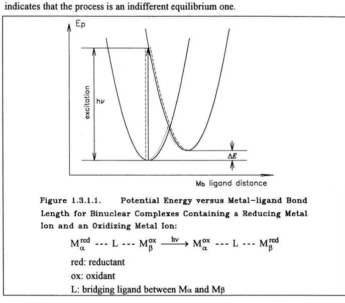
Tris-tetrapyrroles M.^(P)^ M = Lantfaani(fe Metal Ion. 23. Figure 1.3.1.1. Potential Energy versus Metal-Ligand Bond Length
for Binuclear Complexes Contaming a Reducing and
an Oxidizing Metal Ion. 30.
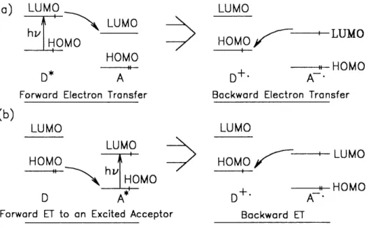
Figure 1.3.1.2. Electron Transitions in a Diatomic Molecule. 31 .
Figure 1.3.1.3. Simplified View of Forward and Backward Electron Transfers involving (a) an Excited Donor, and
(b) an Excited Acceptor. 31.
Figure 2.6.1. Wavevector diagrams of the Four-Wave Interaction
Processes. 50.
Figure 3.1.1. Block Diagram of the Experimental System. 59. Figure 3.1.2. Details of Experimental System. 60.
Figure 3.2.1. Femtosecond Laser Generator and Monitor. 62. Figure 3.3.1. Four-Stage Femtosecond Laser Amplifier. 64. Figure 3.4.0.1. Four-Wave Mbdng System. 65.
Figure 3.4.1.2. Wave Vector Diagram of Four-Wave Mixing Process. 67.
Figure 3.4.2.1. Relative Positions of Pump 1, Pump2, Probe, Diffracted
Signal, and Autograting 1, Autograting2 Beams. 68. Figure 3.5.1. Four-Wave Mixing Signal Detection System. 69. Figure 3.6.1. Four-Wave Mixing Data Acquisition System. 70. Figure 3.7.1. Synchronism Control System. 71. Figure 4.1.2.0.1. Constitution of Octacoordinated Metal Monotetrapyrroles
M(P)L4 and Configurations with A: Monotetrapyrroles, B: Bistetrapyrroles PMPc, e.g. PcCeTPP, M = Lanthanide
Metal Ion. 75.
Figure 4.1.2.0.2. Ground State Absorption Spectra of Sublimated FUms
of the Heteroduner PcCeTPP on a Quartz Substrate. 76. Figure 4.1.2.1.1. TRFWM Signal at Phase-Matching Direction as
a Function ofProbe-to-Pump Delay Time for the
Sublimated Fihn of the Heterodimer PcCeTPP
on a Quartz Substrate in a Time Scale up to 10 ps. 77. Figure 4.1.2.1.2. TRFWM Signal at Phase-Matching Direction as a
Function of Probe-to-Pump Delay Time for the
Sublimated Fihn of the Heterodimer PcCeTPP on a
Quartz Substrate in a Time Scale up to 100 ps. 77. Figure 4.1.2.1.3. TRFWM Signal at Phase-Matching Du-ection as a
Function of Probe-to-Pump Delay Time for the
Sublimated Film of the Heterodimer PcCeTPP on a
Quartz Substrate m a Time Scale up to 500 ps. 78. Figure 4.1.2.1.4. TRFWM Signal at Phase-Matching Direction as a
Function of Probe-to-Pump Delay Time for the
Sublimated Fihn of the Heterodimer PcCeTPP on a
Quartz Substrate in a Time Scale up to 1.3 ps. 78. Figure 4.1.2.2.1. Schematic Presentation of the Relaxation Paths of
the Sublimated Heterodimer Fihn ofPcCeTPP m the Experiment of (a): Transient Absorption (TA), and (b): Time-Resolved Four-Wave Mbdng (TRFWM)
and Two Correlated Vibration Modes co i, T^ and co^, 1:2.
^-Figure 4.1.2.4.1. Singlet Excited State Migration by Ring to Metal Charge Transfer State Coupling between the Neighbouring
Molecules in the Sublimated Fihn of the Heterodimer
Pc2-CeIVTPP2-. 85.
Figure 4.1.2.5.1. Charge Transfer State Migration by Spin-Orbital Coupling
between the Neighbouring Molecules in the Sublimated
Fihn of the Heterodimer Pc2-CeIVTPP2-. 86. Figure 4.1.2.6.1. Excitation Migration by the Excitonic Coupling
between the Adjacent Molecules m the
Subli-mated Film of theHeterodimer Pc2-CeIVTPP2-. 88. Figure 4.1.2.7.1. Oscmating Part of the TRFWM Signal at
Phase-Matching Direction as a Function of
Probe-to-Pump Delay Time Fitted with Two Correlated
Phonon Modes Shifting from One to the Other. 90.
Figure 4.1.2.7.2. Oscillating Part of the TRFWM Signal at Phase-Matching Direction as a Function of
Probe-to-Pump Delay Time Fitted with a Single Phonon
Mode of v = 30 cm-1 and T = 0.90 ps. 90.
Figure 4.1.2.7.3. Oscillating Part of the TRFWM Signal at Phase-Matching Direction as a Function
ofProbe-to-Pump Delay Time Fitted with a Single Phonon
Mode ofv = 40 cm-1 and T= 0.90 ps. 91.
Figure 4.1.2.7.4. OsciUatmg Part of the TRFWM Signal at Phase-Matching Direction as a Function of
Probe-to-Pump Delay Time Fitted with a Single Phonon
Mode of v = 30 cm-1 and T = 8.4 ps. 91.
Figure 4.1.2.7.5. Oscillatmg Part of the TRFWM Signal at Phase-Matching Direction as a Function of
Probe-to-Pump Delay Time Fitted with a Smgle Phonon
Mode of v = 40 cm-1 and T = 8.4 ps. 92.
Figure 4.1.2.7.6. OscUlatmg Part of the TRFWM Signal at Phase-Matching Direction as a Function of
Probe-to-Pump Delay Time Fitted with a Single Phonon
Mode of v = 30 cm-1 and T = 1.3 ns. 92.
Figure 4.1.2.7.7. Osdllating Part of the TRFWM Signal at Phase-Matching Direction as a Function of
Figure 4.1.2.7.8. Oscillating Part of the TRFWM Signal at Phase-Matching Direction as a Function of Probe-to-Pump Delay Time Fitted with a Sumation of Two
Independent Phonon Modes. 93. Figure 4.1.2.7.9. Schematic Presentation of Biplane and Triplane
Mixtures of Celv and Lnm (Lanthanides) with
the Inteq)lanar Distances in A. 95. Figure 4.1.2.8.1. Electron Microscope Image of the Microstructure
of the Sublimated Film of the Heterodimer Pc2-CeIVTPP2-. 102. Figure 4.2.2.1. Molecular Structure of Octacoordinated Metal
Monotetrapyrroles and Configurations with A: Monotetrapyrroles, B: Bistertrapyrroles M(P)2 and C: Tristetrapyrroles, Symmetnc
TrimerPc(CeTPP)2, M = Lanthanied Ion. 108. Figure 4.2.2.2. Ground State Absorption Spectrum of the Sublimated
FUm ofTrimer TPP2-CemPc2-CefflTPP2- on a Quartz
Substrate. 108.
Figure 4.2.2.3. Dif&acted TRFWM Signal as a Function of
Probe-to-Pump Delay Time for the Sublimated Symmetric Trimer Pc(CeTPP)2 on a Quartz Substrate with a Time Scale up
to 10 ps. 109. Figure 4.2.2.4. Dif&acted TRFWM Signal as a Function of
Probe-to-Pump Delay Time for the Sublimated Symmetric Trimer
TPP2-CefflPc2-CefflTPP2" on a Quartz Substi-ate with
a Time Scale up to 100 ps. 109. Figure 4.2.2.5. Dif&acted TRFWM Signal as a Function of
Probe-to-Pump Delay Time for the Sublimated Symmetric Trimer
TPP2-CefflPc2-CefflTPP2' on a Quartz Substrate with
a Time Scale up to 500 ps. 110. Figure 4.2.2.6. Dif&acted TRFWM Signal as a Function of
Probe-to-Pump Delay Time for the Sublimated Symmetric Trimer
TPP2~CefflPc2-CemTPP2~ on a Quartz Substrate with
a Time Scale up to 1.3 ns. 110. Figure 4.2.2.7. Schematic Presentation of the Relaxation Paths of the
TPP2-CefflPc2-CemTPP2-in the Experiment of (a): Transient Absoq)tion, (b):
Time-Resolved Four-Wave Mixing. 113. Figure 4.2.2.8. Electron Microscope Image of the Microstmcture
oftheSublimatedFUmof the Trimer Pc(CeTPP)2. 113.
Figure 4.3.2.1. Molecular Stmcture of the Cobalt PoqAyrin Phthalocyanme Mixed Dimer CoPC^4+ / HsPcTS 4- 119. Figure 4.3.2.2. Ground State Absorption Spectrum of the
Langmuir-Blodgett Film of the Mbced Dimer CoPC^4+ / HzPcTS 4"
on a Glass Substrate. 1 19.
Figure 4.3.2.3. Ground State Absorption Spectrum of the Monomer
CoPC^ , 4Br- in a Solution of DMSO/CHC^ 120.
Fig ure 4.3.2.4. Ground State Absorption Spectrum of the
Unsubsti-tuted Monomer H2PcTS 4~, 4Na+. 120. Figure 4.3.2.5. Four-Wave Mbcing Signal as a Function
ofProbe-to-Pulse Delay Tune for the Langmuir-Blodgett Film of the Mixed Dimer CoPC^4+ / HzPcTS 4" on a Glass
Substrate, with a Time Scale up to 10 ps. 121. Figure 4.3.2.6. Four-Wave Mbdng Signal as a Function
ofProbe-to-Pulse Delay Time for tfae Langmuir-Blodgett Film of
the Mixed DimerCoPC^/H2PcTS4'on a Glass
Substrate, with a Time Scale up to 100 ps. 121. Figure 4.3.2.7. Four-Wave Mixing Signal as a Function
ofProbe-to-Pulse Delay Time for the Langmuir-Blodgett Fihn of the Mixed Dimer CoPC^4+/HzPcTS 4~ on a Glass
Substrate, with a Time Scale up to 500 ps. 122.
Figure 4.3.2.8. Four-Wave Mixing Signal as a Function of
Probe-to-Pulse Delay Time for the Langmuir-Blodgett Fihn of
Figure 4.3.2.9. Four Excited State Populations in LB fihn of the Mixed
Dimer CoPC^4+ / HsPcTS 4" (a): Simplest &, (b): the
Most Complicated Cases. 123.
Figure 4.4.2.0. Molecular Stmctures of Octacoordinated Metal
Mono-tetrapyrroles and Configurations with A: Monotetra-pyrroles, B: Bistetrapyrroless M(P)2 and C: Tristetrapyr-roles M2(P)3, M = Lanthanide Metal Ion to Form, e.g., the
Dimer NdPc2 and the Trimer (NdPc)2TPP, etc. 127.
Figure 4.4.2.1. Ground State Absorption Spectrum of the Sublimated
Fihn of the Dimer NdPc2 on a Quartz Substrate. 128. Figure 4.4.2.2. Ground State Absorption Spectrum of the Sublimated
Film of the Trimer Nd2Pc2TPP on a Quartz Substrate. 128.
Figure 4.4.2.3. Time-Resolved Non-Degenerate Four-Wave Mixing
Signal as a Function of the Probe-to-Pump Delay
Time for the Sublimated Fihn of the Dimer NdPc^ on
a Quartz Substrate with a Tune Scale up to 10 ps. 129.
Figure 4.4.2.4. Time-Resolved Non-Degenerate Four-Wave Mixmg
Signal as a Function of the Probe-to-Pump Delay
Time for the Sublimated Film of the Dimer NdPc^ on
a Quartz Substrate with a Time Scale up to 100 ps. 129.
Figure 4.4.2.5. Time-Resolved Non-Degenerate Four-Wave Mixing
Signal as a Function of the Probe-to-Pump Delay
Time for the Sublimated Fihn of the Dimer NdPc2 on
a Quartz Substrate with a Time Scale up to 500 ps. 130.
Figure 4.4.2.6. Time-Resolved Non-Degenerate Four-Wave Mbung
Signal as a Function of the Probe-to-Pump Delay
Time for the Sublimated Film of the Dimer NdPc^ on
a Quartz Substrate with a Time Scale up to 1.3 ns. 130.
Figure 4.4.2.7. Time-Resolved Non-Degenerate Four-Wave Mixing
Signal as a Function of the Probe-to-Pump Delay Time
for the Sublimated Fihn of the Trimer (NdPc)2TPP on a
on a Quartz Substrate with a Time Scale up to 10 ps. 131.
Figure 4.4.2.8. Time-Resolved Non-Degenerate Four-Wave Mbdng
Signal as a Function of the Probe-to-Pump Delay
Time for the Sublimated Film of the Trimer (NdPc^TPP
Figure 4.4.2.9. Time-Resolved Non-Degenerate Four-Wave Mixing
Signal as a Function of tfae Probe-to-Pump Delay
Time for the Sublimated Fihn of the Trimer (NdP^TPP
on a Quartz Substrate witfa a Time Scale up to 500 ps. 132.
Figure 4.4.2.10. Time-Resolved Non-Degenerate Four-Wave Mixing
Signal as a Function of the Probe-to-Pump Delay
Time for the Sublimated Film of the Trimer (NdP^TPP
on a Quartz Substrate with a Time Scale up to 1.3 ns. 132. Figure 4.4.2.11. Schematic Presentation of the Relaxation Paths of the
Sublimated Fihn ofAe Dimer NdPc^ in the Experiment
of (a): Transient Absorption and (b): Time-Resolved
Four-Wave Mixing. 138.
Figure 4.4.2.12. Schematic Presentation of the Relaxation Paths of the
Sublimated Fihn of the Trimer (NdPc)2TPP in the Experiment of (a): Transient Absorption and (b):
List of Tables
Table 1.1.5.3.1. Major Differences Between the Transient Grating Method and the Transient Absorption Technique. Table 1.2.3.. 1. Specifications for Porphyrins .
Table 1.3.2.1.1. Photorefractive Materials.
Table 4.1.2.9.1. Attribution of the Different Components from the
Experiments of Transient Absorption (TA) and
Time-Resolved Four-Wave Mbdng (TRFWM) /
Transient Grating (TG) for the Sublimated Film of
the Heterodimer PcCeTPP.
Table 4.2.2.1. Attribution of the Different Components from the Experiments of Transient Absorption (TA) and Time-Resolved Four-Wave Mbdng (TRFWM ) /
Transient Grating (TG) for the SublimatedFilm of the Trimer (TPPCe^Pc.
Attribution of the Different Components from the Experiments of Transient Absorption (TA) and Time-Resolved Four-Wave Mixing (TRFWM ) / Transient Grating (TG) for the Sublimated Fihn of
theDunerNdPc2.
Attribution of the Different Components from the Experiments of Transient Absoq)tion (TA) and Time-Resolved Four-Wave Mbdng (TRFWM ) / Transient Grating (TG) for die Sublimated Film of the Dimer (NdP^TPP.
Table 4.5.2.1. Peak Values of the Third-Order Nonliaear Optical
Susceptibilities ^3\
Table 4.4.2.1. Table 4.4.2.2. 16. 24. 34. 103. 112. 135.Table 4.5.2.2. Third-Order Nonlmear Optical Susceptibility %<3) of Some
Metallophthalocyaniaes .
135.
146.
List of Abbreviations
2WM
4WM
A
AS
ATP
BPH
BPS
CeCoPC224+/H2PcTS4-CPM
CT
CTSC
D
DC
DM
EC
EEM
ET
FDFWM
FWDFG
FWI
FWSFG
FWUC
HDM
HBP
Two-Wave Mixing Four-Wave Mixing Acceptor Antena System Adenosine Tri-Phosphate bacteriochlorophyll Bacteria Photosystem Ceriumcobalt porphyrin phthalocyanine mixed dimer Colliding Pulse Mode-locked (dye laser)
Charge Transfer
Charge Transfer State Coupling
Donor
Direct Current Dimer
Excitonic Coupling
Excitonic Excitation Migration
Electron Transfer
Fully-Degenerated Four-Wave Mixing
four-wave difference-frequency generation Four-Wave Interaction
four-wave sum-frequency generation four-wave up-conversion
Heterodimer
1,2,3,4,8,9,10,11,15,16,
HTM
HOMO
ICT
KDP
KTP
LBF
LMCT
Ln LuLUMO
MD
MDM
MLCT
NADP
Nd
NDFWM
NDTRFWM
NLO
NRE
OEP
OMP
OPRM
p
Pc Pc2-CeIVTPP2-Pc2-NdmPce Pc2-NdmTPP2-NdfflPc2-HeterotrimerHighest Occupied Molecular Orbit
Intramolecular Charge Transfer
potassium (K) Dihydrogen Phosphate potassium (K) Titanyl Phosphate
Langmuir-Blodgett Film
Ligand to Metal Charge Transfer Lanthanium
Lutetium
Lowest Unoccupied Molecular Orbit
Mixed Dimer
Molecular Dipole Migration
Metal to Ligand Charge Transfers
Nicotinamide Adenine Dinucleotide Phosphate
Neodium
Non-Degenerated Four-Wave Mbdng
Non-Degenerate Time-Resolve Four-Wave Mudng
Nonlinear Optics
Non-Resonant Excitation
2,3,7,8,12,13,17,18-octaethylporphyrin 2,3,7,8,12,13,17,18-octamethylporphyrm
Organic Phortorefractive Material Porphyrin
Phthalocyanine
cerium poq)hyrm phthalocyanine sandwich mixed heterodimer
neodymium porphyrin phthalocyanine dimer
PDFWM
PR
PRE
PS
PSU
RC
SHG
soc
TA
TAP
TC1P
THG
TM
TPP
TPP2-CefflPc2-CefflTPP2-TRTG
TRFWM
TTP
TRNDFWM
TRTG
Pardally-Degenerated Four-Wave Mixing Photo Refractrvity, Photo Refi-active
Photo Refractive Effect
Photosystem
Photo Synthetic Unit
Reaction Center, Resistance-Capacity Second Harmonic Generation
Spin-Orbit Coupling
Transien Absorption
5,10,15,20-tetra(p-anisyl)porphyrm
5,10,15,20-tetra(p-chlorophenyl)porphyrm Third Harmonic Generation
Trimer
5,10,15,20-tetraphenylporphyrm
cerium porphyrin phthalocyanine symmetnc trimer time-resolved transient grating
Time-Resolved Four-Wave Mixing
5,10,15,20-tetra(p-tolyl)porphyrin
Time-Resolved Non-Degenerate Four-Wave Mbdng Time-Resolved Transient Grating
Introduction
The importance of nonlinear optical phenomena has been known for a long time. Since the 1980s, materials research and development for nonlinear optical applications have rapidly progressed so that several systems are available commercially. To date, the systems have been utilized in information processing, optical switching, optical
frequency conversion, and telecommunications. The advancing development of
optotechnology has encouraged research on suitable nonlinear optical materials. A wide variety of materials, including inorganic and organic crystals, polymers, semiconductors, composites and metal-based systems, possess noticeable nonlinear optical properties. In recent years, organometallic compounds, by their unique characteristics, such as diversity of metals, oxidation states, ligaads, and geometries, have achieved success in and brought a new dimension to the area ofnonlinear optics.
In last six years, investigations of organometallic systems have been greatly intensified, because [1,3]:
1. Organometallic system can possess metal —> ligand or ligand —> metal charge
transfer ( MLCT, LMCT ) bands in the visible region of the spectrum, which are
usually associated with large second-order activity.
2. The compounds have great possibilities for redox changes, a property largely associated with the metal center, which can be electron poor or rich depending on the oxidation state and ligand enviromnent. Facile redox ability can lead to large hyperpolarizability, with the metal center being an extremely strong donor/acceptor in comparison to conventional organic systems.
3. Chromophores, such as phthalocyanines, containmg metal ions, are among the most intensely coloured materials known. The strength of the optical absorption band is also associated with large optical nonlinearities [4].
4. Many organometallic compounds have low-lying excited states with their dipole moments significantly different from their respective ground state dipole moments.
one or more of the associated ligands and have a large osciUator strength. This will
provide a substantial contribution to the hyperpolarizability.
5. Organometallic compounds also have important advantages in the range and mix of non-aromatic ligands that can be attached to the metals. These Ugands can shift the occupied and unoccupied metal d orbitals that interact with the Ti-electron orbitals of the conjugated systems. This provides a mechanism for fine-tunmg and
optimizmg the bulk susceptibility.
6. Furthermore, the metal centers in these molecules can constitute a chiral species [5]. When dissolved, they will form materials that crystallize m
non-centrosymmetric space groups, essential for a non-zero %<2) value.
7. Incorporation of transition metal ions, such as lanthanide ions, can also be expected m some cases to increase the solubility of a material m common organic solvents, therefore enhancing its processability.
The crucial aspect in the progression of nonlinear optical studies has been the development of experimental techniques for investigating and measuring a material's non-linear optical properties. Over the years, several methods have been widely used to make nonlinear optical studies, such as, Kurtz powder technique [6, 9], electric field induced second harmonic generation ( EFISH ) [10 - 13], hyper-Raylejgh scattering
(HRS) [14, 15], third harmonic generation ( THG) [16, 17], and degenerate four-wave
mixing (DFWM) [18 - 22]. More surprisingly, though non-degenerate four-wave mixing (NDFWM), both in resonant and non-resonant situations, is a powerful technique to study dynamical properties of physical systems, very few reported experimental mvesdgations of organometallics have used this important technique. Commonly, a combination of techniques is necessary for a complete investigation of a material's nonUnear optical behaviour.
The third-order nonlinear optical studies on organometallic materials can be divided into six major classes[23]: metalhcenes, metallopolyyne polymers, polysilanes and-germanes, metal dithiolene complexes, thwphenes, and phthalocyaninato metal
The phthalocyamnes and porphyrins are two extremely interesting and useful classes of compounds. Their extensively delocalized two-dimensional Ti-electron systems have broken through some limitations of most other third-order non-linear optical compounds featuring pseudo-one-dimensional Ti-electron conjugation systems. This has led to considerable interest in the two dimensional Ti-conjugated phthalocyanine systems and, in particular, the complexes containing a wide variety of
metals.
Phthalocyanine has a similar macrocycle to porphyrin and its absorption spectrum is complementary to that of porphyrm. Therefore, the dimer formed by linking porphyrin and phthalocyanine will absorb more efficiently the solar energy than the monomer of porphyrm or phthalocyanine [24]. Nevertheless, until now, only Tran-Thi [25-27],
Gaspard [28, 29], and Xu [30, 32] have synthesized porphyrin phthalocyanine
dimer/heterodimer and made observations on energy transfer and charge separation processes. Gaspard and Xu found that these are related to the molecular configurations. Tran-Thi, through the research on Langmuir-Blodgett films of porphyrm phthalocyanines, found that the dimer/heterodimer have a face-to-face geometry, in which there is a strong excitonic coupling between the porphyrm and the phthalocyamne. Compared with the studies on the other polymers of metallo organics and on the metallo monomers ofporphyrm and phfhalocyanine, the research on metaUo multimers ( dimer / heterodimer and trimer / heterotrimer ) of the porphyrm phthalocyanine, is just at its beginning.
Though research on the optical nonlinearity of organometallic complex is very active, the relationship between the structure and the nonlinear optical properties are still unclear. Unlike the design of second-order nonlinear optical materials, which follows some well defined guidelines, models for the synthesis of third-order species are much less developed. Presently, research on the third-order nonlmearities of porphyrm phthalocyanine is focused mainly on the monomers and on the effects of replacing the central metal ion on the optical nonlinearities. A systematic research on the third-order nonlinearities of porphyrm phthalocyanine dimer/heterodimer and trimer/heterotrimer systems has not yet been undertaken.
In this work, we present a third-order nonlmear optical study of sublimated and
dimer/heterodimer and trimer/heterotrimer systems using a non-degenerate
time-resolved four-wave mixing technique.
Chapter 1 introduces some important general consideration in the experiment: (1.1.) ultrafast time-resolved spectroscopy, (1.2.) prototype model system used in the
experiment, (1.3.) two important experimental phenomena encountered, and (1.4.) the
outline of our research work. Chapter 2 presents the theoretical and experimental basics for this work, especially of four-^ave interactions. Chapter 3 describes the experimental system. Chapter 4 presents and discusses the experimental results. Finally, Chapter 5 makes a conclusion on this research work.
Chapter 1
General Consideration
This is an experimental research work on nonlinear optical dynamic behaviours of the sublimated and Langmuir-Blodgett thin fihns of lanthanide porphyrm phthalocyanine multimers. Time-Resolved Non-Degenerated Four-Wave Mudng (TRNDFWM) has been used as a major experimental technique in this work. Nonlinear optical responses of the lanthanide porphyrin phthalocyamne dimers, herodimers and trimers have been investigated by TRNDFWM.
This chapter will give some detailed aspects of the experiment techniques, description of the materials used, and motivation oftfais research work.
1.1. Ultrafast Time-Resolved Spectroscopy
TRNDFWM is one of the most useful ultrafast coherent spectroscopy techniques.
Coherent spectroscopy implies investigations both in the time and frequency
domains, yielding infomiation on dynamic processes as well as Line widths and line positions of the molecular systems under investigation.
Initially started in the mid-sixties, coherent spectroscopy is ultimately connected with the development of intense coherent light sources which, as already mentioned, are prerequisite to the study ofnonlinear optical effects.
Until about the mid-sixties, the performance of the time-resolved techniques was limited to the microsecond region in the case of absoq)tion spectroscopy and to the nanosecond region in the case of fluorescence. During the late sixties, the appearance of giant pulse lasers brought the time resolution into the nanosecond range. A major advance in the generation of ultrashort optical pulses happened after
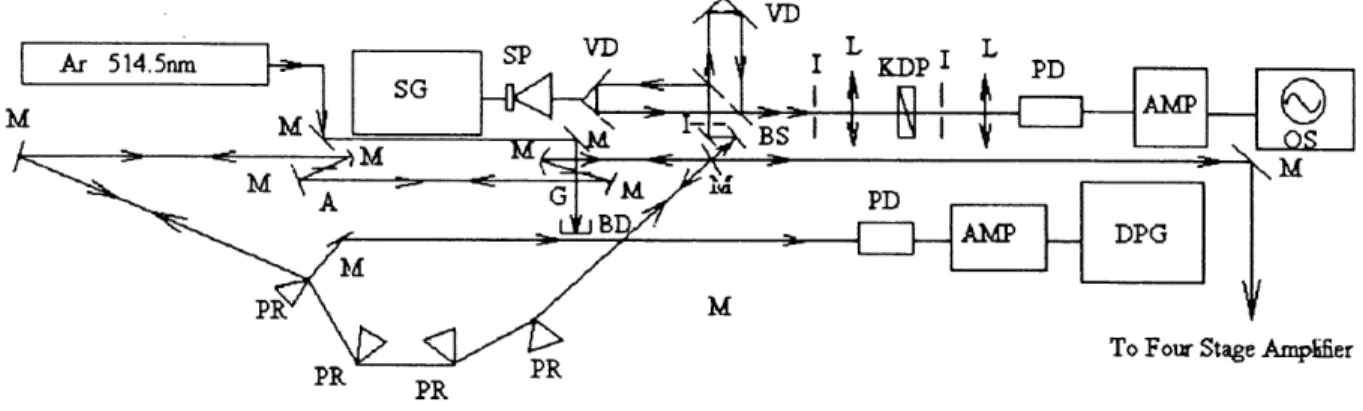
the invention of mode-locking techniques [33, 34]. Passive mode-lockmg of the mby laser by Mocker [35] opened a new era for ultrashort optical pulse generation. Shortly thereafter, the generation of the first optical pulse in the picosecond range with a Nd:glass laser was reported by De Maria [36]. Since that time, there followed a number of dramatic advances in ultrashort laser pulse generation as shown m Fig.
1.1.0.1. . The shortest optical pulse width available at a given year fells at an exponential rate. Each reduction in pulse width has always been accompanied by an advance in technology [37].
Until now, using colliding pulse mode-locking (CPM) [38 - 40], pulse compression [41 - 49], and other techniques, one has been able to reduce the optical pulse width to six femtoseconds [50, 51]. This approaches the ultimate limit m the visible region set by the uncertainty principle.
10-11 10-12 TJ G 0 a <u m ^ ^.
^
0) t»3
0^ ,n d I—I •r-1I
10-13 10-14 10-15 Nd:GlassFlashlamp Dye Laser Continuous Dye Laser
Colliding Pulse Mode Locking
Pulse Compression
1965 1970 1975 1980 1985 1990 Year
Figure 1.1.0.1. Plot of the Logarithm of the Shortest Available Optical Pulse Vidth versus Years.
The ability to generate ultrashort light pulses has extended coherent spectroscopy to real-time measurements of rapid dynamic processes on picosecond and femtosecond time scales. Most of the time-resolved spectroscopy investigations are
experimental capabilities has stimulated theoretical investigations and improved our understanding of the interactions m liquids and solids.
1.1.1. Time-Resolved Spectroscopy
While different sub-Doppler techniques aim at higher spectral resolutions, time-resolved spectroscopic techniques target on higher time resolutions.
The generation of extremely short and intense laser pulses has opened the door to the study of fast transient phenomena. Picosecond and femtosecond light pulses make it possible to investigate ultrafast processes occurring during the excitation and the deactivation of molecular states in solvents and solids.
Optoelectronic detection systems, such as fast photodiodes and sampling oscilloscopes, have reached a time resolution of 10-10 s, or 100 ps. This is stiU too long to resolve many fast transient events on a picosecond and subpicosecond time
scale. Thus, in picosecond and sub-picosecond spectroscopy, one had to develop a
series of new techniques to measure the durations and the profiles of the light pulses, and to probe the ultrafast relaxation processes.
Except for the streak camera which can reach time resolutions of a few picoseconds, most of the methods used to measure pico- and subpico-second phenomena are based on optical delay lines and on some nonlinear optical effects,
such as SHG. In optical delay line systems, as shown in Fig. 1.1.1.1., an ultrashort
laser pulse is divided by a beam splitter into two, or more if necessary, replica pulses which travel different path lengths before they are recombined. Hence, the measurement of a time interval At has been converted into that of a path difference Ax = cAt, where c is the light velocity. For example, in our experiment, the
minimum spatial interval available for the delay line system made by KUNGER of A
x^yn = 0.2 micrometer, is equivalent to the time interval At = Ax /c = 0.2 mic x
3.3564095 fs/mic » 0.7 fs, which is still shorter than the pulse width of the shortest light pulse presently available in the visible region, i.e. 6 fs. Therefore, the resolution
of most detecting systems is now only limited by the pulse duration of the laser source that is used, rather than by the delay line.
VDL BS M L we F
Variable Delay Line Beam Splitter Mirror Lens Water Cell Filter Iris
Figure 1.1.1.1. Optical Delay Line, White Continuum
Generation and Time-Resolved Spectroscopic Measurement
with Probe-Pump Technique in Transient Absorption.
1.1.2. Pump - Probe Technique
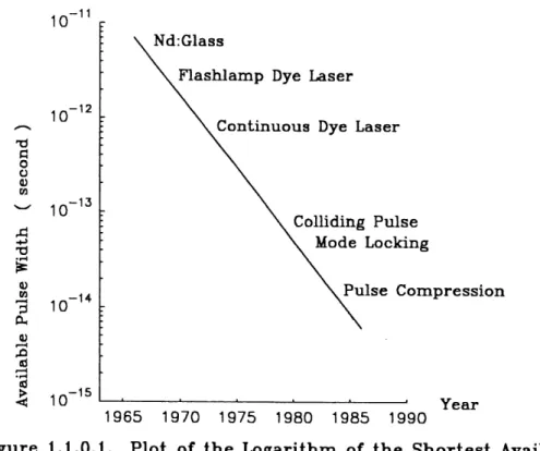
Most of molecular fast relaxation processes are between the nanosecond to picosecond ranges, as shown in Fig. 1.1.2.1. [52]. Thus with ultrashort laser pulses, the limiting factor for time resolution is no longer the pulse duration but the response of the detection systems.
The limitation of the detection system can be overcome by a pump and probe technique. A single ultrashort laser pulse is divided into a group of strong pump beams and a weak probe beam, which can be monochromatic or white continuum.
create an excitation in a molecular system and the probe beam probes the relaxation of the excitation as a function of the delay time At. Now, the time resolution is
limited only by the pump and the probe pulse duration. One of the applications of
pump-probe technique is the measurement of the decay times of short-lived excited states, as shown in Fig. 1.1.2.2. (a). When the probe pulse has a path difference Xi
( < 0 ) with respect to pump pulse (Fig. 1.1.2.2. (a)), the probe pulse will spend less
time |ti = Xi/c| to arrive at the sample than the pump pulse does (Fig. 1.1.2.2. (b)), and this corresponds to a negative delay time Ti (probe-to-pump) (Fig. 1.1.2.2. (c)). At this moment, since when the probe pulse arrives there is no excitation in the sample, the system will give no response at all. If the probe pulse arrives at the sample with a
longer light path X2 (> 0 ) with respect to the pump pulse, the probe pulse will spend
more time t^ = x^ 1c to reach the sample than the pump pulse does and this corresponds to a positive delay time ^ ( probe-to-pump ). At this moment, the probe pulse arrives at the sample in a time ^ after the excitation has been created (Fig. 1.1.2.2. (b)), the energy level 1 has been populated with a non-zero population N1, so
that the probe pulse will feel the decay of this population. Therefore, if delay time
scans from the negative delay (negative light path difference between probe and
pump) to the positive delay (positive light path difference between probe and pump)
in steps, then the detecting system will direcdy measure the response curve point by
point, shown as in Fig. 1.1.2.2. (c).
Although numerous techniques refer to pump-probe methods, the most powerful one is based on white continuum generation.
COLUSTONTTFME
IN LIQUIDS
MOLECULAR ROTATION
ELECTRONIC
DEPHASING
SOLVANT
RELAXATION
VIBRATIONAL DEPHASING
VIBRATIONAL RELAXATION
MBRATIONAL MOTION
10
-1410
-13ELECTRONIC RELAXATION
10
-1210
•n10
-1010
PROTON TRANSFER
ENERGY
TRANSFER IN
PHOTOSYNTHESIS
PHOTOIONIZATION
PROTEIN INTERNAL MOTION
<;
ELECTRONIC TRANSFER
IN PHOTOSYNTHESIS
TORSIONAL
DYNAMICS
OF DNA
-8
10 sec.
PHOTODISSOCIATION
PHOTOCHEMICAL ISOMERISATION
-CAGT
RECOMBINATION
Figure 1.1.2.1. Approximate Time Scales of Elementary
I(x,to)pump/probe
Xl=tlC
(a) Relative Positioh of
Probe—Pump in Spbce. pumpj
probe^
X2=t2C probe^ x -c==^ I(xo. t)pump/probe X2
tl=Xl/C(b) Relative Positioji of pump
Probe—Pump in Tihne. probe^ t2=X2/C probe t1Nt(r)
T1 = ft T2 = t2(c) Time-Resolved
probejpump
Response. T2Ng
Figure 1.1.2-2. Measuring the Decay Time of
Short-lived Excited States by Time-Resolved
1.1.3. White Continuum Generation
White continuum generation is a particularly spectacular application of nonlinear frequency mixing. When a strong laser beam is focused within a glass or within a liquid such as CC^, water or phosphoric acid, there is an emission of a pulse of white light whose duration is comparable to that of the pump pulse.
People now generally agree that this continuum originates from two superposed phenomena. First, high intensity optical pulses propagating through a medium wiU distort the atomic or electronic configurations and change the refractive index appreciably. The refractive index becomes time-dependent so the phase of the optical wave is altered. This generally leads to a broadening of the spectrum. This process is called self-phase modulation [52, 53]. Second, in isotropic liquids, there is a supplementary spectral broadening due to four photon parametric interactions [54
-57].
In fact, white-continuum generation is one of the crucial techniques in ultrafast spectroscopy. The spectrum of the continuum extends from the near ultraviolet to the infrared and thus provides a versatile coherent light source for a huge range of spectroscopic studies. Without it, many ultrafast spectroscopic applications would not be available.
1.1.4. Basic research Opportunities
for Ultrashort Time-Resolved Spectroscopy
From various tune-resolved coherent techniques, we can obtain the following
information [58]:
1.whether a transition is homogeneously broadened or not;
2. the decaying times of homogeneously or inhomogeneously broadened spectral features;
4. the pure dephasing times; 5. the energy relaxation times;
6. the mechanisms and paths of energy relaxation; 7. the collision times;
8. the nonresonant susceptibilities;
9. transition frequencies within conjugated spectra regions;
10. precise frequency differences ofvibrational modes separated by up to 10 x 1012
Hz = 10 THz ( 10 tera Hertz, At-Ao ~ 1 -> Ao-l/At, At > 100 fs = 10-13 s -^ Au
<1013Hz).
A particular technique gives only one or few of the above. A combination of different techniques is usually necessary for obtaining more information.
1.1.5. Transient Grating Method
Among the time-resolved spectroscopic methods which pump-probe techniques, we chose the transient grating method [59] to detect transient responses of our model
systems.
1.1.5.1. Formation of the Transient Grating
In this technique, two pump (excitation) pulses arrive simultaneously at tfae sample. The two sychronized pump beams interfere with each other and produce a
spatially modulated field which is called an interference grating. When a material is
placed mto the interference region of the pump waves, some light-matter interaction, such as absorption, creates a corresponding spatial modulation (grating) of some material properties, such as.:
1. the population of excited electronic states,
3. the space charge and the accompanying field (photorefractive materials),
4. the temperature,
5. the molecular orientation (fluids), and 6. the concentration (mixtures).
Thus a transient grating ( because it will disappear in a finite time after the
excitation) is formed due to population or phase redistribution which occurs under strong light fields. Most of these changes can be described by the population of one,
several, or a whole continuum of excited (e.g. electronic, phonon) states of the
sample material. Therefore, the corresponding gratings are also termed as population
gratings in a general sense.
1.1.5.2. Detection of the Decay of the Transient Grating
In order to make time-resolved measurements of the dynamics of the transient grating, a third beam called probe needs to be used. When arriving at the sample, the probe beam is deflected by the light induced gratmg, producing a signal beam. The intensity of the signal beam is measured as a function of the time delay between the
pump beams and the probe beam which contains information on the dynamics of the
system. This Bragg scattering from the light induced transient grating, in terms of nonlinear optics, is viewed as afour-wave interaction (FWI) or four-\vave mixing
(FWM) process. This will be discussed in details in the following chapters.
The decay of the grating is commonly detected by diffraction of the probe light beam. The diffracted light can either be recorded directly or heterodynedly with a split-off beam from the probe light. The former is simple but requires higher pump
and probe power to get sufficiently strong signals. The latter is more sensitive but the setup more complicated. la our experimental setup, we use only direct detection
scheme.
For ideal plane waves, the dif&action efficiency 77 for the first order diffracted beam can be expressed [60]:
lit
r'=^T^=
probe nA nd 'probejvAnd^ ( 7iAKd\
^ ^ probe ^ ^ probe (1.1.5.2.1.)where An, An, AK are changes m the complex refractive index and its real and imaginary parts in the sample due to transient grating, d is the thickness of the Sample, and ^iffi-acte^ \robe arG the intensides of the diffi-acted beam and the probe
beam. This formula is valid for a grating with a sufficiently small \An\ = \An+iAKf1
and a low absorption material, i.e., Kd «1.
Very small refractive index changes An and optical path changes And can be measured, typically, with a dif&action efficiency T| w 10-15, correspondiag to an optical path change « K/1000. The phase shift in that extreme case has to be measured with interferometric sensitivity.
Amplitude (An = 0) and phase (AK = 0) gratmgs can be differentiated by
illuminating the sample with a parallel beam and observing the self-image appearing at different distances behind the grating [61], or alternatively by heterodyne
detection.
1.1.5.3. Comparison of the Transient Grating Method
with the Transient Absorption Technique
In this work, the post-excitation-dynamic processes of the porphyrm phthalocyaniae dimer/heterodimer tiTmer/heterotrimer have been measured by tiine-resolved four-wave mixing (TRFWM), or equivalently called time-tiine-resolved transient
grating (TRTG) method. The transient absorption (TA) results obtained on the same
samples provide reference data since they give us information about the energy levels (bands) and their related lifetimes. It is therefore useful at this point to compare some crucial aspects of these techniques.
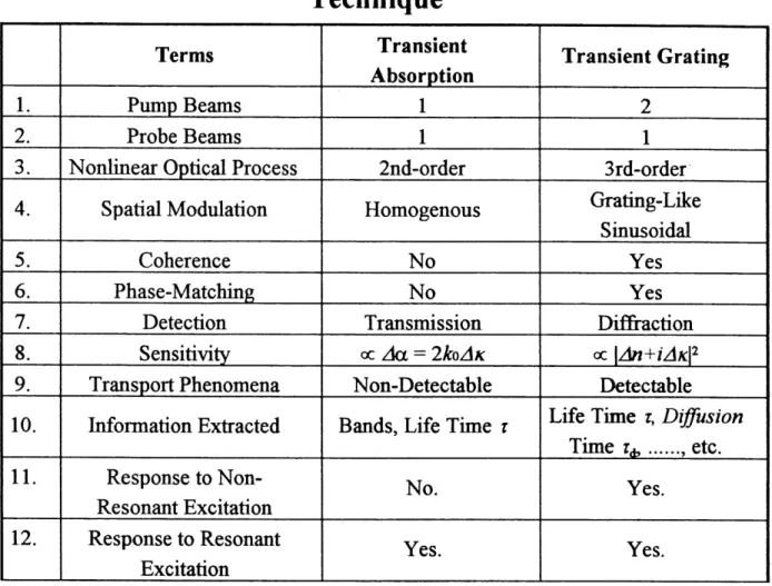
Table 1.1.5.3.1. The Major Differences Between the
Transient Grating Method and the Transient Absorption
Technique
1. 2. 3. 4. 5. 6. 7. 8. 9. 10. 11. 12. Terms Pump Beams Probe Beams Nonlinear Optical ProcessSpatial Modulation Coherence Phase-Matching Detection Sensitivity Transport Phenomena Information Extracted Response to Non-Resonant Excitation Response to Resonant Excitation Transient Absorption
1
1
2nd-order Homogenous No No Transmission oc zkx = 2^ozlA: Non-Detectable Bands, Life Time TNo. Yes. Transient Grating
2
1
Srd-order Gratmg-Like Siausoidal Yes Yes Diffi-action oc \An+iAK\2 DetectableLife Time r. Diffusion
Time T^ ... etc.
Yes.
Yes.
From the table it can be seen that there are only three beams involved in transient absorption, while there are four in the transient grating. The former is a second-order nonlinear optical (NLO) process and the latter is a third-order NLO process. Technically speaking, the latter is more difficult to implement because it involves more beams and needs higher pumping and probing powers. Furtheimore, transient absorption measures the change in absorption coefificient, while transient grating
detects dif&action efficiency which is in proportion to the square of the change of the
complex refracdve index. Therefore TRTG is more sensitive than TA m probing dynamic processes occurring after the excitation is produced.
Due to the difference between the modes of excitation, any transport process with charge transfer or and energy transfer occurring during the de-excitation can only be
picked up by the grating-like modulation of TRTG, and not by TA which has
homogeneous excitations. Hence, the diffusion processes are detectable only for TRTGandnotforTA.
When the frequency of the pump light is not in an absorbing region of the material
(non-resonant excitation, NRE), TA systems will get no signal at all, but TRTG systems will still give a response through the phase grating caused by virtual electron excitations. The signal of the NRE contains infonnation about the pulse duradon of the pump pules and the probe pulse.
In the resonant excitation case, TRTG systems can also feel the relaxation processes that TA systems will feel for the same material. But the TRTG systems may not yield exactly the same response as the TA systems do, because the response of the TRTG system is sensitive to any de-activation process in contrary to the TA
system.
In short, TRTG is a more sensitive and more useful technique to probe the dynamic processes of de-excitation than TA, although the former needs the important
information from the latter about the bands and the life times of the materials under
mvestigation.
Of course, suitable materials have to be available for research in nonlinear optics and various kinds of applications.
1.2. Prototype Model Systems Used in the
Experiment
1.2.1. Nonlinear Optical Materials
All materials, including all forms of matter, gases, liquids, and solids, have nonlinear optical properties. But, the intensity of the optical field needed to make these effects observable, varies over many orders of magnitude. This depends on the detailed nature of the electronic structure, the dynamic behaviour, as well as the symmetry and details of the geometric arrangement of the atomic and molecular components of the medium. From the device point of view, the most important nonlinear optical materials are m solid form and should meet a wide variety of auxiliary material requirements, including extraordinary stability with respect to ambient conditions and to high intensity light source, processability for pattern and
shape definition, and integratibility with additional different materials.
There are a lot of materials possessing nonlinear optical properties: inorganic and
organic crystals, polymers, semiconductors, composites and metal-based systems.
However, none of these has proven to be the "silicon" of nonlinear optics because each material not only has properties that are advantageous for certain applications, but it also has properties that are disadvantageous for others. Therefore, there is still a need for the development of materials satisfying the critical requirements of devices for information processing, optical frequency conversion, integrated optics, and
telecommunications.
We can roughly divide nonlinear optical materials into two major types:
molecular materials and bulk materials.
For the first type, the materials consist of chemically bonded units which interact in bulk through weak Van der Waals forces. Many organic crystals and polymers belong to this class of materials. Nonlmearities in this kind of materials originate primarily from the molecular structure. The micro-nonlmearities in form of the hyperpolarizabilities of constituent molecules can be related to macro-nonlinearities
nonlinearity in this class of materials is at the molecular structure level. For this, one needs to understand the relationship between the molecular electronic structure and the induced molecular nonlinear polarization.
In the second class of materials, nonlinearities are regarded as arising from quasi-free electrons like those in metals and semiconductors. The electronic characteristics
of the bulk medium determines the optical nonlinearities. The origin of the
nonlinearities for this type of materials needs a different theoretical framework. Examples of this kind of materials are: quantum well stmctures derived from GaAs and II-V[ semiconductors, e.g. CdSe, inorganic crystals in which no single molecular
unit in the ionic lattice can be identified, like KDP (potassium dihydrogen phosphate) and KTP (potassium titanyl phosphate).
Compared to inorganic nonlinear optical matenals, the development of organic, or, more generally, molecular nonlinear optical materials is quite recent. Organic and other molecular materials are being more and more recognized as the promisiag materials for the future. These materials are interesting because their various molecular characteristics and their synthetic versatilities can be used to vary and optimize molecular structures in order to maximize nonlinear response, or to optimize the auxiliary properties like mechanical and thermal stability, and laser damage threshold. Organic materials can be cast into thin crystalline films, layer by layer, using the Langmuir-Blodgett technique and other deposition techniques, e.g. sublimation. These fihns show highly oriented structures and optimized nonlinearities. Some of the prominent advantages of many organic materials and high-performance polymers are high mechanical strength as well as excellent environmental and thermal stability, which can be many orders higher that those of inorganic materials. The unique Tc-bonding chemical structure of organic molecular materials leads to extremely large nonresonant ( non-absorptive ) optical nonlinearides, in many cases much higher than those of their morganic counterparts. Devices with nonresonant electronic optical non-linearities will have the fastest
response times limited only by the width of the drivmg laser pulse while also
eliminatmg heat dissipation, beam depletion, thennally induced nonlinearities and other disadvantages ofmorganic nonlinear optical materials. The considerably lower dielectric constant of organic materials yields a low RC time constant. Thus, the
electrooptic modulatmg devices made from these materials can operate with band-widths greater than 10 GHz ( G = 109).
Aside from its potential technological interest, research on nonlinear optical materials also offers challengmg opportunities for a fundamental understanding of the physics ofnonlinear optical interactions. Before one can firmly control the molecular structure in order to enhance optical nonlinearities, there are still many things that must be done to understand completely the relationship between molecular structures and microscopic optical nonlinearities.
One of the most important type of organic nonlinear optical materials is porphyrin phthalocyanine metal complex systems.
1.2.2. Porphyrm and Phthalocyanine
The prototype model systems used in our experiment is based on porphyrin and phthalocyanine metal complexes. Porphyrins and the corresponding metal complexes function as electron transfer agents in many biological systems and play a very important role in many biochemical processes. Porphyrins contain a tetrapyrrolic macrocycle which can be chelated with most metallic ions, even some non-metallic
ions, to form corresponding complexes.
The simplest porphyrm, called porphine, is shown in Fig. 1.2.2.1. .
The natural part ofporphyrins can be obtained when the positions P and P' = 2, 3, 7, 8, 12, 13, 17, 18 of the pyrrole cycles m Fig. 1.2.2.1. are replaced by substitutive groups [62, 63]. There also exist a number of synthetic porphyrins which depend on the nature of the substitutive groups and on the exact replaced positions in the porphine frame. One of the complete artificially synthetic examples ofporphyrms is
phthalocyanine (Pc), shown in Fig. 1.2.2.2. .
Phthalocyanines have a similar structure to porphine, which consists in replacing four carbon atoms at position a, ?, y, § in Fig. 1.2.2.1. by four nitrogen atoms, and substituting y position (?' = 2, 3, 7, 8, 12, 13, 17, 18 in Fig. 1.2.2.1.) on pyrrole
cycles by benzene cycles. This compound was discovered in 1907 by Braun et al.
[64], but was not identified until 1933 by Linstead et al [65, 66].
Other artificial synthetic examples of porphyrins are tetrapyrrole complexes of mono-, bis-, tris- lanthanide porphyrins which were used in our work. These will be discussed in detail in following sections.
3 o( 7
8
Figure 1.2.2.1. Molecular Structure of Porphin
24 2
)3
1.2.3. Lanthanide Porphyrin Phthalocyanine
Dimeric/Heterodimeric Trimeric/Heterotrimeric
Prototype Model Systems
In recent years, the research on optical nonlinearities of organic materials has attracted much attention both in the fundamental aspects to clarify their origins and mechanisms, and in their applications to develop optical devices being capable of processing optical information [67 - 71], etc., using the different frequency -mixing schemes offered by various orders ofnonlinearity. Organic materials offer a number of advantages over mineral compounds. First, they are of superior flexibility m processing and manipulation, which enables us to improve particular optical characteristics by engineering the molecular properties. Moreover, they have large nonlinear optical susceptibility, ultrafast response, thermal and chemical stability. In order to increase the magnitude of the thu-d-order susceptibility ^ \ several methods have been proposed: multilock conjugated polymerization based on organic superlattices, donor-acceptor substitution on Tc-conjugated Imear chains and use of n-conjugated molecules having symmetric sti^icture [72 - 79]. In particular, n-conjugated n -electron systems have been known to have large optical nonlinearities and ultrafast optical responses [80 - 86]. Their nonlinear optical properties can be studied using various techniques such as, second harmonic generation (SHG), third harmonic generation (THG), four-wave mixing (FWM) and transient absorption, etc. [86 - 100].
Many organic materials containing delocalized Ti-electrons have been observed to have large optical nonlinearities and ultrafast responses [101, 102]. Most other third-order nonlinear optical compounds feature Ti-electron conjugation along a backbone and are pseudo-one dimensional systems. Those materials show limitations in their optical nonlinearities. This had led to considerable interest in the two-dimensional
n-conjugated porphyrin [103] and phthalocyanine [104] systems, especially the
complexes with a wide variety of metals. Among these systems, stacked and highly conjugated ones, such as lanthanide bis-/tris-porphyrins and bis-/tris-phthalocyanines, are mterestmg in many respects. These share the common properties for most porphyrins and phthalocyanines, i.e., chemical and thermal stability, versatile and
manufacture of pigments, exhibiting unique characteristics such as catalyst, electrocatalysis, xerography and medical properties [105]. The delocalized Tc-electron in these systems can ser^e as source of charge carriers and is the origin of photoelectnc properties directly related to primitive charge separation processes in photosynfhesis and optical nonlinearides. In this aspect, they provide a series of model systems to study photoinduced electron transfer reactions occurring on ultrafast time scales in photosynthetic reaction centers both in theory and in experiment [86 - 91, 106 - 117]. Their sharp absorption bands in the visible and in the near infrared can be used for resonance enhancement of third-order susceptibility ^3). This makes them promising materials for third-order optical nonlinearities. The compounds are receivmg increased attention. However, very few studies have been done on electrostatic nuxed dimers
obtained by covalently bonding poqAyrm and phthalocyanme moieties bearing oppositely charged substituents [118 - 121].
18
^
L .7
Figure 1.2.3.1. Constitution of Octacoordinated Metal
Monotetrapyrroles M(P)L* and Configurations with A: Monotetrapyrro B: Bistetrapyrroles M(P)2 and C: Tristetrapyrroles M2(P)s, M = Lanthanide Metal Ion
We chose the lanthanide porphyrin dimer/heterodimer and trimer/heterotrimer as prototype model systems in which to study dynamic processes such as electron transfer, energy transfer, related nonlinear optical properties, and their relationship to the molecular structure.
Table 1.2.3.1. Specifications ofPorphyrins
No.0
1
2
3
4
5
6
7
8
PinM( P )L.
p
PcTTP
OEP
TPP
TC1P
HBP
OMP
TAP
NameofthePmM(P)L, a general porphyrin, phthalocyanme phthalocyanine 5,10,15,20-tetra(^-tolyl)porpliymi 2,3,7,8,12,13,17,18-octaethylporphyrin 5,10,15,20-tetraphenylporphyrm 5,10,15,20-tetra(p-chlorophenyl)porphyTm 1,2,3,4,8,9,10,11,15,15,17, lg,22J 3, 24,25,-hexadecahyrotetrabenzor^^/^lporphyrm
2,3,7,8,12,13,17,18-octamethylporphyrin 5,10,15,20-tetra(p-anisyl)porphyrm R in position 2~20 in Fig. 1.2.3.1. N in 5, 10, 15, 20; QH^ in 2/3, 7/8, 12/13, 17/18 p CH^ C,H, m 5, 10, 15,20
CA in 2, 3, 7, 8, 12, 13, 17, 18 C^Hs in 5, 10, 15, 20 pClC^ in 5, 10, 15, 20 ( CH^ \ in 2/3, 7/8, 12/13, 17/18 CH3 in 2, 3, 7, 8, 12, 13, 17, 18 p CH30C6H4 in 5, 10, 15,20A complete understanding of the photophysical behaviour of porphyrins and related chromophores within the Van der Waals will provide us with a deep insight into how the electron donor in photosynttoic reaction centers influences the initial stages of charge separation processes [12Z 123]. Moreover this knowledge will enable us to modify molecular systems m a controllable manner so as to maximize their nonlinearities. This is needed by and various kinds of other applications.
Lanfhanide porphyrm dimers/heterodimers md tamers/hetCTOtTuners represent a series of ideal molecular systems which can be used to study the electronic structure


![Table 1.3.2.1.1. Photorefractive Materials Materials Ferroelectrics f 179] BaTi03 KNb03 LiNb03, LiTa03 Sr,.,Ba,Nb,0](https://thumb-eu.123doks.com/thumbv2/123doknet/5428558.127115/58.918.149.794.150.761/table-photorefractive-materials-materials-ferroelectrics-bati-linb-lita.webp)