2015 ESC/ERS Guidelines for the diagnosis
and treatment of pulmonary hypertension
The Joint Task Force for the Diagnosis and Treatment of Pulmonary
Hypertension of the European Society of Cardiology (ESC) and the
European Respiratory Society (ERS)
Endorsed by: Association for European Paediatric and Congenital
Cardiology (AEPC), International Society for Heart and Lung
Transplantation (ISHLT)
Authors/Task Force Members: Nazzareno Galie`
*
(ESC Chairperson) (Italy),
Marc Humbert
*
a(ERS Chairperson) (France), Jean-Luc Vachiery
c(Belgium),
Simon Gibbs (UK), Irene Lang (Austria), Adam Torbicki (Poland), Ge´rald Simonneau
a(France), Andrew Peacock
a(UK), Anton Vonk Noordegraaf
a(The Netherlands),
Maurice Beghetti
b(Switzerland), Ardeschir Ghofrani
a(Germany),
Miguel Angel Gomez Sanchez (Spain), Georg Hansmann
b(Germany), Walter Klepetko
c(Austria), Patrizio Lancellotti (Belgium), Marco Matucci
d(Italy), Theresa McDonagh
(UK), Luc A. Pierard (Belgium), Pedro T. Trindade (Switzerland), Maurizio Zompatori
e(Italy) and Marius Hoeper
a(Germany)
*Corresponding authors: Nazzareno Galie`, Department of Experimental, Diagnostic and Specialty Medicine – DIMES, University of Bologna, Via Massarenti 9, 40138 Bologna, Italy,
Tel:+39 051 349 858, Fax: +39 051 344 859, Email:nazzareno.galie@unibo.it
Published on behalf of the European Society of Cardiology. All rights reserved.&2015 European Society of Cardiology & European Respiratory Society.
This article is being published concurrently in the European Heart Journal (10.1093/eurheartj/ehv317) and the European Respiratory Journal (10.1183/13993003.01032-2015). The articles are identical except for minor stylistic and spelling differences in keeping with each journal’s style. Either citation can be used when citing this article.
Marc Humbert, Service de Pneumologie, Hoˆpital Biceˆtre, Universite´ Paris-Sud, Assistance Publique Hoˆpitaux de Paris, 78 rue du Ge´ne´ral Leclerc, 94270 Le Kremlin-Bicetre, France,
Tel:+33 145217972, Fax: +33 145217971, Email:marc.humbert@aphp.fr
ESC Committee for Practice Guidelines (CPG) and National Cardiac Societies document reviewers: listed in Appendix
a
Representing the European Respiratory Society;b
Representing the Association for European Paediatric and Congenital Cardiology;c
Representing the
Inter-national Society for Heart and Lung Transplantation;dRepresenting the European League Against Rheumatism; andeRepresenting the European Society of
Radiology.
ESC entities having participated in the development of this document:
ESC Associations: Acute Cardiovascular Care Association (ACCA), European Association for Cardiovascular Prevention & Rehabilitation (EACPR), European Association of Cardio-vascular Imaging (EACVI), European Association of Percutaneous CardioCardio-vascular Interventions (EAPCI), European Heart Rhythm Association (EHRA), Heart Failure Association (HFA). ESC Councils: Council for Cardiology Practice (CCP), Council on Cardiovascular Nursing and Allied Professions (CCNAP), Council on Cardiovascular Primary Care (CCPC). ESC Working Groups: Cardiovascular Pharmacotherapy, Cardiovascular Surgery, Grown-up Congenital Heart Disease, Pulmonary Circulation and Right Ventricular Function, Valvular Heart Disease.
The content of these European Society of Cardiology (ESC) and European Respiratory Society (ERS) Guidelines has been published for personal and educational use only. No com-mercial use is authorized. No part of the ESC/ERS Guidelines may be translated or reproduced in any form without written permission from the ESC and/or ERS. Permission can be obtained upon submission of a written request to Oxford University Press, the publisher of the European Heart Journal or from the European Respiratory Journal and the party author-ized to handle such permissions on behalf of the ESC and ERS.
Disclaimer: The ESC/ERS Guidelines represent the views of the ESC and ERS and were produced after careful consideration of the scientific and medical knowledge and the evidence available at the time of their publication. The ESC and ERS are not responsible in the event of any contradiction, discrepancy and/or ambiguity between the ESC/ERS Guidelines and any other official recommendations or guidelines issued by the relevant public health authorities, in particular in relation to good use of healthcare or therapeutic strategies. Health profes-sionals are encouraged to take the ESC/ERS Guidelines fully into account when exercising their clinical judgment, as well as in the determination and the implementation of preventive, diagnostic or therapeutic medical strategies; however, the ESC/ERS Guidelines do not override, in any way whatsoever, the individual responsibility of health professionals to make appropriate and accurate decisions in consideration of each patient’s health condition and in consultation with that patient and, where appropriate and/or necessary, the patient’s caregiver. Nor do the ESC/ERS Guidelines exempt health professionals from taking into full and careful consideration the relevant official updated recommendations or guidelines issued by the competent public health authorities, in order to manage each patient’s case in light of the scientifically accepted data pursuant to their respective ethical and professional obligations. It is also the health professional’s responsibility to verify the applicable rules and regulations relating to drugs and medical devices at the time of prescription.
by guest on January 14, 2016
http://eurheartj.oxfordjournals.org/
Document Reviewers: Victor Aboyans (CPG Review Coordinator) (France), Antonio Vaz Carneiro (CPG Review Coordinator) (Portugal), Stephan Achenbach (Germany), Stefan Agewall (Norway), Yannick Allanored(France),
Riccardo Asteggiano (Italy), Luigi Paolo Badano (Italy), Joan Albert Barbera`a(Spain), He´le`ne Bouvaist (France),
He´ctor Bueno (Spain), Robert A. Byrne (Germany), Scipione Carerj (Italy), Grac¸a Castro (Portugal), Çetin Erol (Turkey), Volkmar Falk (Germany), Christian Funck-Brentano (France), Matthias Gorenflob(Germany),
John Grantonc(Canada), Bernard Iung (France), David G. Kiely (UK), Paulus Kirchhof (Germany/UK),
Barbro Kjellstrom (Sweden), Ulf Landmesser (Switzerland), John Lekakis (Greece), Christos Lionis (Greece), Gregory Y. H. Lip (UK), Stylianos E. Orfanosa(Greece), Myung H. Parkc(USA), Massimo F. Piepoli (Italy),
Piotr Ponikowski (Poland), Marie-Pierre Revele(France), David Rigaua(ERS methodologist) (Switzerland),
Stephan Rosenkranz (Germany), Heinz Vo¨ ller (Germany), and Jose Luis Zamorano (Spain)
The disclosure forms of all experts involved in the development of these guidelines are available on the ESC website
http://www.escardio.org/guidelines
Online publish-ahead-of-print 29 August 2015
-Keywords Guidelines † Pulmonary hypertension † Pulmonary arterial hypertension † Chronic thromboembolic pulmonary hypertension † Congenital heart disease † Connective tissue disease † Heart failure † Respiratory failure † Endothelin receptor antagonists † Phosphodiesterase type 5 inhibitors † Prostacyclin analogues † Lung disease † Left heart disease
Table of Contents
Abbreviations and acronyms . . . 69
1. Preamble . . . 70
2. Introduction . . . 71
3. Definitions and classifications . . . 72
3.1 Definitions . . . 72
3.2. Classifications . . . 72
4. Epidemiology and genetics of pulmonary hypertension . . . 74
4.1 Epidemiology and risk factors . . . 74
4.2 Genetics . . . 75
5. Pulmonary hypertension diagnosis . . . 75
5.1 Diagnosis . . . 75
5.1.1 Clinical presentation . . . 75
5.1.2 Electrocardiogram . . . 75
5.1.3 Chest radiograph . . . 76
5.1.4 Pulmonary function tests and arterial blood gases . . 76
5.1.5 Echocardiography . . . 76
5.1.6 Ventilation/perfusion lung scan . . . 78
5.1.7 High-resolution computed tomography, contrast enhanced computed tomography, and pulmonary angiography . . . 78
5.1.8 Cardiac magnetic resonance imaging . . . 78
5.1.9 Blood tests and immunology . . . 78
5.1.10 Abdominal ultrasound scan . . . 79
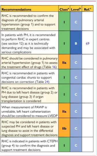
5.1.11 Right heart catheterization and vasoreactivity . . . . 79
5.1.12 Genetic testing . . . 80
5.2 Diagnostic algorithm . . . 81
6. Pulmonary arterial hypertension (group 1) . . . 82
6.1 Clinical characteristics . . . 82
6.2 Evaluation of severity . . . 82
6.2.1 Clinical parameters, imaging and haemodynamics . . 82
6.2.2 Exercise capacity . . . 83
6.2.3 Biochemical markers . . . 83
6.2.4 Comprehensive prognostic evaluation and risk assessment . . . 84
6.2.5 Definition of patient status . . . 85
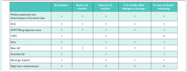
6.2.6 Treatment goals and follow-up strategy . . . 85
6.3 Therapy . . . 86
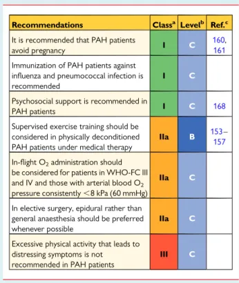
6.3.1 General measures . . . 86
6.3.1.1 Physical activity and supervised rehabilitation . . . 86
6.3.1.2 Pregnancy, birth control, and post-menopausal hormonal therapy . . . 87 6.3.1.3 Elective surgery . . . 87 6.3.1.4 Infection prevention . . . 87 6.3.1.5 Psychosocial support . . . 87 6.3.1.6 Adherence to treatments . . . 87 6.3.1.7 Travel . . . 87 6.3.1.8 Genetic counselling . . . 87 6.3.2 Supportive therapy . . . 87 6.3.2.1 Oral anticoagulants . . . 87 6.3.2.2 Diuretics . . . 88 6.3.2.3 Oxygen . . . 88
6.3.2.4 Digoxin and other cardiovascular drugs . . . 88
6.3.2.5 Anaemia and iron status . . . 88
6.3.3 Specific drug therapy . . . 88
6.3.3.1 Calcium channel blockers . . . 88
6.3.3.2 Endothelin receptor antagonists . . . 89
6.3.3.3 Phosphodiesterase type 5 inhibitors and guanylate cyclase stimulators . . . 89
6.3.3.4 Prostacyclin analogues and prostacyclin receptor agonists . . . 90
6.3.3.5 Experimental compounds and strategies . . . 92
6.3.4 Combination therapy . . . 92
6.3.5 Drug interactions . . . 93
6.3.6 Balloon atrial septostomy . . . 94
by guest on January 14, 2016
http://eurheartj.oxfordjournals.org/
6.3.7 Advanced right ventricular failure . . . 94
6.3.7.1 Intensive care unit management . . . 94
6.3.7.2 Right ventricle assistance . . . 94
6.3.8 Transplantation . . . 94
6.3.9 Treatment algorithm . . . 95
6.3.10 Diagnosis and treatment of pulmonary arterial hypertension complications . . . 97
6.3.10.1 Arrhythmias . . . 97
6.3.10.2 Haemoptysis . . . 97
6.3.10.3 Mechanical complications . . . 97
6.3.11 End of life care and ethical issues . . . 97
7. Specific pulmonary (arterial) hypertension subsets . . . 97
7.1 Paediatric pulmonary arterial hypertension . . . 97
7.1.1 Diagnosis . . . 98
7.1.2 Therapy . . . 98
7.2 Pulmonary arterial hypertension associated with adult congenital heart disease . . . 99
7.2.1 Diagnosis . . . 99
7.2.2 Therapy . . . 99
7.3 Pulmonary arterial hypertension associated with connective tissue disease . . . 100
7.3.1 Diagnosis . . . 101
7.3.2 Therapy . . . 101
7.4 Pulmonary arterial hypertension associated with portal hypertension . . . 101
7.4.1 Diagnosis . . . 102
7.4.2 Therapy . . . 102
7.5 Pulmonary arterial hypertension associated with human immunodeficiency virus infection . . . 102
7.5.1 Diagnosis . . . 103
7.5.2 Therapy . . . 103
7.6 Pulmonary veno-occlusive disease and pulmonary capillary haemangiomatosis . . . 103
7.6.1 Diagnosis . . . 104
7.6.2 Therapy . . . 104
8. Pulmonary hypertension due to left heart disease (group 2) . . 104
8.1 Diagnosis . . . 105
8.2 Therapy . . . 106
9. Pulmonary hypertension due to lung diseases and/or hypoxia (group 3) . . . 106
9.1 Diagnosis . . . 107
9.2 Therapy . . . 107
10. Chronic thromboembolic pulmonary hypertension (group 4) 108 10.1 Diagnosis . . . 108
10.2 Therapy . . . 109
10.2.1 Surgical . . . 109
10.2.2 Medical . . . 110
10.2.3 Interventional . . . 110
11. Pulmonary hypertension with unclear and/or multifactorial mechanisms (group 5) . . . 111
12. Definition of a pulmonary hypertension expert referral centre 111 12.1 Facilities and skills required for a expert referral centre 111 13. To do and not to do messages from the guidelines . . . 112
14. Web addenda . . . 113
15. Appendix . . . 113
16. References . . . 113
Abbreviations and acronyms
ALAT alanine aminotransferase
ASAT aspartate aminotransferase
APAH associated pulmonary arterial hypertension
BAS balloon atrial septostomy
BMPR2 bone morphogenetic protein receptor 2
BNP brain natriuretic peptide
BPA balloon pulmonary angioplasty
BREATHE Bosentan Randomised trial of Endothelin
Antagonist THErapy
CAV1 caveolin-1
CCB calcium channel blocker
cGMP cyclic guanosine monophosphate
CHD congenital heart disease
CI cardiac index
CMR cardiac magnetic resonance
CO cardiac output
COPD chronic obstructive pulmonary disease
Cpc-PH combined post-capillary and pre-capillary
pulmonary hypertension
CPET cardiopulmonary exercise testing
CPFE combined pulmonary fibrosis and emphysema
CT computed tomography
CTD connective tissue disease
CTPA computed tomography pulmonary angiogram
CTEPH chronic thromboembolic pulmonary
hypertension
DLCO diffusing capacity of the lung for carbon monoxide
DPAH drug-induced pulmonary arterial hypertension
DPG diastolic pressure gradient (diastolic PAP 2 mean
PAWP)
EACVI European association of cardiovascular imaging
ECG electrocardiogram
ECMO extracorporeal membrane oxygenation
EIF2AK4 eukaryotic translation initiation factor 2 alpha
kinase 4
EMA European Medicines Agency
ERA endothelin receptor antagonist
FC functional class
FDA US Food and Drug Administration
HAART highly active antiretroviral therapy
HIV human immunodeficiency virus
HF-pEF heart failure with preserved left ventricular
ejec-tion fracejec-tion
HPAH heritable pulmonary arterial hypertension
HRCT high resolution computed tomography
ICU intensive care unit
INR international normalized ratio
IPAH idiopathic pulmonary arterial hypertension
Ipc-PH isolated post-capillary pulmonary hypertension
IPF idiopathic pulmonary fibrosis
i.v. intravenous
IVC inferior vena cava
LA left atrium/atrial
by guest on January 14, 2016
http://eurheartj.oxfordjournals.org/
LHD left heart disease
LV left ventricle/ventricular
MR magnetic resonance
NYHA New York Heart Association
NO nitric oxide
NT-proBNP N-terminal pro-brain natriuretic peptide
PA pulmonary artery
PaCO2 arterial carbon dioxide pressure
PaO2 arterial oxygen pressure
PAH pulmonary arterial hypertension
PAP pulmonary arterial pressure
PAPm mean pulmonary arterial pressure
PAPs systolic pulmonary arterial pressure
PAWP pulmonary artery wedge pressure
PASP pulmonary artery systolic pressure
PCH pulmonary capillary haemangiomatosis
PDE-5i phosphodiesterase type 5 inhibitor
PE pulmonary embolism
PEA pulmonary endarterectomy
PFTs pulmonary function tests
PH pulmonary hypertension
PoPH porto-pulmonary hypertension
PPHN persistent pulmonary hypertension of the
newborn
PVOD pulmonary veno-occlusive disease
PVR pulmonary vascular resistance
RA right atrium
RAP right atrial pressure
RCT randomized controlled trial
RHC right heart catheterization
RV right ventricle/ventricular
6MWD/6MWT 6-minute walking distance/6-minute walking test
SCD sickle cell disease
sGC soluble guanylate cyclase
SSc systemic sclerosis
SvO2 mixed venous oxygen saturation
SVR systemic vascular resistance
TAPSE tricuspid annular plane systolic excursion
t.i.d. three times a day
TGF-b transforming growth factor b
TPG transpulmonary pressure gradient (mean PAP 2
mean PAWP)
TRV tricuspid regurgitant velocity
VE/VCO2 minute ventilation – carbon dioxide production
relationship
V/Q ventilation/perfusion
WHO-FC World Health Organization functional class
WU Wood units
1. Preamble
Guidelines summarize and evaluate all available evidence on a par-ticular issue at the time of the writing process, with the aim of as-sisting health professionals in selecting the best management strategies for an individual patient with a given condition, taking into account the impact on outcome, as well as the risk – benefit
ratio of particular diagnostic or therapeutic means. Guidelines and recommendations should help health professionals to make decisions in their daily practice. However, the final decisions con-cerning an individual patient must be made by the responsible health professional(s) in consultation with the patient and care-giver as appropriate.
A great number of Guidelines have been issued in recent years by the European Society of Cardiology (ESC) and by the European Respiratory Society (ERS), as well as by other societies and organi-sations. Because of the impact on clinical practice, quality criteria for the development of guidelines have been established in order to make all decisions transparent to the user. The recommendations for formulating and issuing ESC Guidelines can be found on the
ESC website (http://www.escardio.org/Guidelines-&-Education/
Clinical-Practice-Guidelines/Guidelines-development/Writing-ESC-Guidelines). ESC Guidelines represent the official position of the ESC on a given topic and are regularly updated.
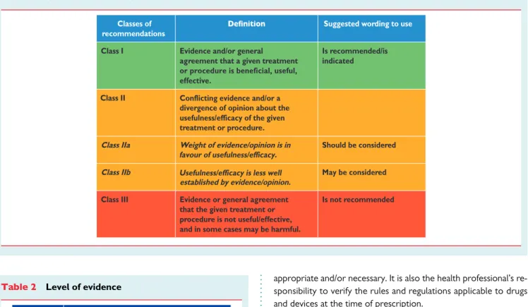
Members of this Task Force were selected by the ESC and ERS to represent professionals involved with the medical care of patients with this pathology. Selected experts in the field undertook a comprehensive review of the published evidence for management (including diagnosis, treatment, prevention and rehabilitation) of a given condition according to ESC Committee for Practice Guidelines (CPG) policy and approved by the ERS. A critical evaluation of diagnostic and therapeutic procedures was performed, including assessment of the risk – benefit ratio. Estimates of expected health outcomes for larger populations were included, where data exist. The level of evidence and the strength of the recommendation of particular management options were weighed and graded according to predefined scales,
as outlined in Tables1and2.
The experts of the writing and reviewing panels provided declar-ation of interest forms for all reldeclar-ationships that might be perceived as real or potential sources of conflicts of interest. These forms were
compiled into one file and can be found on the ESC website (http://
www.escardio.org/guidelines). Any changes in declarations of inter-est that arise during the writing period must be notified to the ESC and ERS and updated. The Task Force received its entire financial support from the ESC and ERS without any involvement from the healthcare industry.
The ESC CPG supervises and coordinates the preparation of new Guidelines produced by task forces, expert groups or consensus pa-nels. The Committee is also responsible for the endorsement pro-cess of these Guidelines. The ESC Guidelines undergo extensive review by the CPG and external experts, and in this case by ERS-appointed experts. After appropriate revisions the Guidelines are approved by all the experts involved in the Task Force. The fina-lized document is approved by the CPG and by ERS for publication in the European Heart Journal and in the European Respiratory Jour-nal. The Guidelines were developed after careful consideration of the scientific and medical knowledge and the evidence available at the time of their dating.
The task of developing ESC/ERS Guidelines covers not only integration of the most recent research, but also the creation of educational tools and implementation programmes for the recom-mendations. To implement the guidelines, condensed pocket guide-line versions, summary slides, booklets with essential messages,
by guest on January 14, 2016
http://eurheartj.oxfordjournals.org/
summary cards for non-specialists and an electronic version for digital applications (smartphones, etc.) are produced. These ver-sions are abridged and thus, if needed, one should always refer to the full text version, which is freely available on the ESC website. The National Societies of the ESC are encouraged to endorse, translate and implement all ESC Guidelines. Implementation pro-grammes are needed because it has been shown that the outcome of disease may be favourably influenced by the thorough application of clinical recommendations.
Surveys and registries are needed to verify that real-life daily prac-tice is in keeping with what is recommended in the guidelines, thus completing the loop between clinical research, writing of guidelines, disseminating them and implementing them into clinical practice.
Health professionals are encouraged to take the ESC/ERS Guide-lines fully into account when exercising their clinical judgment, as well as in the determination and the implementation of preventive, diagnostic or therapeutic medical strategies. However, the ESC/ERS Guidelines do not override in any way whatsoever the individual re-sponsibility of health professionals to make appropriate and accur-ate decisions in consideration of each patient’s health condition and in consultation with that patient and the patient’s caregiver where
appropriate and/or necessary. It is also the health professional’s re-sponsibility to verify the rules and regulations applicable to drugs and devices at the time of prescription.
2. Introduction
Pulmonary hypertension (PH) is a pathophysiological disorder that may involve multiple clinical conditions and can complicate the major-ity of cardiovascular and respiratory diseases. The composition of the guidelines task force reflects the multidisciplinary nature of PH, includ-ing members of different medical societies, associations and workinclud-ing groups. The current document follows the two previous ESC and ERS Guidelines, published in 2004 and 2009, focusing on clinical manage-ment of PH. A systematic literature review was performed from MEDLINEw to identify new studies published since 2009 concerning the topic of PH. Task force members selected studies based on rele-vance and appropriateness. The main changes and adaptations as compared with the 2009 ESC and ERS PH guidelines are as follows: † The table of contents structure has been simplified, with three
initial general chapters including classifications, basic aspects and differential diagnosis, two chapters for pulmonary arterial hypertension (PAH) and one chapter each for PH due to left heart disease (LHD), lung disease and/or hypoxia, chronic thromboembolic pulmonary hypertension (CTEPH) and unclear and/or multifactorial mechanisms.
† New wordings and parameters for the haemodynamic definition of post-capillary PH subgroups have been adopted. Pulmonary vascular resistance (PVR) has been included in the haemodynamic definition of PAH.
† An updated common clinical classification for adult and paediatric patients is reported.
† New advances in pathology, pathobiology, genetics, epidemi-ology and risk factors are reported.
Table 1 Classes of recommendations
Classes of recommendations
Suggested wording to use
Class I Evidence and/or general
agreement that a given treatment or procedure is beneficial, useful, effective.
Is recommended/is indicated
Class II
divergence of opinion about the Conflicting evidence and/or a
usefulness/efficacy of the given
favour of usefulness/efficacy. Usefulness/efficacy is less well treatment or procedure.
Class IIa Weight of evidence/opinion is in Should be considered
Class IIb
established by evidence/opinion.
May be considered
Class III Evidence or general agreement
that the given treatment or procedure is not useful/effective, and in some cases may be harmful.
Is not recommended
Table 2 Level of evidence
Level of evidence A
Data derived from multiple randomized clinical trials or meta-analyses. Level of
evidence B
Data derived from a single randomized clinical trial or large non-randomized studies.
Level of evidence C
Consensus of opinion of the experts and/ or small studies, retrospective studies, registries.
by guest on January 14, 2016
http://eurheartj.oxfordjournals.org/
† An updated diagnostic algorithm has been provided in an inde-pendent chapter and novel screening strategies are proposed in the web addenda.
† The importance of expert referral centres in the management of PH patients has been highlighted in both the diagnostic and treat-ment algorithms.
† New developments on PAH severity evaluation and on treat-ments and treatment goals are reported, including combination therapy and two new recently approved drugs. The treatment al-gorithm has been updated accordingly.
† The chapters on PH due to LHD and lung diseases have been up-dated. The term ‘out of proportion PH’ has been abandoned in both conditions.
† New diagnostic and treatment algorithms are reported in the CTEPH chapter, including general criteria for operability and bal-loon pulmonary angioplasty (BPA) and a newly approved drug. † A short chapter on PH due to unclear and/or multifactorial
mechanisms has been added.
3. Definitions and classifications
3.1 Definitions
PH is defined as an increase in mean pulmonary arterial pressure
(PAPm)≥25 mmHg at rest as assessed by right heart
catheteriza-tion (RHC).1Available data have shown that the normal PAPm at
rest is 14 + 3 mmHg with an upper limit of normal of approximately
20 mmHg.1,2The clinical significance of a PAPm between 21 and 24
mmHg is unclear. Patients presenting with a pulmonary artery pressure (PAP) in this range should be carefully followed when they are at risk for developing PAH [e.g. patients with connective tissue disease (CTD) or family members of patients with heritable
PAH (HPAH)].1
Due to the lack of reliable data that define which levels of exercise-induced changes in PAPm or PVR have prognostic implica-tions, a disease entity ‘PH on exercise’ cannot be defined and should
not be used.1A recent retrospective study has proposed a definition
of PH on exercise with the combination of PAPm and total PVR
data, but no outcome prospective validation has been provided.3
The term PAH describes a group of PH patients characterized haemodynamically by the presence of pre-capillary PH, defined by
a pulmonary artery wedge pressure (PAWP)≤15 mmHg and a
PVR .3 Wood units (WU) in the absence of other causes of pre-capillary PH such as PH due to lung diseases, CTEPH or other rare
diseases.1
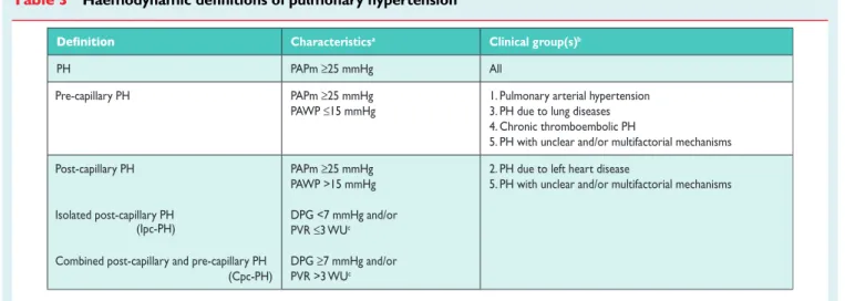
According to various combinations of PAP, PAWP, cardiac output (CO), diastolic pressure gradient (DPG) and PVR, assessed in stable clinical conditions, different haemodynamic definitions of PH are
shown in Table 3 together with their corresponding clinical
classification (Table4).1,4The reasons for the updated definitions
of post-capillary PH are reported in the specific section (8.0).
3.2 Classifications
The clinical classification of PH is intended to categorize multiple clinical conditions into five groups according to their similar clinical presentation, pathological findings, haemodynamic characteristics
and treatment strategy.5The clinical classification may be updated
when new data are available on the above features or when add-itional clinical entities are considered. A comprehensive version of
the clinical classification is presented in Table4.6A condensed
ver-sion is provided in a web addenda (Web Table I). The new findings are as follows:
† New conditions that are frequently found in children have been in-cluded in different clinical groups in order to provide a comprehen-sive classification appropriate to both adult and paediatric patients. † Recently identified gene mutations have been included in the HPAH subgroup of clinical group 1 (PAH). The new mutations are more rare as compared with the traditional bone
morpho-genetic protein receptor 2 (BMPR2) mutations (Table4).
† Pre-capillary PH associated with chronic haemolytic anaemia appears to be significantly different from other forms of PAH in Table 3 Haemodynamic definitions of pulmonary hypertensiona
Characteristicsa Clinical group(s)b
PH PAPm ≥25 mmHg All
Pre-capillary PH PAPm ≥25 mmHg
PAWP ≤15 mmHg
1. Pulmonary arterial hypertension 3. PH due to lung diseases 4. Chronic thromboembolic PH
5. PH with unclear and/or multifactorial mechanisms Post-capillary PH
Isolated post-capillary PH
Combined post-capillary and pre-capillary PH (Cpc-PH) PAPm ≥25 mmHg PAWP >15 mmHg DPG <7 mmHg and/or PVR ≤3 WUc DPG ≥7 mmHg and/or PVR >3 WUc
2. PH due to left heart disease
5. PH with unclear and/or multifactorial mechanisms
(Ipc-PH)
CO ¼ cardiac output; DPG ¼ diastolic pressure gradient (diastolic PAP – mean PAWP); mPAP ¼ mean pulmonary arterial pressure; PAWP ¼ pulmonary arterial wedge pressure; PH ¼ pulmonary hypertension; PVR ¼ pulmonary vascular resistance; WU ¼ Wood units.
a
All values measured at rest; see also section 8.0.
b
According to Table4.
c
Wood Units are preferred to dynes.s.cm25.
by guest on January 14, 2016
http://eurheartj.oxfordjournals.org/
regard to pathological findings (absence of plexiform lesions), haemodynamic characteristics (low PVR and high CO) and re-sponse to PAH-specific therapies (no demonstration of efficacy). Therefore these clinical conditions have been moved from group 1 (PAH) to group 5 (unclear and/or multifactorial mechanisms). † Group 1’ [pulmonary veno-occlusive disease (PVOD) and/or
pulmonary capillary haemangiomatosis (PCH)] has been expanded and includes idiopathic, heritable, drug-, toxin- and radiation-induced and associated forms.
Table 4 Comprehensive clinical classification of
pulmonary hypertension (updated from Simonneau et al.5)
1. Pulmonary arterial hypertension
1.1 Idiopathic 1.2 Heritable
1.2.1 BMPR2 mutation 1.2.2 Other mutations 1.3 Drugs and toxins induced 1.4 Associated with:
1.4.1 Connective tissue disease 1.4.3 Portal hypertension
1.4.4 Congenital heart disease (Table 6) 1.4.5 Schistosomiasis
1’. Pulmonary veno-occlusive disease and/or pulmonary capillary haemangiomatosis
1’.1 Idiopathic 1’.2 Heritable
1’.2.1 EIF2AK4 mutation 1’.2.2 Other mutations
1’.3 Drugs, toxins and radiation induced 1’.4 Associated with:
1’.4.1 Connective tissue disease 1’.4.2 HIV infection
1”. Persistent pulmonary hypertension of the newborn 2. Pulmonary hypertension due to left heart disease
2.1 Left ventricular systolic dysfunction 2.2 Left ventricular diastolic dysfunction 2.3 Valvular disease
obstruction and congenital cardiomyopathies 2.5 Congenital /acquired pulmonary veins stenosis
3. Pulmonary hypertension due to lung diseases and/or hypoxia
3.1 Chronic obstructive pulmonary disease 3.2 Interstitial lung disease
3.3 Other pulmonary diseases with mixed restrictive and obstructive pattern
3.4 Sleep-disordered breathing 3.5 Alveolar hypoventilation disorders 3.6 Chronic exposure to high altitude 3.7 Developmental lung diseases (Web Table III)
4. Chronic thromboembolic pulmonary hypertension and other pulmonary artery obstructions
4.1 Chronic thromboembolic pulmonary hypertension 4.2 Other pulmonary artery obstructions
4.2.1 Angiosarcoma
4.2.2 Other intravascular tumors 4.2.3 Arteritis
4.2.4 Congenital pulmonary arteries stenoses 4.2.5 Parasites (hydatidosis)
5. Pulmonary hypertension with unclear and/or multifactorial mechanisms
5.1 Haematological disorders: chronic haemolytic anaemia, myeloproliferative disorders, splenectomy
5.2 Systemic disorders: sarcoidosis, pulmonary histiocytosis, lymphangioleiomyomatosis, neurofibromatosis 5.3 Metabolic disorders: glycogen storage disease, Gaucher disease, thyroid disorders
5.4 Others: pulmonary tumoral thrombothic microangiopathy, osing mediastinitis, chronic renal failure (with/without dialysis), segmental pulmonary hypertension
BMPR2 ¼ bone morphogenetic protein receptor, type 2; EIF2AK4 ¼ eukaryotic. translation initiation factor 2 alpha kinase 4; HIV ¼ human immunodeficiency virus.
Table 5 Important pathophysiological and clinical definitions
1. Pulmonary hypertension (PH) is a haemodynamic and pulmonary arterial pressure ≥25 mmHg at rest as assessed by right heart catheterization (Table 3). PH can be found in multiple clinical conditions (Table 4).
2. Pulmonary arterial hypertension (PAH, group 1) is a clinical condition characterized by the presence of pre-capillary PH (Table 3) and pulmonary vascular resistance >3 Wood units, in the absence of other causes of pre-capillary PH such as PH due to lung diseases, chronic thromboembolic PH, or other rare diseases (Table 4). PAH includes different forms that share a similar clinical picture and virtually identical pathological changes of the lung microcirculation (Table 4).
exercise’. 3.
Table 6 Clinical classification of pulmonary arterial hypertension associated with congenital heart disease
(updated from Simonneau et al.5)
1. Eisenmenger’s syndrome
Includes all large intra- and extra-cardiac defects which begin as systemic-to-pulmonary shunts and progress with time to severe elevation of PVR and to reversal (pulmonary-to-systemic) or bidirectional shunting; cyanosis, secondary erythrocytosis, and multiple organ involvement are usually present.
2. PAH associated with prevalent systemic-to-pulmonary shunts
• Correctablea • Non-correctable
Includes moderate to large defects; PVR is mildly to moderately increased, systemic-to-pulmonary shunting is still prevalent, whereas cyanosis at rest is not a feature.
3. PAH with small/coincidental defects b
Marked elevation in PVR in the presence of small cardiac defects (usually ventricular septal defects <1 cm and atrial septal defects <2 cm of effective diameter assessed by echo), which themselves do not account for the development of elevated PVR; the clinical picture is very similar to idiopathic PAH. Closing the defects is contra-indicated.
4. PAH after defect correction
Congenital heart disease is repaired, but PAH either persists immediately after correction or recurs/develops months or years haemodynamic lesions.
PAH ¼ pulmonary arterial hypertension; PVR ¼ pulmonary vascular resistance.
a
With surgery or intravascular percutaneous procedure.
b
The size applies to adult patients. However, also in adults the simple diameter may be not sufficient for defining the haemodynamic relevance of the defect and also the pressure gradient, the shunt size and direction, and the pulmonary to systemic flows ratio should be considered (Web Table II).
by guest on January 14, 2016
http://eurheartj.oxfordjournals.org/
† Persistent PH of the newborn (PPHN) includes a heterogeneous group of conditions that may differ from classical PAH. As a
con-sequence, PPHN has been subcategorised as group I′′.7–9
† Paediatric heart diseases such as congenital or acquired left heart inflow or outflow tract obstruction and congenital cardiomyop-athies have been included in group 2 (PH due to LHD). † No changes are proposed for group 3 (PH due to lung diseases
and/or hypoxia).
† Group 4 has been renamed as ‘CTEPH and other pulmonary artery (PA) obstructions’, which includes CTEPH, pulmonary angiosarcoma, other intravascular tumours, arteritis, congenital
pulmonary arteries stenoses and parasites (Table4).
† Segmental PH is observed in discrete lung areas perfused by aorto-pulmonary collaterals in congenital heart diseases such as pulmon-ary or tricuspid atresia. This very unusual haemodynamic condition has been included in group 5 (unclear and/or multifactorial mechanisms).
† Some pathological and pathophysiological information on the clinical groups are reported in the web addenda.
Important pathophysiological and clinical definitions are reported in
Table5. A clinical classification of PAH associated with congenital
heart disease (CHD) is reported in Table6.
An anatomical – pathophysiological classification of congenital systemic-to-pulmonary shunts associated with PAH is presented in Web Table II. A list of developmental lung diseases associated with PH is presented in Web Table III.
4. Epidemiology and genetics
of pulmonary hypertension
4.1 Epidemiology and risk factors
Reporting in the literature of PH incidence data at the global level is poor. In the UK, a prevalence of 97 cases per million with a female:-male ratio of 1.8 has been reported. The age-standardized death rate in the USA ranges between 4.5 and 12.3 per 100,000 population. Comparative epidemiological data on the prevalence of the different groups of PH are not widely available, but it is clear that LHD (group 2) is believed to be the most common cause of PH, although severe PH is relatively uncommon in this setting. Although patients belonging to groups 2 and 3 represent an important part of the clinical practice, there is disproportionately little information about the demographics and clinical course of this segment of the PH population, suggesting that registry database methodology may be useful for these groups. Globally, schistosomiasis-associated PAH and high altitude– related PH represent an important burden to mankind.
† Group 1 (PAH): Several registries have described the epidemiology
of PAH.10–12The lowest estimate of the prevalence of PAH and
idiopathic PAH (IPAH) are 15 cases and 5.9 cases per million adult population, respectively. The lowest estimate of PAH incidence is 2.4 cases per million adult population per year. In Europe, PAH prevalence and incidence are in the range of 15–60 subjects per
mil-lion population and 5–10 cases per milmil-lion per year, respectively.11
In registries, around half of PAH patients have idiopathic, heritable or drug-induced PAH. In the subgroup of associated PAH conditions
(APAH), the leading cause is CTD, mainly systemic sclerosis (SSc).10
PAH may occur in different settings depending on associated
clinical conditions.13IPAH corresponds to sporadic disease, without
any familial history of PAH or known triggering factor. While the mean age of patients with IPAH in the first US National Institutes of Health registry created in 1981 was 36 years, PAH is now more frequently diagnosed in elderly patients, resulting in a mean age at diagnosis between 50 and 65 years in current registries. Furthermore, the female predominance is quite variable among registries and may not be present in elderly patients, and survival ap-pears to have improved over time.
A number of risk factors for the development of PAH has been identified and are defined as any factor or condition that is suspected to play a predisposing or facilitating role in disease development. Risk factors were classified as definite, likely or possible, based on the strength of their association with PH and their probable causal
role.13A definite association is acknowledged in the case of either an
epidemic, such as occurred with appetite suppressants, or if large, multicentre epidemiological studies demonstrate an association between the clinical condition or drug and PAH. A likely association is acknowledged if a single-centre case– control study or multiple case series demonstrate an association or if clinical and haemo-dynamic recovery occurs after stopping exposure, such as oc-curred in dasatinib-induced PAH. A possible association can be suspected, for example, for drugs with similar mechanisms of action as those in the definite or likely category but which have not yet been studied, such as drugs used to treat attention deficit disorder.
Definite clinical associations are listed among APAH in Table4and
the risk level of different drugs and toxins are listed in Table7.6,14–16
† Group 2 (PH due to LHD): The prevalence of PH in patients with chronic heart failure increases with the progression of functional class (FC) impairment. Up to 60% of patients with severe left ven-tricular (LV) systolic dysfunction and up to 70% of patients with heart failure with preserved ejection fraction may present with PH. In left-sided valvular diseases, the prevalence of PH increases with the se-verity of the defect and of the symptoms. PH can be found in virtually all patients with severe symptomatic mitral valve disease and in up to
65% of those with symptomatic aortic stenosis.17–19
† Group 3 (PH due to lung diseases and/or hypoxaemia): Mild PH is common in both severe interstitial lung disease and severe chronic
Table 7 Updated risk level of drugs and toxins known to induce pulmonary arterial hypertension
Likely Possible
• Aminorex • F • Dexf
• Toxic rapeseed oil
• ex • Selective serotonin reuptake inhibitorsa • Amphetamines • Dasatinib • L-tryptophan • Methamphetamines • Cocaine • Phenylpropanolamine • St John’s Wort • Amphetamine-like drugs • Interferon α and β • Some chemotherapeutic agents such as alkylating agents (mytomycine C, cyclophosphamide)b a
Increased risk of persistent pulmonary hypertension in the newborns of mothers with intake of selective serotonin reuptake inhibitors.
b
Alkylating agents are possible causes of pulmonary veno-occlusive disease.
by guest on January 14, 2016
http://eurheartj.oxfordjournals.org/
obstructive pulmonary disease (COPD),20while severe PH is
un-common.21Severe PH can be seen in the combined emphysema/
fibrosis syndrome, where the prevalence of PH is high.22
† Group 4 [CTEPH and other PA obstructions]: In the Spanish PH Registry, CTEPH prevalence and incidence were 3.2 cases per
million and 0.9 cases per million per year, respectively.23Even
though a prevalence of CTEPH of 3.8% has been reported in sur-vivors of acute pulmonary embolism (PE), the true incidence of
CTEPH after acute PE is lower, in the range of 0.5 – 2%.24A
his-tory of acute PE was reported for 74.8% of patients from the
International CTEPH Registry.25Associated conditions included
thrombophilic disorders (lupus anticoagulant/antiphospholipid antibodies, protein S and C deficiency, activated protein C resist-ance including factor V Leiden mutation, prothrombin gene mu-tation, antithrombin III deficiency and elevated factor VIII) in 31.9% of patients and splenectomy in 3.4%.
4.2 Genetics
† Group 1 (PAH): Heterozygous BMPR2 mutations account for approximately 75% of familial PAH and up to 25% of apparently
sporadic PAH cases.26BMPR2 encodes a type 2 receptor for
bone morphogenetic proteins involved in the control of vascular cell proliferation. Mutations of genes coding for activin receptor-like kinase 1 and endoglin have been identified in PAH patients with a personal or family history of hereditary haemorrhagic telangiectasia, as well as in BMPR1B and SMAD9, supporting a prominent role for
transforming growth factor b (TGF-b) family members in PAH.26
Whole exome sequencing has identified rare heterozygous muta-tions in genes coding for proteins such as caveolin 1 (CAV1) and the
potassium channel subfamily K member 3 (KCNK3).26,27
† Group 1: Heritable PVOD/PCH has been recognized in consan-guineous families, suggesting recessive transmission. Whole gen-ome sequencing demonstrated that bi-allelic mutations in eukaryotic translation initiation factor 2 alpha kinase 4 (EIF2AK4) were present in all familial PVOD/PCH and in 25% of histologically
confirmed sporadic PVOD/PCH.28EIF2AK4 encodes a
serine-threonine kinase present in all eukaryotes that can induce changes in gene expression in response to amino acid deprivation. † Group 2 (PH due to LHD): No specific genetic linkage has been
identified.18
† Group 3 (PH due to lung diseases and/or hypoxaemia): Gene polymorphism might contribute towards determining the
sever-ity of PH in hypoxaemic patients with COPD.29
† Group 4 (CTEPH and other PA obstructions): No specific genet-ic mutations have been linked to the development of CTEPH. † Group 5 (PH with unclear and/or multifactorial mechanisms):
The heterogeneity of this group prevents an appropriate descrip-tion of genetics, epidemiology and risk factors in these guidelines.
5. Pulmonary hypertension
diagnosis
5.1 Diagnosis
The diagnosis of PH requires a clinical suspicion based on symptoms and physical examination and review of a comprehensive set of
investigations to confirm that haemodynamic criteria are met and to describe the aetiology and the functional and haemodynamic severity of the condition. The interpretation of these investigations requires, at the very least, expertise in cardiology, imaging and respiratory medicine and may best be discussed at a multidisciplin-ary team meeting. This is particularly important for identifying patients who may have more than one cause of PH. The main cause of PH should be identified according to the clinical
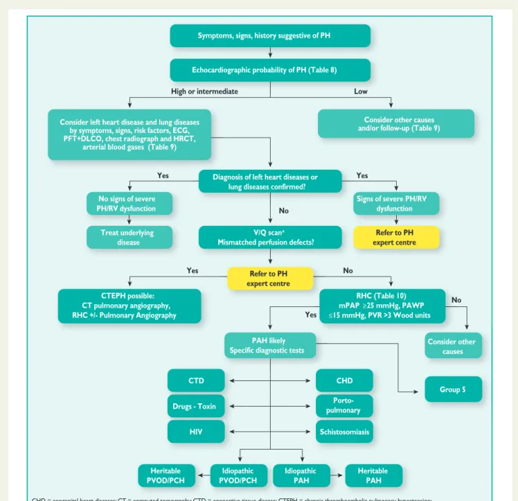
classifica-tion in Table4. An algorithm for reaching a diagnosis is shown in
Figure1.
5.1.1 Clinical presentation
The symptoms of PH are non-specific and mainly related to progres-sive right ventricular (RV) dysfunction. Initial symptoms are typically induced by exertion. They include shortness of breath, fatigue, weakness, angina and syncope. Less commonly patients may also de-scribe dry cough and exercise-induced nausea and vomiting. Symp-toms at rest occur only in advanced cases. Abdominal distension and ankle oedema will develop with progressing RV failure. The presen-tation of PH may be modified by diseases that cause or are asso-ciated with PH as well as other concurrent diseases.
In some patients the clinical presentation may be related to mech-anical complications of PH and the abnormal distribution of blood flow in the pulmonary vascular bed. These include haemoptysis re-lated to rupture of hypertrophied bronchial arteries, as well as symptoms attributable to pulmonary arterial dilatation such as hoarseness caused by compression of the left recurrent laryngeal nerve, wheeze caused by large airway compression and angina due to myocardial ischaemia caused by compression of the left main coronary artery. Significant dilation of the PA may result in its rupture or dissection, leading to signs and symptoms of cardiac tamponade.
The physical signs of PH include left parasternal lift, an accentu-ated pulmonary component of the second heart sound, an RV third heart sound, a pansystolic murmur of tricuspid regurgitation and a diastolic murmur of pulmonary regurgitation. Elevated jugular ven-ous pressure, hepatomegaly, ascites, peripheral oedema and cool extremities characterize patients with advanced disease. Wheeze and crackles are usually absent.
Clinical examination may suggest an underlying cause of PH. Telangiectasia, digital ulceration and sclerodactyly are seen in sclero-derma, inspiratory crackles may point towards interstitial lung dis-ease and spider naevi, testicular atrophy, and palmar erythema suggest liver disease. When digital clubbing is encountered, PVOD, cyanotic CHD, interstitial lung disease or liver disease should be considered.
5.1.2 Electrocardiogram
An electrocardiogram (ECG) may provide supportive evidence of PH, but a normal ECG does not exclude the diagnosis. An abnormal ECG is more likely in severe rather than mild PH. ECG abnormalities may include P pulmonale, right axis deviation, RV hypertrophy, RV strain, right bundle branch block, and QTc prolongation. While RV hypertrophy has insufficient sensitivity (55%) and specificity (70%)
to be a screening tool, RV strain is more sensitive.30Prolongation
of the QRS complex and QTc suggest severe disease.31,32The
ECG differential diagnosis includes anterolateral myocardial
by guest on January 14, 2016
http://eurheartj.oxfordjournals.org/
ischaemia. In contrast to PH, ECG changes in ischaemia more com-monly affect the lateral and inferior leads, and when present in the anterior chest leads are usually accompanied by a Q wave in V1 to V3, and rarely cause right axis deviation.
Supraventricular arrhythmias may occur in advanced disease, in particular atrial flutter, but also atrial fibrillation, with a cumulative
incidence in 25% of patients after 5 years.33Atrial arrhythmias
com-promise CO and almost invariably lead to further clinical deterior-ation. Ventricular arrhythmias are rare.
5.1.3 Chest radiograph
In 90% of patients with IPAH the chest radiograph is abnormal at the
time of diagnosis.34Findings in patients with PAH include central
pulmonary arterial dilatation, which contrasts with ‘pruning’ (loss) of the peripheral blood vessels. Right atrium (RA) and RV enlarge-ment may be seen in more advanced cases. A chest radiograph may assist in differential diagnosis of PH by showing signs suggesting lung
disease (group 3, Table4) or pulmonary venous congestion due to
LHD (group 2, Table4). Chest radiography may help in distinguishing
between arterial and venous PH by respectively demonstrating
in-creased and dein-creased artery:vein ratios.35
Overall, the degree of PH in any given patient does not correlate with the extent of radiographic abnormalities. As for ECG, a normal chest radiograph does not exclude PH.
5.1.4 Pulmonary function tests and arterial blood gases Pulmonary function tests and arterial blood gases identify the con-tribution of underlying airway or parenchymal lung disease. Patients with PAH have usually mild to moderate reduction of lung volumes
related to disease severity.36,37Although diffusion capacity can be
normal in PAH, most patients have decreased lung diffusion capacity for carbon monoxide (DLCO). An abnormal low DLCO, defined as
,45% of predicted, is associated with a poor outcome.36,37
The differential diagnosis of a low DLCO in PAH includes PVOD, PAH associated with scleroderma and parenchymal lung disease. Although airflow obstruction is unusual, peripheral airway obstruc-tion can be detected. Due to alveolar hyperventilaobstruc-tion at rest,
arter-ial oxygen pressure (PaO2) remains normal or is only slightly lower
than normal and arterial carbon dioxide pressure (PaCO2) is
decreased.38
COPD as a cause of hypoxic PH is diagnosed on the evidence of irreversible airflow obstruction together with increased residual
vo-lumes and reduced DLCO.39Arterial blood gases of COPD patients
show a decreased PaO2with normal or increased PaCO2.40A
de-crease in lung volume combined with dede-creased diffusion capacity
for carbon monoxide may indicate interstitial lung disease.39The
se-verity of emphysema and of interstitial lung disease can be diagnosed using high-resolution computed tomography (CT). Combined em-physema and pulmonary fibrosis may pseudonormalize spirometry, although the DLCO is almost always reduced, emphasizing the need to interpret pulmonary function alongside lung imaging.
The prevalence of nocturnal hypoxaemia and central sleep
ap-noeas are high in PAH (70 – 80%).41,42Overnight oximetry or
poly-somnography should be performed where obstructive sleep apnoea syndrome or hypoventilation are considered.
5.1.5 Echocardiography
Transthoracic echocardiography is used to image the effects of PH on the heart and estimate PAP from continuous wave Doppler mea-surements. Echocardiography should always be performed when PH is suspected and may be used to infer a diagnosis of PH in pa-tients in whom multiple different echocardiographic measurements are consistent with this diagnosis. When treatment of PH itself is being considered, echocardiography alone is not sufficient to sup-port a treatment decision and cardiac catheterization is required. Detailed guidelines describing the echocardiographic assessment of the right heart can be found in documents created and/or en-dorsed by the European Association of Cardiovascular Imaging
Table 8A Echocardiographic probability of
pulmonary hypertension in symptomatic patients with a suspicion of pulmonary hypertension
Peak tricuspid regurgitation velocity (m/s) Presence of other echo ‘PH signs’a Echocardiographic probability of pulmonary hypertension ≤2.8 or not ≤2.8 or not measurable No Low
measurable Yes Intermediate
2.9–3.4 No 2.9–3.4 Yes High >3.4 Not required PH ¼ pulmonary hypertension. a See Table8B.
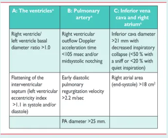
Table 8B Echocardiographic signs suggesting pulmonary hypertension used to assess the probability of pulmonary hypertension in addition to tricuspid
regurgitation velocity measurement in Table8A
A: The ventriclesa B: Pulmonary
arterya
C: Inferior vena cava and right
atriuma Right ventricle/
left ventricle basal diameter ratio >1.0
Right ventricular acceleration time <105 msec and/or midsystolic notching
Inferior cava diameter >21 mm with decreased inspiratory collapse (<50 % with a sniff or <20 % with quiet inspiration) Flattening of the interventricular septum (left ventricular eccentricity index >1.1 in systole and/or diastole) Early diastolic pulmonary regurgitation velocity >2.2 m/sec
Right atrial area (end-systole) >18 cm2
PA diameter >25 mm.
PA ¼ pulmonary artery.
a
Echocardiographic signs from at least two different categories (A/B/C) from the list should be present to alter the level of echocardiographic probability of pulmonary hypertension.
by guest on January 14, 2016
http://eurheartj.oxfordjournals.org/
(EACVI), a registered branch of the ESC, and the reader is referred
to these for further instruction.43,44
The estimation of systolic PAP is based on the peak tricuspid re-gurgitation velocity (TRV) taking into account right atrial pressure (RAP) as described by the simplified Bernoulli equation. RAP can be estimated by echocardiography based on the diameter and re-spiratory variation in diameter of the inferior vena cava (IVC): an IVC diameter ,2.1 cm that collapses .50% with a sniff suggests a normal RA pressure of 3 mmHg (range 0 – 5 mmHg), whereas an IVC diameter .2.1 cm that collapses ,50% with a sniff or ,20% on quiet inspiration suggests a high RA pressure of 15 mmHg (range 10 – 20 mmHg). In scenarios in which the IVC diameter and collapse do not fit this paradigm, an intermediate value of 8 mmHg (range 5 – 10 mmHg) may be used. The EACVI recom-mends such an approach rather than using a fixed value of 5 or 10 mmHg for PA systolic pressure (PASP) estimations. However, given the inaccuracies of RAP estimation and the amplification of meas-urement errors by using derived variables, we recommend using the continuous wave Doppler measurement of peak TRV (and not the estimated PASP) as the main variable for assigning the echo-cardiographic probability of PH.
When peak TRV is technically difficult to measure (trivial or mild tricuspid regurgitation) some laboratories use contrast echocardi-ography [e.g. agitated saline administered by intravenous (i.v.) injec-tion], which may improve the Doppler signal, allowing measurement of peak TRV velocity. Unfortunately, despite the strong correlation of TRV with a tricuspid regurgitation pressure gradient, Doppler-derived pressure estimation may be inaccurate in the individual pa-tient. In patients with severe tricuspid regurgitation, TRV may be sig-nificantly underestimated and cannot be used to exclude PH.
Overestimation may also occur.44PH cannot be reliably defined
by a cut-off value of TRV. Consequently, estimation of PAP based
solely on Doppler transthoracic echocardiography measurements is not suitable for screening for mild, asymptomatic PH. Other echo-cardiographic variables that might raise or reinforce suspicion of PH independent of TRV should always be sought.
Conclusions derived from an echocardiographic examination should aim to assign a level of probability of PH. This ESC Guideline suggests grading the probability of PH based on TRV at rest and on the presence of additional pre-specified echocardiographic variables
suggestive of PH (Table8A). The probability of PH may then be
judged as high, intermediate or low. When interpreted in a clinical context, the echocardiographic result is required to decide the need for cardiac catheterization in individual patients. In order to facilitate and standardize assignment to the level of probability of PH, several additional echocardiographic signs are proposed in addition to
cri-teria based on TRV (Table8B). These signs provide assessment of
the RV size and pressure overload, the pattern of blood flow vel-ocity out of the RV, the diameter of the PA and an estimate of
RAP.43–45Their measurement has been defined in
recommenda-tions endorsed by the EACVI.43,44
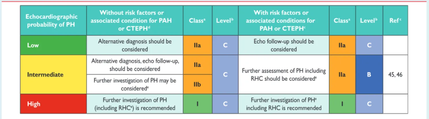
The recommended plan for further patient investigation based on
echocardiographic probability of PH is shown in Table9for
symp-tomatic patients. In the Web addendum, a similar table (Web Table IX) for screening for asymptomatic patients with risk factors for PAH or with incidental findings suggesting the possibility of PH on ECG or lung imaging is provided.
Echocardiography can be helpful in detecting the cause of sus-pected or confirmed PH. Two-dimensional, Doppler and contrast examinations can be used to identify CHD. High pulmonary blood flow found on pulsed wave Doppler in the absence of a detectable shunt or significant dilatation of proximal PA despite only moderate PH may warrant transoesophageal examination with contrast or cardiac magnetic resonance (CMR) imaging to exclude sinus
Table 9 Diagnostic management suggested according to echocardiographic probability of pulmonary hypertension in patients with symptoms compatible with pulmonary hypertension, with or without risk factors for pulmonary arterial hypertension or chronic thromboembolic pulmonary hypertension
Echocardiographic probability of PH
Without risk factors or associated condition for PAH
or CTEPHd
Classa Levelb
With risk factors or associated conditions for
PAH or CTEPHc
Classa Levelb Refc
Low Alternative diagnosis should be
considered IIa C
Echo follow-up should be
considered IIa C
Intermediate
Alternative diagnosis, echo follow-up, should be considered IIa
C Further assessment of PH including
RHC should be considerede IIa B 45, 46 Further investigation of PH may be
considerede IIb
High Further investigation of PH
(including RHCe) is recommended I C
Further investigation of PHe
including RHC is recommended I C
CTEPH ¼ chronic thromboembolic pulmonary hypertension; Echo ¼ echocardiographic; PAH ¼ pulmonary arterial hypertension; PH ¼ pulmonary hypertension; RHC ¼ right heart catheterization. a Class of recommendation. b Level of evidence. c
Reference(s) supporting recommendations.
d
These recommendations do not apply to patients with diffuse parenchymal lung disease or left heart disease.
e
Depending on the presence of risk factors for PH group 2, 3 or 5.
Further investigation strategy may differ depending on whether risk factors/associated conditions suggest higher probability of PAH or CTEPH – see diagnostic algorithm.
by guest on January 14, 2016
http://eurheartj.oxfordjournals.org/
venosus atrial septal defect and/or anomalous pulmonary venous re-turn. In cases of suspicion of LV diastolic dysfunction, Doppler echo-cardiographic signs should be assessed even if their reliability is considered low. RHC should be considered when the diagnosis re-mains uncertain after non-invasive investigations (see section 8.1). The practical clinical value of exercise Doppler echocardiography in the identification of cases with PH limited to exercise is uncertain because of the lack of validated criteria and prospective confirmatory data.
5.1.6 Ventilation/perfusion lung scan
A ventilation/perfusion (V/Q) lung scan should be performed in pa-tients with PH to look for CTEPH. The V/Q scan has been the screening method of choice for CTEPH because of its higher sensi-tivity compared with CT pulmonary angiogram (CTPA), especially in
inexperienced centres.47A normal- or low-probability V/Q scan
ef-fectively excludes CTEPH with a sensitivity of 90 – 100% and a spe-cificity of 94 – 100%; however, many V/Q scans are not diagnostic. While in PAH the V/Q lung scan may be normal, it may also show small peripheral unmatched and non-segmental defects in perfusion. A caveat is that unmatched perfusion defects may also be seen in other pulmonary vascular disease such as PVOD. While a V/Q scan is still recommended as the screening test of choice, ventilation scans are often replaced with either a recent chest radiograph or a recent high-resolution CT of the lungs, but such practices are not really evidence-based. Also, CT is preferred in many centres since it is more readily available. A few studies suggest that single photon emission CT, also a nuclear medicine technique, could be superior to V/Q planar scan and CTPA, but these results need more
exten-sive evaluation.48More recently, newer techniques such as
three-dimensional magnetic resonance (MR) perfusion mapping, have been demonstrated to be as sensitive as traditional perfusion scintig-raphy in screening for CTEPH; MR can also be used as a
radiation-free modality to assess both ventilation and perfusion in CTEPH.49
5.1.7 High-resolution computed tomography, contrast-enhanced computed tomography, and pulmonary angiography
CT imaging is a widely available tool that can provide important in-formation on vascular, cardiac, parenchymal and mediastinal abnor-malities. It may suggest the diagnosis of PH (PA or RV enlargement), identify a cause of PH such as CTEPH or lung disease, provide clues as to the form of PAH (e.g. oesophageal dilation in SSc or congenital cardiac defects such as anomalous pulmonary venous drainage) and
also provide prognostic information.50
CT may raise a suspicion of PH in symptomatic patients or those examined for unrelated indications by showing an increased PA
diameter (≥29 mm) and pulmonary:ascending aorta diameter ratio
(≥1.0). A segmental artery:bronchus ratio .1 : 1 in three or four
lobes has been reported to have high specificity for PH.51,52
High-resolution CT provides detailed views of the lung paren-chyma and facilitates the diagnosis of interstitial lung disease and em-physema. High-resolution CT may also be very helpful where there is a clinical suspicion of PVOD. Characteristic changes of interstitial oedema with diffuse central ground-glass opacification and thicken-ing of interlobular septa support the diagnosis of PVOD; additional findings may include lymphadenopathy, pleural shadows and
effusions.53Pulmonary capillary haemangiomatosis is suggested by
diffuse bilateral thickening of the interlobular septa and the presence of small, centrilobular, poorly circumscribed nodular opacities. However, ground-glass abnormalities are also present in PAH,
oc-curring in more than one-third of patients.50
Contrast CT angiography of the PA is helpful in determining whether there is evidence of surgically accessible CTEPH. It can de-lineate the typical angiographic findings in CTEPH, such as complete obstruction, bands and webs and intimal irregularities, as accurately
and reliably as digital subtraction angiography.54,55With this
tech-nique, collaterals from bronchial arteries can be identified. Traditional pulmonary angiography is required in most patients for the workup of CTEPH to identify those who may benefit from
pulmonary endarterectomy (PEA) or BPA.56,57Angiography can be
performed safely by experienced staff in patients with severe PH using modern contrast media and selective injections. Angiography may also be useful in the evaluation of possible vasculitis or pulmon-ary arteriovenous malformations, but CT angiography has similar or
even higher accuracy for both diagnoses, and is less invasive.58,59
5.1.8 Cardiac magnetic resonance imaging
CMR imaging is accurate and reproducible in the assessment of RV size, morphology and function and allows non-invasive assessment of blood flow, including stroke volume, CO, pulmonary arterial dis-tensibility and RV mass.
In patients with suspected PH, the presence of late gadolinium en-hancement, reduced pulmonary arterial distensibility and retrograde flow have high predictive value for the identification of PH; however,
no single CMR measurement can exclude PH.60–62In patients with
PH, CMR may also be useful in cases of suspected CHD if echocar-diography is not conclusive.
Contrast-enhanced and unenhanced MR angiography have a po-tential in the study of the pulmonary vasculature in patients with sus-pected CTEPH, particularly in clinical scenarios such as sussus-pected chronic embolism in pregnant women, young patients or when
iodine-based contrast media injection is contraindicated.63
CMR provides useful prognostic information in patients with PAH
both at baseline and at follow-up.64–66
5.1.9 Blood tests and immunology
Blood tests are not useful in diagnosing PH, but are required to iden-tify the aetiology of some forms of PH as well as end organ damage. Routine biochemistry, haematology and thyroid function tests are required in all patients, as well as a number of other specific blood tests. Liver function tests may be abnormal because of high hepatic venous pressure, liver disease and/or endothelin receptor antagon-ist (ERA) therapy. Hepatitis serology should be performed if clinical abnormalities are noted. Thyroid disease is common in PAH and may develop during the course of the disease. This should always be considered in cases of abrupt deterioration.
Serological testing is required to detect underlying CTD, hepatitis and human immunodeficiency virus (HIV). Up to 40% of patients with IPAH have elevated antinuclear antibodies usually in a low titre (1:80). It is important to look for evidence of SSc since this disease has a relatively high prevalence of PAH. Limited scleroderma typic-ally has antinuclear antibodies, including anti-centromere, dsDNA, anti-Ro, U3-RNP, B23, Th/To and U1-RNP. Diffuse scleroderma is
by guest on January 14, 2016
http://eurheartj.oxfordjournals.org/