R E S E A R C H
Open Access
Variation of inflammatory dynamics and
mediators in primiparous cows after
intramammary challenge with
Escherichia coli
Adel Pezeshki
1, Philippe Stordeur
2, Hugues Wallemacq
3, Frédéric Schynts
2, Mieke Stevens
1, Philippe Boutet
3,
Luc J Peelman
1, Bart De Spiegeleer
1, Luc Duchateau
1, Fabrice Bureau
3, Christian Burvenich
1*Abstract
The objective of the current study was to investigate (i) the outcome of experimentally induced Escherichia coli mastitis in primiparous cows during early lactation in relation with production of eicosanoids and inflammatory indicators, and (ii) the validity of thermography to evaluate temperature changes on udder skin surface after experimentally induced E. coli mastitis. Nine primiparous Holstein Friesian cows were inoculated 24 ± 6 days (d) after parturition in both left quarters with E. coli P4 serotype O32:H37. Blood and milk samples were collected before and after challenge with E. coli. The infrared images were taken from the caudal view of the udder following challenge with E. coli. No relationship was detected between severity of mastitis and changes of
thromboxane B2 (TXB2), leukotriene B4 (LTB4) and lipoxin A4 (LXA4). However, prostaglandin E2 (PGE2) was related to systemic disease severity during E. coli mastitis. Moreover, reduced somatic cell count (SCC), fewer circulating basophils, increased concentration of tumor necrosis factor-a (TNF-a) and higher milk sodium and lower milk potassium concentrations were related to systemic disease severity. The thermal camera was capable of detecting 2-3°C temperature changes on udder skin surface of cows inoculated with E. coli. Peak of udder skin temperature occurred after peak of rectal temperature and appearance of local signs of induced E. coli mastitis. Although infrared thermography was a successful method for detecting the changes in udder skin surface temperature following intramammary challenge with E. coli, it did not show to be a promising tool for early detection of mastitis.
Introduction
The severity of coliform mastitis is of much more con-cern than its incidence [1]. Pathogen, cow and environ-ment are three interdependent factors which influence the mastitis susceptibility [1]. From the various bacterial virulence factors studied during Escherichia coli mastitis [2], only a few have been found to play an important role in the outcome of the disease. It has been accepted that the type of E. coli strain is not the main factor in classification of severity. Preventive treatments which are efficient against contagious mastitis have been shown to be inefficient in the control of E. coli mastitis [3]. The severity of bovine E. coli mastitis is mainly
determined by cow factors rather than by the patho-genecity of the invading pathogen and management [1]. It is known that the growth of E. coli in the udder cis-tern is specially related with the period of lactation and parity of cows. E. coli mastitis with severe clinical symp-toms is more frequently observed around calving and during early lactation in dairy cows, whereas symptoms are mild to moderate during mid and late lactation. Because of hormonal, metabolic and nutritional altera-tions associated with pregnancy, immune system is com-promised around calving (reviewed by Pezeshki et al. [4]). Cow parity is another important physiological factor that influences the severity of clinical coliform mastitis [5,6]. Clinical severe cases of coliform mastitis are mostly seen among multiparous cows rather than primiparous cows during early lactation. To our best knowledge the inflammatory status of primiparous cows
* Correspondence: [email protected]
1Department of Comparative Physiology and Biometrics, Laboratory of
Genetics, Drug Quality and Registration Group, Ghent University, B-9000 Ghent, Belgium
Full list of author information is available at the end of the article
© 2011 Pezeshki et al; licensee BioMed Central Ltd. This is an Open Access article distributed under the terms of the Creative Commons Attribution License (http://creativecommons.org/licenses/by/2.0), which permits unrestricted use, distribution, and reproduction in any medium, provided the original work is properly cited.
ranking based on severity after intramammary infection of E. coli is poorly understood during early lactation. Physiological factors have been mainly studied in multi-parous cows ranging from second lactation to sixth lactation [6-10].
Thromboxanes (TX), prostaglandins (PG), leukotriens (LT) and lipoxines (LX) which are the enzymatically generated products of cyclooxygenases (COX) and lipox-ygenases are generated during inflammation and act as major pro-inflammatory mediators. There is evidence that TXB2 and PGE2 might have an important role in severity of E. coli mastitis in primiparous cows during early lactation [11]. Although not significantly, nonster-oidal anti-inflammatory drug treatment reduced the concentration of TXB2 and PGE2 in milk, while it had no effect on chemotactic inflammatory mediators and early innate immune molecules [11]. LTB4 is known to induce infiltration and sequestration of the neutrophils in the bovine mammary gland, whereas LXA4 serve as neutrophil“stop signals” in inflammation [12]. As exces-sive recruitment of neutrophils can exacerbate the inflammatory process via releasing detrimental intracel-lular products for tissues, persistence of LTB4 produc-tion or its ratio with LXA4 could be of particular importance in severity of the disease. We hypothesize that the magnitude of eicosanoids response plays an important role in the susceptibility to E. coli mastitis during early lactation in primiparous cows and can influence the resolution of this disease.
An automated method for early detection of coliform mastitis would prevent its progression and likely reduce the costs brought about by repeated treatments. Inflam-mation of the udder or mastitis results in increase of the temperature. The radiated heat emitted by the skin, reflecting subcutaneous circulation and tissue metabo-lism could be measured using infrared thermography. Although the thermography technique has been tested for early detection of experimentally induced acute clini-cal mastitis using E. coli endotoxin [13] or subcliniclini-cal mastitis [14], the technique has not yet been validated for evaluating the temperature changes of the udder’s skin surface after challenge with E. coli.
The objectives of the current study were to investigate (i) the outcome of experimental E. coli mastitis in primi-parous cows during early lactation in relation with pro-duction of eicosanoids and inflammatory indicators in blood and milk and to determine (ii) whether thermo-graphy is a valid method for evaluating the temperature changes of the affected udder.
Materials and methods
Experimental animals and study facilities
In total, 9 primiparous Holstein Friesian cows in early lactation ranging in weight (one week after calving)
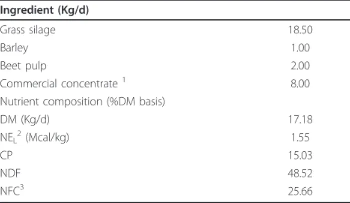
from 511 to 615 kg were used. Cows arrived at the experimental dairy farm (Center of Rural Economy-CER-Marloie, Belgium) 15 days before expected calving date (actual day was 22 ± 6 days (d)). The age of cows was between 23 to 34 months at calving. Cows were milked twice a day at 06:30 and 18:30 using a quarter milking device [8]. After parturition, cows were fed a daily ration according to the National Research Council system (NRC, 2001). The forages were delivered in two equal meals at 07:00 and 19:00 while the concentrate was fed in several times, according to milk production. The composition and ingredient of the lactation diet used during the experiment is reported in Table 1. Water was provided ad libitum.
Experimental infections were approved by the Ethical Committee of the CER (No. META-DVA-1). After an adaptation period, animals were inoculated intramamma-rily with the E. coli suspension at 24 ± 6 days in milk. Bacteriological examination of the milk was carried out according to International Dairy Federation (IDF, 1981) recommendations. The cows were clinically healthy and free of major mastitis pathogens through three consecu-tive bacteriologically negaconsecu-tive examinations, with a quar-ter foremilk somatic cell count (SCC) below 200 000 cells/mL. Daily quarter milk production (QMP) was recorded at d -7, -4, -3, -2, -1, 0, +1, +2, +3 and +6, rela-tive to day of inoculation. On average, the concentration of preinfectionb-hydroxybutyrate (BHBA), glucose and non-esterified fatty acids (NEFA) was 1.09 ± 0.42, 3.70 ± 0.42 and 0.33 ± 0.17 mmol/L, respectively.
Bacteria and intramammary inoculation
The strain used in this study, to reproduce clinical signs of mastitis, was the E. coli strain P4, serotype O32:H37.
Table 1 Ingredient and nutrient compositions of the diet administered during the experimental period
Ingredient (Kg/d)
Grass silage 18.50
Barley 1.00
Beet pulp 2.00 Commercial concentrate1 8.00 Nutrient composition (%DM basis)
DM (Kg/d) 17.18 NEL2(Mcal/kg) 1.55 CP 15.03 NDF 48.52 NFC3 25.66 1
Contained 18% CP, 3% fat, 13% cellulose, 8000 UI/kg Vitamin A, 1200 UI/kg Vitamin D3, 15 mg/kg Vitamin E, 9.6 mg/kg CU2SO4.
2
Calculated according to NRC (2001). NELwas estimated based on a cows
weighing 650 kg and average 21.8 kg dry matter intake per day.
3
NFC calculated as 100 - [(NDF - NDFCP) + CP + ether extract + ash] (NRC, 2001).
The strain was maintained in a stock on lyophilization medium at -20°C. A stock of bacteria was sub-cultured in brain-heart infusion broth (CM225; Oxoid, Nepean, ON, Canada) during three consecutive days at 37°C. Subsequently, subcultures were washed 3 times with pyrogen-free PBS and resuspended in PBS [11]. Immedi-ately before inoculation, a final concentration of 1 × 10⁴ cfu/mL bacterial suspension in pyrogen-free PBS was prepared. On d0 (the day of challenge), 30 min after morning milking, the bacterial suspension was inocu-lated aseptically into the left quarter’s teat cistern of all 9 cows by means of a sterile, pyrogen-free teat cannula (length, 7 cm, I.D., 2 mm; Me.Ve.Mat, Deinze, Belgium). The total volume of inoculation was 10 mL, consisting of 1 mL of bacterial suspension and 9 mL of pyrogen-free saline solution (NaCl 0.9%; Baxter N.V., Lessines, Belgium) per quarter [5,15].
Sampling procedure
Blood samples were collected in the morning on d -2, +1, +2, +3, +6 relative to the day of challenge. On the day of challenge, blood samples were collected at -0.5, +0.5, +3, +6, +9, +12, +15, +18, +21 hours (h) relative to the time of challenge. Blood samples (3 × 9 mL) were drawn aseptically from the external jugular vein by veni-puncture into 3 vacutainer tubes (BD Biosciences, USA) containing Lithium-Heparin (10 IU/L), EDTA or no anticoagulant. Plasma was separated from the blood samples in tubes containing Lithium-Heparin and stored at -20°C until analysis of metabolites. Serum was har-vested from blood samples in dry tubes and stored at -20°C for later analysis of haptoglobin. The blood sam-ples in tubes containing EDTA were used for the deter-mination of hematologic parameters.
Quarter milk samples were taken on d -7, -4, -2, +1, +2, +3, +6 relative to the day of challenge. On the day of challenge, milk samples were collected at -0.5, +0.5, +3, +6, +9, +12, +15, +18, +21 h relative to the time of challenge. Equal volumes of milk samples were collected for each specific analysis within first 12 h after the chal-lenge and at other time points mentioned above. Fore-milk (5 mL) was aseptically collected for quantification of E. coli (cfu/mL), SCC and milk composition and stored at 4°C until analysis. Morning milk samples (20 mL) were collected for determination of eicosanoids and stored at -70°C prior to analysis. The collected morning milk samples (5 mL) for quantification of ions were stored at -20°C before the analysis begins.
Clinical examinations
The cows were examined and scored generally for rectal temperature, skin turgor, rumen motility, general atti-tude [5] and specifically for udder temperature, quarter swelling, pain, teat relaxation, milk leakage and milk
appearance [11] until six days after the intramammary inoculation of E. coli.
Infrared thermographic imaging
The infrared images were taken from the caudal view of the experimental and control quarters at 0, 3, 6, 9, 10, 11, 12, 13, 14, 15 PIH. To ensure integrity of the ther-mal data and reduce the effect of environmental factors, some considerations were standardized in the present study: (i) all infrared images were captured within the stall and (ii) at the same distance (1.5-2 m) from the subject, when (iii) cows were in rest with no exercise. (iv) Circadian rhythm effects were controlled by scan-ning the animals at the same time of the day. Moreover, any extraneous debris and foreign materials like manure, straw and moisture were removed from the body surface to have clear infrared images from the skin of the udder. A hand-held portable infrared camera (ThermaCAM® E2, FLIR SYSTEMS) with thermal sensitivity of 0.12°C was used to take images of all animals. The camera was calibrated to ambient temperature and absorptive condi-tions on each scanning time. An emissivity value of 0.93 was set on the camera before performing the scanning. Analytical methods
The concentration of milk eicosanoids was determined with a commercially available competitive ELISA kit (Neogen, Lexington, KY, USA). Briefly, quarter milk sam-ples were filtered using a 70-μm cell strainer (Becton Dickinson; Erembodegem, Belgium) to discard cell clus-ters. Milk sample was diluted with distilled water (50:50 v/v), and 1 mL of the mixture was loaded on a condi-tioned 100-mg C18column (Varian, St.-Katelijne-Waver,
Belgium). After washing the column subsequently with distilled water, methanol: distilled water (30:70 v/v), and hexane, the column was centrifuged at 3200 × g for 3 min to remove any trace of hexane. Finally, eicosanoids were eluted from the C18column with 1 mL of methanol.
The collected elute was dried under a stream of nitrogen. Dried samples were reconstituted in an appropriate volume of assay buffer and manufacturer’s instructions were followed for measuring the concentration of eicosanoids.
The number of E. coli (cfu/mL) in milk was determined by plating out the prepared dilutions in PBS on drigalski agar (Biokar Diagnostic, Beauvois, France) and counting the colonies after 24 h of incubation at 37°C. Milk sodium, chlorine and potassium were measured using an ion-selective electrode analyzer (Ilyte; Instrumentation Laboratories, Milan, Italy). Milk SCC was determined using a fluoropto electronic method (Fossomatic 400 cell counter; Foss Electrics, HillerØ, Denmark). Milk fat, pro-tein and lactose were determined using mid-infrared-photospectrometry (MilkoScan 4000; Foss Electrics,
HillerØ, Denmark). Total blood leukocyte count was determined using an electronic particle counter (Coulter Counter Z2; Coulter Electronics Ltd., Luton, UK). Differential blood leukocyte count was performed on blood smears using bright field microscopy as pre-viously described [16]. Plasma tumor necrosis factor-a (TNF-a) and Interleukin-1b (IL-1b) were quantified with ELISA assays as previously described [17]. Mouse anti-recombinant bovine TNF-a and mouse anti-ovine IL-1b antibody were commercially available (Serotec Ltd., Oxford, UK). Serum was assayed for haptoglobin using the hemoglobin-binding assay method. Blood metabolites were analyzed at 37°C using a clinical auto-analyzer (ILAB 600, Instrumentation Laboratory, Lexington, MA, USA).
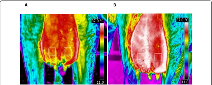
Infrared images
The output of the infrared camera was un-calibrated digitized images with pixel points. Pixel points were ana-lyzed with appropriate computer software (FLIR Quick-Report 1.2) to generate mean temperature of the skin surface. The mean temperature variation of four differ-ent areas between the groove of the udder and the hind leg and median suspensory ligament on caudal udder skin surface was examined. The areas were selected above the infected and control teats and centered according to the teats. The mean temperature of rectan-gles of 25 × 25 pixels drawn by the help of the camera software was measured (Figure 1). The two areas just above the teats showed the smallest variation in the temperature of the udder (Figure 1). An example of the
infrared images at 3 and 12 PIH is shown in Figure 1A and Figure 1B, respectively.
Statistical analysis
The main objective was to study the disease severity in relation with production of different inflammatory med-iators/indicators and clinical parameters. Disease severity was defined as the percentage decrease of QMP in unin-fected quarters at 48 PIH compared to QMP in the same quarters at -24 PIH [8]. To investigate the rela-tionship of various parameters analyzed in milk and blood and disease severity, a mixed model was used with cow as random effect, and time, disease severity and their interaction as fixed effects. To explore the relationship between disease severity and systemic and local inflammatory responses, the Kendall’s correlation coefficient between categorical parameters and disease severity was calculated. To study the kinetics for tem-perature of udder skin in infected and uninfected quar-ters, a mixed model was used, with temperature as response variable, cow as random effect, and PIH, udder point and their interaction as categorical fixed effects. To compare the measurements at different time points with time zero, the mixed model was used, with mea-sured parameters as response variable, cow as random effect and PIH as categorical fixed effect. For milk pro-duction, measurements at all times were compared with time -48. Significance level for multiple comparisons was adjusted by Dunnett’s procedure. A P value of <0.05 was considered significant. Values in the graphs shown are mean ± SEM.
B
A
Figure 1 Thermal images of the caudal part of the udder of a primiparous cow after challenge withE. coli strain P4, serotype O32: H37 (1 × 10⁴cfu) at post infusion hour (PIH) 3 (A) and PIH 12 (B) during early lactation. Two fixed 25 × 25 pixel areas above the teats used to extract the data at various time points following intramammary infection. Left quarters were infected at PIH 0 and right quarters were control quarters. There was a significant increase for udder skin temperature between PIH 0 and PIH 12 in both infected (P = 0.02) and control quarters (P = 0.009).
Results
Preinfection milk and blood constituents and disease severity
A relationship was found between preinfection SCC and disease severity (P = 0.002), with higher preinfection SCC corresponding to higher disease severity. We failed to detect a significant relationship between preinfection milk fat, protein, lactose, and milk production and dis-ease severity. No correlation was found between the severity of experimental coliform mastitis and preinfec-tion concentrapreinfec-tions of BHBA, glucose and NEFA. Disease severity and local and systemic inflammatory response
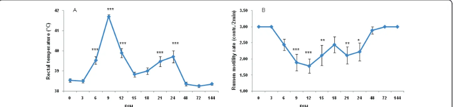
Following intramammary E. coli inoculation, rectal tem-perature (RT), skin turgor score and general attitude score rapidly increased to reach a maximum at PIH 9. There was a relapse of RT and RT increased up to 39.7°C again at PIH 24 (Figure 2A). There was no relationship between disease severity and RT changes. Rumen motility was reduced in all cows from PIH 3 onward (Figure 2B). The depression of reticulorumen motility was more pro-nounced at PIH 9, with 7 animals showing a slight decrease in motility (1 to 2×/2 min); one animal showed a complete absence of reticulorumen activity. Reticuloru-men motility was normal for all cows PIH 48 onward. A significant negative correlation was found between the rumen motility rate and disease severity (r = -0.56; P= 0.04).
Local clinical signs at the level of the infected mam-mary gland were also investigated. Cows became restless and showed symptoms of pain. The first changes in milk appearance (flakes, color, etc.) occurred at PIH 9, with two animals showing some flakes in milk. Two most severe responders showed more flakes and clots and slightly watery milk at PIH 48 and 72. Although milk appearance was normal for all moderate responders at PIH 144, watery milk with more flakes and clots was
observed for the most severely diseased cow at this time point. Slight swelling of the infected quarters occurred in 3 cows at PIH 3. Seven cows showed moderate ling of the infected quarters at PIH 9. Pronounced swel-ling disappeared gradually, and was already minimal (8 quarters with slight swelling) at PIH 48. Teat relaxation and milk leakage were only observed in a small number of cows.
Infrared thermography
The temperature of the udder skin on the infected quar-ter was significantly higher than that of the control quarter (P = 0.04). The mean udder temperature of infected quarters was increased from 3 PIH onward (Figure 3). However, skin temperature of the control quarters was increased from 9 PIH onward only. A peak of udder skin temperature in infected and control quar-ters occurred after the peak of rectal temperature (12 vs. 9 PIH, respectively). The temperature of udder skin at 12 PIH for both infected and uninfected quarters was significantly higher compared to 0 PIH (P = 0.02 and P= 0.009, respectively). There were no significant differ-ences between udder skin temperatures at other time points when compared to 0 PIH. Thereafter, the udder skin temperature returned to normal in both quarters. Disease severity and milk eicosanoids
In milk, the mean PGE2 concentration immediately after intramammary inoculation was 2572.9 ± 1155.6 pg/mL. Overall, there was a significant time effect for PGE2 (P < 0.01; Figure 4A). There was a relationship between disease severity and PGE2 concentration (P < 0.0001), with higher PGE2 concentrations corresponding to higher disease severity. The mean concentration of milk TXB2 immediately after infusion of E. coli was 1504.3 ± 1267.7 pg/mL. After infusion of E. coli, TXB2 changed significantly over time (P < 0.0001; Figure 4B). TXB2 started to increase from PIH 9 onward and returned to
Figure 2 Rectal temperature (A) and rumen motility rate (B) of 9 primiparous cows challenged intramammarily in 18 quarters (left quarters) with 1 × 10⁴cfu
E. coli strain P4, serotype O32:H37 at post infusion hour (PIH) 0 during early lactation. Means (± SEM) are shown. The clinical symptoms were scored according to Vangroenweghe et al. [5].* P < 0.05, ** P < 0.01, ***P < 0.001: significance of difference from the control value.
its normal level after PIH 24. Immediately after infusion of E. coli, the mean concentration of LTB4 was 2270.7 ± 776.5 pg/mL. There was no significant effect of time for concentration of LTB4 in milk (Figure 4C). In milk, the mean concentration of LXA4 immediately after infusion was 3513.9 ± 2108.1 pg/mL. The effect of time on the
concentration of LXA4 was significant (P < 0.0001; Figure 4D). LXA4 decreased gradually after intramam-mary infusion of E. coli and did not normalize before 144 PIH. We failed to detect any relationship between disease severity and concentrations of TXB2, LTB4 and LXA4.
Disease severity and indicators of inflammation in milk and blood
The infected and uninfected QMP of cows used in the current study is shown in Figure 5A and 5B, respec-tively. In the infected quarters, QMP decreased on d 0, the day of intramammary inoculation (Figure 5A). Infected QMP decreased the lowest level on d+1, rela-tive to day of challenge. On d+3, the recovery in the infected quarters was not complete compared to the preinfection.
The effect of time on the number of E. coli in inocu-lated quarters was significant (P < 0.0001). Overall, the number of E. coli increased to peak values of 4.96 log10
cfu/100 mL at PIH 6. No significant correlation was found between disease severity and the number of E. coli. SCC increased following the intramammary E. coli infection. There was a significant relationship
+ ++ ++ ++
Figure 3 Rectal temperature (lozenge) and udder skin temperature of the infected (square) and control quarter (triangle). Swelling of infected quarters: +mild, ++moderate. See legend of Figure 2 for more details.
Figure 4 Prostaglandin E2 (A), thromboxane B2 (B), leukotriene B4 (C) and lipoxin A4 (D) in secreta of all infused quarters at various times following intramammary infection. See legend of Figure 2 for more details.
between disease severity and SCC, with lower SCC values observed for more heavily diseased cows (P = 0.03).
Following the intramammary challenge, marked changes in number of blood neutrophils (P < 0.0001), basophils (P < 0.0001) and eosinophils (P = 0.005) hap-pened over time. The number of neutrophils started to decrease from PIH 6 onward, with nadir at PIH 9. The number of circulating neutrophils increased after PIH 9 and reached initial values by PIH 24. The number of cir-culating eosinophils decreased gradually after PIH 3 to reach its minimal level at PIH 24. There was a signifi-cant relationship between disease severity and the num-ber of basophils (P = 0.003), with more diseased cows having lower basophil levels. Following intramammary challenge, marked changes in the number of lympho-cytes and monolympho-cytes were observed (P < 0.0001). Although not significantly, lymphocytes showed a tem-porary increase at PIH 3 and decreased from PIH 6 onward, with nadir at PIH 12. The number of lympho-cytes gradually recovered from PIH 24 onward. Immedi-ately after intramammary inoculation of E. coli, the number of monocytes started to decrease and reached its lowest level at PIH 9. The number of monocytes increased from PIH 9 onward until the end of the study. There were significant differences in the numbers of white blood cells (WBC) over time (P < 0.0001). WBC decreased from 6 PIH onward, reached its nadir at PIH 12 and started to return to its normal level from PIH 24 onward. Kinetic of the WBC was not related signifi-cantly with disease severity.
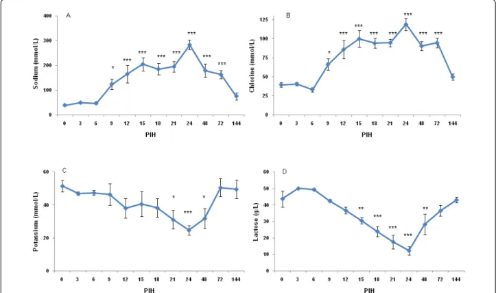
After intramammary inoculation of E. coli, marked changes in concentration of indicators for the disinte-gration of the blood-milk barrier (i.e. milk sodium, chloride, potassium and lactose) occurred (Figure 6A, B, C, D). Following the E. coli challenge, the lactose con-centration started to decrease after PIH 6, reaching nadir at PIH 24. Changes of chlorine and lactose were not related to disease severity. However, there was a
significant relationship between disease severity and potassium (P < 0.0001) and sodium (P = 0.03) levels, with more diseased cows having higher sodium and lower potassium in milk. Milk fat and protein was chan-ged over time following intramammary inoculation of E. coli(P < 0.0001).
The kinetics of plasma TNF-a and IL-1 did not differ significantly over time after the challenge. Following infusion of E. coli, a significant relationship between dis-ease severity and concentrations of TNF-a (P < 0.0001) and IL-1 (P < 0.0001) was found. More severely diseased cows had lower IL-1 but higher TNF-a levels. The con-centration of haptoglobin changed significantly over time (P < 0.0001). Serum haptoglobin in the infected quarters increased from PIH 15 onward, peaked at PIH 72 and returned to its initial levels at PIH 144. No sig-nificant relationship between disease severity and the concentration of haptoglobin was detected.
Discussion
As expected, similar to animals receiving the same dose (1 × 10⁴ cfu) of E. coli in previous experiments [5,15], quarter swelling, fever, depression of reticuloru-men motility, changed milk appearance and presence of pain in udder was observed following intramam-mary E. coli challenge. In line with results of Van-groenweghe et al. [5,11], the variation of clinical responses in primiparous cows was quite small in this study. The preinfection concentration of BHBA, NEFA and glucose indicated that the cows in the present study were not ketotic at the time of challenge [9]. In contrast to previous research [9,10], no correlation existed between the severity of experimental E. coli mastitis and preinfection concentrations of BHBA, glu-cose and NEFA. The clinical outcome of the disease in terms of severity was determined using a day-to-day method [8,10] (i.e. QMP loss in uninfected quarters) in the current study.
We quantified TXB2, PGE2, LXA4 and LTB4 in the milk of cows challenged intramammarily with E. coli and studied the relationship between these variables and severity of mastitis. The eicosanoids are derived from arachidonic acid and are important mediators and mod-ulators of inflammation. They are released locally during coliform and LPS mastitis [18-20]. We showed that milk PGE2 and TXB2 concentrations were increased after intramammary challenge of E. coli. In the present study, the concentration of PGE2 was linked to the severity of mastitis; however there was no relationship between severity of the disease and changes of TXB2, LXA4 and LTB4. A significant increase in PGE2 [20] and TXB2 [18-21] has been reported in milk after intramammary E. colichallenge. Although Vangroenweghe et al. [11] could not detect any significant difference in milk PGE2 and TXB2 concentrations between carprofen- and the saline-treated animals, the concentration of TXB2 and PGE2 remained lower in carprofen-treated animals with more moderate responses. The severity of E. coli masti-tis seems to be linked to the activity of specific key enzymes, i.e. COX that are involved in the synthesis of prostaglandins and thromboxanes. Schmitz et al. [22] demonstrated that the mRNA synthesis of COX-2 was increased in LPS-treated quarters and reached its
highest level 3 h after infusion of LPS. The activity of this enzyme in connection to severity of coliform masti-tis remains to be investigated in primiparous cows dur-ing early lactation.
In the present study, an attempt was made to gain further insights into the possible interrelationships between the various indicators of inflammation and severity of experimentally induced E. coli mastitis in pri-miparous cows during early lactation. The negative cor-relation found between rumen motility rate and severity of the disease could be due to elevated concentrations of TNF-a in the blood plasma of severely diseased cows. A positive relationship was found between depressed circulating basophils and severity of coliform mastitis. No significant association between the number of total white blood cells, neutrophils, eosinophils, lymphocytes and monocytes and severity might be explained by the limited number of animals in this study. The recruit-ment of neutrophils into the challenged quarters as reflected by SCC is an important aspect of the defense mechanism against E. coli/endotoxin mastitis. We found a negative relationship between SCC and severity of the disease. It is presumed that the lower SCC is associated with a relative slow influx of PMN and clearance of the bacteria from the glands of the most severe cases of
Figure 6 Sodium (A), chlorine (B), potassium (C) and lactose (D) in secreta of all infused quarters at various times following intramammary infection. See legend of Figure 2 for more details.
coliform mastitis. In contrast to previous research [10], the severity of coliform mastitis was not linked to the number of bacteria that grow in the gland cistern in the present study.
Our results showed that the thermal camera was suc-cessful in detecting the 2-3°C changes in temperature of the udder skin associated with clinical mastitis. Similarly, previous research demonstrated that a thermal camera is capable of detecting 1-1.5°C [13] and 2.3°C [23] tempera-ture changes of udder surface of cows intramammarily infused with endotoxin. In contrast with their results, the temperature of the udder skin of the experimental and control quarters was not increased simultaneously with rectal temperature and there was a delay in the peak of udder skin temperature in our study. The coincidence of increase in skin temperature with decrease in body tem-perature is probably due to peripheral cutaneous vasodi-lation during defervescence (abatement of fever). In fact, the generated heat during inflammation is transmitted to the skin surface through augmented capillary blood flow and dissipated as infrared energy [24]. As a result of increased permeability of capillaries, plasma leaked into the interstitium and maximal swelling occurred between PIH 9 and 15 (Figure 1B). In an earlier study [25], we demonstrated that after infusion of LPS, the increase in blood flow showed a biphasic aspect, both in goats and cows, with a return to normal values between the 2 peaks. It appears that the peak observed for udder skin temperature in the current study coincides with the sec-ond peak in the study of Dhsec-ondt et al. [25]. We found that the temperature of udder skin is increased both in experimental and control quarters which was in accor-dance with results of Hovinen et al. [13]. Scott et al. [23] demonstrated a clear rise in temperature of the infected quarters and a lower rise in the control quarter 1 h after inoculation of LPS. Increase of udder temperature in the infected and control quarters reflects a systemic effect of E. coli. The thermal camera showed increased udder skin surface temperature at PIH 12 which was later than the appearance of systemic and local signs at PIH 9. There-fore, infrared thermography was not a successful method in early detection of experimentally induced E. coli mastitis.
In conclusion, intramammary PGE2 production was related to bacterial growth and intensity of the inflam-matory reaction. This is in agreement with the general concepts of acute inflammation. Moreover, severity of E. coli mastitis in primiparous cows was linked to SCC, circulating basophils, concentration of plasma TNF-a and milk sodium and potassium concentrations which might be the result of underlying mechanisms which are responsible for individual variation in the susceptibility to E. coli mastitis. Infrared thermography was a success-ful technique to detect the changes in temperature of
the udder skin surface in both infected and control quarters after challenge with E. coli. However, this increase in udder skin surface temperature recorded by thermal images occurred after the appearance of local signs, indicating that the infrared thermography techni-que was not promising for early detection of E. coli mastitis. The increase in skin temperature coincided with the decrease in body temperature which is probably the result of the peripheral cutaneous vasodilation dur-ing defervescence.
Acknowledgements
This study was subsidized by grants of Ghent University (FWO grant # G.0050.06N), The University of Liège and Boehringer Ingelheim Animal Health GmbH (Ingelheim, Germany). We greatly acknowledge Prof. Max J Paape and Prof. Douglas D Bannerman (Bovine Functional Genomics Laboratory, USDA-ARS, Beltsville, MD, USA) for providing the antibodies and Prof. Alan Hill previously working at Compton (UK) for providing the E. coli strain. We wish to thank Dr Philippe Delahaut (Laboratoire d’Hormonologie, Centre d’Économie Rurale, Marloie, Belgium) for providing the facilities. We also thank Carolien Rogiers from Ghent University for her skilled technical assistance.
Author details
1Department of Comparative Physiology and Biometrics, Laboratory of
Genetics, Drug Quality and Registration Group, Ghent University, B-9000 Ghent, Belgium.2Departement Biotechnologie, plateforme Infectiologie
Expérimentale, Centre d’Economie Rurale, B-6900 Marloie, Belgium.
3Laboratory of Cellular and Molecular Physiology, GIGA-Research and Faculty
of Veterinary Medicine, University of Liège, B-4000 Liège, Belgium. Authors’ contributions
AP carried out the field study and drafted the manuscript. PS carried out the field study. HW participated in the sample analysis. FS provided the facilities in the experimental farm. MS participated in scientific discussions of the results. PB participated in the sample analysis and scientific discussions. LJP and BDS participated in design of the study and coordination. LD performed the statistical analysis. FB provided the facilities for sample analysis. CB designed the study and participated in the scientific discussions. All authors read and approved the final draft.
Competing interests
The authors declare that they have no competing interests. Received: 27 May 2010 Accepted: 20 September 2010 Published: 24 January 2011
References
1. Burvenich C, Van Merris V, Mehrzad J, Diez-Fraile A, Duchateau L: Severity ofE. coli mastitis is mainly determined by cow factors. Vet Res 2003, 34(05):521-564.
2. Sanchez-Carlo V, McDonald JS, Packer RA: Virulence factors ofEscherichia coli isolated from cows with acute mastitis. Am J Vet Res 1984, 45:1775-1777.
3. Oliver SP, King SH, Lewis MJ, Torre PM, Matthews KR, Dowlen HH: Efficacy of chlorhexidine as a postmilking teat disinfectant for the prevention of bovine mastitis during lactation. J Dairy Sci 1990, 73:2230-2235. 4. Pezeshki A, Capuco AV, De Spiegeleer B, Peelman L, Stevens M, Collier RJ,
Burvenich C: An integrated view on how the management of the dry period length of lactating cows could affect mammary biology and defence. J Anim Physiol Anim Nutr 2010, 94:e7-30.
5. Vangroenweghe F, Duchateau L, Burvenich C: Moderate inflammatory reaction during experimentalE. coli mastitis in primiparous cows. J Dairy Sci 2004, 87:886-895.
6. van Werven T, Noordhuizen-Stassen EN, Daemen AJJM, Schukken YH, Brand A, Burvenich C: Preinfection in vitro chemotaxis, phagocytosis, oxidative burst, and xpression of CD11/CD18 receptors and their
predictive capacity on the outcome of mastitis induced in dairy cows withEscherichia coli. J Dairy Sci 1997, 80:67-74.
7. Dosogne H, Burvenich C, van Werven T, Roets E, Noordhuizen-Stassen EN, Goddeeris B: Increased surface expression of CD11b receptors on PMN is not sufficient to sustain phagocytosis duringEscherichia coli mastitis in early postpartum dairy cows. Vet Immunol Immunopathol 1997, 60:47-59. 8. Heyneman R, Burvenich C, Vercauteren R: Interaction between the
respiratory burst activity of neutrophil leukocytes and experimentally inducedE. coli mastitis in cows. J Dairy Sci 1990, 73:985-994. 9. Kremer WDJ, Noordhuizen-Stassen EN, Grommers FJ, Schukken YH,
Heeringa R, Brand A: Severity of experimentalEscherichia coli mastitis in ketonemic and nonketonemic dairy cows. J Dairy Sci 1993, 76:3428-3436. 10. Vandeputte-Van Messom G, Burvenich C, Roets E, Massart-Leën A-M,
Heyneman R, Kremer WDJ, Brand A: Classification of newly calved cows into moderate and severe responders to experimentally induced Escherichia coli mastitis. J Dairy Res 1993, 60:19-29.
11. Vangroenweghe F, Duchateau L, Boutet P, Lekeux P, Rainard P, Paape MJ, Burvenich C: Effect of carprofen treatment following experimentally inducedEscherichia coli mastitis in primiparous cows. J Dairy Sci 2005, 88:2361-2376.
12. Levy BD, Clish CB, Schmidt B, Gronert K, Serhan CN: Lipid mediator class switching during acute inflammation: signals in resolution. Nat Immunol 2001, 2:612-619.
13. Hovinen M, Siivonen J, Taponen S, Hänninen L, Pastell M, Aisla A-M, Pyörälä S: Detection of clinical mastitis with the help of a thermal camera. J Dairy Sci 2008, 91:4592-4598.
14. Barth K: Basic investigations to evaluate a highly sensitive infrared-thermograph-technique to detect udder inflammation in cows. Milchwissenschaft 2000, 55:607-609.
15. Vangroenweghe F, Rainard P, Paape MJ, Duchateau L, Burvenich C: Increase ofEscherichia coli inoculum doses induce faster innate immune response in primiparous cows. J Dairy Sci 2004, 87:4132-4144. 16. Mehrzad J, Dosogne H, Meyer E, Heyneman R, Burvenich C: Respiratory
burst activity of blood and milk neutrophils in dairy cows during different stages of lactation. J Dairy Res 2001, 68:399-415.
17. Bannerman DD, Paape MJ, Lee J, Zhao X, Hope JC, Rainard P:Escherichia coli and Staphylococcus aureus elicit differential innate immune response following intramammary infection. Clin Diagn Lab Immunol 2004, 11:463-472.
18. Anderson KL, Kindahl H, Petroni A, Smith AR, Gustafsson BK: Arachidonic acid metabolites in milk of cows during acute mastitis. Am J Vet Res 1985, 46:1573-1577.
19. Burvenich C, Peeters G: Effect of prostaglandin synthetase inhibitors on mammary blood flow during experimentally induced mastitis in lactating goats. Arch Int Pharmacodyn Thér 1982, 258:128-137. 20. Peter AT, Clark PW, Van Roekel DE, Luker CW, Gaines JD, Bosu WT:
Temporal changes in metabolites of prostanoids in milk of heifers after intramammary infusion ofEscherichia coli organisms. Prostaglandins 1990, 39:451-457.
21. Anderson KL, Kindahl H, Smith AR, Davis LE, Gustafsson BK: Endotoxin-induced bovine mastitis: arachidonic acid metabolites in milk and plasma and effects of flunixin meglumine. Am J Vet Res 1986, 47:1373-1377.
22. Schmitz S, Pfaffl MW, Meyer HHD, Bruckmaier RM: Short-term changes of mRNA expression of various inflammatory factors and milk proteins in mammary tissue during LPS-induced mastitis. Domest Anim Endocrinol 2004, 26:111-126.
23. Scott SL, Schaefer AL, Tong AKW, Lacasse P: Use of infrared thermography for early detection of mastitis in dairy cows. Can J Anim Sci 2000, 80:764-765.
24. Kunc P, Knizkova I, Prikryl M, Maloun J: Infrared thermography as a tool to study the milking process: a review. Agricultura Tropica et Subtropica 2007, 40:29-32.
25. Dhondt G, Burvenich C, Peeters G: Mammary blood flow during experimentalEscherichia coli endotoxin induced mastitis in goats and cows. J Dairy Res 1977, 44:433-440.
doi:10.1186/1297-9716-42-15
Cite this article as: Pezeshki et al.: Variation of inflammatory dynamics and mediators in primiparous cows after intramammary challenge with Escherichia coli. Veterinary Research 2011 42:15.
Submit your next manuscript to BioMed Central and take full advantage of:
• Convenient online submission • Thorough peer review
• No space constraints or color figure charges • Immediate publication on acceptance
• Inclusion in PubMed, CAS, Scopus and Google Scholar • Research which is freely available for redistribution
Submit your manuscript at www.biomedcentral.com/submit