Vascular endothelial growth factor (VEGF) in
endometriosis
Jacques Donnez
1, Pierre Smoes, Ste´phane Gillerot,
Franc¸oise Casanas-Roux and Michelle Nisolle
Department of Gynaecology, St Luc’s University Hospital, Avenue Hippocrate, 10, B-1200, Brussels, Belgium1To whom correspondence should be addressed
Angiogenesis is likely to be involved in the pathogenesis of endometriosis. According to the transplantation theory, when the exfoliated endometrium is attached to the periton-eal layer, the establishment of a new blood supply is essential for the survival of the endometrial implant and development of endometriosis. From the known angiogenic factors, vascular endothelial growth factor (VEGF) has emerged as a pivotally important regulator of normal angiogenesis and pathological neovascularization. The VEGF protein was evaluated immunohistochemically in the eutopic endometrium of 10 women without endometriosis (group I) at laparoscopy and the eutopic endometrium and peritoneal endometriotic lesions of 43 women with endometriosis (group II). VEGF histological scores were 9.7 � 4.3 and 4.0 � 2.6 respectively in the epithelium and stroma of the eutopic endometrium of group I women, and 10.3 � 2.3 and 3.6 � 2.3 respectively in women of group II. In red lesions, the VEGF scores were 11.1 � 3.0 in the epithelium and 5.1 � 3.0 in the stroma, and in black lesions were 8.6 � 2.7 and 1.6 � 1.6, respectively. Signific-antly lower values were observed in black lesions as compared with eutopic endometrium and red lesions, the values of which were similar. Scores were also evaluated according to the phase of the cycle. In eutopic as well as ectopic endometrium, no significant cyclic variations were observed throughout the cycle. However, VEGF content was found to be higher in the eutopic glandular epithelium of women with endometriosis during the late secretory phase, possibly suggesting a more likely tendency to implant. In contrast, significantly higher VEGF content was noted in red lesions as compared with black lesions. During all phases of the cycle, the VEGF content in stromal cells of red lesions was higher than in black lesions. Similarities in VEGF content were observed in the glandu-lar epithelium of the eutopic endometrium of women with endometriosis and red lesions, suggesting that endo-metriosis probably arises from the peritoneal seeding of viable endometrial cells during retrograde menstruation and that red lesions can be considered as the first stage of implantation. After the attachment phase, the high VEGF levels could provoke an increase in the subperitoneal vascular network and facilitate implantation and viability in the retroperitoneal space. Lower VEGF levels in black
lesions explain the decrease in both stromal vascularization, followed by fibrosis and inactivation of the implant.
Key words: black endometriotic lesions/eutopic endometrium/
red endometriotic lesions/VEGF
Introduction
According to the transplantation theory, when exfoliated endo-metrium is attached to the peritoneal layer, the establishment of a new blood supply is essential for the survival of the endometrial implant and the development of endometriosis (Sampson, 1927; Nisolle and Donnez, 1996).
Angiogenesis is a fundamental process by which new blood vessels are formed. The highly regulated angiogenesis that occurs within the female reproductive tract is critical for normal reproduction, including follicular maturation, selection and normal function of the corpus luteum, and endometrial growth and remodelling (Folkman and Klagsbrun, 1987; Gordon et al., 1995). Unregulated angiogenesis is involved in non-neoplastic diseases such as diabetic blindness and rheumatoid arthritis.
Nowadays, several angiogenic factors have been identified, including acidic and basic fibroblast growth factors (FGF-α, FGF-β), platelet-derived endothelial cell growth factor (PD-ECGF), transforming growth factors-α and -β (α, TGF-β), tumour necrosis factor-α (TNF-α) and vascular endothelial growth factor (VEGF) (Folkman and Shing, 1992).
VEGF, also known as vascular permeability factor, is a heparin-binding glycoprotein with potent angiogenic, endothel-ial cell-specific mitogenic and vascular permeability activities. It has been suggested that VEGF is an important angiogenic factor in many physiological and pathological conditions and has recently been incriminated in the pathogenesis of capillary leakage in ovarian hyperstimulation syndrome (Abramov et al., 1997). The presence of VEGF has also been demonstrated in human endometrium and it may be important in both physiological and pathological angiogenesis (Charnock-Jones
et al., 1993; Smith, 1996).
Several authors have demonstrated higher peritoneal concen-trations of VEGF in women with moderate to severe endo-metriosis than in women without the disease (McLaren et al., 1996b; Shifren et al., 1996). Nevertheless, the presence of VEGF in endometriotic tissue is less detailed in the literature and there is a discrepancy between the conclusions of these studies. Indeed, although McLaren et al. (1996b) found that VEGF expression was limited in endometriotic tissue and only seen in individual tissue macrophages distributed throughout
the stroma, Shifren et al. (1996) demonstrated similar VEGF expression in both endometriosis and eutopic endometrium.
The aim of this study was to compare the VEGF content in eutopic endometrium and black and red peritoneal lesions throughout the menstrual cycle in order to clarify the role of VEGF in the pathogenesis of endometriosis.
Materials and methods
In this study, 10 women without endometriosis at laparoscopy (group I) and 43 women with laparoscopy-proven endometriosis (group II) were evaluated. Among group II women, peritoneal biopsies of 3–5 mm in size were taken from areas with endometriotic lesions using a biopsy punch forceps (26175 DN; Storz, Tuttligen, Germany). Biopsies of both typical black lesions (n � 29) and red flame-like lesions (n � 24) were taken. All patients had regular (28–30 days) ovulatory cycles, and none was given hormonal therapy for at least 3 months before surgery. The mean age was 31.5 � 5.0 years. In all patients, accurate menstrual dating could be carried out according to the last and next menstrual period and basal body temperature, and corroborated with appropriate histological dating of endometrial biopsies according to the criteria of Noyes et al. (1950). The endometrial biopsies (n � 43) were classified as follows: proliferative phase (PP) (days 4–14; n � 16); early secretory phase (ESP) (days 15–19; n � 13); mid to late secretory phase (LSP) (days 20–28; n � 14).
Biopsies from patients in group I were also classified according to the same criteria (Noyes et al., 1950).
Measurement of VEGF staining on tissue was based on immunolab-elling with a polyclonal antibody, which recognizes the 165, 189 and 121 amino acid splice variants of VEGF of human origin, that are located in the cytoplasm of endometrial cells, myometrium, macrophages of the peritoneal fluid, vascular smooth muscle and endothelium and various cell lines (Tischer et al., 1991; Charnock-Jones et al., 1993; McLaren et al., 1996a; Smith, 1996).
VEGF labelling was performed by immunoperoxidase techniques using the peroxidase–antiperoxidase (PAP) complex which increases the reliability and sensitivity of detection. Tissue samples were fixed in 4% formaldehyde and embedded in paraffin. Thick tissue sections (6 µm) were mounted on Superfrost Plus slides (Menzel-Gla¨zer, Germany) and stained using the following technique. After rehydration of the sections and inhibition of endogenous peroxidases in 0.3% hydrogen peroxide solution, retrieval of VEGF antigens was achieved by the microwave technique in citrate buffer. Non-specific reactivities were inhibited by a 30 min incubation in a solution of 10% normal goat serum (NGS; Pan Systems, NTL, Brussels, Belgium) and 1% bovine serum albumin (BSA; Sigma, Bornem, Belgium). The sections were then incubated in 1/100 dilution of polyclonal rabbit anti-VEGF primary IgG antibody [anti-VEGF (a-20); cat# sc-152, Santa Cruz Biotechnology, Santa Cruz, California, USA) in 1% NGS/0.1% BSA solution overnight at 4°C. The tissue was then incubated with 1% goat anti-rabbit second antibody (code Z0421, Dako A/S, Copenhagen, Denmark) followed by the PAP complex (code Z0113, Dako), and then diaminobenzidine (DAB; S3000, Dako) and hydrogen peroxide until a stable non-diffusable brown precipitate product was detectable. Slides were lightly counterstained in hemalum. In each case, negative controls were prepared which consisted of one section incubated without anti-VEGF antibody.
Fluorescence double-staining for VEGF-positive macrophage detec-tion was carried out on some tissue secdetec-tions. After the VEGF had been identified, slides were incubated for 30 min in a 10% NGS/1% BSA solution and then overnight at 4°C with the monoclonal mouse
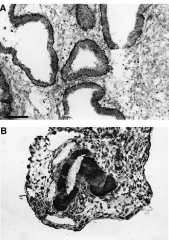
Figure 1. VEGF immunostaining. (A) In eutopic endometrium
during the early secretory phase. Bar � 360 µm. (B) In a red peritoneal lesion during the early secretory phase.
anti-human macrophage, CD68 antibody (dilution 1/50; clone KP1, Dako). The slides were then incubated for 30 min in the dark with a 1/20 dilution of FITC-conjugated rabbit anti-mouse Ig (Dako), washed and mounted in aqueous mounting medium.
All sections were examined on a blind basis using a Leitz Orthoplan light microscope (Leitz, Wetzlar, Germany) with the 40� objective. A semi-quantitative analysis was obtained by determination of the distribution and the intensity of the staining within the glandular epithelium and the stroma. The VEGF histological score (H) was calculated as follows: H � Σ Pi, where i is the intensity from 0 (negative cells) to 3 (high staining intensity); and P is the percentage of stained cells for each given i, where P values of 1, 2, 3, 4 and 5 indicate �15%, 15–50%, 50–85%, �85% and 100% positive-staining cells, respectively.
Bivariate analysis of variance was used for statistical evaluation.
Results
Biopsies taken from typical black and red peritoneal lesions showed the presence of endometrial elements (glands and stroma) in all cases. VEGF immunoreactivity was detected in both the glandular epithelium and stroma of the endometrium and peritoneal endometriotic lesions. Staining was homogen-eous in epithelial cells but heterogenhomogen-eous in stromal cells (Figure 1).
Tables I and II represent the VEGF histological (H)-scores found in the glandular and stromal cells of eutopic and ectopic endometrium according to the phase of the menstrual cycle.
Table I. VEGF histological score in the glandular cells of eutopic and ectopic endometrium according to the
phase of the menstrual cycle
Patient Over whole Proliferative Early secretory Late secretory
cycle phase phase phase
Women without endometriosis (group I; n � 10)
Eutopic endometrium 9.7 � 4.3 10.3 � 0.6 10.7 � 3.0 6.0 � 6.6
(n � 10) (n � 3) (n � 4) (n � 3)
Women with endometriosis (group II; n � 43) Eutopic endometrium 10.3 � 2.3 9.9 � 2.6 10.6 � 2.2a 10.1 � 1.9b (n � 43) (n � 16) (n � 13) (n � 14) Black lesions 8.6 � 2.7 8.8 � 1.2 8.4 � 2.7 8.8 � 3.6 (n � 29) (n � 8) (n � 12) (n � 9) Red lesions 11.1 � 3.0 9.8 � 4.0 12.0 � 2.7a 11.3 � 2.7b (n � 22) (n � 6) (n � 6) (n � 10)
Values are means � SD.
aSignificantly different from the values observed in black lesions (P � 0.04).
bSignificantly different from the values observed in the eutopic endometrium of group I (P � 0.04).
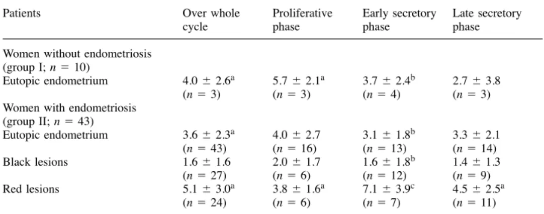
Table II. VEGF histological score in the stromal cells of eutopic and ectopic endometrium according to the
phase of the menstrual cycle
Patients Over whole Proliferative Early secretory Late secretory
cycle phase phase phase
Women without endometriosis (group I; n � 10)
Eutopic endometrium 4.0 � 2.6a 5.7 � 2.1a 3.7 � 2.4b 2.7 � 3.8
(n � 3) (n � 3) (n � 4) (n � 3)
Women with endometriosis (group II; n � 43) Eutopic endometrium 3.6 � 2.3a 4.0 � 2.7 3.1 � 1.8b 3.3 � 2.1 (n � 43) (n � 16) (n � 13) (n � 14) Black lesions 1.6 � 1.6 2.0 � 1.7 1.6 � 1.8b 1.4 � 1.3 (n � 27) (n � 6) (n � 12) (n � 9) Red lesions 5.1 � 3.0a 3.8 � 1.6a 7.1 � 3.9c 4.5 � 2.5a (n � 24) (n � 6) (n � 7) (n � 11)
Values are means � SD.
aSignificantly different from the values observed in black lesions (P � 0.03). bSignificantly different from the values observed in red lesions (P � 0.03). cSignificantly different from the proliferative phase (P � 0.05).
Group I (women without endometriosis)
The glandular VEGF H-score of eutopic endometrium was 9.7 � 4.3 and similar during the different phases of the menstrual cycle (PP, 10.3 � 0.6; ESP, 10.7 � 3.0; LSP, 6.0 � 6.6). In the stroma, the VEGF H-score was 4.0 � 2.6 and found to be lower during the late secretory phase when compared with the proliferative and early secretory phases, though the difference was not significant.
Group II (women with endometriosis)
In the eutopic endometrium, the glandular VEGF H-score was 10.3 � 2.3 and similar during the different phases of the menstrual cycle (PP, 9.9 � 2.6; ESP, 10.6 � 2.2; LSP, 10.1 � 1.9). In the stroma, the VEGF H-score was 3.6 � 2.3, there being no significant cyclic variations in score throughout the menstrual cycle (PP, 4.0 � 2.7; ESP, 3.1 � 1.8; LSP, 3.3 � 2.1).
In black peritoneal lesions, the H-score was 8.6 � 2.7 in the epithelium and 1.6 � 1.6 in the stroma. No significant cyclic variations in VEGF H-score occurred in the glandular
epithelium (PP, 8.8 � 1.2; ESP, 8.4 � 2.7; LSP, 8.8 � 3.6) or in the stroma (PP, 2.0 � 1.7; ESP, 1.6 � 1.8; LSP, 1.4 � 1.3). In red peritoneal lesions, the glandular VEGF H-score was 11.1 � 3.0 and there were no significant cyclic variations in the glandular epithelium (PP, 9.8 � 4.0; ESP, 12.0 � 2.7; LSP, 11.3 � 2.7). In the stroma, the VEGF H-score was 5.1 � 3.0, significantly higher (P � 0.03) than that observed in black lesions (1.6 � 1.6). A significant (P � 0.05) increase was observed during the early secretory phase when compared with the proliferative phase (H-scores of 7.1 � 3.9 and 3.8 � 1.6, respectively).
Comparative VEGF expression of eutopic endometrium in patients with and without endometriosis
During the proliferative and early secretory phases, the glandu-lar VEGF H-scores in eutopic endometrium were simiglandu-lar in both groups. However, during the LSP, the VEGF H-score was significantly higher in the eutopic endometrium of endo-metriosis patients than in control eutopic endometrium (10.1 � 1.9 and 6.0 � 6.6, respectively; P � 0.04).
between the VEGF H-scores found in the eutopic endometrium of the two groups of patients throughout the menstrual cycle.
Comparative VEGF expression of black and red peritoneal lesions
In the glandular epithelium, the VEGF H-score was signific-antly (P � 0.004) higher in red than in black peritoneal lesions during the ESP (12.0 � 2.7 and 8.4 � 2.7, respectively). During the LSP, the VEGF H-scores in red lesions (11.3 � 2.7) and in the eutopic endometrium (10.1 � 1.9) of women with endometriosis (group II) were significantly (P � 0.04) higher than in the eutopic endometrium of women without endo-metriosis (6.0 � 6.6) (group I).
In the stroma, the VEGF H-score was significantly (P � 0.03) higher in red lesions during the LSP than in black lesions (4.5 � 2.5 and 1.4 � 1.3, respectively). Macrophages were present and they were VEGF- and CD68-positive; however, the presence of VEGF-positive and CD68-negative cells in our study proved the immunoreactivity of the stromal cells in the endometrium as well as in the peritoneal endometriotic lesions.
Discussion
Although the aetiology of endometriosis is unknown, it is generally accepted that the condition is a result of the implanta-tion of exfoliated endometrium, deposited in the peritoneal cavity following retrograde menstruation (Sampson, 1927). When the exfoliated endometrium enters the peritoneal cavity and becomes attached to the mesothelial layer through attach-ment proteins like the cadherins, a process of angiogenesis is essential for further implantation and the development of peritoneal endometriosis (Nisolle and Donnez, 1996). Angio-genesis is dependent on soluble factors released from cells (Folkman and Klagsbrun, 1987; Gordon et al., 1995; Smith, 1996). Several peptide growth factors, including α, FGF-β, PD-ECGF and VEGF, stimulate vascular endothelial cell growth in vitro and angiogenesis in vivo (Gordon et al., 1995). VEGF is a member of a family of heparin-binding proteins that acts directly on endothelial cells to induce proliferation and angiogenesis (Folkman and Klagsbrun, 1987; Folkman and Shing, 1992; Gordon et al., 1995) and was found to be the only growth factor which stimulated the growth of human decidual endothelial cells maintained in culture; thus, VEGF was considered essential in uterine angiogenesis (Grimwood
et al., 1995). Several sources of VEGF have been suggested,
including peritoneal macrophages, the number and activation/ secretory activity of which are increased in uterine endomet-rium and endometriotic tissue (Gordon et al., 1995; Smith, 1996; Smith et al., 1996).
In the human endometrium, Smith (1996) and Shifren et al. (1996) described cyclic changes in the distribution of VEGF and mRNA expression throughout the cycle and suggested that VEGF expression is under steroid control, being up-regulated in response to oestradiol (Greb et al., 1997). In contrast, McLaren et al. (1996a) observed low expression of VEGF, mostly seen in macrophages distributed throughout the stroma of endometriotic lesions.
In the present study, the VEGF content in the glandular
epithelium and stroma of eutopic endometrium and endometri-otic lesions was compared immunohistochemically, and thus permitted a clear distinction between black and red peritoneal lesions (Donnez et al., 1996; Nisolle and Donnez, 1997).
In the group of patients without endometriosis, there were no significant variations in VEGF content in the eutopic glandular epithelium or stroma throughout the menstrual cycle. However, differences were noted between the eutopic endomet-rium of women with and without endometriosis. Indeed, in the present study, VEGF content was significantly higher in the eutopic glandular epithelium of endometriosis patients during the late secretory phase, suggesting that the endomet-rium of women with endometriosis is more likely to implant than that of women without endometriosis.
During the luteal phase, significantly higher levels of VEGF immunostaining were observed in both the glandular epithelium and stroma of red peritoneal endometriotic lesions as compared with black lesions, suggesting an active angiogenic process in red lesions, like that in eutopic endometrium. In contrast, black lesions were characterized by a poor angiogenesis, a fact which confirms the differences in the stromal vascularization index between red and black lesions observed in a previous study (Nisolle et al., 1993). The similar VEGF content found in red lesions and eutopic glandular epithelium is another argument in favour of the transplantation theory. Red lesions must therefore be considered as the first stage of endometriosis and as freshly implanted lesions.
Our results are not in accordance with those of McLaren
et al. (1996a) who reported that, in ectopic endometrium,
VEGF immunoreactivity was localized mainly on isolated cells within the stroma and only weak staining was present on the glandular epithelium. Moreover, using double immunofluores-cence staining with the macrophage marker CD14 and VEGF, they also demonstrated that individual VEGF-positive cells within the stroma were macrophages. However, in their study, only eight endometriotic lesions were analysed and the macro-scopic appearance was not mentioned. It is possible that only black lesions were analysed. By contrast, our study clearly demonstrated that in red lesions, VEGF-positive cells included not only macrophages, but also numerous stromal cells.
The fact that red lesions are characterized by high concentra-tions of VEGF and the presence of matrix metalloproteinases (MMP-1) throughout the cycle (Kokorine et al., 1997) could be the key point of the transplantation theory. Indeed, under the influence of MMP-1, partial shedding of red lesions and their implantation elsewhere in the peritoneal cavity under the influence of VEGF could explain the development of peritoneal endometriosis.
On the basis of our data, we suggest that the eutopic endometrium itself plays a crucial role in the histogenesis of endometriosis. Indeed, during retrograde menstruation, endo-metrial implants with high VEGF-expressing glandular cells enter the peritoneal cavity. This high VEGF content found in the glandular cells of menstruating endometrium was also noted by Smith (1996). That the attachment phase is influenced by the presence of attachment proteins and MMP-1 has been confirmed in two of our other studies (Beliard et al., 1997; Kokorine et al., 1997). Indeed, we observed a correlation
Figure 2. Hypothesis of the histogenesis of peritoneal
endometriosis: the role of eutopic endometrium.
between MMP-1 expression and the activity of endometriotic tissue, suggesting its involvement in tissue remodelling and bleeding, and possibly also in the secondary shedding and reimplantation of endometriotic lesions. In tumour angiogen-esis, the tumour cells release angiogenic molecules such as VEGF but they may not be the sole source of angiogenic molecules. Tumours may recruit macrophages and then activate them to secrete angiogenic activity—the same mechanism as can be suggested in endometriosis. After the attachment phase, high VEGF concentrations could provoke an increase in the subperitoneal vascular network and facilitate implantation and viability. Moreover, VEGF, in addition to being angiogenic, causes increased permeability of the capillary bed and, in similar fashion to the mechanisms involved in pathologic angiogenesis, this could suggest that in freshly implanted endometriotic lesions, the higher expression of VEGF might explain a higher permeability of the capillary bed (Folkman and Shing, 1992). This may lead to a leakage of fibrin products into the extracellular space which will increase the recruitment of macrophages, the angiogenic activity of which is increased by the secretion of TNF-α, that is known to be secreted by macrophages when these cells are activated by large molecules such as bacterial endotoxins or fibrin products (Figure 2).
It could be hypothesized that active red endometriosis undergoing cycling and with high concentrations of VEGF may help the implant to revascularize and proliferate and this, to some extent, explains the high concentrations of VEGF in peritoneal fluid observed in several studies (Shifren et al., 1996; McLaren et al., 1996b).
In conclusion, angiogenesis is considered as an important step in the implantation and development process of menstrual endometrial fragments entering the peritoneal cavity. The high content of VEGF, as demonstrated in our study, has led to the hypothesis that VEGF-induced angiogenesis may be a critical aspect of the pathophysiology of this disease and suggests, as Smith (1996) already has, that anti-angiogenic therapies could be considered as a new clinical approach to this disease. A recent animal study demonstrated the anti-angiogenic effects of mifepristone via suppression of VEGF production (Greb
et al., 1997) and there is now a need to initiate prospective
clinical studies to support this hypothesis. References
Abramov, Y., Barak, V., Nisman, B. and Schenker, J.G. (1997) Vascular endothelial growth factor plasma levels correlate to the clinical picture in severe ovarian hyperstimulation syndrome. Fertil. Steril., 67, 261–265. Beliard, A., Donnez, J., Nisolle, M. and Foidart, J.M. (1997) Localization
of laminin, fibronectin, E-cadherin, and integrins in endometrium and endometriosis. Fertil. Steril., 67, 266–271.
Charnock-Jones, D., Sharkey, A., Raffut-Williams, J. et al. (1993) Identification and localization of alternately spliced mRNAs for vascular endothelial growth factor in human uterus and estrogen regulation in endometrial carcinoma cell lines. Biol. Reprod., 48, 1120–1128.
Donnez, J., Nisolle, M., Smoes, P. et al. (1996) Peritoneal endometriosis and ‘endometriotic’ nodule of the rectovaginal septum are two different entities. Fertil. Steril., 66, 362–368.
Folkman, J. and Klagsbrun, M. (1987) A family of angiogenic peptides. Nature, 329, 671–672.
Folkman, J. and Shing, Y. (1992) Angiogenesis. J. Biol. Chem., 267, 10931–10934.
Gordon, J., Shifren, J., Foulk, R. et al. (1995) Angiogenesis in the human female reproductive tract. Obstet. Gynecol. Surv., 50, 688–697.
Greb, R.R., Heikinheimo, O., Williams, R.F. et al. (1997) Vascular endothelial growth factor in primate endometrium is regulated by oestrogen-receptor and progesterone-receptor ligands in vivo. Hum. Reprod., 12, 1280–1292. Grimwood, J., Bicknell, R. and Rees, M.C.P. (1995) The isolation,
characterization and culture of human decidual endothelium. Hum. Reprod.,
10, 2142–2148.
Kokorine, I., Nisolle, M., Donnez, J. et al. (1997) Expression of interstitial collagenase (matrix metalloproteinase-1) is related to the activity of human endometriotic lesions. Fertil. Steril., 68, 246–251.
McLaren, J., Prentice, A., Charnock-Jones, D.S. et al. (1996a) Vascular endothelial growth factor is produced by peritoneal fluid macrophages in endometriosis and is regulated by ovarian steroids. J. Clin. Invest., 98, 482–489.
McLaren, J., Prentice, A., Charnock-Jones, D.S. and Smith, S.K. (1996b) Vascular endothelial growth factor (VEGF) concentrations are elevated in peritoneal fluid of women with endometriosis. Hum. Reprod., 11, 220–223. Nisolle, M. and Donnez, J. (eds) (1996) Peritoneal, Ovarian and Recto-vaginal Endometriosis. The Identification of Three Separate Diseases. Parthenon Publishing, Carnforth.
Nisolle, M. and Donnez, J. (1997) Peritoneal, ovarian endometriosis and adenomyotic nodule of the recto-vaginal septum are three different entities. Fertil. Steril., 68, 585–596..
Nisolle, M., Casanas-Roux, F., Anaf, V. et al. (1993) Morphometric study of the stromal vascularization in peritoneal endometriosis. Fertil. Steril., 59, 681–684.
Noyes, R.W., Hertig, A.T. and Rock, J. (1950) Dating the endometrial biopsy. Fertil. Steril., 1, 3–25.
Sampson, J.A. (1927) Peritoneal endometriosis due to menstrual dissemination of endometrial tissue into the pelvic cavity. Am. J. Obstet. Gynecol., 14, 422–469.
Shifren, J.L., Tseng, J.F., Zaloudek, C.J. et al. (1996) Ovarian steroid regulation of vascular endothelial growth factor in the human endometrium: implications for angiogenesis during the menstrual cycle and in the pathogenesis of endometriosis. J. Clin. Endocrinol. Metab., 81, 3112–3118. Smith, S.K. (1996) Vascular endothelial growth factor and the endometrium.
Hum. Reprod., 11, 56–61.
Smith, S.K., McLaren, J., Sharkey, A. and Charnock-Jones, D.S. (1996) Angiogenesis in endometriosis. Proceedings, Vth World Congress on Endometriosis, Yokohama, Japan, p. 6.
Tischer, E., Mitchell, R., Hartman, T. et al. (1991) The human gene for vascular endothelium growth factor. J. Biol. Chem., 266, 11947–11954. Received on November 10, 1997; accepted on March 26, 1998