HAL Id: inserm-01691531
https://www.hal.inserm.fr/inserm-01691531
Submitted on 24 Jan 2018HAL is a multi-disciplinary open access archive for the deposit and dissemination of sci-entific research documents, whether they are pub-lished or not. The documents may come from teaching and research institutions in France or abroad, or from public or private research centers.
L’archive ouverte pluridisciplinaire HAL, est destinée au dépôt et à la diffusion de documents scientifiques de niveau recherche, publiés ou non, émanant des établissements d’enseignement et de recherche français ou étrangers, des laboratoires publics ou privés.
MicroRNA-based Drugs for Brain Tumours
Shubaash Anthiya, Audrey Griveau, Claire Loussouarn, Patrick Baril, Martin
Garnett, Jean-Paul Issartel, Emmanuel Garcion
To cite this version:
Shubaash Anthiya, Audrey Griveau, Claire Loussouarn, Patrick Baril, Martin Garnett, et al.. MicroRNA-based Drugs for Brain Tumours. Trends in Cancer, Cell Press, 2018, 4 (3), pp.222-238. �10.1016/j.trecan.2017.12.008�. �inserm-01691531�
Trends in Cancer
MicroRNA-based Drugs for Brain Tumours
--ManuscriptDraft--Manuscript Number: TRECAN-D-17-00183R1
Article Type: Review
Corresponding Author: Emmanuel Garcion FRANCE
First Author: Shubaash Anthiya, PhD
Order of Authors: Shubaash Anthiya, PhD
Audrey Griveau, PhD Claire Loussouarn, PhD Patrick Baril, PhD Martin Garnett, PhD Jean-Paul Issartel, PhD Emmanuel Garcion, PhD, HDR
Abstract: MicroRNAs (miRNAs) are key regulatory elements coded by the genome. A single miRNA can downregulate expression of multiple genes involved in diverse functions. As cancer is a disease with multiple gene aberrations, developing novel approaches to identify and modulate miRNA-pathways may result in a breakthrough for cancer treatment. With a special focus on glioblastoma, this review provides an up-to-date understanding of miRNA biogenesis, role of miRNA in cancer resistance, essential tools for modulating miRNA expression, emerging list of clinically promising RNAi delivery systems and how they can be adapted for therapy.
1
MicroRNA-based Drugs for Brain Tumours
1
Shubaash ANTHIYA1,2*, Audrey GRIVEAU1*, Claire LOUSSOUARN1, Patrick BARIL3,
2
Martin GARNETT2, Jean-Paul ISSARTEL4 and Emmanuel GARCION1,
3 4
1CRCINA, INSERM, Université de Nantes, Université d'Angers, Angers, France.
5
2School of Pharmacy, University Park, University of Nottingham, Nottingham NG7 2RD, U.K.
6
3Centre de Biophysique Moléculaire, CNRS UPR4301, Université d’Orléans, 45071, Orléans,
7
France. 8
4INSERM U1216, Grenoble Institut des Neurosciences, Université Joseph Fourier, CHU
9
Michallon, 38043, Grenoble, France. 10
11
*These authors contributed equally to this work.
12 13 14 15 Corresponding author: 16 Dr Emmanuel Garcion 17 [email protected] 18
CRCINA, INSERM U1232 Team 17, 19
Design and Application of Innovative Local treatments in Glioblastoma (GLIAD), 20 Université d’Angers, 21 IBS - CHU, 22 4 Rue Larrey, 23 F-49933 Angers, France 24 +33 (0) 2 44 68 85 43 25
Manuscript revised free of annotation Click here to download Manuscript Garcion Manuscript-Final revised free of annotations.docx
2
Abstract (91/120 words)
26
MicroRNAs (miRNAs) are key regulatory elements coded by the genome. A single miRNA can 27
downregulate expression of multiple genes involved in diverse functions. As cancer is a disease 28
with multiple gene aberrations, developing novel approaches to identify and modulate miRNA-29
pathways may result in a breakthrough for cancer treatment. With a special focus on 30
glioblastoma, this review provides an up-to-date understanding of miRNA biogenesis, role of 31
miRNA in cancer resistance, essential tools for modulating miRNA expression, emerging list of 32
clinically promising RNAi delivery systems and how they can be adapted for therapy. 33
34
Keywords: glioblastoma, cancer resistance, RNAi, drug delivery, brain targeting, argonaute,
35
exosomes, nanoparticles 36
37 38
3
MicroRNA therapeutics for Glioblastoma
39 40
Glioblastomas (GBM) are aggressive grade IV primary tumours of the central nervous system [1, 41
2]. Standard therapy consists of maximal surgical resection followed by external radiotherapy 42
and chemotherapy with temozolomide that confers a median survival of about 15 months [2]. 43
Tumour-treating fields (TTFields) is the latest improvement in GBM treatment which uses a 44
non-invasive external device (Optune® transducer-array) to create a low intensity electric field 45
of intermediate frequency (~200kHz) which interferes with mitosis, hinders cell division and 46
consequently induces cell death in dividing cells [3]. The combination of TTFields plus 47
temozolomide after the standard chemoradiotherapy significantly increased the overall median 48
survival to 20.5 months, with better quality of life [3]. Other treatment options for GBM include 49
chemotherapy other than temozolomide, immunotherapy and therapies targeting specific 50
oncogenic pathways, for review see Seystahl et al [4]. All currently available treatments are 51
palliative, not curative [2, 3]. Therapeutic failure of GBM can be attributed to the sub-optimal 52
delivery of drugs and aberrant expression of multiple genes resulting in tumour heterogeneity, 53
aggressive infiltrative behaviour and treatment resistance of GBM cells. It is important to 54
identify multiple drug targets for developing effective therapies. 55
MicroRNAs (miRNAs or miRs) are natural RNA interference (RNAi) molecules produced by 56
the cells (for Biogenesis and Functions of miRNAs, see Box-1 and Figure-1). MiRNAs are 57
involved in GBM pathophysiology, tumor plasticity, and resistance to therapy, see Figure 2 [5-58
7]. Differential expression of miRNAs is observed in the tumour mass versus control brain tissue 59
between glioma subtypes [8, 9], and even between different cell subpopulations of the same 60
tumour [7]. These differences in miRNA expression can be used as diagnostic and prognostic 61
4 biomarkers [7, 10]. The major therapeutic advantage of miRNAs arises from the fact that a single 62
miRNA can target multiple genes involved in distinct cellular functions. For example, increasing 63
the expression of a tumour-suppressor miRNA (ts-miRNA) like 128-3p [11-17] or miR-64
145-5p [18-23] in GBM can block cell proliferation, self-renewal, invasion, metastasis, 65
angiogenesis and drug resistance by selectively down-regulating the expression of multiple 66
genes, Figure-2. Similarly, repeated systemic treatment with miR-138 blocks multiple key 67
immune-checkpoints proteins in cells (CTLA-4, PD-1 and FoxP3), resulting in significant T-68
cell mediated tumour regression and increased survival in orthotropic brain-tumour model 69
expressing PD-L1 ligand [24]. Conversely, inhibiting the function or blocking the expression of 70
an oncogenic miRNA (oncomiR) in cancer cells can reactivate multiple tumour-suppressor genes 71
leading to tumour regression (e.g. miR-21-3p [25] or miR-21-5p [26]) or even to tumour 72
eradication, as in the case of miR-10b [27]. These examples illustrate that the targeting of 73
miRNAs in GBM either by inhibition, replacement or modulation of their activity is a promising 74
therapeutic approach [28, 29], Figure-2. Shea et al reviewed an extensive list of GBM relevant 75
miRNAs [30]. 76
The miRNA database (miRBase, version 21) enlists 1881 precursors and 2588 mature human 77
miRNAs [31], of which several of them are identified preferentially as tumour-suppressor 78
miRNA (ts-miRs), as oncomiRs, or as both, depending on the cell type and function of miRNAs 79
in those cells [19, 32]. For example, Fareh et al showed forced expression of miR-302/367 80
cluster in GBM cells resulted in repression of self-renewal properties and in vivo inhibition of 81
tumour development [33]. Conversely, Guo et al reported that self-renewal and tumour-82
promoting properties were enhanced by the expression of miR-302/367 cluster in prostate cancer 83
cells [34, 35]. On the other hand, blocking oncogenic miR-21 expression in immune cells, 84
5 instead of in tumour cells, might block T-cell activation and the anti-tumour immune response 85
[36]. Hence, it is important to understand the holistic context of miRNA activity [32] and direct 86
the miRNA therapeutics towards the target cells to maximize therapeutic success. 87
Nanomedicines provides essential delivery systems to optimize the biodistribution and activity of 88
miRNA-modulating drugs [37, 38]. 89
Here, we review an up-to-date understanding of miRNA biogenesis and functions (Box-1 and 90
Figure-1), role of miRNA in GBM and treatment resistance (Figure-2), various tools available
91
for modulating miRNA expression (Figure-1), emerging list of clinically promising RNA 92
delivery systems and how they can be adopted for brain-tumour treatment (Figure-3). 93
6
Strategies to Modulate MicroRNAs
95
MiRNA could be modulated either by affecting its biogenesis or mode of action, see Figure-1b. 96
97
Intervening on microRNA biogenesis 98
Global reduced levels of mature miRNAs are a common feature of cancer, and is usually 99
associated with aggressive metastatic phenotypes, drug resistance and poor prognosis [61-63]. 100
Mutations in p53 are a common occurrence in cancer. Mutant-p53 sequesters cofactors of Drosha 101
(e.g. p68, p72) and interferes with the nuclear cleavage of certain pri-miRNAs, leading to 102
epithelial to mesenchymal transition (EMT), cell migration and survival [63]. Hypoxia 103
downregulates the expression of both Drosha and Dicer and reduces miRNA biogenesis, 104
resulting in tumour progression and infiltration in ovarian and breast cancer [64]. Targeting 105
either Hypoxia-inducible factor 1-alpha (HIF1a) or combined blockade of transcription factors 106
Avian Erythroblastosis Virus E26 Oncogene Homolog-1 (ETS1) and ETS Transcription Factor 107
(ELK1) rescued miRNA biogenesis and reduced tumour growth in ovarian cancer [64]. 108
Exportin-5 (XPO5) acts as a tumour suppressor by mediating the nucleocytoplasmic export of
109
pre-miR and increasing the levels of mature miRNA in the cytoplasm [61]. Activation of 110
Extracellular Signal-Regulated Kinase 2 (ERK) in liver cancer reduces XPO5 activity, resulting 111
in tumour progression and drug resistance[62]. Inducing XPO5 expression by pharmacologically 112
blocking ERK restores sensitivity to chemotherapy and reduces tumour progression [61, 62]. 113
Although restoring miRNA biogenesis in cancer is beneficial in many instances, abrogating 114
microRNA biogenesis may also have beneficial effects. For example, knockdown of argonaute-2 115
(AGO2) by siRNA in myeloid leukaemia induces cell apoptosis [65]. However, such treatments 116
may cause several side effects in clinical settings since they can affect the homeostasis of 117
7 miRNA biogenesis in the entire body. Hence, miRNA-biogenesis blocking treatments should 118
only be used if the if the treatment benefits out-weigh the risks. Conventional chemotherapy act 119
by damaging the DNA, hence causing a permanent damage to the body. It is possible that these 120
miRNA-biogenesis blocking drugs could be safer compared to conventional genotoxic drugs. 121
Similarly, drugs that selectively inhibit the biosynthesis of specific microRNAs can be used. For 122
example, a synthetic peptide blocks pre-miR-21 [66] and TargapremiR-210 blocks pre-miR-210 123
biogenesis [67], see Figure 1. Alternatively, chemicals that activate the expression of 124
microRNAs can also be of use. Phenformin, an anti-diabetic drug, increases the expression of 125
let-7, miR-124 and miR-137 resulting in the inhibition of GBM stemness and growth [68]. 126
Silibinin, a plant based flavonoid, treatment upregulated miR-494 in head and neck cancer, 127
which downregulated A Disintegrin And Metalloprotease Domain 10 (ADAM10) and B 128
Lymphoma Mo-MLV Insertion Region 1 Homolog (BMI1) resulting in inhibition of tumour 129
growth and self-renewal properties. Blocking the protease activity of ADAM10, an enzyme 130
responsible for Neuroligin-3 (NLGL3) secretion, potently inhibits in vivo glioma growth [69]. 131
Hence, silibinin may be useful in GBM treatment. Erismodegib, a phase III drug for 132
medulloblastoma, upregulates the tumour suppressor miR-128, upregulates miR-200 family 133
which suppresses epithelial to mesenchymal transition (EMT), and suppresses the anti-apoptotic 134
miR-21 in glioma-initiating cells [70]. An epigenetic modifier drug decitabine (5-aza-2ʹ-135
deoxycytidine) and a polyphenolic compound curcumin upregulate miR-145 expression [21, 71]. 136
Curcumin also blocks the expression of a long-non-coding RNA, LncRNA-ROR, a competitive 137
endogenous RNA (ceRNA) that blocks miR-145 activity [72]. Klinger et al points out the 138
importance and desperate need of clinical trials with curcumin for glioblastoma and other brain 139
tumours [73]. Taken together, some Food and Drug Administration (FDA)-approved small-140
8 molecule drugs have the ability to control the expression of beneficial miRNAs. Small molecule 141
drugs usually have better absorption, stability and biodistribution compared to nucleic acid 142
therapeutics, hence preferably used for miRNA modulation. 143
144 145
Gene therapy 146
Clustered Regularly Interspaced Short Palindromic Repeats/ CRISPR-associated 9 147
(CRISPR/Cas9) Genome editing enables permanent changes in specific locations on the 148
genome. Cas9 is a bacterial endonuclease guided by a dual-RNA system comprising a CRISPR 149
RNA (crRNA) and a trans-activating crRNA (tracrRNA). Jinek et al engineered a single guide 150
RNA (sgRNA) to replace the dual RNA [74]. sgRNA/Cas9 nucleoprotein complex creates 151
double strand breaks (DSBs) in a specific genome location complimentary to sgRNA and a 152
“NGG” triple-nucleotide protospacer adjacent motif (PAM) [74]. These DSBs are fixed by one 153
of the two DNA repair mechanisms, error-prone non-homologues end-joining (NHEJ), or 154
template-mediated homology directed repair (HDR). NHEJ creates insertion/deletion mutations 155
(INDEL). Such mutations in the stemloop structure of a specific pri-miRNA disrupt its 156
biogenesis and this strategy is used for selective knock-out of onco-miRNAs [27, 75]. For 157
example, ablation of miR-10 expression in GBM caused the death of tumour cells but not of 158
normal cells [27]. Similarly, a missing gene (e.g. ts-miRNA) can be introduced by harnessing the 159
HDR mechanism and supplying a DNA template. However, CRISPR/Cas9 mediated gene 160
insertion is relatively less common due to the challenges associated with the co-delivery of 161
DNA-template [76, 77]. The limitation of viral-vectors is the random gene insertion into the 162
genome. For in vitro applications, electroporation can be used for delivering Cas9 encoded 163
9 plasmid DNA and minimize the risk of gene insertion [77]. Hirosawa et al developed miR-Cas9-164
ON and OFF systems for cell-type specific genome editing controlled by endogenous miRNA 165
levels [78]. This study also used Cas9 to cause several DSBs using a sgRNA targeting an 166
abundant repetitive DNA sequence found in the genome (Arthrobacter luteus restriction 167
endonuclease cleavable region, Alu1)[78]. 168
To develop GBM treatments using genome editing tools, it would be desired to direct the Cas9-169
expression only in desired set of tissues (e.g. in cancer cells) when using viral or non-viral 170
vectors. As human miRNA expression profiles and miRNA promotors are mapped, it is possible 171
to engineer tissue-specific expression of Cas9-gene. For example, if Cas9 expression is not 172
required in a specific tissue type (e.g. normal brain cells), a miRNA that is abundantly expressed 173
in that tissue can be identified (e.g. miR-128, a brain enriched miRNA) and its miRNA-target 174
site can be introduced in the Cas9-gene. This strategy can be multiplexed for different tissue 175
types by introducing several miRNA target site [78]. Conversely, RNAi-Inducible Luciferase 176
Expression System (RILES) is a selective gene expression system responding to the presence of 177
specific miRNAs. This system uses a bacterial transcriptional repressor, Cysteine metabolism 178
repressor (CymR), to suppress a gene-of-interest under the control of an operator sequence. By 179
introducing a miRNA target sequence in the CymR-gene, CymR expression can be reduces in a 180
specific miRNA rich environment, which in-turn leads to the expression of the gene-of-181
interest[79]. Zika virus has shown glioblastoma stem-like cells (GSC)-specific replication and 182
oncolytic activity [80]. If the molecular mechanism behind selective viral replication in GSCs are 183
identified, those strategies also can be used for directing other viral vectors to selectively target 184
GBM cells. T-VISA-miR-34a plasmid system that overexpresses the encoded miRNA is also 185
modified to specifically express the miRNA only in the cells that express high levels of hTERT, 186
10 hence targeting cancer cells [81]. This system has a prolonged miRNA activity at the therapeutic 187
level, one week compared to 2 days for miRNA mimics approach [81]. MicroRNA-sponges are 188
artificial sequences encoded in a plasmid that potently inhibit miRNA function like the natural 189 ceRNAs [82]. 190 191 Oligonucleotide Therapy 192
MiRNA replacement therapy aims at increasing the expression of a specific miRNA in a target 193
cell. This can be achieved by introducing synthetic double stranded RNA molecules called 194
miRNA mimics with identical sequence as natural miRNAs. Yu et al showed that free 195
metabolically stabilized single-stranded-RNA (ssRNA) can function like ds-siRNA. When 196
stereotactically injected into mouse brain ss-RNAs combined with AGO2 and inhibited target 197
genes without any carrier system [83]. 198
Conversely, anti-miRNA therapy aims to block the expression or inhibit the function of an 199
oncogenic miRNA (oncomiR). Anti-sense oligonucleotides (ASOs) are ssRNAs that binds to 200
mature miRNA and compromises its function [84]. ASOs are referred by different terminologies 201
such as antagomiRs, anti-miRs, anti-microRNA oligonucleotides (AMOs) or Locked Nucleic 202
Acids (LNA). Small RNA zippers are a new class of ssDNA-LNA designed to block a miRNA 203
by connecting the 3ʹ-end sequences of a mature miRNA to the 5ʹ-end sequences of the adjacent 204
miRNA molecule [85]. Most of the clinical trials with synthetic oligonucleotides (ODNs) are 205
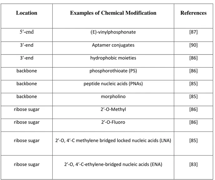
conducted with naked RNA, i.e. without a carrier system. ODNs are chemically modified to 206
achieve desired properties for RNA-delivery to target cells [83, 86]. Most often, they contain a 207
mix of chemical modifications optimised for function [83, 86-88]. To be clinically active, these 208
ODNs must survive endo and exonucleases in the body, avoid immune activation, avoid 209
11 sequestration by the reticuloendothelial system, stay longer in circulation, access its target site 210
(passive diffusion), be preferentially taken up by the target cells (active targeting), and access the 211
cytoplasm (site of action) [86]. Additional requirements for effective delivery of miRNA-mimics 212
includes successful RISC-loading, passenger strand separation, target interaction, translational 213
suppression, and release [86]. 214
Cancer cells can develop resistance to miRNA replacement therapy. The possible mechanisms 215
are discussed here. After being released, miRNA mimics might require RNA editing enzymes 216
like ADAR (Adenosine deaminase acting on RNA) and APOBEC1 (apolipoprotein B 217
mRNA editing enzyme, catalytic polypeptide-like-1) to function like natural miRNAs [89]. 218
ADAR2 acts on dsRNA and mediates adenosine-to-inosine (A-to-I) editing, which changes the 219
miRNA target specificity and is essential for stability and normal functions 220
of certain miRNAs [89]. ADAR2 is downregulated in GBM resulting in reduced conversion of 221
A-to-I editing of miRNA[89]. This problem can be overcome if the supplied miR-mimics already 222
contain inosine at appropriate positions. Some other problems associated with miRNA-resistance 223
includes the expression of competitive endogenous RNAs (ceRNAs) [72] and competitive 224
endogenous RNA-binding proteins (ceRBPs) can bind to miRNA-target sites (MRE) preventing 225
access to RISC and hence block miR-mediated gene-silencing [90]. In a 3D in-vivo setting, gap-226
junction (connexin) mediated intracellular transport of miRNA [52] could dilute the miRNA 227
concentration in the target cells, but also could help to spread the gene-silencing activity to 228
neighbouring cells [52]. All these points must be considered to achieve successful miRNA 229
function. Chemical modifications are discussed in Table 1 [84, 86-88, 91] and clinical trials are 230
discussed in Box 1. 231
12 Blood and extracellular matrix contain abundant extracellular miRNA stabilized by exosomes 232
and argonaute protein complexes and they are biologically active after cellular entry [50, 51, 92] 233
indicating that chemical modifications can be beneficial but not a requirement to achieve RNAi 234
effects when combined with RNA-stabilizing nanocarriers derived from both natural and 235
synthetic sources. 236
It is very difficult to understand what is being presented and discussed in this entire section. It 237
needs to be heavily re-written. 238
239
Natural Nanomedicine
240
Exosomes and microvesicles are naturally secreted miRNA loaded vesicles that act as natural
241
delivery and signalling systems in cell to cell communication [93]. These vesicles can be charged 242
with miRNA, either by transfecting large amounts of exogenous miRNA into the packaging cells 243
or by directly transfecting the exosomes by electroporation or chemical methods [93-95]. Cell 244
mediated miRNA delivery has been reported where mesenchymal stem cells (MSCs) or 245
glioblastoma cancer cells were modified ex vivo to over-express certain tumour-suppressor 246
miRNAs. These modified cells produced exosomes loaded with the specific ts-miRNA. When 247
these cells were injected in the tumour vicinity, they secreted exosomes loaded with ts-miRNA 248
and inhibited tumour growth [33]. 249
Argonaute-2 (AGO2) is a the major functional element of miRNA. The evidences showing that 250
AGO2 can be used for miRNA delivery is discussed in Box-2. 251
252
Bio-mimetic Delivery Systems
253 254
13 Bacterial-mediated RNAi delivery (transkingdom RNAi) has also been explored. The microRNA 255
of interest can be expressed in bacteria through a plasmid encoding its hairpin loop structure as 256
described for in vitro siRNA delivery [97]. The virus-like particles (VLPs) via bacteriophage 257
MS2 can deliver a miRNA. MS2 VLPs are biocompatible, biodegradable, stable and the 258 synthesis is simple [98]. 259 260 Synthetic Nanoparticles 261
Spherical nucleic acids (SNA) consist of a core gold nanoparticle (AuNP) with its surface
262
densely packed with oligonucleotides attached via thiolate-Au interaction. The passenger strand 263
bearing a thiol (SH) group can react with gold nanoparticles to deliver miRNA or siRNA. The 264
guide strand can be separated from the passenger strand inside the cells by the RNAi machinery 265
[99]. These nanoparticles show high transfection efficiency, low toxicity and ability to cross the 266
blood brain barrier (BBB) without a targeting ligand [99]. 267
Cell penetrating peptides (CPP) and other cationic peptides can be complexed with nucleic acids
268
to form highly efficient delivery systems. Introducing a thiol group in the peptide helps to create 269
a bioreducible polymeric peptide that can be specifically released in the intracellular 270
environment and deliver both siRNA and plasmid DNA [100]. 271
A liposome containing siRNA-CPP complexes and magnetic nanoparticles were used where
272
magnetic field was used to concentrate the nanoparticles at the site tumour and later the release 273
of siRNA-CPP was triggered using an electric field [101]. This strategy is interesting for 274
selective release of RNAi molecules using external triggers. 275
14
Lipid nanocapsules (LNCs) have an oily hydrophobic core stabilized by surfactants.
276
Oligonucleotides like LNAs can be delivered with a cationic peptide grafted on LNC’s surface 277
[102]. 278
Polyethylenimine (PEI) is a gold standard cationic polymer used in the nucleic acid delivery. Use
279
in the clinic is limited by toxicity [103]. Toxicity can be overcome by chemically modifying 280
more toxic primary and secondary amines into less toxic secondary and tertiary amines. High 281
molecular weight (HMW) and branched cationic polymers show high toxicity while the low 282
molecular weight (LMW) and linear polymers show poor nucleic acid binding and transfection 283
efficiency [103]. One way to overcome this is by using hybrid polymers, e.g. linear PEI-chitosan 284
hybrid nanoparticles show better transfection efficiency and improved safety profiles [103]. 285
Another way is by cross-linking LMW polymers with bioreducible disulphide linkages. For 286
example, thiol cross-linked LMW-PEI polyplexes conjugated with brain targeting rabies virus 287
glycoprotein (RVG) were useful for the delivery of miR-124a [104]. These nanoparticles 288
displayed low toxicity and brain targeting capabilities. 289
Endosomal escape and release from nanocarrier are two major factors that impact the efficiency 290
of synthetic nanocarriers. The polymeric delivery systems can destabilize the late endosomal 291
(LE) compartment by proton-sponge effect during its acidification (pH 5 – 6) [105]. Therefore, 292
endosomal escape of polymer-nucleic acid complex should accompany the escape of 293
oligonucleotides from the polymers for a functional activity. Similarly, the fate of ~70 % of 294
siRNA delivered by lipid nanoparticles reach late endosomes (LE), packed into exosomes for 295
exocytosis [106] and the remaining would be degraded after fusion with lysosome [105, 107]. 296
Only a small fraction (1 – 2%) of the oligonucleotides reach the cytoplasm that account for the 297
functional activity. This cytoplasmic release was predicted to happen either by direct fusion of 298
15 liposomal delivery systems with cell membrane or destabilization of the endosomal bilayer 299
causing the oligonucleotides to leak out into the cytoplasm [107, 108]. Hence, strategies that 300
allow the intra-cellular release of oligonucleotides after internalization should be addressed more 301
seriously to achieve highly functional delivery systems [109]. 302
This section needs to be heavily re-written for clarity and grammar. 303
304
Crossing the BBB and Locoregional Delivery
305
The blood brain barrier (BBB) prevents the access of drugs and nanoparticles from the blood 306
stream to the brain, and represents a major challenge to deliver therapeutic amounts of drugs to 307
intracranial tumours. Several strategies have been devised to overcome the BBB including 308
ligand-mediated transcytosis [110], temporary physical or chemical disruption [111], or 309
convection enhanced delivery [112]. Yu et al created an optimised bi-specific antibody targeting 310
human transferrin receptor that crossed the BBB in mice and monkeys, while also targeting an 311
intra-brain enzyme β-secretase [110]. Optimal affinity between ligand and receptor is an 312
important factor to increase brain uptake, as it prevents receptor degradation and allows multiple 313
rounds of transcytosis [110]. In clinical trials, Carpentier et al implanted an ultrasound (US) 314
transducer in the skull of GBM patients and achieved safe, reversible, loco-regional BBB 315
opening and observed no dose-limiting toxicities to increasing intensities of ultrasound [111]. 316
US-mediated BBB opening enabled the crossing of gadolinium contrast agent (1kDa) and 317
hydrophilic carboplatin (0.3kDa) [111]. As this study indicates that the intensity of ultrasound 318
can be safely increased further, this technique can also be used to facilitate brain entry of ASOs 319
(8kDa), miRNAs (~15kDa) or nanoparticles (>100kDa). 320
16 siR-LODER is a PLGA based biodegradable polymeric implant loaded with siRNA polyplexes 321
that releases siRNA-polyplexes in the local environment for extended period [2 – 5 months] 322
[113]. A phase I study reported good progress and Silenseed Ltd. is starting a Phase II clinical 323
trials with siG12D-LODER (siRNA against mutated KRAS oncogene) in combination with 324
gemcitabine for pancreatic cancer patients (NCT01676259). Such long-term release systems can 325
be very helpful for loco-regional delivery of RNAi molecules for GBM treatment. 326
327
Concluding Remarks
328
GBM patients have a median survival of less than two years due to the lacks effective curative 329
treatments. The importance of miRNA and its therapeutic benefits in GBM are increasingly 330
documented in pre-clinical studies. Further developments in this field can lead novel treatments 331
for GBM. MiRNA-modulating strategies can either act as stand-alone therapeutics or be used in 332
combination with conventional therapies as sensitizing agents. Many of these strategies use 333
plasmid DNA or oligonucleotides and delivering nucleic acids to cells is an important technical 334
problem. Simpler delivery strategies are more likely to enter clinical trials, hence naked RNA 335
delivery is attractive. Chemical modifications on RNAi molecules have improved in vivo 336
stability of naked RNA, however further improvements in cellular uptake and intracellular 337
release are needed to expand its applications beyond liver. Synthetic nanoparticles often show 338
promising results in in vitro and in vivo, but fail in human applications due issues with stability, 339
toxicity, targeting and efficacy. Use of patient derived exosomes might be more biocompatible 340
and safe in clinical applications. The potential of AGO2 to excel as a RNAi delivery system is 341
high (refs?) [114-116]. If argonaute-mediated miRNA delivery is properly explored, AGO2 can 342
be a breakthrough for RNAi, like CRISPR-Cas9 is for genome editing. AGO2 will not however 343
17 solve the delivery concerns with ASOs, hence other strategies such as chemical modifications 344
and nanoparticle mediated RNA delivery are equally important. Such as?? Combining RNAi, 345
nanomedicine and locoregional delivery can result in effective cancer therapeutics for GBM. 346
18
Acknowledgements
348
This work was supported by Inca (Institut National du Cancer) through the PL-BIO 2014-2020 349
INCA project MARENGO - “MicroRNA agonist and antagonist Nanomedicines for 350
GliOblastoma treatment: from molecular programmation to preclinical validation”. It is also 351
related to the LabEx IRON “Innovative Radiopharmaceuticals in Oncology and Neurology” as 352
part of the french government “Investissements d’Avenir” program. The authors are also 353
thankful to the European support in the frame of the NanoFar consortium, an Erasmus Mundus 354
Joint Doctorate (EMJD) program in nanomedicine and pharmaceutical innovation. European 355
Commission (NanoFar EMJD), “Le Conseil Général de Maine-et-Loire” and Inca financially 356
supported SA, AG and CL, respectively. 357
Conflict of Interest
358
The authors declare that they have no conflict of interest. 359 360 361 362 363 364 365 366 367
19
References
368
1. Louis, D.N. et al. (2016) The 2016 World Health Organization Classification of Tumors of the 369
Central Nervous System: a summary. Acta Neuropathol 131 (6), 803-20. 370
2. Stupp, R. et al. (2005) Radiotherapy plus concomitant and adjuvant temozolomide for 371
glioblastoma. N Engl J Med 352 (10), 987-96. 372
3. Stupp, R. et al. (2015) Maintenance Therapy With Tumor-Treating Fields Plus Temozolomide 373
vs Temozolomide Alone for Glioblastoma: A Randomized Clinical Trial. JAMA 314 (23), 2535-374
43. 375
4. Seystahl, K. et al. (2016) Therapeutic options in recurrent glioblastoma--An update. Crit Rev 376
Oncol Hematol 99, 389-408. 377
5. Zhao, J. (2016) Cancer stem cells and chemoresistance: The smartest survives the raid. 378
Pharmacol Ther 160, 145-58. 379
6. Hanahan, D. and Weinberg, R.A. (2011) Hallmarks of cancer: the next generation. Cell 144 380
(5), 646-74. 381
7. Godlewski, J. et al. (2017) MicroRNA Signatures and Molecular Subtypes of Glioblastoma: 382
The Role of Extracellular Transfer. Stem Cell Reports 8 (6), 1497-1505. 383
8. Lages, E. et al. (2011) MicroRNA and target protein patterns reveal physiopathological 384
features of glioma subtypes. PLoS One 6 (5), e20600. 385
9. Piwecka, M. et al. (2015) Comprehensive analysis of microRNA expression profile in 386
malignant glioma tissues. Mol Oncol 9 (7), 1324-40. 387
10. Ye, X. et al. (2017) Identification of microRNAs associated with glioma diagnosis and 388
prognosis. Oncotarget 8 (16), 26394-26403. 389
11. Shi, Z.-M. et al. (2012) MiR-128 inhibits tumor growth and angiogenesis by targeting 390
p70S6K1. PLoS One 7 (3), e32709. 391
12. Papagiannakopoulos, T. et al. (2012) Pro-neural miR-128 is a glioma tumor suppressor that 392
targets mitogenic kinases. Oncogene 31 (15), 1884-1895. 393
13. Shan, Z.N. et al. (2016) miR128-1 inhibits the growth of glioblastoma multiforme and 394
glioma stem-like cells via targeting BMI1 and E2F3. Oncotarget 7 (48), 78813-78826. 395
14. Rooj, A.K. et al. (2017) MicroRNA-Mediated Dynamic Bidirectional Shift between the 396
Subclasses of Glioblastoma Stem-like Cells. Cell Rep 19 (10), 2026-2032. 397
15. Evangelisti, C. et al. (2009) MiR-128 up-regulation inhibits Reelin and DCX expression and 398
reduces neuroblastoma cell motility and invasiveness. FASEB J. 23 (12), 4276-4287, 399
10.1096/fj.09-134965. 400
16. Godlewski, J. et al. (2008) Targeting of the Bmi-1 Oncogene/Stem Cell Renewal Factor by 401
MicroRNA-128 Inhibits Glioma Proliferation and Self-Renewal. Cancer Res. 68 (22), 9125-402
9130. 403
17. (2014) MicroRNA 128a Increases Intracellular ROS Level by Targeting Bmi-1 and Inhibits 404
Medulloblastoma Cancer Cell Growth by Promoting Senescence. 405
18. Alvarado, A.G. et al. (2016) Coordination of self-renewal in glioblastoma by integration of 406
adhesion and microRNA signaling. Neuro Oncol 18 (5), 656-66. 407
19. Cioce, M. et al. (2016) Mir 145/143: tumor suppressor, oncogenic microenvironmental factor 408
or ...both? Aging (Albany NY). 409
20. Li, C. et al. (2017) The lincRNA-ROR/miR-145 axis promotes invasion and metastasis in 410
hepatocellular carcinoma via induction of epithelial-mesenchymal transition by targeting ZEB2. 411
Sci Rep 7 (1), 4637. 412
20 21. Zhu, X. et al. (2014) miR-145 sensitizes ovarian cancer cells to paclitaxel by targeting Sp1 413
and Cdk6. Int J Cancer 135 (6), 1286-96. 414
22. Zheng, H. et al. (2014) Fas signaling promotes chemoresistance in gastrointestinal cancer by 415
up-regulating P-glycoprotein. Oncotarget. 416
23. Xu, Q. et al. (2012) MiR-145 directly targets p70S6K1 in cancer cells to inhibit tumor 417
growth and angiogenesis. Nucleic Acids Res 40 (2), 761-74. 418
24. Wei, J. et al. (2016) MiR-138 exerts anti-glioma efficacy by targeting immune checkpoints. 419
Neuro Oncol 18 (5), 639-48. 420
25. Shang, C. et al. (2015) MiR-21 up-regulation mediates glioblastoma cancer stem cells 421
apoptosis and proliferation by targeting FASLG. Mol Biol Rep 42 (3), 721-7. 422
26. Ge, Y. et al. (2016) Strand-specific in vivo screen of cancer-associated miRNAs unveils a 423
role for miR-21( *) in SCC progression. Nat Cell Biol 18 (1), 111-21. 424
27. El Fatimy, R. et al. (2017) Genome Editing Reveals Glioblastoma Addiction to MicroRNA-425
10b. Mol Ther 25 (2), 368-378. 426
28. Lee, R.C. et al. (1993) The C. elegans heterochronic gene lin-4 encodes small RNAs with 427
antisense complementarity to lin-14. Cell 75 (5), 843-54. 428
29. Lee, Y. et al. (2004) MicroRNA genes are transcribed by RNA polymerase II. Embo j 23 429
(20), 4051-60. 430
30. Shea, A. et al. (2016) MicroRNAs in glioblastoma multiforme pathogenesis and therapeutics. 431
Cancer Med 5 (8), 1917-46. 432
31. Kozomara, A. and Griffiths-Jones, S. (2014) miRBase: annotating high confidence 433
microRNAs using deep sequencing data. Nucleic Acids Res 42 (Database issue), D68-73. 434
32. Svoronos, A.A. et al. (2016) OncomiR or Tumor Suppressor? The Duplicity of MicroRNAs 435
in Cancer. Cancer Res 76 (13), 3666-70. 436
33. Fareh, M. et al. (2017) Cell-based therapy using miR-302-367 expressing cells represses 437
glioblastoma growth. Cell Death Dis 8 (3), e2713. 438
34. Gao, Z. et al. (2015) The miR-302/367 cluster: a comprehensive update on its evolution and 439
functions. Open Biol 5 (12), 150138. 440
35. Guo, Y. et al. (2017) miR-302/367/LATS2/YAP pathway is essential for prostate tumor-441
propagating cells and promotes the development of castration resistance. Oncogene. 442
36. He, W. et al. (2017) MiR-21 is required for anti-tumor immune response in mice: an 443
implication for its bi-directional roles. Oncogene 36 (29), 4212-4223. 444
37. Nel, A.E. et al. (2009) Understanding biophysicochemical interactions at the nano-bio 445
interface. Nature Materials 8 (7), 543-557. 446
38. Peer, D. et al. (2007) Nanocarriers as an emerging platform for cancer therapy. Nature 447
Nanotechnology 2 (12), 751-760. 448
39. Bartel, D.P. (2004) MicroRNAs: genomics, biogenesis, mechanism, and function. Cell 116 449
(2), 281-97. 450
40. Finnegan, E.F. and Pasquinelli, A.E. (2013) MicroRNA biogenesis: regulating the regulators. 451
Crit Rev Biochem Mol Biol 48 (1), 51-68. 452
41. Han, J. et al. (2006) Molecular basis for the recognition of primary microRNAs by the 453
Drosha-DGCR8 complex. Cell 125 (5), 887-901. 454
42. Lund, E. et al. (2004) Nuclear export of microRNA precursors. Science 303 (5654), 95-8. 455
43. Wilson, R.C. et al. (2015) Dicer-TRBP complex formation ensures accurate mammalian 456
microRNA biogenesis. Mol Cell 57 (3), 397-407. 457
21 44. Frank, F. et al. (2010) Structural basis for 5'-nucleotide base-specific recognition of guide 458
RNA by human AGO2. Nature 465 (7299), 818-22. 459
45. Bartel, D.P. (2009) MicroRNAs: target recognition and regulatory functions. Cell 136 (2), 460
215--33. 461
46. Eichhorn, S.W. et al. (2014) mRNA destabilization is the dominant effect of mammalian 462
microRNAs by the time substantial repression ensues. Mol Cell 56 (1), 104-15. 463
47. Jonas, S. and Izaurralde, E. (2015) Towards a molecular understanding of microRNA-464
mediated gene silencing. Nat Rev Genet 16 (7), 421-33. 465
48. Weinmann, L. et al. (2009) Importin 8 is a gene silencing factor that targets argonaute 466
proteins to distinct mRNAs. Cell 136 (3), 496-507. 467
49. Nishi, K. et al. (2013) Human TNRC6A is an Argonaute-navigator protein for microRNA-468
mediated gene silencing in the nucleus. RNA 19 (1), 17-35. 469
50. Makarova, J.A. et al. (2016) Intracellular and extracellular microRNA: An update on 470
localization and biological role. Prog Histochem Cytochem 51 (3-4), 33-49. 471
51. Arroyo, J.D. et al. (2011) Argonaute2 complexes carry a population of circulating 472
microRNAs independent of vesicles in human plasma. Proc Natl Acad Sci U S A 108 (12), 5003-473
8. 474
52. Zong, L. et al. (2016) Gap junction mediated miRNA intercellular transfer and gene 475
regulation: A novel mechanism for intercellular genetic communication. Sci Rep 6, 19884. 476
53. Adlakha, Y.K. and Saini, N. (2014) Brain microRNAs and insights into biological functions 477
and therapeutic potential of brain enriched miRNA-128. In Mol Cancer, p. 33. 478
54. Zhang, X. et al. (2017) Identification of miRNA-7 by genome-wide analysis as a critical 479
sensitizer for TRAIL-induced apoptosis in glioblastoma cells. Nucleic Acids Res 45 (10), 5930-480
5944. 481
55. Tian, R. et al. (2017) Differential expression of miR16 in glioblastoma and glioblastoma 482
stem cells: their correlation with proliferation, differentiation, metastasis and prognosis. 483
Oncogene. 484
56. Guessous, F. et al. (2013) Oncogenic effects of miR-10b in glioblastoma stem cells. J 485
Neurooncol 112 (2), 153-63. 486
57. Fabbri, M. et al. (2012) MicroRNAs bind to Toll-like receptors to induce prometastatic 487
inflammatory response. Proc Natl Acad Sci U S A 109 (31), E2110-6. 488
58. Hu, J. et al. (2016) MiR-215 Is Induced Post-transcriptionally via HIF-Drosha Complex and 489
Mediates Glioma-Initiating Cell Adaptation to Hypoxia by Targeting KDM1B. Cancer Cell 29 490
(1), 49-60. 491
59. Mucaj, V. et al. (2015) MicroRNA-124 expression counteracts pro-survival stress responses 492
in glioblastoma. Oncogene 34 (17), 2204-14. 493
60. Khalil, S. et al. (2016) miRNA array screening reveals cooperative MGMT-regulation 494
between miR-181d-5p and miR-409-3p in glioblastoma. Oncotarget 7 (19), 28195-206. 495
61. Li, Y. et al. (2016) Downregulation and tumor-suppressive role of XPO5 in hepatocellular 496
carcinoma. Mol Cell Biochem 415 (1-2), 197-205. 497
62. Sun, H.L. et al. (2016) ERK Activation Globally Downregulates miRNAs through 498
Phosphorylating Exportin-5. Cancer Cell 30 (5), 723-736. 499
63. Gurtner, A. et al. (2016) Dysregulation of microRNA biogenesis in cancer: the impact of 500
mutant p53 on Drosha complex activity. J Exp Clin Cancer Res 35, 45. 501
64. Rupaimoole, R. et al. (2014) Hypoxia-mediated downregulation of miRNA biogenesis 502
promotes tumour progression. Nat Commun 5, 5202. 503
22 65. Naoghare, P.K. et al. (2011) Knock-down of argonaute 2 (AGO2) induces apoptosis in 504
myeloid leukaemia cells and inhibits siRNA-mediated silencing of transfected oncogenes in 505
HEK-293 cells. Basic Clin Pharmacol Toxicol 109 (4), 274-82. 506
66. Shortridge, M.D. et al. (2017) A Macrocyclic Peptide Ligand Binds the Oncogenic 507
MicroRNA-21 Precursor and Suppresses Dicer Processing. ACS Chem Biol 12 (6), 1611-1620. 508
67. Costales, M.G. et al. (2017) Small Molecule Inhibition of microRNA-210 Reprograms an 509
Oncogenic Hypoxic Circuit. J Am Chem Soc 139 (9), 3446-3455. 510
68. Jiang, W. et al. (2016) Repurposing phenformin for the targeting of glioma stem cells and the 511
treatment of glioblastoma. Oncotarget 7 (35), 56456-56470. 512
69. Venkatesh, H.S. et al. (2017) Targeting neuronal activity-regulated neuroligin-3 dependency 513
in high-grade glioma. Nature advance online publication. 514
70. Fu, J. et al. (2013) NPV-LDE-225 (Erismodegib) inhibits epithelial mesenchymal transition 515
and self-renewal of glioblastoma initiating cells by regulating miR-21, miR-128, and miR-200. 516
Neuro Oncol 15 (6), 691-706. 517
71. Zaman, M.S. et al. (2010) The functional significance of microRNA-145 in prostate cancer. 518
Br J Cancer 103 (2), 256-64. 519
72. Liu, T. et al. (2017) Curcumin suppresses proliferation and in vitro invasion of human 520
prostate cancer stem cells by ceRNA effect of miR-145 and lncRNA-ROR. Gene. 521
73. Klinger, N.V. and Mittal, S. (2016) Therapeutic Potential of Curcumin for the Treatment of 522
Brain Tumors. Oxid Med Cell Longev 2016, 9324085. 523
74. Jinek, M. et al. (2012) A programmable dual-RNA-guided DNA endonuclease in adaptive 524
bacterial immunity. Science 337 (6096), 816-21. 525
75. Chang, H. et al. (2016) CRISPR/cas9, a novel genomic tool to knock down microRNA in 526
vitro and in vivo. Sci Rep 6, 22312. 527
76. Chen, Z.H. et al. (2017) Targeting genomic rearrangements in tumor cells through Cas9-528
mediated insertion of a suicide gene. Nat Biotechnol 35 (6), 543-550. 529
77. Meca-Cortes, O. et al. (2017) CRISPR/Cas9-Mediated Knockin Application in Cell Therapy: 530
A Non-viral Procedure for Bystander Treatment of Glioma in Mice. Mol Ther Nucleic Acids 8, 531
395-403. 532
78. Hirosawa, M. et al. (2017) Cell-type-specific genome editing with a microRNA-responsive 533
CRISPR-Cas9 switch. Nucleic Acids Res 45 (13), e118. 534
79. Ezzine, S. et al. (2013) RILES, a novel method for temporal analysis of the in vivo regulation 535
of miRNA expression. Nucleic Acids Res 41 (20), e192. 536
80. Zhu, Z. et al. (2017) Zika virus has oncolytic activity against glioblastoma stem cells. J Exp 537
Med. 538
81. Li, L. et al. (2012) Targeted expression of miR-34a using the T-VISA system suppresses 539
breast cancer cell growth and invasion. Mol Ther 20 (12), 2326-34. 540
82. Ma, S. et al. (2017) Combination of AAV-TRAIL with miR-221-Zip Therapeutic Strategy 541
Overcomes the Resistance to TRAIL Induced Apoptosis in Liver Cancer. Theranostics 7 (13), 542
3228-3242. 543
83. Yu, D. et al. (2012) Single-stranded RNAs use RNAi to potently and allele-selectively inhibit 544
mutant huntingtin expression. Cell 150 (5), 895-908. 545
84. Ariyoshi, J. et al. (2015) Development of Novel Antisense Oligonucleotides for the 546
Functional Regulation of RNA-Induced Silencing Complex (RISC) by Promoting the Release of 547
microRNA from RISC. Bioconjug Chem 26 (12), 2454-60. 548
23 85. Meng, L. et al. (2017) Small RNA zippers lock miRNA molecules and block miRNA 549
function in mammalian cells. Nat Commun 8, 13964. 550
86. Khvorova, A. and Watts, J.K. (2017) The chemical evolution of oligonucleotide therapies of 551
clinical utility. Nat Biotechnol 35 (3), 238-248. 552
87. Nikan, M. et al. (2016) Docosahexaenoic Acid Conjugation Enhances Distribution and 553
Safety of siRNA upon Local Administration in Mouse Brain. Mol Ther Nucleic Acids 5 (8), 554
e344. 555
88. Elkayam, E. et al. (2017) siRNA carrying an (E)-vinylphosphonate moiety at the 5 end of the 556
guide strand augments gene silencing by enhanced binding to human Argonaute-2. Nucleic 557
Acids Res 45 (6), 3528-3536. 558
89. Paul, D. et al. (2017) A-to-I editing in human miRNAs is enriched in seed sequence, 559
influenced by sequence contexts and significantly hypoedited in glioblastoma multiforme. Sci 560
Rep 7 (1), 2466. 561
90. Degrauwe, N. et al. (2016) The RNA Binding Protein IMP2 Preserves Glioblastoma Stem 562
Cells by Preventing let-7 Target Gene Silencing. Cell Rep 15 (8), 1634-47. 563
91. Esposito, C.L. et al. (2016) A combined microRNA-based targeted therapeutic approach to 564
eradicate glioblastoma stem-like cells. J Control Release 238, 43-57. 565
92. Li, L. et al. (2012) Argonaute 2 complexes selectively protect the circulating microRNAs in 566
cell-secreted microvesicles. PLoS One 7 (10), e46957. 567
93. Wahlgren, J. et al. (2016) Delivery of Small Interfering RNAs to Cells via Exosomes. 568
Methods Mol Biol 1364, 105-25. 569
94. Lunavat, T.R. et al. (2016) RNAi delivery by exosome-mimetic nanovesicles - Implications 570
for targeting c-Myc in cancer. Biomaterials 102, 231-8. 571
95. Haraszti, R.A. et al. (2017) Loading of Extracellular Vesicles with Chemically Stabilized 572
Hydrophobic siRNAs for the Treatment of Disease in the Central Nervous System. Bio Protoc 7 573
(20). 574
96. Lee, H.K. et al. (2013) Mesenchymal stem cells deliver synthetic microRNA mimics to 575
glioma cells and glioma stem cells and inhibit their cell migration and self-renewal. Oncotarget 4 576
(2), 346-61. 577
97. Ahmed, O. et al. (2015) Delivery of siRNAs to cancer cells via bacteria. In Methods Mol 578
Biol (2014/10/17 edn), pp. 117-29. 579
98. Ashley, C.E. et al. (2011) Cell-specific delivery of diverse cargos by bacteriophage MS2 580
virus-like particles. ACS Nano 5 (7), 5729-45. 581
99. Kouri, F.M. et al. (2015) miR-182 integrates apoptosis, growth, and differentiation programs 582
in glioblastoma. Genes Dev 29 (7), 732-45. 583
100. Yoo, J. et al. (2017) Bioreducible branched poly(modified nona-arginine) cell-penetrating 584
peptide as a novel gene delivery platform. J Control Release 246, 142-154. 585
101. Yang, Y. et al. (2016) Thermal and magnetic dual-responsive liposomes with a cell-586
penetrating peptide-siRNA conjugate for enhanced and targeted cancer therapy. Colloids Surf B 587
Biointerfaces 146, 607-15. 588
102. Griveau, A. et al. (2013) Silencing of miR-21 by locked nucleic acid-lipid nanocapsule 589
complexes sensitize human glioblastoma cells to radiation-induced cell death. Int J Pharm. 590
103. Tripathi, S.K. et al. (2012) Linear polyethylenimine-graft-chitosan copolymers as efficient 591
DNA/siRNA delivery vectors in vitro and in vivo. Nanomedicine 8 (3), 337-45. 592
104. Hwang do, W. et al. (2011) A brain-targeted rabies virus glycoprotein-disulfide linked PEI 593
nanocarrier for delivery of neurogenic microRNA. Biomaterials 32 (21), 4968-75. 594
24 105. Gujrati, M. et al. (2016) Multifunctional pH-Sensitive Amino Lipids for siRNA Delivery. 595
Bioconjug Chem 27 (1), 19-35. 596
106. Sahay, G. et al. (2013) Efficiency of siRNA delivery by lipid nanoparticles is limited by 597
endocytic recycling. Nat Biotechnol 31 (7), 653-8. 598
107. Juliano, R.L. and Carver, K. (2015) Cellular uptake and intracellular trafficking of 599
oligonucleotides. Adv Drug Deliv Rev 87, 35-45. 600
108. Gilleron, J. et al. (2013) Image-based analysis of lipid nanoparticle-mediated siRNA 601
delivery, intracellular trafficking and endosomal escape. Nat Biotechnol 31 (7), 638-46. 602
109. Gilleron, J. et al. (2015) Identification of siRNA delivery enhancers by a chemical library 603
screen. Nucleic Acids Research. 604
110. Yu, Y.J. et al. (2014) Therapeutic bispecific antibodies cross the blood-brain barrier in 605
nonhuman primates. Sci Transl Med 6 (261), 261ra154. 606
111. Carpentier, A. et al. (2016) Clinical trial of blood-brain barrier disruption by pulsed 607
ultrasound. Sci Transl Med 8 (343), 343re2. 608
112. Halle, B. et al. (2016) Convection-enhanced delivery of an anti-miR is well-tolerated, 609
preserves anti-miR stability and causes efficient target de-repression: a proof of concept. J 610
Neurooncol 126 (1), 47-55. 611
113. Zorde Khvalevsky, E. et al. (2013) Mutant KRAS is a druggable target for pancreatic 612
cancer. Proc Natl Acad Sci U S A 110 (51), 20723-8. 613
114. Prud'homme, G.J. et al. (2016) Neuropilin-1 is a receptor for extracellular miRNA and 614
AGO2/miRNA complexes and mediates the internalization of miRNAs that modulate cell 615
function. Oncotarget 7 (42), 68057-68071. 616
115. Kang, T. et al. (2016) Synergistic targeting tenascin C and neuropilin-1 for specific 617
penetration of nanoparticles for anti-glioblastoma treatment. Biomaterials 101, 60-75. 618
116. Ferreira, R. et al. (2014) Argonaute-2 promotes miR-18a entry in human brain endothelial 619
cells. J Am Heart Assoc 3 (3), e000968. 620
117. Bartel, D. (2004) MicroRNAsGenomics, Biogenesis, Mechanism, and Function. Cell 116 621
(2), 281--297. 622
118. Toscano-Garibay, J.D. and Aquino-Jarquin, G. (2014) Transcriptional regulation 623
mechanism mediated by miRNA-DNA*DNA triplex structure stabilized by Argonaute. Biochim 624
Biophys Acta 1839 (11), 1079-83. 625
119. Wang, Z. (2011) The principles of MiRNA-masking antisense oligonucleotides technology. 626
Methods Mol Biol 676, 43-9. 627
120. Weil, S. et al. (2017) Tumor microtubes convey resistance to surgical lesions and 628
chemotherapy in gliomas. Neuro Oncol 19 (10), 1316-1326. 629
121. Hong, X. et al. (2015) Gap junctions modulate glioma invasion by direct transfer of 630
microRNA. Oncotarget 6 (17), 15566-77. 631
122. van der Ree, M.H. et al. (2016) Miravirsen dosing in chronic hepatitis C patients results in 632
decreased microRNA-122 levels without affecting other microRNAs in plasma. Aliment 633
Pharmacol Ther 43 (1), 102-13. 634
123. Van Der Ree, M. et al. LO7 : A single subcutaneous dose of 2mg/kg or 4mg/kg of RG-101, 635
a GalNAc-conjugated oligonucleotide with antagonist activity against MIR-122, results in 636
significant viral load reductions in chronic hepatitis C patients. Journal of Hepatology 62, S261. 637
124. Grimm, D. (2011) The dose can make the poison: lessons learned from adverse in vivo 638
toxicities caused by RNAi overexpression. Silence 2, 8. 639
25
Legend - Figure 1: Biogenesis and Modulating Strategies of miRNAs
641
(a) Canonical Biogenesis of miRNA: (adapted and modified with permission [117]) MiRNA
642
genes are transcribed by RNA polymerases in the nucleus forming large primary-miRNA (> 500 643
bases) which harbours one or more stem-loop structures [29]. The RNA-binding protein (Di 644
George syndrome Critical Region gene 8, DGCR8/Pasha) and a RNAse III endonuclease 645
(Drosha) recognizes the stemloop structures and releases the stem-loop Precursor miRNA (Pre-646
miRNA, ~70 nucleotides) [41]. Pre-miRNA is then transported out of the nucleus through 647
nuclear pore complexes (NPC) by binding to Exportin-5 (XPO5) and Ran-GTPase [42]. The pre-648
miRNA has two arms (5ʹ and 3ʹ) each might encode an active miRNA sequence named as miR-649
X-5p (Red strand) or miR-X-3p (Black strand) respectively. Once in the cytoplasm, the pre-650
miRNA loads into a pre-RISC (RNA induced silencing complex) consisting of Dicer, TAR RNA 651
binding protein (TRBP), one of the argonautes (AGO1 - AGO4) and chaperones (Heat Shock 652
Proteins, HSP70/HSP90) [39, 40, 43]. Dicer cleaves the stem-loop structure creating a ~23bp 653
miRNA duplex which loads into an argonaute (AGO) [39, 40, 43]. Either miR-5p or miR-3p 654
strand loads into the AGO [26, 44]. Binding of seed sequence (2 – 7 nucleotides at the 5ʹ-end of 655
the miRNA, highlighted in green) to the 3ʹ-untranslated region (3ʹ-UTR) of the target mRNA 656
results in the inhibition of protein synthesis either by messenger RNA (mRNA) destabilization 657
(>75%) and translational repression [46, 47]. 658
(b) Other functions of miRNA and RISC: RISC complex also exerts nuclear functions by
659
shuttling between the cytoplasm and the nucleus via Exportin 1 and Importin 8 [48, 49, 118], 660
and possibly also exerts mitochondrial functions [50]. MicroRNA can be secreted from the cell, 661
either in extracellular vesicles or bound to AGO2 [7, 50, 51], and function as endocrine 662
signalling molecules. 663
26
(c) MicroRNA Modulation Strategies: Genome editing [27, 75]; Small Molecule miRNA
664
Inhibitors (e.g. TargapremiR) [67]; MicroRNA mimics and ss-siRNA [83, 86]; Antisense 665
Oligonucleotides (ASO) [85]; MicroRNA Sponges (Gene Therapy) [82]; MicroRNA Masks 666
[119]. 667
(d) Can recombinant-AGO2 with targeting ligands acts as a RNAi delivery system?
27
Legend - Figure 2: Role of miRNA in the Hallmarks of Cancer and Treatment Resistance
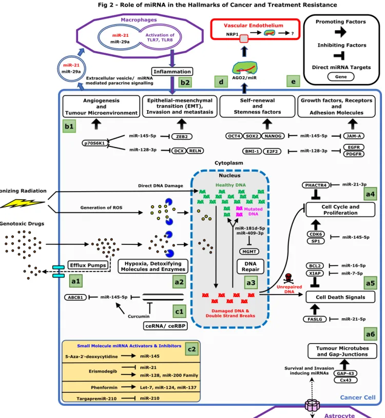
669
(a) Role of miRNA in Treatment Resistance: Standard glioblastoma treatment involves radio
670
and chemotherapy which damage DNA and create double strand breaks (DSBs). Various 671
resistance mechanisms lead to treatment failure: (1) Efflux pumps reduce intracellular drug 672
concentration [22]; (2) Detoxifying enzymes inactivate the drug [5]; (3) DNA repair reverses 673
lethal DSBs and DNA damages; MGMT is a DNA repair enzyme targeted by miR-181d and 674
miR-409 [60]; (4, 5) Damaged DNA induces cell death signals and stop cell cycle progression. 675
For detailed list of resistance genes and mechanisms, see review [5]; (6) Tumour-microtubes and 676
connexin mediated invasion through exchange of resistance factors (e.g. miRNAs) helps cancer 677
cells to adapt to genotoxic treatments and surgery [120, 121]. 678
(b) Role of miRNA in other cancer functions: (1) Role of ts-miRNAs (128 [11-17],
miR-679
145 [18-23], miR-7 [54], miR-16 [55]) and oncomiRs (miR-21-5p [25] and miR-21-3p [26]) in 680
controlling other hallmarks of cancer. (2) Exosomal miRNAs (especially miR-21 and miR-29a) 681
can directly bind to Toll-like receptors (TLR7 and TLR8) in the endosomal compartment of 682
macrophages resulting in pro-metastatic inflammation [57]; 683
(c) Intrinsic and Extrinsic Regulation of miRNA function: (1) Intracellular competitive
684
endogenous RNAs (ceRNAs) and competitive endogenous RNA binding proteins (ceRBPs) can 685
inhibit ts-miRNA [72, 90]. Lnc-RNA-ROR, a ceRNA inhibiting miR-145 is presented as an 686
example. Curcumin inhibits Lnc-RNA-ROR and hence restoring miR-145 function; (2) Small 687
molecule drugs controlling the expression of ts-miRNAs and oncomiRs [21, 67, 68, 70, 72]; 688
28
(d) Role of Argonaute-miRNA complex: Vascular endothelial cells express neurophilin1
689
(NRP1) which acts a receptor for AGO2-miRNA complexes [114-116]. Functional significance 690
of AGO2-miRNA complexes in GBM is yet unknown. 691
(e) Legend explaining the symbols.
692
Abbreviations: EGFR (Epidermal growth factor receptor); PDGFR (Platelet-derived growth 693
factor receptor); BMI1 (B Lymphoma Mo-MLV Insertion Region 1 Homolog); E2F3 (E2F 694
Transcription Factor 3); DCX (Doublecortex), RELN (Reelin); OCT4 (Octamer-Binding 695
Transcription Factor 3); SOX2 [SRY (Sex-Determining Region Y)-Box 2]; KLF4 (Kruppel-Like 696
Factor 4); JAM-A (a junction adhesion molecule), ZEB2 (Zinc Finger E-Box Binding 697
Homeobox 2), ABCB1 (ATP Binding Cassette Subfamily B Member 1), HIF-α (hypoxia-698
inducible factor 1); TRAIL (TNF-related apoptosis-inducing ligand); XIAP (X-linked inhibitor 699
of apoptosis protein); BCL2 (B-Cell CLL/Lymphoma 2), CDK6 (Cyclin-dependent kinase-6); 700
PHACTR4 (Phosphatase and actin regulator 4); FASLG (FAS ligand); TLR7 and TLR8 (Toll-701
like receptor 7 and 8); KDM1B (Lysine Demethylase 1B); Cx43 (Connexin 43); GAP43 702
(Growth Associated Protein 43). 703
704 705
29
Legend - Figure-3: RNA nanomedicine based strategies for GBM treatment
706
Solid GBM tumours are facing two main situations, unresectable tumour and the resection 707
cavity. Two major modalities of treatment using innovative nanomedicines may impact the 708
modulation of miRNA contingents and thus GBM outcome: (a) loco-regional treatments and (b) 709
systemic delivery. 710
(a) For loco-regional treatment, although intratumoural stereotaxic infusion (a1) is explored, 711
intrathecal, intranasal and CSF delivery can also be used for infusion of natural (a2) or synthetic 712
nanomedicine (a3). 713
(a2) Natural Delivery Systems: Argonaute-2 (AGO-2)-miRNA complex and exosomes are
714
presented as examples. Pros: Biocompatible and safe. Cons: Delivery and targeting efficiency 715
needs further improvement. 716
(a3) Synthetic Delivery Systems: Gold nanoparticles (AuNPs) have a metallic core. The RNA
717
is usually loaded on the surface using strong thiol-gold interaction (S-Au); Pros: Facile synthesis 718
and biocompatible. Cons: Require chemical modification as the RNA are exposed to the surface 719
[99]; Lipid nanocapsules (LNCs) have an oily hydrophobic core. LNC’s surface can be modified 720
with cationic polymers or peptides which be used for RNA binding. Alternatively, lipoplexes 721
(cationic lipid+RNA complexes) can be prepared and embedded into its core; Pros: High cellular 722
uptake. Cons: Low RNA loading capacity and toxicity from surfactants. Chemical conjugates are 723
chemically modified naked RNA linked to a ligand to facilitate cellular uptake and increase 724
delivery efficacy; Pros: Simple design. Cons: Optimization needed to reach organs other than 725
liver. Liposomes can hold the RNA in its shell or in the aqueous core; Pros: High cellular 726
uptake. Cons: Low endosomal escape. Cationic polymers condense the nucleic acid by 727
30 electrostatic interaction to form nanoparticles. Pros: Can load very high quantities of RNA per 728
nanoparticle due to ionic condensation. Cons: Toxicity and release of RNA from the polymer 729
after cellular entry is not clearly understood. The surface of synthetic and natural nanoparticles 730
can be easily engineered with polyethylene glycol (PEG) and desired ligand. 731
(a4) Modalities of administration: As in the case of delivery of radiopharmaceticals
732
{Vanpouille-Box, 2011 #5048}, it is important to reach optimal therapeutic index and to define 733
therapeutic time windows where the treatment is more efficient. Drug distribution and clearance 734
information help to decide dose fractionation and schedule of administration of miRNA-735
nanomedicine {Ezzine, 2013 #567}. The volume of distribution would depend on the mode of 736
administration (Bolus or CED). (Part of the image adapted from Servier Medical Art, available 737
under creative commons attributions 3.0). 738
(a5) Alternatively, long term release implant might be used for sustained release like siRLODER
739
[113]. 740
(b) Systemic delivery and crossing the blood-brain barrier (BBB): The brain endothelium
741
with tight junctions and the surrounding supporting cells form a selective barrier isolating most 742
of the blood components from accessing the delicate brain tissue. Ultrasound transducers placed 743
inside the cranium can reversibly disrupt the BBB causing the leakage of blood components into 744
the brain tissue [111]. This strategy can be used for facilitating the entry of intravenously 745
injected nanoparticles into the brain tissue. (b2) Nanoparticles conjugated with anti-transferrin 746
antibodies can enter the brain tissue by transcytosis through the endothelial cells [110]. Pink dots 747
represent nanoparticles without any ligand on its surface, while the Green dots represent 748
transferrin-conjugated nanoparticles. 749
31 750
32
Box 1: Biogenesis and functions of miRNAs
751
The biogenesis and functions of human miRNAs are represented in Figure 1 [39, 40]. MiRNA 752
genes are transcribed mainly from the nuclear genome (Primary-miRNA transcript, pri-miR) 753
[29], processed and transported out of the nucleus (Stem-loop structured Precursor-miRNA, pre-754
miR) [41, 42], trimmed in the cytoplasm (Duplex miRNA, miR-5p/miR-3p) [43] and loaded onto 755
argonaute proteins to form the RISC complex (single-stranded Mature miRNA). Either or both 756
miRNA-strands can be loaded onto individual RISC complexes [26, 44]. MiRNA-guided RISC 757
binds to target messenger-RNAs (primarily in the 3ʹ-untranslated region, at specific sites called 758
miRNA recognition elements (MREs)) and blocks protein expression by mRNA destabilization 759
(~80%) and repressing translation (~20%) [45-47]. The RISC complex also exerts miRNA-760
mediated gene-silencing in the nucleus [48, 49] and mitochondria [50]. MiRNAs can also 761
function like endocrine or paracrine signalling molecules as they can be transported to adjacent 762
cells via connexins [52] or secreted from the cell, either in extracellular vesicles (EVs) or bound 763
to argonaute proteins (e.g. AGO2) [7, 50, 51]. 764