HAL Id: dumas-01245798
https://dumas.ccsd.cnrs.fr/dumas-01245798
Submitted on 17 Dec 2015HAL is a multi-disciplinary open access
archive for the deposit and dissemination of sci-entific research documents, whether they are pub-lished or not. The documents may come from
L’archive ouverte pluridisciplinaire HAL, est destinée au dépôt et à la diffusion de documents scientifiques de niveau recherche, publiés ou non, émanant des établissements d’enseignement et de
Prevalence and risk factors of peripheral retinal changes
in the elderly using ultra-wide field imaging: the alienor
study
Camille Seguy
To cite this version:
Camille Seguy. Prevalence and risk factors of peripheral retinal changes in the elderly using ultra-wide field imaging: the alienor study. Human health and pathology. 2015. �dumas-01245798�
Université Bordeaux
U.F.R DES SCIENCES MEDICALES
Année 2015 thèse n°3158
Thèse pour l'obtention du
DIPLOME D'ETAT DE DOCTEUR EN MEDECINE
Présentée et soutenue publiquement
Le 19 octobre 2015
Par Camille SEGUY
Née le 15 août1985 à Bordeaux
Directeur
Madame Cécile DELCOURT
Rapporteur
Monsieur le Professeur Carl ARNDT
Jury
Monsieur le Professeur Jean-Francois KOROBELNIK Président
Madame le Professeur Marie-Noelle DELYFER Juge
Monsieur le Professeur David TOUBOUL Juge
Madame le Docteur Marie-Bénédicte ROUGIER Juge
PREVALENCE AND RISK FACTORS OF
PERIPHERAL RETINAL CHANGES IN THE
ELDERLY USING ULTRA-WIDE FIELD IMAGING :
REMERCIEMENTS
A mon Maitre et Président de jury,
Monsieur le Professeur Jean-Francois KOROBELNIK
Professeur des Universités d' Ophtalmologie
Chef de service d'ophtalmologie
Chef de l’unité médicale de rétine, uvéites et neuro-ophtalmologie, CHU de Bordeaux
Votre expérience chirurgicale, vos qualités professionnelles et humaines sont des exemples pour
moi.
Merci de m'accorder votre confiance et de m'accueillir dans votre service en tant que chef de
clinique. J'ai hâte de continuer ce parcours dans votre service à vos côtés.
Soyez assuré de ma reconnaissance et de mon profond respect.
A mon Maitre et Juge de thèse
Madame le Professeur Marie-Noelle DELYFER
Professeur des Universités
Praticien Hospitalier d'Ophtalmologie
Unité médicale de rétine, uvéites et neuro-ophtalmologie, CHU de Bordeaux
Vous me faites l'honneur de juger ce travail.
Je vous remercie pour votre disponibilité, votre bienveillance et votre soutien au quotidien.
Votre rigueur ainsi que votre conscience professionnelle et vos qualités chirurgicales sont des
exemples pour moi.
Soyez assurée de ma sincère admiration et de mon profond respect.
A mes Juges de thèse
Monsieur le Professeur David TOUBOUL
Professeur des Universités
Praticien Hospitalier Ophtalmologie
Chef de l’unité médicale cataracte, chirurgie réfractive, cornée, glaucome et lentilles, CHU de
Bordeaux
Tu me fais l'honneur de juger ce travail. Je te remercie de m'avoir fait partager ton expérience
lorsque j'étais interne à tes côtés. Tes qualités chirurgicales restent un exemple pour moi.
Sois assuré de ma profonde reconnaissance et de mon profond respect.
Madame le Docteur Marie-Bénédicte ROUGIER
Praticien hospitalier d'Ophtalmologie
Unité médicale de rétine, uvéites et neuro-ophtalmologie, CHU de Bordeaux
tant en neuro-ophtalmologie qu'en inflammation intra-oculaire. Je suis ravie de pouvoir continuer
à apprendre à vos cotés.
Soyez assurée de ma sincère reconnaissance et de mon profond respect.
A ma Juge et directrice de thèse
Madame Cécile DELCOURT
Chargé de recherche CR1
ISPED INSERM U 897 Université Victor Segalen Bordeaux 2
Tu m'as fait l'honneur de diriger, juger et corriger ce travail.
Je te remercie pour tout le temps que tu y as consacré. Ta disponibilité, tes conseils avisés et ton
soutien m'ont été d'une aide précieuse.
Sois assurée de ma profonde reconnaissance et de mon profond respect.
A mon rapporteur de thèse
Monsieur le Professeur Carl Arndt
Professeur des Universités
Praticien Hospitalier Ophtalmologie
Service d'ophtalmologie , CHU de Reims
Vous me faites l'honneur d'avoir accepté d'évaluer l'intérêt scientifique de ce travail et je vous en
remercie.
Soyez assuré de mon profond respect et ma sincère reconnaissance.
A Mélanie Le Goff
Statisticienne, ISPED INSERM U 897, Université Victor Segalen Bordeaux 2.
Je te remercie pour ton aide. Ta contribution a été essentielle dans l'élaboration de ce travail.
Sois assurée de ma sincère reconnaissance.
A Brigitte Gontier
Orthoptiste
Je te remercie pour ta patience et ta gentillesse à toute épreuve!
A mes Maitres d'internat
Messieurs les Docteurs W. Williamson et Y. Ailem
Vous avez accompagné avec patience et bienveillance mes premiers pas en ophtalmologie.
Soyez assurés de ma reconnaissance.
Monsieur le Docteur C. Schweitzer
Merci de me faire partager ton expérience et tes conseils.
Monsieur le Docteur Bazin et l'équipe d'ophtalmolgie de l'Hopital Robert Piqué.
Vos conseils et votre bienveillance m'ont beaucoup apporté tant sur le plan médical que
chirurgical.
Soyez assuré de ma sincère reconnaissance et de mon profond respect.
Monsieur le Docteur F. Léger
Merci de m'avoir fait partager vos connaissances et votre expertise en anatomo-pathologie mais
également pour votre accueil et votre confiance tout au long du semestre passé à vos cotés.
Soyez assuré de mon profond respect.
Monsieur le Professeur H. Loiseau
Merci pour votre accueil et ces moments inoubliables !
A mes Anciens chefs
Les Docteur N. Mesplié, S Leoni, M. Blaizeau, O. Chatoux, C. Paya. J. Pechmeja, D. Smadja.
Merci pour votre patience, disponibilité et vos conseils avisés.
A mes Co internes
Climi, Clem, Sarra, Marion,Yona, Valentine, Antoine B, Julien, Hélène, Marc, Quentin, Audrey,
Marie-Victoire.
Merci pour votre aide, et ces bons moments passés ensemble malgré le travail et la fatigue parfois!
A l'équipe d'Orthoptistes :
Merci pour votre compétence , votre disponibilité et votre aide au quotidien.
Aux Secrétaires
Soyez assurées de ma sincère reconnaissance et de mon respect pour votre travail et votre aide au
quotidien.
A Isa et Mumu
Je vous remercie pour votre efficacité à toute épreuve et la qualité de votre travail.
A toute l'équipe d'infirmières et d'aides soignants
J'apprécie votre professionnalisme et votre bonne humeur.
Merci de votre aide au quotidien.
A ma famille
A mes parents
Pour leur amour et leur soutien indéfectible tout au long de ces années de médecine.
A mon père pour tous ces week-ends passés à sillonner la France (...et plus..) à me suivre et me
soutenir dans ma vie d'escrimeuse.
A ma mère pour sa bienveillance et sa patience . Ton sens de l'organisation et ton soutien
logistique infaillible ont été précieux pour moi.
A mes frères
A mon frère Benjamin : ton intelligence, tes qualités de travail, ton dévouement et ta détermination
professionnels seront toujours des modèles pour moi.
A mon frère Paul-Henri, merci pour ta bienveillance, ta franchise et ton soutien.
Ta vie d'étudiant en médecine ne fait que commencer... tu as tout mon soutien et ma fierté !
A Aurore et la Cacahouète Team : Eve et Marcel
Merci pour votre joie, spontanéité et ces bons moments passés ensemble !!... qu'il y en ait beaucoup
d'autres!!
A mes grands-parents
Vous êtes pour moi des modèles de tolérance et d'amour.
Merci pour tous ces moments passés ensemble.
A Annie, ma marraine,
Tu as toujours été présente à mes cotés, merci pour ta présence, ta bienveillance et ton soutien.
Je suis fière de t'avoir comme marraine.
A Alexandra et Philippe
Je vous remercie de m'avoir accueillie parmi vous, avec affection et bienveillance. Je suis très
heureuse et honorée de votre présence à mes côtés.
A Antoine,
J'ai une chance infinie de t'avoir à mes cotés. Tant de chose à vivre et à construire ensemble...
Et tant de chose à te dire....que je te réserve à toi seul...
A mes amis
A Anne-Co, merci pour ta franchise, tes conseils avisés et debriefing...(si possible au Pepponne !
C'est toujours mieux...). Je suis heureuse de t'avoir comme amie. Merci pour ton amitié.
A Nico, Climi, Pilou... pour ces moments inoubliables passés à l'Internat de Pau mais aussi tous
les autres depuis... je suis heureuse de vous avoir comme amis!
Aux Charliettes : pour nos sessions films, photos, voyages...et corvette bien sur!
Aux Boriskan : merci pour tous ces bons moments passés ensemble...et toutes ces découvertes
Prevalence and risk factors of peripheral retinal changes in
the elderly using ultra-wide field imaging: the ALIENOR
study.
Camille Séguy 1,2, Christine Tadros 3, Mélanie Le Goff 1,4, Noëlle Delyfer 1,2,4,
Marie-Bénédicte Rougier 2, Tunde Peto 3, Cécile Delcourt 1,4, Jean-François Korobelnik 1,2,4.
1- Univ. Bordeaux, ISPED, F-33000 Bordeaux France.
2- CHU de Bordeaux, Service d'Ophtalmologie, F-33000 Bordeaux, France.
3-
NIHR Biomedical Research Centre at Moorfields Eye Hospital NHS Foundation Trust and UCL
Institute of Ophthalmology, Head of Reading Centre, London, UK.
ABSTRACT Purpose:
To determine the prevalence and associated risk factors of peripheral retinal lesions resembling age-related macular degeneration (AMD) using ultra-wide field (UWF) imaging.
Methods
376subjects, aged 75 years or more, underwent UWF imaging by Optomap P200C (Optos plc, United Kingdom) between 2009 and 2011. Presence of peripheral soft drusen, hard drusen, retinal pigment epithelial (RPE) changes, atrophy or choroidal neovascularisation (CNV) was graded at the Moorfields Eye Hospital Reading Center using a standardized grading protocol. Associations of these peripheral lesions with AMD (graded according to the international classification from non mydriatic colour retinal photographs) and AMD risk factors were estimated using Generalized Estimating Equation logistic regressions.
Results
61% of the participants had peripheral retinal lesions resembling AMD, 51% had peripheral soft drusen, 31.3% peripheral hard drusen, 12.2% peripheral RPE changes and 4.5% peripheral atrophy. There was no peripheral lesion like PED (pigment epithelial detachment) or CNV. After adjustment for age and gender, peripheral soft drusen were associated with atrophic AMD (OR=4.66, p=0.0085) while peripheral RPE changes were associated with neovascular AMD (OR =4.12, p=0.014). Peripheral soft drusen were positively associated with complement factor H (CFH) Y402 H polymorphism (OR=1.85, p=0.008 for TC genotype and OR=2.47, p=0.01 for CC) and negatively associated with high BMI (OR= 0.47, p=0.02).
Conclusions
Phenotyping peripheral changes with UWF imaging confirms the presence of widespread age-related
changes outside the macular area, which are more frequent in subjects with AMD, as well as in subjects with genetic susceptibility to AMD, conferred by the CFH Y402 polymorphism.
Age-related macular degeneration (AMD) is the leading cause of blindness in industrialized
countries (1,2) . It is a multifactorial degenerative disorder of the macula which distorts the central vision (3) and can cause severe visual impairments. There are two late AMD phenotypes, neovascular and atrophic AMD, generally preceded by several morphological changes (drusen and pigmentary abnormalities) called early AMD. Based on the EUREYE study (4), the prevalence of late AMD amongst the European elderly is estimated to be 3.32% and it is on the increase due to aging of the population and changes in environmental factors (5).
AMD has been described for over 100 years (Hutchison and Tay, 1875) and until now, attention has largely focused on macular changes as these are the leading reasons for visual loss. In addition, there are difficulties in examining the peripheral retina (dilation quality, patient's cooperation, possibility and quality of the indentation) and the lack of ophthalmic equipment capable of detecting and documenting peripheral retinal changes. Therefore, monitoring changes in the periphery has been difficult and is only now becoming an essential tool for our understanding of AMD natural history and aetiology. Once better characterised and understood, the role of peripheral changes in relation to normal and pathological aging could influence our ability to treat AMD patients more appropriately.
Since a few years the Optos Panoramic 200 system (Optos plc, United Kingdom) offers ultra-wide field (UWF) fundus imaging of 200 degrees with high resolution, allows for a reliable and fast capture of good quality images of the periphery even without dilation (6–8) and has a wide literature on its usefulness in the clinical management of retinal diseases (9–26). However, little is know about the frequency of peripheral lesions in the general elderly populations. By using UWF imaging, we assessed the prevalence of peripheral lesions resembling AMD in a population-based sample of French elderly subjects and studied their associations with risk factors known to be associated with AMD.
SUBJECTS AND METHODS
Study Purpose
The Alienor (Antioxydants, LIpides Essentiels, Nutrition and maladies OculaiRes) Study is a prospective, single center, population-based epidemiological study focusing on age-related eye diseases (AMD, glaucoma, cataract, dry eye syndrome) and their associations with nutritional factors (in particular antioxidants, macular pigment and fatty acids), gene polymorphisms, environmental and vascular factors (27).
Study Sample
Subjects of the Alienor Study were recruited from an ongoing population-based study on the vascular risk factors for dementia, the Three-City (3C) Study (28). The 3C Study included 9 294 subjects aged 65 years or more from three French Cities (Bordeaux, Dijon and Montpellier), among whom 2104 were recruited in Bordeaux. They were initially recruited from electoral rolls in 1999-2001 and followed-up about every two years since. Data collected at each examination included cognitive testing with diagnoses of dementia and assessment of vascular risk factors. Fasting blood and DNA samples were collected and kept frozen at − 80°C. In addition, in Bordeaux, nutritional data were collected, including measurements of plasma concentrations of antioxidants, omega 3 fatty acids and carotenoids as well as a food questionnaire. The Alienor Study consists of eye examinations, which are offered to all participants of the 3C cohort in Bordeaux since the third follow-up (2006-2008). Among the 1450 participants re-examined between October 2006 and May 2008, 963 (66.4%) participated in the Alienor Study’s baseline eye examination. Among those, 624 (59 deaths, 265 refusals, 15 moving) participed in the Alienor study's second eye examination (wave 2, between 2009 and 2011), the main focus of the present study, since UWF imaging was introduced at this examination.
This research followed the tenets of the Declaration of Helsinki. Participants gave written consent for the participation in the study. The design of the Alienor study has been approved by the Ethical Committee of Bordeaux (Comité de Protection des Personnes Sud-Ouest et Outre-Mer III) in May 2006.
Eye Examination
The eye examinations took place in the Department of Ophthalmology of the University Hospital of Bordeaux (France). It included a recording of ophthalmological history, measures of visual acuity,
refraction, two 45° nonmydriatic color retinal photographs (one centered on the macula, the other centered on the optic disc), measures of intraocular pressure and central corneal thickness, and break-up time test. In addition, from 2009, one UWF retinograph (Optos Panoramic 200C, Optos plc, Dunfermline,
Scotland,United Kingdom) and Optical Coherence Tomography (OCT) examination (Heidelberg Engineering Spectralis®) were performed in each eyes.
UWF images were taken according to a standard protocol through an undilated pupil and were performed by the same qualified technician. Subjects looked into the instrument with one eye and adjusted their eye position to see the fixation pattern. The orthoptist could check the patient’s eye position and guide the patient to the correct position and vertex distance. After having verified the quality of the images and allocated an anonymization number to each participant, the pictures were sent to the Moorfields Eye Hospital Reading Center to be graded.
UWF image interpretation
Images were interpreted by a trained technician (TC) between August 2011 and November 2011 using dedicated hardware and software. All images were reviewed using the Optos V2 Vantage Pro Review software version 2.6.3.2. Grader selected the better image, placed the digital grading grid appropriately onto the fundus image and started the interpretation looking for peripheral lesions resembling early and late AMD as:
-hard drusen defined by a diameter lower than 125µm and a number higher than 10, -soft drusen defined by a diameter greater than 125µm, -retinal pigment epithelial changes defined as areas of hyperpigmentation and/or hypopigmentation without visibility of choroidal vessels,
-choroidal neovascularisation defined as subretinal or sub-RPE haemorrhages and fibrous scar tissue. (Examples on Figure 1).
Lesions were described in the mid-periphery (zone 4: areas inside a ring centered on the fovea, with a 11000 µm diameter and outside temporal vascular arcades) and in the far-periphery (zone 5: all areas outside zone 4) (Figure 2). Currently, there is no agreement about the grading of the peripheral retinal UWF changes; these zones were arbitrarily defined. An adjustment was made by a retina specialist and senior grader (TP) if necessary. None of the persons involved in the grading of UWF images had any access to other clinical data (including AMD diagnosis) and risk factors data of the subjects.
Classification of AMD on the 45° retinal photographs
Retinal photographs were performed using a high-resolution digital non mydriatic retinograph (TRC NW6S; Topcon, Tokyo, Japan). Photographs were interpreted at the Bordeaux University Hospital, in duplicate by two specially trained technicians. Inconsistencies between the two interpretations were adjudicated by a retina specialist for classification of AMD and other retinal diseases and by a glaucoma specialist for classification of glaucoma. All cases of late AMD, other retinal diseases, and glaucoma were reviewed and confirmed by specialists.
Retinal photographs were interpreted according to the international classification (29) and to a modification of the grading scheme used in the Multi-Ethnic Study of Atherosclerosis for drusen size, location and area (30). Eyes were classified according into one of the three exclusive groups: no AMD, early AMD, late AMD. Early AMD was defined by the presence of soft distinct drusen and/or soft indistinct drusen and/or reticular drusen and/or pigmentary abnormalities, within the central 3000µm. Soft distinct and indistinct drusen were larger than 125 microns in diameter and with uniform density and sharp edges or decreasing density from the center outwards and fuzzy edges, respectively. Pigmentary abnormalities were defined as areas of hyperpigmentation and/or hypopigmentation (without visibility of choroidal vessels). Late AMD was defined by the presence of neovascular AMD or geographic atrophy within the central 3000µm. Neovascular AMD included serous or hemorrhagic detachment of the retinal pigment epithelium (RPE) or sensory retina, subretinal or sub-RPE hemorrhages and fibrous scar tissue. Geographic atrophy was defined as a discrete
area of retinal depigmentation, 175 microns in diameter or larger, characterized by a sharp border and the presence of visible choroidal vessels.
In addition, at wave 2, a Spectral-Domain Ocular Coherence Tomography (SD-OCT) examination was performed using Heidelberg Spectralis® HRA+OCT (Heidelberg Engineering). Macular scans (vertical and horizontal lines, macular volume) were interpreted by a retina specialist (JFK) for signs of retinal atrophy and neovascular AMD (subretinal fluid, subretinal tissue, pigment epithelium detachment, intra-retinal fluid). Finally, classification of late atrophic and neovascular AMD at wave 2 was based on all available
information (ophthalmological history and treatments, retinal photographs, OCT scans).
Other Variables
Other studied variables were those significantly associated with AMD in the Alienor study: age, smoking and other cardiovascular risk factors, plasma omega 3 polyunsatured fatty acids (PUFA) and genetics polymorphisms : Complement Factor H (CFH) Y402H (rs1061170), Age-Related Maculopathy
Susceptibility 2 (ARMS2) A69S (rs10490924), Apolipoproteins E2 (ApoE2), Apolipoproteins E4 (ApoE4), Hepatic lipase (LIPC) (rs10468017), LIPC (rs493258), Lipoprotein Lipase (LPL) (rs12678919), ATP-Binding Cassette transporter A1 (ABCA1) (rs1883025), Cholesteryl Ester Transfer Protein CETP
(rs3764261) polymorphisms (31–37).
Data were collected during a face-to-face interview using a standardized questionnaire administered by a trained psychologist or nurse. At baseline, general data included demographic characteristics,
educational level, and smoking. BMI (kg/m2) was calculated as weight/ height2 using weight and height measured at baseline. Two separate measures of blood pressure in a seated position were performed in all participants. Hypertension was defined as average systolic >= 140 mmHg and/or average diastolic
>=90mmHg and/or antihypertensive medication use at baseline examination. Diabetes was defined as fasting blood glucose >= 6.1mmol/L or nonfasting glycemia >= 11.0mmol/l or antidiabetic medication.
Plasma lipids were measured at the Biochemistry Laboratory of the University Hospital of Dijon from baseline fasting blood samples. Plasma fatty acid composition was determined from the fasting blood samples collected at baseline examination of the 3C Study (1999-2001), after storage at -80°C for 36 months
(35,36) which could guarantee the stability of the analyzes, as previously described (35).
Genetic polymorphisms were determined by the Lille Génopôle, from the DNA samples collected at baseline (1999–2001).
Statistical Analyses
Associations of peripheral soft drusen and peripheral RPE changes with covariables were estimated using logistic Generalized Estimating Equation (GEE) models, taking into account the data from both eyes and their intra-individual correlation.
The chi2 test was used to test for significant differences between various groups. The level of significance was defined as p< 0.05 for all analyses. All statistical analyses were performed using SAS version 9.2 (SAS Institute Inc, Cary, NC; procedure GENMOD for the GEE analysis).
RESULTS
Among the 624 subjects of the second eye examination of the Alienor study , subjects were aged 82.2 years on average, the proportion of women was 62.7% (n= 391) and the prevalence of late AMD was 10.4% (n=56). For technical and logistical reasons, UWF images were available in 376 of the 624
participants (60.3%).
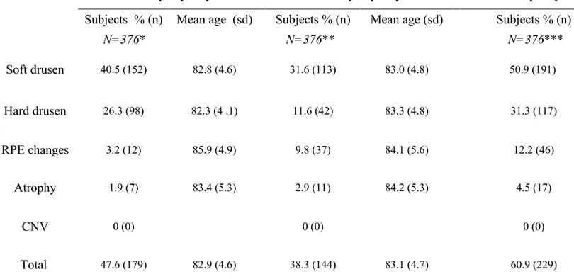
As shown in Table 1, in the mid-periphery (zone 4), 152 (40.5%) subjects had soft drusen in at least one eye, 98 (26.3%) hard drusen, 12 (3.2%) RPE changes, 7 (1.9%) atrophy and no CNV. In the
far-periphery (zone 5) we observed 113 (31.6%) subjects with soft drusen, 42 (11.6%) with hard drusen, 37 (9.8%) with RPE changes, 11 (2.9%) with atrophy and no CNV.
A total of 191 patients (51%) showed soft drusen in the periphery (zone 4 and/or 5), 117 (31.3%) hard drusen, 46 (12.2%) RPE changes, 17 (4.5%) atrophy and no CNV. Overall, 179 patients (47.6%) had at least one peripheral AMD-like lesion in the mid periphery, 144 (38.3%) in the far periphery and 229 (61%) had at least one peripheral abnormalities resembling AMD.
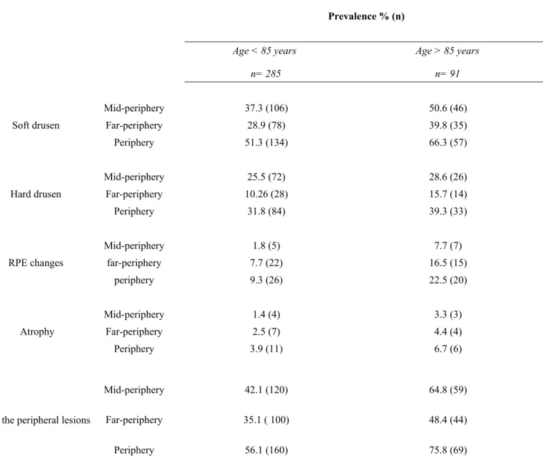
The prevalence of all types of lesions were higher in subjects aged more than 85 years, by comparison with younger subjects, both in the mid and far-periphery (Table 2). In participants aged 75-84 years, 42.1% had at least one abnormality in the mid-periphery, 35.1% in the far-periphery and 56.1% in the periphery (zone 4+5). In participants older than 85 years, 64.8% had lesions in the mid-periphery, 48.4% in the far-periphery and 75.8% in the periphery (zone 4+5).
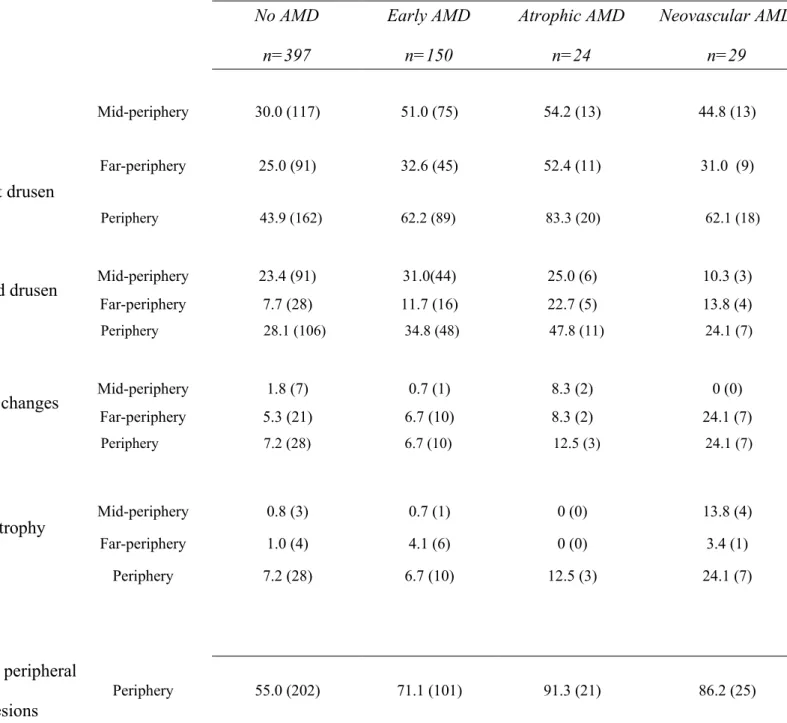
In Table 3, we report the frequency of abnormalities according to the stage of AMD. For all the items, eyes with signs of early or late AMD had more frequently peripheral abnormalities as compared with those without AMD. The frequency of peripheral lesions increased from 55% in eyes without AMD, to 71% in those with early AMD, 91% in those with atrophic AMD and 86% in those with neovascular AMD. In
other words, the prevalence of peripheral retinal abnormalities increased with the severity of the stage of the AMD and nearly all the atrophic forms (91%) were associated with peripheral lesions resembling AMD.
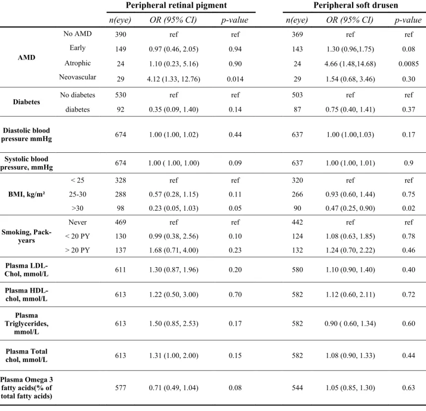
However, a
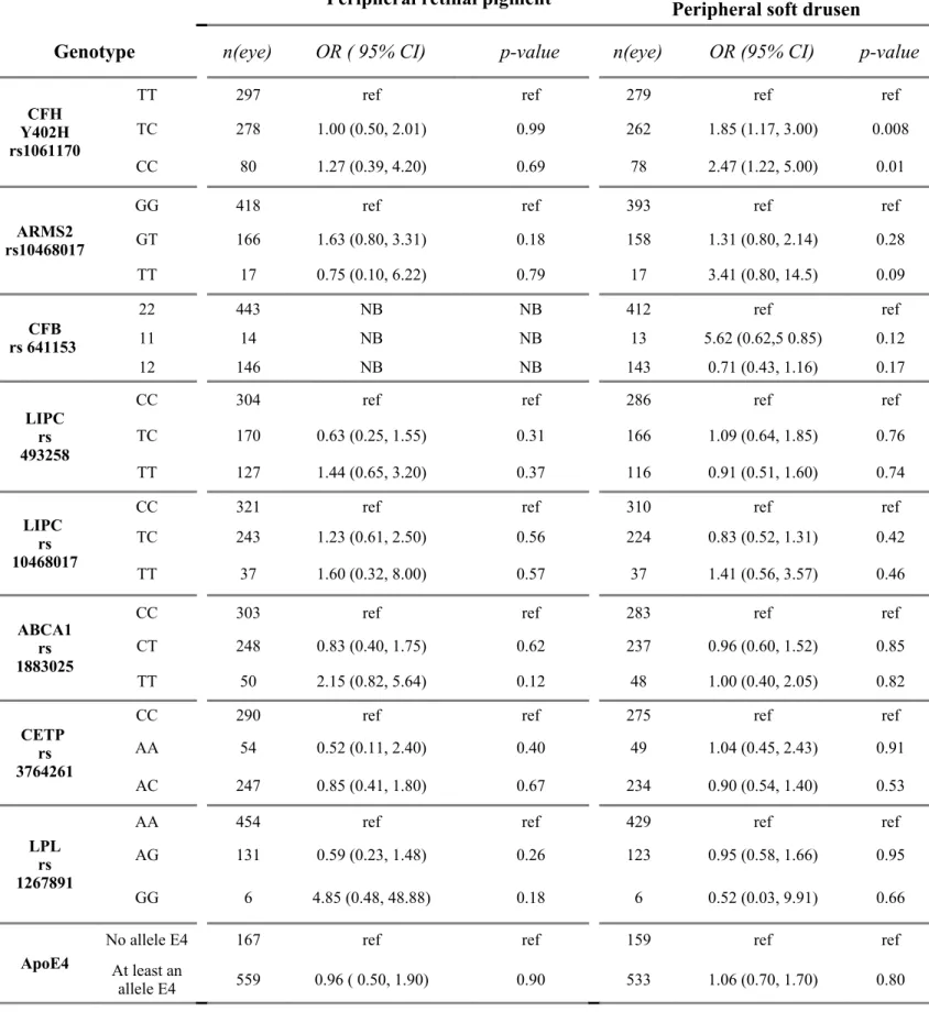
fter adjustment for age and gender, only the associations of peripheral soft drusen with atrophic AMD remained significant (OR=4.66, p= 0.0085) as well as RPE changes with neovascular AMD (OR= 4.12, p=0.014) (Table 4) suggesting that increased frequency of peripheral lesions in AMD subjects were partly due to the older age of these subjects.With regard to AMD risk factors, we found significant age and gender adjusted associations of peripheral soft drusen with CFH Y402H polymorphism (OR= 1.85, p=0.008 and OR=2.47, p=0.01 respectively for TC and CC phenotypes), but not with other AMD-related genes ( ARMS2, LIPC, ABCA1, CETP, LPL, ApoE4, ApoE2). Moreover, we found an opposite statistical association -compared with AMD- between higher BMI (> 30 kg/m²) and peripheral soft drusen (OR= 0.47, p= 0.02 ). No significant associations were found with smoking status, high blood pressure, diabetes, plasma lipids or omega 3 fatty acids or genetic polymorphisms .(Table 5)
DISCUSSION
Since a few years, reliable UWF imaging system is feasible with the Optomap Panoramic 200Tx system (Optos PLC, Dunfermline, Fife, Scotland, UK) which is an ultra-wide field scanning laser
ophthalmoscope (SLO) with two laser wavelengths scanning at 532nm (‘‘green laser separation’’) and 633nm (‘‘red laser separation’’). The two images can be viewed separately or superimposed by specific software (Vantage V2, Optos PLC, Dunfermline, Fife, Scotland, UK) to yield semirealistic colour image. This nonmydriatic imaging system provides up to a 200-degree field of view of the retina (82%) without contact, quickly and with good quality images even if there are lens opacities. This is made possible by an ellipsoidal mirror of the scanning laser ophthalmoscope designed by optos and which can capture UWF of the retina. Prevalence of peripheral abnormalities with UFW imaging has been described in few publications and to our knowledge, this study is the first assessment of the associations between UFW peripheral changes
resembling early and late AMD disease and so many AMD risk factors. With regards to AMD, previous publications described mainly autofluorescence peripheral changes in Optomap and relation with CFH genotypes (24,38–43)
.
Tan&Al reported color and autofluorescence photographic peripheralabnormalities resembling AMD but in a population exclusively of AMD patients (24); Heussen&al (40) in a population of patients consulting for one symptom and Munch&Al (43) only peripheral drusen without UWF imaging system. Recently Lengyel&Al (44) presented the prevalence of peripheral lesions resembling AMD on Optomap and the associations with macular lesions but not with AMD risk factors.
We report a high prevalence of peripheral abnormalities resembling AMD in this elderly population, especially soft drusen in mid-periphery. 61% of subjects presented peripheral abnormalities, 51% soft drusen, 31% hard drusen, 12% RPE changes, 4.5% atrophy, no CNV or PED.
It is very close to what Heussen reported on UWF color image (68.5% of the subjects had at least one lesion in the periphery). It is similar to what Tan reported for drusen (51.7% of eyes) compared to our atrophic AMD population for soft drusen but we found much lower RPE changes (35% of eyes) and peripheral atrophy (16.8% of eyes) except for the dry AMD patients in zone 4. Munch described much lower drusen
(3.3% of the patients) but without UWF system and in a younger population.Compared to Lengyel, who used very similar methodology, we found similar prevalence of peripheral lesions (67.5% of the eyes), a little less of peripheral drusen (67.0%), more RPE changes (5.3%) and similar peripheral atrophy (4.5%); by taking into account that results were given in eyes while provided prevalence by subjects.
Peripheral changes were more prevalent in older participants and in AMD participants as reported by Munch. After adjustment for age and gender, the presence of peripheral soft drusen was statistically
associated with atrophic AMD [OR=4.66, p=0.0085] while RPE changes were associated with neovascular AMD [OR=4.12, p=0.014]. We also found a statistically significant association between peripheral soft drusen and the CFH Y402H polymorphism, a major risk factor for AMD (OR=1.85, p=0.008 for the TC phenotypes and OR=2.47, p=0.01 for the CC phenotypes). However, other risk factors that were strongly associated with macular early abnormalities in the Alienor study (in particular ARMS2, LIPC and LPL genes, plasma HDL-cholesterol), were not associated with peripheral drusen or RPE abnormalities (33,36,37). Our results are consistent with a previous study, which showed an association of peripheral drusen with late AMD and the CFH Y402H polymorphism, but not with the ARMS2 locus (41).Finally, a lower risk of peripheral soft drusen was found in subjects with high BMI >30 kg/m² [OR=0.47, p=0.02], by contrast with the highest risk for AMD generally found in these subjects (45). Overall, our results suggest that the natural history of peripheral abnormalities may be partially different from that of macular lesions.
Our study analyses UWF imaging in a population-based sample of elderly subjects, with 63% of women, an average of age of 82 years and a prevalence of AMD similar to the one observed in the same age group in other European studiesand other industrialized countries (4,5,46)
.
The same qualified technician used the same camera and images were interpreted by a reference reading center; graders were unaware of any findings on the clinical examinations or risk factor exposures of the study participants, in order to minimize classification bias.This can explain why missing data are mostly in far periphery, especially for soft drusen and hard drusen. Acquiring images may be easier with retraction of eyelids, as reported by Csutak and Inoue (6,47). The relatively small number of AMD cases in our series may limit statistical power for analysis of
subcategories (atrophic/neovascular). Only one grader performed the picture interpretation and there was no agreement test, but the grader was in a trained and experienced reading center. Hard drusen (< 125µm) includes drusens <63 µm but their significance is not proven. We did not find any Pigment Epithelial Detachment or peripheral CNV.
CONCLUSION
There are many age-related changes found in the periphery of elderly subjects, that may be correlated to the development of macular lesions. However, peripheral changes appear related only to a subgroup of AMD risk factors (in particular CFH polymorphisms), suggesting that their etiology may be partially different. Further studies with UFW imaging are needed to confirm these findings, in particular in large prospective population-based studies.
Acknowledgments: The autors thank, the Optos clinical research team (Anne Marie Cairns), the staff at the Moorfields Eye Hospital Reading center , and the medical staff at the Department of ophthalmology of the CHU of Bordeaux, France (Brigitte Gontier).
REFERENCES
1. Resnikoff S, Pascolini D, Etya’ale D, Kocur I, Pararajasegaram R, Pokharel GP, et al. Global data on visual impairment in the year 2002. Bull World Health Organ. nov 2004;82(11):844-51.
2. Klaver CC, Wolfs RC, Vingerling JR, Hofman A, de Jong PT. Age-specific prevalence and causes of blindness and visual impairment in an older population: the Rotterdam Study. Arch Ophthalmol. mai 1998;116(5):653-8.
3. Lim LS, Mitchell P, Seddon JM, Holz FG, Wong TY. Age-related macular degeneration. Lancet. 5 mai 2012;379(9827):1728-38.
4. Augood CA, Vingerling JR, de Jong PTVM, Chakravarthy U, Seland J, Soubrane G, et al. Prevalence of age-related maculopathy in older Europeans: the European Eye Study (EUREYE). Arch
Ophthalmol. avr 2006;124(4):529-35.
5. Friedman DS, O’Colmain BJ, Muñoz B, Tomany SC, McCarty C, de Jong PTVM, et al. Prevalence of age-related macular degeneration in the United States. Arch Ophthalmol. avr 2004;122(4):564-72.
6. Csutak A, Lengyel I, Jonasson F, Leung I, Geirsdottir A, Xing W, et al. Agreement between image grading of conventional (45°) and ultra wide-angle (200°) digital images in the macula in the Reykjavik eye study. Eye Lond Engl. oct 2010;24(10):1568-75.
7. Cheng SCK, Yap MKH, Goldschmidt E, Swann PG, Ng LHY, Lam CSY. Use of the Optomap with lid retraction and its sensitivity and specificity. Clin Exp Optom J Aust Optom Assoc. juill
2008;91(4):373-8.
8. Friberg TR, Pandya A, Eller AW. Non-mydriatic panoramic fundus imaging using a non-contact scanning laser-based system. Ophthalmic Surg Lasers Imaging Off J Int Soc Imaging Eye. déc 2003;34(6):488-97.
the management of non-infectious retinal vasculitis. J Ophthalmic Inflamm Infect. 2013;3(1):30.
10. Anderson L, Friberg TR, Singh J. Ultrawide-angle retinal imaging and retinal detachment. Semin Ophthalmol. mars 2007;22(1):43-7.
11. Jain A, Shah SP, Tsui I, McCannel TA. The value of Optos Panoramic 200MA imaging for the monitoring of large suspicious choroidal lesions. Semin Ophthalmol. févr 2009;24(1):43-4.
12. Meyer CH, Saxena S. Non-mydriatic imaging of a giant retinal tear with the Optos Optomap Panoramic 200MA. Clin Experiment Ophthalmol. mai 2010;38(4):427.
13. Kernt M, Schaller UC, Stumpf C, Ulbig MW, Kampik A, Neubauer AS. Choroidal pigmented lesions imaged by ultra-wide-field scanning laser ophthalmoscopy with two laser wavelengths (Optomap). Clin Ophthalmol Auckl NZ. 2010;4:829-36.
14. Mudvari SS, Virasch VV, Singa RM, MacCumber MW. Ultra-wide-field imaging for cytomegalovirus retinitis. Ophthalmic Surg Lasers Imaging Off J Int Soc Imaging Eye. juin 2010;41(3):311-5.
15. Prasad PS, Oliver SCN, Coffee RE, Hubschman J-P, Schwartz SD. Ultra wide-field angiographic characteristics of branch retinal and hemicentral retinal vein occlusion. Ophthalmology. avr 2010;117(4):780-4.
16. Shah SP, Jain A, Tsui I, McCannel TA. Optos Optomap Panoramic 200MA imaging of a serous choroidal detachment responsive to furosemide. Semin Ophthalmol. févr 2009;24(1):40-2.
17. Theodoropoulou S, Ainsworth S, Blaikie A. Ultra-wide field imaging of retinopathy of prematurity (ROP) using Optomap-200TX. BMJ Case Rep. 2013;2013.
18. Neubauer AS, Kernt M, Haritoglou C, Priglinger SG, Kampik A, Ulbig MW. Nonmydriatic screening for diabetic retinopathy by ultra-widefield scanning laser ophthalmoscopy (Optomap). Graefes Arch Clin Exp Ophthalmol Albrecht Von Graefes Arch Für Klin Exp Ophthalmol. févr 2008;246(2):229-35.
19. Dai S, Chow K, Vincent A. Efficacy of wide-field digital retinal imaging for retinopathy of prematurity screening. Clin Experiment Ophthalmol. janv 2011;39(1):23-9.
20. Campbell JP, Leder HA, Sepah YJ, Gan T, Dunn JP, Hatef E, et al. Wide-field retinal imaging in the management of noninfectious posterior uveitis. Am J Ophthalmol. nov 2012;154(5):908-11.e2.
21. Coffee RE, Jain A, McCannel TA. Ultra wide-field imaging of choroidal metastasis secondary to primary breast cancer. Semin Ophthalmol. févr 2009;24(1):34-6.
22. Cho M, Kiss S. Detection and monitoring of sickle cell retinopathy using ultra wide-field color photography and fluorescein angiography. Retina Phila Pa. avr 2011;31(4):738-47.
23. Wessel MM, Aaker GD, Parlitsis G, Cho M, D’Amico DJ, Kiss S. Ultra-wide-field angiography improves the detection and classification of diabetic retinopathy. Retina Phila Pa. avr
2012;32(4):785-91.
24. Tan CS, Heussen F, Sadda SR. Peripheral autofluorescence and clinical findings in neovascular and non-neovascular age-related macular degeneration. Ophthalmology. juin 2013;120(6):1271-7.
25. Silva PS, Cavallerano JD, Sun JK, Soliman AZ, Aiello LM, Aiello LP. Peripheral lesions identified by mydriatic ultrawide field imaging: distribution and potential impact on diabetic retinopathy severity. Ophthalmology. déc 2013;120(12):2587-95.
26. Piffer A-LL, Boissonnot M, Gobert F, Zenger A, Wolf S, Wolf U, et al. Relevance of wide-field autofluorescence imaging in Birdshot retinochoroidopathy: descriptive analysis of 76 eyes. Acta Ophthalmol (Copenh). sept 2014;92(6):e463-9.
27. Delcourt C, Korobelnik J-F, Barberger-Gateau P, Delyfer M-N, Rougier M-B, Le Goff M, et al. Nutrition and age-related eye diseases: the Alienor (Antioxydants, Lipides Essentiels, Nutrition et maladies OculaiRes) Study. J Nutr Health Aging. déc 2010;14(10):854-61.
28. 3C Study Group. Vascular factors and risk of dementia: design of the Three-City Study and baseline characteristics of the study population. Neuroepidemiology. déc 2003;22(6):316-25.
29. Bird AC, Bressler NM, Bressler SB, Chisholm IH, Coscas G, Davis MD, et al. An international classification and grading system for age-related maculopathy and age-related macular degeneration. The International ARM Epidemiological Study Group. Surv Ophthalmol. avr 1995;39(5):367-74.
30. Klein R, Klein BEK, Knudtson MD, Wong TY, Cotch MF, Liu K, et al. Prevalence of age-related macular degeneration in 4 racial/ethnic groups in the multi-ethnic study of atherosclerosis. Ophthalmology. mars 2006;113(3):373-80.
31. Delcourt C, Delyfer M-N, Rougier M-B, Amouyel P, Colin J, Le Goff M, et al. Associations of complement factor H and smoking with early age-related macular degeneration: the ALIENOR study. Invest Ophthalmol Vis Sci. juill 2011;52(8):5955-62.
32. Merle B, Delyfer M-N, Korobelnik J-F, Rougier M-B, Colin J, Malet F, et al. Dietary omega-3 fatty acids and the risk for age-related maculopathy: the Alienor Study. Invest Ophthalmol Vis Sci. juill 2011;52(8):6004-11.
33. Delcourt C, Delyfer M-N, Rougier M-B, Lambert J-C, Amouyel P, Colin J, et al. ARMS2 A69S polymorphism and the risk for age-related maculopathy: the ALIENOR study. Arch Ophthalmol. août 2012;130(8):1077-8.
34. Cougnard-Grégoire A, Delyfer M-N, Korobelnik J-F, Rougier M-B, Malet F, Le Goff M, et al. Long-term blood pressure and age-related macular degeneration: the ALIENOR study. Invest Ophthalmol Vis Sci. mars 2013;54(3):1905-12.
35. Merle BMJ, Delyfer M-N, Korobelnik J-F, Rougier M-B, Malet F, Féart C, et al. High concentrations of plasma n3 fatty acids are associated with decreased risk for late age-related macular degeneration. J Nutr. avr 2013;143(4):505-11.
of HDL-related loci with age-related macular degeneration and plasma lutein and zeaxanthin: the Alienor study. PloS One. 2013;8(11):e79848.
37. Cougnard-Grégoire A, Delyfer M-N, Korobelnik J-F, Rougier M-B, Le Goff M, Dartigues J-F, et al. Elevated high-density lipoprotein cholesterol and age-related macular degeneration: the Alienor study. PloS One. 2014;9(3):e90973.
38. Reznicek L, Wasfy T, Stumpf C, Kampik A, Ulbig M, Neubauer AS, et al. Peripheral fundus autofluorescence is increased in age-related macular degeneration. Invest Ophthalmol Vis Sci. avr 2012;53(4):2193-8.
39. Witmer MT, Kozbial A, Daniel S, Kiss S. Peripheral autofluorescence findings in age-related macular degeneration. Acta Ophthalmol (Copenh). sept 2012;90(6):e428-33.
40. Heussen FM, Tan CS, Sadda SR. Prevalence of peripheral abnormalities on ultra-widefield greenlight (532 nm) autofluorescence imaging at a tertiary care center. Invest Ophthalmol Vis Sci. sept
2012;53(10):6526-31.
41. Seddon JM, Reynolds R, Rosner B. Peripheral retinal drusen and reticular pigment: association with CFHY402H and CFHrs1410996 genotypes in family and twin studies. Invest Ophthalmol Vis Sci. févr 2009;50(2):586-91.
42. Shuler RK, Schmidt S, Gallins P, Hauser MA, Scott WK, Caldwell J, et al. Peripheral reticular pigmentary change is associated with complement factor H polymorphism (Y402H) in age-related macular degeneration. Ophthalmology. mars 2008;115(3):520-4.
43. Munch IC, Ek J, Kessel L, Sander B, Almind GJ, Brøndum-Nielsen K, et al. Small, hard macular drusen and peripheral drusen: associations with AMD genotypes in the Inter99 Eye Study. Invest Ophthalmol Vis Sci. mai 2010;51(5):2317-21.
44. Lengyel I, Csutak A, Florea D, Leung I, Bird AC, Jonasson F, et al. A Population-Based Ultra-Widefield Digital Image Grading Study for Age-Related Macular Degeneration-Like Lesions at the Peripheral Retina. Ophthalmology. 11 avr 2015.
45. Chakravarthy U, Wong TY, Fletcher A, Piault E, Evans C, Zlateva G, et al. Clinical risk factors for age-related macular degeneration: a systematic review and meta-analysis. BMC Ophthalmol. 2010;10:31.
46. Vingerling JR, Dielemans I, Hofman A, Grobbee DE, Hijmering M, Kramer CF, et al. The prevalence of age-related maculopathy in the Rotterdam Study. Ophthalmology. févr 1995;102(2):205-10.
47. Inoue M, Yanagawa A, Yamane S, Arakawa A, Kawai Y, Kadonosono K. Wide-field fundus imaging using the Optos Optomap and a disposable eyelid speculum. JAMA Ophthalmol. févr 2013;131(2):226.
Table 1. Prevalence of retinal lesions for each peripheral zone, in subjects of the Alienor study (%(n)).
Mid-periphery
Far-periphery
Periphery
Subjects % (n) Mean age (sd)
Subjects % (n)
Mean age (sd)
Subjects % (n)
N=376* N=376**
N=376***
Soft drusen
40.5 (152) 82.8 (4.6) 31.6 (113) 83.0 (4.8) 50.9 (191)Hard drusen
26.3 (98) 82.3 (4 .1) 11.6 (42) 83.3 (4.8) 31.3 (117)RPE changes
3.2 (12) 85.9 (4.9) 9.8 (37) 84.1 (5.6) 12.2 (46)Atrophy
1.9 (7) 83.4 (5.3) 2.9 (11) 84.2 (5.3) 4.5 (17)CNV
0 (0) 0 (0) 0 (0)Total
47.6 (179) 82.9 (4.6) 38.3 (144) 83.1 (4.7) 60.9 (229)Data are frequencies (%) of the total group ( the numbers of subjects), unless otherwise specified sd= standard deviation
* Missing values for the Mid-periphery, respectively for Soft Drusen, Hard Drusen and RPE changes: 1, 3, 1. ** Missing values for the Far-periphery, respectively for Soft Drusen and Hard Drusen: 18, 14,
Table 2. Prevalence of peripheral retinal abnormalities according to age, in subjects of the Alienor study
(% (n)).
Prevalence % (n) Age < 85 years n= 285 Age > 85 years n= 91 Soft drusen Mid-periphery 37.3 (106) 50.6 (46) Far-periphery 28.9 (78) 39.8 (35) Periphery 51.3 (134) 66.3 (57) Hard drusen Mid-periphery 25.5 (72) 28.6 (26) Far-periphery 10.26 (28) 15.7 (14) Periphery 31.8 (84) 39.3 (33) RPE changes Mid-periphery 1.8 (5) 7.7 (7) far-periphery 7.7 (22) 16.5 (15) periphery 9.3 (26) 22.5 (20) Atrophy Mid-periphery 1.4 (4) 3.3 (3) Far-periphery 2.5 (7) 4.4 (4) Periphery 3.9 (11) 6.7 (6)Any the peripheral lesions
Mid-periphery 42.1 (120) 64.8 (59)
Far-periphery 35.1 ( 100) 48.4 (44)
Periphery 56.1 (160) 75.8 (69)
Table 3. Proportion of peripheral retinal changes according to the stage of AMD (%(n)).
Prevalence (eyes) % (n)
No AMD
n=397
Early AMD
n=150
Atrophic AMD
n=24
Neovascular AMD
n=29
Soft drusen
Mid-periphery 30.0 (117) 51.0 (75) 54.2 (13) 44.8 (13) Far-periphery 25.0 (91) 32.6 (45) 52.4 (11) 31.0 (9) Periphery 43.9 (162) 62.2 (89) 83.3 (20) 62.1 (18)Hard drusen
Mid-periphery 23.4 (91) 31.0(44) 25.0 (6) 10.3 (3)Far-periphery 7.7 (28) 11.7 (16) 22.7 (5) 13.8 (4) Periphery 28.1 (106) 34.8 (48) 47.8 (11) 24.1 (7)
RPE changes
Mid-periphery 1.8 (7) 0.7 (1) 8.3 (2) 0 (0)Far-periphery 5.3 (21) 6.7 (10) 8.3 (2) 24.1 (7) Periphery 7.2 (28) 6.7 (10) 12.5 (3) 24.1 (7)
Atrophy
Mid-periphery 0.8 (3) 0.7 (1) 0 (0) 13.8 (4)Far-periphery 1.0 (4) 4.1 (6) 0 (0) 3.4 (1) Periphery 7.2 (28) 6.7 (10) 12.5 (3) 24.1 (7)
All the peripheral
lesions
Table 4. Associations of AMD-like lesions at the peripheral retina and risk factors of the AMD in the Alienor study.
Peripheral retinal pigment
Peripheral soft drusen
n(eye)
OR (95% CI)
p-value
n(eye)
OR (95% CI)
p-value
AMD
No AMD 390 ref ref 369 ref ref
Early 149 0.97 (0.46, 2.05) 0.94 143 1.30 (0.96,1.75) 0.08 Atrophic 24 1.10 (0.23, 5.16) 0.90 24 4.66 (1.48,14.68) 0.0085 Neovascular 29 4.12 (1.33, 12.76) 0.014 29 1.54 (0.68, 3.46) 0.30
Diabetes No diabetes 530 ref ref 503 ref ref
diabetes 92 0.35 (0.09, 1.40) 0.14 87 0.75 (0.40, 1.41) 0.37 Diastolic blood pressure mmHg 674 1.00 (1.00, 1.02) 0.44 637 1.00 (1.00,1.03) 0.17 Systolic blood pressure, mmHg 674 1.00 ( 1.00, 1.00) 0.09 637 1.00 (1.00, 1.01) 0.9 BMI, kg/m²
< 25 328 ref ref 320 ref ref
25-30 288 0.57 (0.28, 1.15) 0.11 266 0.93 (0.60, 1.44) 0.75 >30 98 0.23 (0.05, 1.03) 0.05 90 0.47 (0.25, 0.90) 0.02
Smoking, Pack-years
Never 469 ref ref 442 ref ref
< 20 PY 130 0.99 (0.38, 2.56) 0.10 124 1.08 (0.63, 1.85) 0.78 > 20 PY 137 1.68 (0.71, 4.00) 0.23 132 1.24 (0.70, 2.22) 0.46 Plasma LDL-Chol, mmol/L 611 1.30 (0.87, 1.96) 0.20 580 1.10 (0.90, 1.40) 0.40 Plasma HDL-chol, mmol/L 613 1.22 (0.50, 3.00) 0.70 582 1.12 (0.60, 2.11) 0.72 Plasma Triglycerides, mmol/L 613 1.50 (0.85, 2.53) 0.17 582 0.90 ( 0.60, 1.34) 0.60 Plasma Total chol, mmol/L 613 1.31 (1.00, 2.00) 0.15 582 1.08 (0.90, 1.33) 0.44 Plasma Omega 3 fatty acids(% of
Table 5. Associations between genotypes and peripheral retinal drusen and pigment in the Alienor Study.
Peripheral retinal pigment
Peripheral soft drusen
Genotype
n(eye)
OR ( 95% CI)
p-value
n(eye)
OR (95% CI)
p-value
CFH Y402H rs1061170
TT 297 ref ref 279 ref ref
TC 278 1.00 (0.50, 2.01) 0.99 262 1.85 (1.17, 3.00) 0.008 CC 80 1.27 (0.39, 4.20) 0.69 78 2.47 (1.22, 5.00) 0.01
ARMS2 rs10468017
GG 418 ref ref 393 ref ref
GT 166 1.63 (0.80, 3.31) 0.18 158 1.31 (0.80, 2.14) 0.28 TT 17 0.75 (0.10, 6.22) 0.79 17 3.41 (0.80, 14.5) 0.09 CFB rs 641153 22 443 NB NB 412 ref ref 11 14 NB NB 13 5.62 (0.62,5 0.85) 0.12 12 146 NB NB 143 0.71 (0.43, 1.16) 0.17 LIPC rs 493258
CC 304 ref ref 286 ref ref
TC 170 0.63 (0.25, 1.55) 0.31 166 1.09 (0.64, 1.85) 0.76 TT 127 1.44 (0.65, 3.20) 0.37 116 0.91 (0.51, 1.60) 0.74
LIPC rs 10468017
CC 321 ref ref 310 ref ref
TC 243 1.23 (0.61, 2.50) 0.56 224 0.83 (0.52, 1.31) 0.42 TT 37 1.60 (0.32, 8.00) 0.57 37 1.41 (0.56, 3.57) 0.46
ABCA1 rs 1883025
CC 303 ref ref 283 ref ref
CT 248 0.83 (0.40, 1.75) 0.62 237 0.96 (0.60, 1.52) 0.85 TT 50 2.15 (0.82, 5.64) 0.12 48 1.00 (0.40, 2.05) 0.82
CETP rs 3764261
CC 290 ref ref 275 ref ref
AA 54 0.52 (0.11, 2.40) 0.40 49 1.04 (0.45, 2.43) 0.91 AC 247 0.85 (0.41, 1.80) 0.67 234 0.90 (0.54, 1.40) 0.53
LPL rs 1267891
AA 454 ref ref 429 ref ref
AG 131 0.59 (0.23, 1.48) 0.26 123 0.95 (0.58, 1.66) 0.95 GG 6 4.85 (0.48, 48.88) 0.18 6 0.52 (0.03, 9.91) 0.66
ApoE4
No allele E4 167 ref ref 159 ref ref
At least an
ApoE2
No allele E2 164 ref ref 158 ref ref
At least an
allele E2 562 0.80 (0.40,1.60) 0.52 534 1.3 (0.82, 2.14) 0.25 NB: could not be determined because of low number of observations.
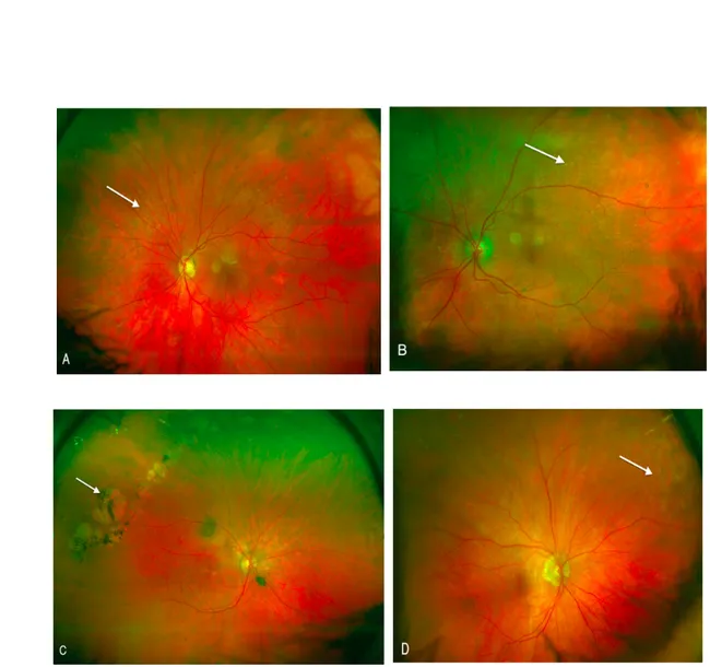
Figure 1: Color photos illustrating peripheral fundus abnormalities
A: Photography illustrating hard drusen
B: Image showing soft drusen C : Areas of GA D: Areas of RPE changes in the periphery of the
Figure 2:
Projected grid on image taken by Optos P200C UWF laser scanning ophtalmoscope.
Acknowledgments to the authors and the AAO to allow us to use this image.Lengyel I, Csutak A, Florea D, Leung I, Bird AC, Jonasson F, et al. A Population-Based Ultra-Widefield Digital Image Grading Study for Age-Related Macular Degeneration-Like Lesions at the Peripheral Retina. Ophthalmology. 11 avr 2015.
SERMENT D’HIPPOCRATE
Au moment d’etre admis(e) à exercer la médecine, je promets et je jure d’etre fidèle aux lois de l’honneur et de la probité.
Mon premier souci sera de rétablir, de préserver ou de promouvoir la santé dans tous ses éléments, physiques et mentaux, individuels et sociaux.
Je respecterai toutes les personnes, leur autonomie et leur volonté, sans aucune discrimination selon leur état ou leurs convictions. J’interviendrai pour les protéger si elles sont affaiblies,
vulnérables ou menacées dans leur intégrité ou leur dignité. Meme sous la contrainte, je ne ferai pas usage de mes connaissances contre les lois de l’humanité.
J’informerai les patients des décisions envisagées, de leurs raisons et de leurs conséquences. Je ne tromperai jamais leur confiance et n’exploiterai pas le pouvoir hérité des circonstances pour forcer les consciences.
Je donnerai mes soins à l’indigent et à quiconque me les demandera. Je ne me laisserai pas influencer par la soif du gain ou la recherche de la gloire.
Admis(e) dans l’intimité des personnes, je tairai les secrets qui me seront confiés. reçu(e) à l’intérieur des maisons, je respecterai les secrets des foyers et ma conduite ne servira pas à corrompre les moeurs.
Je ferai tout pour soulager les souffrances. Je ne prolongerai pas abusivement les agonies. Je ne provoquerai jamais la mort délibérément.
Je préserverai l’indépendance nécessaire à l’accomplissement de ma mission. Je
n’entreprendrai rien qui dépasse mes compétences. Je les entretiendrai et les perfectionnerai pour assurer au mieux les services qui me seront demandés.
J’apporterai mon aide à mes confrères ainsi qu’à leurs familles dans l’adversité.
Que les hommes et mes confrères m’accordent leur estime si je suis fidèle à mes promesses ; que je sois déshonoré(e) et méprisé(e) si j’y manque.