Effects of Holothuroid Ichtyotoxic Saponins on the
Gills of Free-Living Fishes and Symbiotic Pearlfishes
IGOR EECKHAUT1*, GUILLAUME CAULIER1, LOLA BRASSEUR1, PATRICK FLAMMANG1,
PASCAL GERBAUX2, AND ERIC PARMENTIER3*
1
Biology of Marine Organisms and Biomimetics, University of Mons, B-7000 Mons, Belgium;2Synthesis and Organic Mass Spectrometry, University of Mons, B-7000 Mons, Belgium; and3Laboratory of
Morphology, University of Lie`ge, B6 Sart Tilman, B-4000 Lie`ge, Belgium
Abstract. Several carapid fishes, known as pearlfishes, are endosymbiotic in holothuroids and asteroids. These echinoderms contain a strong concentration of saponins that are efficient membranolytic repellents to predators. We compared the effects of exposure to saponins from the sea cucumber body wall and from the Cuvierian tubules on the behavior and gill ultrastructure of pearlfishes and free-living fishes. Saponins were extracted from the body wall of two holothuroids, the Mediterranean Holothuria forskali and the tropical Bohadschia atra, and from the water surrounding the Cuvierian tubules of B. atra. Five species of carapids that live in symbiosis with holothuroids and seven species of free-living fishes were exposed to these extracts. The free-living fishes exhibited a stress response and died about 45 times faster than pearlfishes when exposed to the same quantity of saponins. Cuvierian tubules and saponins ex-tracted from the body wall were lethal to the free-living fishes, whereas the carapids were much less sensitive. The carapids did not exhibit a stress response.The high toxicity shown by Cuvierian tubules was not explained by the nature of the saponins that were identified by mass spectrometry, but it is likely due to the higher concentration of saponins in the tubules. Histology and scanning and transmission elec-tron microscopy of the gills of the free-living fishes and pearlfishes showed that saponins act at the level of the secondary lamellae where they induce the detachment of the epithelia, create edema at the level of the epithelia, and induce pores in the epithelial cells that lead to their destruc-tion and the invasion of inner cells (pillar cells and red
blood cells). This sequence of events happens 5 min after saponin exposure in free-living fishes and after 1 h in carapids.
Introduction
Coral reefs form complex marine ecosystems where thousands of animal associations occur, such as those involving invertebrates and fishes. Among them, the anemone/clownfish symbiosis is probably the best known (e.g., Mebs, 1994; Elliott and Mariscal, 1997; Cleveland
et al., 2011), but another extraordinary example, though
less known, is the symbiosis between carapid fishes (Ophidiiformes) and various invertebrates. Although sev-eral species are free-living, those belonging to the genera
Carapus, Encheliophis, and Onuxodon are able to enter
and live inside different invertebrates, especially echino-derms such as holothuroids and asteroids. These remark-able associations account for the notoriety of the group (Trott, 1981; Markle and Olney, 1990). Species belong-ing to the genera Carapus and Onuxodon are commen-sals, whereas Encheliophis species are regarded as para-sites (Parmentier and Das, 2004; Parmentier and Michel, 2013). Parasitic species reside inside their hosts and eat their internal tissues (gonads, digestive glands); commen-sals use their hosts as shelters and feed outside them (Parmentier and Vandewalle, 2003; Parmentier and Das, 2004).
It is well known that holothuroid tissues contain a high quantity of saponins (Nigrelli, 1952; Van Dyck et al., 2009, 2010), secondary metabolites that act as a predator deter-rent. They are abundant in the body wall and are also
Received 16 July 2014; accepted 22 April 2015.
* To whom correspondence should be addressed. E-mail: igor.eeckhaut@ umons.ac.be or e.parmentier@ulg.ac.be
Abbreviations: OM, opercular movements. © 2015 Marine Biological Laboratory
particularly concentrated in the Cuvierian tubules, a special-ized defense system developed by some sea cucumber spe-cies of the family Holothuriidae (Matsuno and Iba, 1966; Elyakov et al., 1973). This organ, located in the posterior part of the animal, consists of multiple tubules that can be expelled out of the cloacum after stimulation (Hamel and Mercier, 2000; Flammang et al., 2002). Expelled tubules lengthen into sticky white threads that may entangle poten-tial predators.
Saponins are an important class of natural products first discovered in higher plants, where they are widely distributed (for example in the families Dioscoreaceae, Araliaceae, Legu-minosae, Cucurbitaceae) (Li et al., 2006). In these plants, saponins are found in different compartments such as the rhizome, the leaves, and the cortex (Guo et al., 2006). Owing to their wide variety of biological and pharmacological activ-ities, vegetal saponins are part of the most efficient compounds in popular Chinese medicine (Guo et al., 2006). Nigrelli (1952) and Yamanouchi (1955) were the first to investigate the sapo-nins of sea cucumbers (in Actinopyga agassizi and Holothuria
leucospilota, respectively). Since these contributions, saponins
have been isolated from numerous species of Aspidochirotida and Dendrochirotida and also from sea stars (Mackie and Turner, 1970; Kitagawa and Kobayashi, 1977; Demeyer et al., 2014). More recently, they have also been noted in sponges (Thompson et al., 1985; Kubanek et al., 2000) and in chrysomelid beetles (Plasman et al., 2000a, b). Saponins have been reported to possess a wide range of biological activities including hemolytic, antitumoral, anti-inflammatory, antifun-gal, anti-bacterial, cytostatic, and ichthyotoxic properties (Ka-linin et al., 1996a, b, 2008; Prokofeva et al., 2003; Bordbar et
al., 2011). Many of these activities are the result of their
cytotoxic action—that is, their capacity to induce disturbances, up to lysis, in cellular membranes (Kalinin et al., 1996a). It has been established that a majority of the saponin activities are the consequence of their interaction with cell membrane⌬5sterols, especially cholesterol (Popov et al., 1983; Stonik et al., 1999). This interaction induces significant changes in the physico-chemical properties of cell membranes, such as variations in their stability, microviscosity, and permeability. Saponins form complexes with membrane sterols, leading to cell disruption by the formation of pores. Due to this irreversible interaction, the selective permeability of cell membranes is impaired and cell compounds are transferred into the extracellular matrix, ulti-mately resulting in cell death (Segal and Schlo¨sser, 1975). The toxic activity of saponins on organisms is well known. Ya-manouchi (1955) found that holothuroid saponins kill marine and freshwater fishes, and also earthworms. He observed that freshwater fishes are more resistant to saponins than are marine fishes, and he suggested that it is because the gills of the latter are less permeable. The people of Guam traditionally used holothuroids to poison coral fishes: they cut the holothuroids and squeezed out their contents to render the fishes weak and less active (Bakus, 1973).
Pearlfishes are less sensitive to the ichtyotoxic holothu-roid saponins than are other fishes (Parmentier and Vande-walle, 2005). Parmentier and Vandewalle (2005) found that the time required for carapids to die when exposed to holothuroid saponins is 2 to 16 times longer than for other fishes and suggested that the resistance of pearlfishes to holothuroid toxins may be related to specializations of the gills. Mucus coating may also assist the fish in resisting the sticky substances emitted by the Cuvierian tubules (Trott, 1970). Here, we compare the effects of exposure to Cuvier-ian tubules and to saponin extracts on the ultrastructure of the gills of free-living fish species and pearlfishes (the gills are assumed to be the fish organs most affected by mem-branolytic saponins). We also compare the survival times and motion behavior of these fishes when they are exposed to the various saponins, and we use mass spectrometry to characterize the nature of the saponin cocktails that were used.
Materials and Methods
Samples
Twelve fish species, including 5 carapids and 7 species of free-living fishes, were used in the analyses. The pearlfishes
Carapus homei (Richardson, 1846), C. mourlani (Petit,
1934), and Encheliophis gracilis (Bleeker 1856) were col-lected from the Great Reef of Toliara (Madagascar) in January and February 2008 and 2009; C. boraborensis (Kaup, 1856) and other individuals of C. homei from the Opunohu Bay of Moorea (French Polynesia) in October 2008; and C. acus (Bru¨nnich, 1768) from the Bay of Calvi (Corsica) in June 2008 and 2009. The free-living fishes were used as a control to assess potential specialization of the pearlfishes. These species included Amphiprion akallopisos Bleeker 1853 (Pomacentridae), Dascyllus aruanus (Lin-naeus, 1853) (Pomacentridae), from Toliara; D. flavicaudus Randall and Allen, 1977 (Pomacentridae), D. trimaculatus (Ru¨ppell, 1829) (Pomacentridae), Neoniphon sammara (Forsskaal,1775) (Holocentridae), and other individuals of
Dascyllus aruanus from Moorea (French Polynesia); Sym-phodus ocellatus (Linnaeus, 1758) (Labridae) and Coris julis (Linnaeus, 1758) (Labridae) from Calvi (Corsica).
The carapids were collected by scuba diving (3–15-m depth), along with their holothuroid hosts (Bohadschia
ar-gus Jaeger, 1833, Bohadschia atra Massin, Rasolofonirina,
Conand & Samyn, 1999, Thelenota ananas (Jaeger, 1833), or Holothuria forskali Delle Chiaje 1823). The other species were collected with hand nets from sea anemones
(Am-phiprion akallopisos) or coral reef habitat. The animals
were then kept in tanks of 20 l at 27 °C, with running seawater, for 2 days before being exposed to saponins or the presence of Cuvierian tubules. At the end of each test, some fishes were placed in the freezer to anesthetize and were then euthanized. Samples of their gills were taken
and fixed with Bouin’s fluid or 3% glutaraldehyde solu-tion in cacodylate buffer for histology and scanning and transmission electron microscopy. Animals used in our experiments were maintained and treated in compliance with the guideline specified by the Belgian Ministry of Trade and Agriculture.
Saponin extraction and identification
Saponins of Bohadschia atra (from Toliara, Madagas-car) and Holothuria forskali (from Calvi, Corsica) were used during the experiments. These species were col-lected at a depth of 20 to 40 m by scuba diving. Once in the laboratory, the holothurians were dissected and a piece of body wall was placed in 70% ethanol and stored at ⫺20 °C until extraction. Extracts were prepared as detailed in Van Dyck et al. (2009) and Caulier et al. (2013a, b). Basically, the purification consists in liquid-liquid extractions with different organic solvents (hexane, dichloromethane, chloroform, isobutanol) and a solid phase chromatography (Xad 4 Amberlite). Some extracts were analyzed with mass spectrometry, and oth-ers were used in experiments to test the resistance of fishes to saponins. Cuvierian tubules of B. atra (from 4.7 to 37.3 g) were removed from dissected individuals and placed in 1 liter of seawater for 1 h. The saponin cocktails extracted from the holothuroid body wall and from the water conditioned by the Cuvierian tubules of B. atra were identified to determine similarities or differences that could help to explain the various effects observed. Mass spectrometry (Waters QToF Premier mass spec-trometer) was performed in the positive ion mode, using
the MALDI or the Electrospray ionization (ESI) sources (see also Van Dyck et al., 2009; Caulier et al., 2013a, b).
Behavioral responses of fishes to saponin extract exposure
The behavioral responses of 12 species of fishes (5 spec-imens for each) to saponin extracts was investigated (see Table 1). These fishes were exposed to saponins from the body wall of Bohadschia atra (for the tropical fish species) or Holothuria forskali (for the Mediterranean fish species). The final saponin concentration used in the 1-liter aerated aquaria was 200 g/ml, quantification being obtained through the weighing of the dried extract. The chosen con-centration corresponds to the range of saponin concon-centration previously measured in the water conditioned by H. forskali (290 g/l; Van Dyck et al., 2011) and by Bohadschia
vitiensis (250 g/l; Eeckhaut, unpubl. data). The motion
behavior of fishes was observed for 2 h, and the respiration rate, estimated by the frequency of opercular movements (abbreviated to OM hereafter), was recorded with a hand-held counter after 1 h of exposure. Fishes were first left in the aquarium for 15 min before the beginning of the test to allow them to get used to the new container, and saponins were then introduced.
Behavioral responses of fishes to Cuvierian tubules exposure
For nine tropical species (see Table 2), exposure to Cu-vierian tubules was tested. Each replicate (n⫽ 3) consisted of placing one specimen of Carapus (three tested species) or
Table 1
Morphological data on fishes and fish gills
Species Collection Site Length (cm) Weight (g)
Opercular Rhythm (n/min) Normal Exposed CARAPIDAE
Carapus acus Calvi 12.68⫾ 4.04 4.98⫾ 0.90 21.49⫾ 7.19 24.27⫾ 6.63
Carapus boraborensis Moorea 17.60⫾ 5.50 9.22⫾ 7.08 22.92⫾ 3.55 11.42⫾ 7.28
Carapus homei Moorea 13.26⫾ 1.14 3.38⫾ 1.38 48.91⫾ 6.98 48.27⫾ 13.48
Carapus mourlani Toliara 7.72⫾ 1.82 1.94⫾ 0.91 63.48⫾ 22.42 29.96⫾ 26.58
Encheliophis gracilis Toliara 21.07⫾ 2.39 8.30⫾ 2.83 69.87⫾ 6.78 75.08⫾ 7.77 CONTROL FISHES
Amphiprion akallopisos Toliara 7.97⫾ 1.88 16.01⫾ 12.09 182.96⫾ 24.01 212.56⫾ 17.59
Dascyllus aruanus Moorea 4.70⫾ 0.95 4.23⫾ 4.31 167.07⫾ 49.25 191.46⫾ 33.40
Dascyllus flavicaudus Moorea 4.60⫾ 1.39 2.70⫾ 2.60 174.08⫾ 27.39 188.78⫾ 27.67
Dascyllus trimaculatus Moorea 4.42⫾ 2.02 2.83⫾ 5.22 166.89⫾ 38.98 196.78⫾ 30.02
Neoniphon sammara Toliara 6.00⫾ 0.22 3.11⫾ 0.38 184.06⫾ 26.87 195.56⫾ 18.38
Coris julis Calvi 9.12⫾ 0.35 13.64⫾ 8.53 150.10⫾ 7.10 188.02⫾ 10.7
Symphodus ocellatus Calvi 5.07⫾ 0.36 2.96⫾ 0.82 146.00⫾ 12.4 194.04⫾ 8.9 Opercular rhythms were obtained when fishes were exposed to 200g/l of saponins (extracted from Bohadschia atra or Holothuria forskali; see Materials and Methods) for 1 h.
one of Encheliophis (one tested species) with one free-living fish (five tested species) in a 2.5-l tank with Cuvierian tubules (from 4.7 to 37.3 g). Pearlfishes were tested with free-living fishes so as to expose them to the same tubule quantity and the same experimental conditions. The tubules used for the experiment were either directly expelled by the sea cucumber B. atra or removed from individuals after dissection. The times to mortality of fishes were recorded and their weights were taken at the end of the experiments.
Statistics
Nonparametric Mann-Whitney U-tests were performed (Statistica 7.0) to compare the averages of opercular movements between fish when exposed to saponin ex-tracts. Comparisons were made between (i) each species of fish, (ii) free-living fishes and carapids, and (iii) Med-iterranean and tropical species. The same tests were also applied to compare the average survival times between fishes exposed to Cuvierian tubules. Statistical signifi-cance was set to 5%.
Morphological analyses of the gills
The gill morphology of the five species of carapids (C.
homei, C. acus, C. mourlani, E. gracilis, and A. akallopisos)
was examined with light (3 individuals) or electron microscopy (1 individual) before and after a 5-min exposure to saponin extract (free-living fishes) or 1 h (carapids) to Cuvierian tu-bules. The surface of the gills for the five species was com-pared with scanning electron microscopy; the histology in C.
acus and A. akallopisos was compared, and the ultrastructure
of the secondary lamellae of C. mourlani, E. gracilis, and A.
akallopisos was observed with transmission electron
micros-copy after treatment.
Light microscopy. The samples were fixed in Bouin’s fluid
(with acetic acid) for 8 h and decalcified overnight in a 1:1 mixture of a 2% ascorbic acid solution and NaCl 0.3 mol l–1 at 4 °C (Dietrich and Fontaine, 1975). The sam-ples were then dehydrated through a graded series of ethanol (70%, 90%, and 100%), embedded in Paraplast, and cut into 7m thick sections that were stained with a Masson trichrome (Ganter and Jolle`s, 1969 –1970). The images were captured with an Olympus photomicroscope connected to a computer with the ImageJ, ver. 1.48, program.
Scanning electron microscopy (SEM). The samples were
fixed in Bouin’s fluid (without acetic acid) for 8 h, dehy-drated with a graded series of ethanol (70%, 90%, and 100%) and critical-point dried. They were then mounted on aluminum stubs, coated with gold in a sputter-coater, and observed with a JEOL JSM-6100 scanning electron micro-scope.
Transmission electron microscopy (TEM). The samples
were fixed in a 3% glutaraldehyde solution in 0.1 mol l–1 cacodylate buffer (pH 7.8) for 3 to 24 h at 4 °C. They were washed in the buffer before post-fixation in a 1% osmium tetroxide solution in the same buffer at 4 °C for 1 h. Pieces were then decalcified for 1 week in a mixture of a 2% ascorbic acid solution and NaCl 0.3 mol l–1at 4 °C. Decalcified samples were dehydrated through a graded series of ethanol (70%, 90%, 100%), transferred into Spuur’s resin at room temperature for 24 h followed by another 24 h at 60 °C, and cut with a Leica ultracut UCT ultramicrotome. Sections were stained with lead citrate and uranyl acetate and observed with a Zeiss LEO 906E electron microscope.
Table 2
Average time to mortality of the fishes (controls and carapids; n⫽ 5) when exposed to Cuvierian tubules from Bohadschia argus (for French Polynesian fishes) and B. atra (for Malgachian fishes)
Time to Mortality per Individual
Time to Mortality/Weight of Individual
Time to Mortality/Weight of Individual/Weight Tubules Species Time (min) SD MIN MAX Time (min) SD MIN MAX Time (min) SD MIN MAX
Amphiprion akallopisos 7 1.7 5 8 0.8 0.6 0.3 1.5 0.03 0.11 0.02 0.04 Dascyllus aruanus 4.7 0.6 4 5 3.1 1.6 1.7 4.8 0.58 0.45 0.14 1.04 Dascyllus flavicaudus 5.2 1.3 4 6.5 3.1 3.1 0.8 6.6 0.61 0.7 0.04 1.4 Dascyllus trimaculatus 5.5 1.8 4 7.5 0.4 0.1 0.3 0.6 0.05 0.03 0.02 0.08 Neoniphon sammara 4 0.9 3 4.9 1.8 0.8 0.9 2.6 0.24 0.27 0.08 0.55 Carapus boraborensis 280.7 127 154 392 43.2 30.4 10.9 80 6.53 7.33 0.54 17.02 Carapus homei 226.2 105.8 124 323 82.7 25.3 47.1 103.5 12.29 10.54 2.32 22.03 Carapus mourlani 155 52.2 120 215 64.9 21.3 52.4 89.6 3.3 2.9 1.4 6.64 Encheliophis gracilis 282.2 138.4 193 488 51.7 43.6 24 116.8 7.23 11.74 0.85 24.84 Takes into account (i) the individual, (ii) the weight of individuals, and (iii) the weight of individuals⫹ the weight of Cuvierian tubules.
Results
Behavior of free-living fishes and pearlfishes to saponin exposure
Effect of saponin extract exposure. The recorded normal (i.e.,
non-exposed to saponins) opercular frequencies of free-living fishes varied between 146 and 184 mov/min (Table 1). There was no significant difference between the normal opercular
frequencies of Polynesian and Malagasy fishes (P⬎ 0.05), but there was between the tropical and Mediterranean fishes (P⬍ 0.05). When exposed to saponins, the opercular movements (OM) of free-living fishes increased up to 200% during the first 10 to 15 min and then dropped to between 105% and 133% until the fishes died (Table 1). During the first 15 min, the fishes showed a strong reaction in which they swam vigorously and randomly around the aquarium. After this first phase, the
Figure 1. The number of opercular movements/minute in carapids and free-living fishes (n⫽ 5 for each
species), exposed and nonexposed to saponins from holothuroids. Error bars correspond to standard deviation.
Figure 2. The time to mortality (minutes) of carapids and free-living fishes (n⫽ 3) exposed to saponins of
free-living fish species became quiescent and then died within 2 h of exposure. Therefore, the first sign of saponin toxicity on free-living fishes was an increase in the frequency of OM (Table 1; Fig. 1). However, this increase was statistically significant only in Coris julis and Symphodus ocellatus (P⬍ 0.05).
The opercular frequencies of the carapids were much lower than those recorded for free-living fishes (21 to 70 OM/min) with the lowest in Carapus acus and the highest in Encheliophis gracilis (Table 1). OM of exposed car-apids remained similar (P ⬎ 0.05) except in Carapus
mourlani and Carapus boraborensis, where a decrease in
the frequency of OM was observed (Table 1; Fig. 1). The decrease was very obvious in exposed C. mourlani, with the individuals totally stopping OM for several minutes.
Effects of Cuvierian tubules exposure. The observed effects
were similar to those obtained with body wall extracts (i.e., the increase in OM in the free-living fishes was not observed in carapids), and the free-living fishes were
affected much faster, all dying within 10 min of exposure to Cuvierian tubules. Death occurred even more quickly when the fish became entangled in the tubules (210 s. on average). In contrast, the carapids swam freely across Cuvierian tubules without being entangled in them. How-ever, even though they were more resistant than free-living fishes, they also died after a minimum of 2.5 h. The average times to death of all tested fishes are summarized in Table 2 and illustrated in Figure 2. Table 2 shows (i) the average time to death of individuals (free-living and carapid) and the average lifetimes when these values were normalized by taking into account (ii) the weight of the individuals and (iii) the weight of the individuals and the weight of the Cuvierian tubules. In the three cases, the carapids were clearly much more resistant than the other fishes to the toxins expelled by Cuvierian tubules. The free-living fish
Neoniphon sammara, which died on average 4 min after
ex-posure, was the least resistant, while the most resistant was E.
gracilis, which died after 282 min. Taking into account the
Figure 3. Full-scan (⫹) MALDI-ToF mass spectrum analysis of the total saponin mixture extracted from the
body wall of Bohadschia atra. Saponin signals are highlighted by boxed m/z ions [M⫹Na⫹]. The y axis corresponds to the relative intensity.
Figure 4. Full-scan (⫹) MALDI-ToF mass spectrum analysis of the total saponin mixture extracted from the
water sample with the Cuvierian tubules of Bohadschia atra. Saponin signals are highlighted by boxed m/z ions [M⫹Na⫹]. The y axis corresponds to the relative intensity.
weight of individuals and the weight of Cuvierian tubules,
Carapus homei appeared to be the most resistant fish. On
average, the free-living fishes died 45 times faster than the carapids. There was no significant difference between saponin resistance among free-living species (P⬎ 0.05), but there was between carapid species (P⬍ 0.05). C. mourlani was the most sensitive, while the other three, C. homei, C. boraborensis, and
E. gracilis, were very resistant to the action of Cuvierian
tubules. However, the time to death of the carapids varied greatly within species, as indicated by the standard deviations (Fig. 2). In all tests with all fishes, the fish secreted a lot of mucus from their bodies and gills.
Identification of the saponin extracts
The full-scan MALDI mass spectrum of the saponin extract obtained from the body wall of B. atra displays nine intense peaks that correspond to triterpene saponins. Other peaks either correspond to plastic polymers or matrix complexes. The nine ions were detected at m/z [M⫹Na⫹] 1125, 1141, 1157, 1287, 1303, 1433, 1447, 1449, and 1465 (Fig. 3). Further mass spectrometric analyses, on an ESI-HPLC spectrometer, showed different isomers corresponding to lothurinoside C, Desholothurin A, Holothurinoside K1, Ho-lothurinoside E, HoHo-lothurinoside A, HoHo-lothurinoside F,
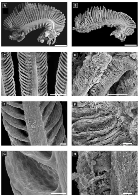
Figure 5. Scanning electron microscopy of a second gill arch of Amphiprion akallopisos unexposed to holothurian
saponins in A, C, E, G and exposed to holothurian saponins in B, D, F, H. (A) and (B) general views of the second gill arch; (C) and (D) views of the primary lamellae; (E) and (F) views of the secondary lamellae; (G) and (H) details of a secondary lamella. Scale bar is 1 mm in A and B; 100m in C, D; 20 m in E and F; 10 m in G and H.
Marmoratoside A, Holothurinoside G, and Arguside C, respec-tively. The MALDI mass spectrum obtained for the saponins extracted from the water where the Cuvierian tubules of B. atra were incubated displayed eight intense peaks that correspond to triterpene saponins (Fig. 4). The eight ions were detected at m/z [M⫹Na⫹] 1125, 1141, 1287, 1303, 1329 (new), 1449, 1463 (Holothurinoside H) and 1479 (Ho-lothurinoside I). Six of them were similar to the body wall mass spectrum, but the relative intensities between both spectra were different. The MALDI mass spectrum of the saponin extract obtained from the body wall of Holothuria forskali (not
shown) displayed the eight ions that were also detected at the same m/z signals for the saponin extract of the Cuvierian tubules of Bohadschia atra, but the relative intensities between both spectra were different.
Histological and ultrastructural characterization of the gills after saponin exposure
A comparison of the histology of gills of untreated and treated fishes showed that the carapid gills were much less altered than those of the other fishes (Figs. 5, 6, 7). After the
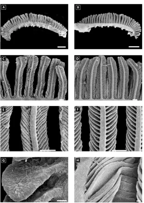
Figure 6. Scanning electron microscopy of a second gill arch of Carapus homei unexposed to
holothu-rians saponin in A, C, E, G and exposed to holothurian saponins in B, D, F, and H. (A) and (B) general views of the second gill arch; (C) and (D) views of the primary lamellae; (E) and (F) views of the secondary lamellae; (G) and (H) details of a secondary lamella. Scale bar is 1 mm in A and B; 100m in C, D, E, and F; and 20m in G and H.
exposure to saponins, the gills of Amphiprion akallopisos were damaged mostly at the surface of the secondary la-mellae (Fig. 5B, D, F, H). Most of the gill filaments were curled and bent. The secondary lamella were swollen, and the space between them was reduced (Fig. 5D). Red blood cells were often observed outside blood vessels, indicating vascular disruption (Fig. 5H). In contrast, the surface of carapid gills did not appear to be affected by exposure to saponins (Fig. 6A to H; Fig. 7).
Histology and TEM observations revealed three cell types in the epithelium of secondary lamellae of C.
mour-lani, E. gracilis, and A. akallopisos: epithelial cells (Figs.
8A, 9B), pillar cells (Figs. 8B, 9A, B), and red blood cells (Fig. 8A to C). Epithelial cells are flat cells about 2 m thick in the middle part, which includes a flattened nucleus (Figs. 7, 9A). The epidermis of secondary lamellae is un-derlined by a thick basal lamina of 500 nm (Figs. 8A to C, 9C). The epidermis covers pillar cells and red blood cells
(Figs. 7, 8A, 9A, B). A pillar cell is an I-shaped cell with a central core running from one side of the lamella to the other (Figs. 7, 8B, 9A, B). The shape of pillar cells creates cavities that form the blood vessels (Figs. 7, 8A to C, 9B). A longitudinal section through a secondary lamella shows 13 (in A. akallopisos) to 16 (in the carapid species) blood vessels flanked by the core of the pillar cells (Fig. 7). The thickness of lamella (i.e., the length of pillar cells) is about 6m.
When exposed to saponins, ultrastructural changes were seen in all fish species. In the carapids, small bulges appeared on the surface of the epithelial cells (Fig. 9B, C) and parts of the epithelia appeared contracted (Fig. 9A). In many places, epithelial cells were detached from the basal lamina (Fig. 9B), and many vacuoles also appeared in the cytoplasm of the epithelial and pillar cells (Figs. 8B, 9B, 9C). The red blood cells of the carapids did not appear to be affected by exposure to saponins (Figs. 8A,
Figure 7. Cross section of a second gill arch and filaments from Carapus acus in A, C, E and from Amphiprion
akallopisos in B, D, F; (A) and (B) second gill arch; filaments and secondary lamellae unexposed (C and D) and
exposed (E and F) to saponins; AV: afferent vessel; C: central venus sinus; GF: gill filament; SL: secondary lamella; 1: epithelial cell; 2: pillar cell; 3: red blood cell. Scale bar is 100m in A and B; 50 m in C and D; 10m in E and F. Black arrows indicate epithelial alterations.
C, 9B). The disturbances in A. akallopisos were always more severe: the TEM observations showed a total dis-organization of the tissues with the disintegration of the epithelial cells, pillar cells, and red blood cells (Fig. 9D). Epithelial cells completely disappeared in many places, and multivesicular structures were present instead of the core of pillar cells (Fig. 9D, F). The red blood cells were also damaged and had electron-dense cytoplasm and py-cnotic nuclei (Fig. 9E).
Discussion
Here it has been observed that holothuroid Cuvierian tubules and saponins extracted from the integument of
Bo-hadschia atra and Holothuria forskali are lethal for
free-living fishes under experimental conditions. It has also been demonstrated that carapids are much less sensitive to Cu-vierian tubules as they die later than free-living fishes, and they do not seem to be harmed by the saponins of the holothuroid integument. Mass spectrometry showed that there were three cocktails made of nine (body wall of B.
atra) or eight saponins (body wall of H. forskali and water
with B. atra Cuvierian tubules) with six saponins in com-mon for the three extracts. The high toxicity showed by Cuvierian tubules does not, therefore, seem to be explained by the qualitative nature of the molecules in the cocktails. Rather, it may be due to the higher concentration of sapo-nins present in the Cuvierian tubules, as shown by Van Dyck et al. (2011).
Parmentier and Vandewalle (2005) were the first to test carapid resistance to the presence of Cuvierian tubules. They placed Cuvierian tubules of three individuals of B.
argus with the pearlfishes C. homei and C. boraborensis and
some tropical fishes used as controls, in one aquarium. They observed the death of all the fishes after less than 5 min, except for the carapids, which resisted up to 160 minutes (Parmentier and Vandewalle, 2005). The experiments de-tailed here show similar results. The less resistant pearlfish was C. mourlani, which is mainly found in association with asteroids and less so with holothuroids (Parmentier, 2003). Sea stars also possess saponins, but they differ structurally from holothuroid saponins.
Histology and ultrastructure analyses showed that sapo-nins had a negative effect on the secondary lamellae of fish gills, where they caused the detachment of the epithelia, edema and pore formation at the level of the epithelial cells and pillar cells leading to their destruction. In the labora-tory, this sequence of events happened quickly in the free-living fish species and very slowly in carapids. In situ, predatory fishes that approach holothuroids are surrounded by saponins, which are constantly secreted (VanDyck et al., 2011). If these predators disturb the holothuroids, the latter emit higher concentrations and specific saponin congeners into the water (Van Dyck et al., 2011). Moreover, several species also expel some of their Cuvierian tubules through the cloacum, increasing the concentration of saponins in the surrounding water. If predators are still interested in eating the holothuroids, the latter can orientate their bodies to
Figure 8. Transmission electron microscopy of a secondary lamella of Encheliophis gracilis unexposed to
saponins, showing the tip of the lamella with red blood cells inside vessels and the external epithelium (A), the pillar cells with the departure of the pillar cell processes forming the wall of the blood vessels (B) and red blood cells with details of the vessel wall. BL: basal lamina; EC: epithelial cell; N: nucleus; PC: pillar cell; PCP: pillar cell process; RBC: red blood cell. Scale bar is 2m.
release more Cuvierian tubules in which the predators could finally become stuck. These predators are then in close contact with saponins, which should be enough to repel most of them. Oddly, carapids penetrate the cloacum of holothuroids and place themselves in the respiratory trees without provoking the expulsion of Cuvierian tubules (Par-mentier and Vandewalle, 2005). In the laboratory, carapids were unaffected by the glue of the tubules and swam be-tween them without getting stuck. Indeed, most of the saponin content would be conserved inside Cuvierian tu-bules because they are not expelled outside the holothurian. It was observed that, while carapids are less sensitive to
saponins than other fishes, they are affected by high saponin concentrations and, as they are permanently in contact with holothuroids, they must have particular, efficient adapta-tions to protect themselves.
Parmentier and Vandewalle (2005) suggested that a mechanism occurs at the level of the gills of carapids to explain their high resistance to saponins. Trott (1970) ob-served an increase in mucus production by Encheliophis
vermicularis in response to extracts of Cuvierian tubules.
The present results show that the structure of secondary lamellae in carapids is similar to that of other fishes: they are of similar length and thickness, possess a similar number
Figure 9. Transmission electron microscopy of secondary lamellae after saponin exposure. Detailed views of
secondary lamellae of Carapus acus (A) and C. mourlani (B); details of the vacuoles of epithelial cells, basal membrane and blood vessel outline of C. mourlani (C); details of the alterations in Amphiprion akallopisos showing the destructuration of a secondary lamella (D), red blood cell pycnosis in A. akallopisos (E) and vacuolization in pillar cell in A. akallopisos (F). BL: basal lamina; BV: blood vessel; EC: epithelial cell; PC: pillar cell; PCP: pillar cell process; RBC: red blood cell; SL, saponin-attacking lamina; V: vacuole. Scale bar is 10m in A and B; 5 m in C, D, E, F and G; and 1 m in H. Arrows and asterisks on part B indicate the forming vacuoles and the deformities on the surface of epithelial cells.
of blood vessels, and are made of similar cells (epithelial cells, pillar cells, and red blood cells). As the gill structure does not explain the resistance of carapids to saponins, three other hypotheses can be suggested: (i) the quantity of mucus secreted in carapids is higher than in other fishes, and the thickness of the mucus creates a natural barrier against the toxin, (ii) the nature of the mucus in carapids is different from that of other fishes and includes some molecules, such as enzymes, that can act on saponins and suppress their noxious effects, and (iii) saponins cannot act on carapid gills because the cytoplasmic membrane of their epithelial cells possesses special sterols, as is the case for their hosts. Indeed, the cell membranes of holothuroids are resistant to their own saponins (Stonik et al.,1999). This resistance was proved to be related to the low content of free sterols in membranes of sea cucumbers, to the presence of ⌬7 and ⌬9(11)
-sterols as well as sulfated and glycosylated sterol conjugates instead of⌬5-sterols (Popov et al., 1983 ; Stonik and Elyakov, 1988).⌬5-sterols are the main site of action of saponins (Mackie,1976). It is therefore possible that the composition of carapid cell membrane is similar to that of their echinoderm hosts and that carapids possess a lot more ⌬7
or ⌬9(11)-sterols than ⌬5-sterols. This already explains why echinoderms are unaffected by high levels of saponins in their tissues, and why exposure to saponin solutions, lethal to all other fishes, would be without effect on Car-apidae. Further experiments should investigate these three hypotheses to highlight the mechanism that results in car-apids being unaffected by host saponins.
Acknowledgments
The authors thank the IH.SM (Toliara, Madagascar) for having allowed them to carry out their experiments within their institute and the Madagascar Holothurie team for help-ing to obtain C. mourlani, E. gracilis, E. vermiops, and the free-living fish species. Thanks also go to the STARESO team (Calvi, France) for helping to obtain C. acus, to S. Planes (CRIOBE, Moorea, French Polynesia) for helping to obtain C. boraborensis and C. homei, and to M. Todesco for technical assistance. This work was supported by a FRFC project (FNRS, Fonds National de la Recherche Scienti-fique). This study is a contribution by the Centre Interuni-versitaire de Biologie Marine (CIBIM).
Literature Cited
Bakus, G. J. 1973. The biology and ecology of tropical holothurians. Pp. 325–367 in Biology and Geology of Coral Reefs 2, A. O. Jones and R. Endean, eds. Academic Press, London.
Bordbar, S., F. Anwar, and N. Saari. 2011. High-value components and bioactives from sea cucumbers for functional foods—a review.
Mar. Drugs 9: 1761–1805.
Caulier, G., P. Flammang, P. Gerbaux, and I. Eeckhaut. 2013a.
When a repellent becomes an attractant: harmful saponins are kairo-mones attracting the symbiotic harlequin crab. Sci. Rep. 3: 1–5.
Caulier, G., P. Flammang, P. Rakotorisoa, P. Gerbaux, M. Demeyer, and I. Eeckhaut. 2013b. Preservation of the bioactive saponins of
Holothuria scabra through the processing of trepang. Cah. Biol. Mar.
54: 685– 690.
Cleveland, A., E. A. Verde, and R. W. Lee. 2011. Nutritional exchange in a tropical tripartite symbiosis: direct evidence for the transfer of nutrients from anemonefish to host anemone and zooxanthellae. Mar.
Biol. 158: 589 – 602.
Demeyer, M., J. De Winter, G. Caulier, I. Eeckhaut, P. Flammang, and P. Gerbaux. 2014. Molecular diversity and body distribution of saponins in the sea star Asterias rubens by mass spectrometry. Comp.
Biochem. Physiol. B 168: 1–11.
Dietrich, H., and A. Fontaine. 1975. A decalcification method for ultrastructure of echinoderm tissues. Biotech. Histochem. 50: 351–354.
Elliott, J. K., and R. N. Mariscal. 1997. Ontogenetic and interspecific variation in the protection of anemonefishes from sea anemones. J. Exp.
Mar. Biol. Ecol. 208: 57–72.
Elyakov, G., V. Stonik, E. Levina, V. Slanke, T. Kuznetsova, and V. Levin. 1973. Glycosides of marine invertebrates. II. A comparative study of the glycoside fractions of Pacific sea cucumbers. Comp.
Biochem. Physiol. B 44: 325–336.
Flammang, P., J. Ribesse, and M. Jangoux. 2002. Biomechanics of adhesion in sea cucumber Cuvierian tubules (Echinodermata, Holothu-roidea). Integr. Comp. Biol. 42: 1107–1115.
Ganter, P., and G. Jolle´s. 1969 –1970. Histochimie Normale et Pathologique. Gauthier-Villars, Paris.
Guo, M., F. Song, Z. Liu, and S. Liu. 2006. Characterization of triterpenoidic saponin mixture in crude extracts from leaves of
Acan-thopanax senticosus harms by saponin structural correlation and mass
spectrometry. Anal. Chim. Acta 557: 198 –203.
Hamel, J. F., and A. Mercier. 2000. Cuvierian tubules in tropical holothurians: usefulness and efficiency as a defence mechanism. Mar.
Freshw. Behav. Physiol. 33: 115–139.
Kalinin, V. I., M. Anisimov, N. Prokofieva, S. Avilov, S. Afiyatullov, and V. Stonik. 1996a. Biological activities and biological role of triterpene glycosides from holothuroids (Echinodermata). Pp. 139 –181 in Echinoderm Studies 5, M. J. Jangoux, ed. CRC Press, Boca Raton, FL.
Kalinin, V. I., N. Prokofieva, G. Likhatskaya, E. Schentsova, I. Aga-fonova, S. Avilov, and O. Drozdova. 1996b. Hemolytic activities of triterpene glycosides from the holothurian order Dendrochirotida: some trends in the evolution of this group of toxins. Toxicon 34: 475– 483.
Kalinin, V. I., D. L. Aminin, S. A. Avilov, A. S. Silchenko, and V. A. Stonik. 2008. Triterpene glycosides from sea cucucmbers (holothu-rioidea, echinodermata). Biological activities and functions. Stud. Nat.
Prod. Chem. 35: 135–196.
Kitagawa, I., and M. Kobayashi. 1977. On the structure of the major saponin from the starfish Acanthaster planci. Tetrahedron Lett. 18: 859 – 862.
Kubanek, J., J. R. Pawlik, T. M. Eve, and W. Fenical. 2000. Triter-pene glycosides defend the Caribbean reef sponge Erylus formosus from predatory fishes. Mar. Ecol. Prog. Ser. 207: 69 –77.
Li, R., Y. Zhou, Z. Wu, and L. Ding. 2006. ESI-QqTOF-MS/MS and APCI-IT-MS/MS analysis of steroid saponins from the rhizomes of
Dioscorea panthaica. J. Mass Spectrom. 41: 1–22.
Mackie, A. M., and A. Turner. 1970. Partial characterization of a biologically active steroid glycoside isolated from the starfish
Marthas-terias glacialis. Biochem. J. 117: 543–550.
Mackie, G. O. 1976. Propagated spikes and secretion in a coelenterate glandular epithelium. J. Gen. Physiol. 68: 313–325.
Markle, D. F., and J. E. Olney. 1990. Systematics of the pearlfish (Pisces: Carapidae). Bull. Mar. Sci. 47: 269 – 410.
Matsuno, T., and J. Iba. 1966. Studies on the saponins of the sea cucumber. Yakugaku Zasshi 86: 637.
Mebs, D. 1994. Anemonefish symbiosis: vulnerability and resistance of fish to the toxin of the sea anemone. Toxicon 32: 1059 –1068.
Nigrelli, R. 1952. The effects of holothurin on fish, and mice with sarcoma 180. Zoologica NY 37: 89 –90.
Parmentier, E. 2003. Contribution to the study of the relations between fish of the family of Carapidae and their invertebrate hosts: a multi-disciplinary approach. Ph.D. thesis, University of Liege, Belgium. 316 pp.
Parmentier, E., and K. Das. 2004. Commensal vs. parasitic relation-ship between Carapini fish and their hosts: some further insight through ␦13C and␦15N measurements. J. Exp. Mar. Biol. Ecol. 310: 47–58.
Parmentier, E., and L. Michel. 2013. Boundary lines in symbiosis forms. Symbiosis 60: 1–5.
Parmentier, E., and P. Vandewalle. 2003. Morphological adaptations of pearlfish (Carapidae) to their various habitats. Pp. 259 –274 in Fish
Adaptations, A. L. Val and B. G. Kapoor, eds. Science Publishers,
Enfield, NH.
Parmentier, E., and P. Vandewalle. 2005. Further insight on carapid— holothuroid relationships. Mar. Biol. 146: 455– 465.
Plasman, V., J. C. Braekman, D. Daloze, M. Luhmer, D. Windsor, and J. Pasteels. 2000a. Triterpene saponins in the defensive secretion of a chrysomelid beetle, Platyphora ligata. J. Nat. Prod. 63: 646 – 649.
Plasman, V., J. C. Braekman, D. Daloze, D. Windsor, and J. Pasteels. 2000b. Triterpene saponins, quaternary ammonium compounds, phosphatidyl cholines, and amino acids in the pronotal and elytral secretions of Platyphora opima and Desmogramma subtropica. J. Nat.
Prod. 63: 1261–1264.
Popov, A., N. Kalinovskaia, T. Kuznetsova, I. Agafonova, and M. Anisimov. 1983. Role of sterols in the membranotropic activity of triterpene glycosides. Antibiotiki 28: 656 – 659.
Prokof’eva, N. G., E. L. Chaikina, A. A. Kicha, and N. V. Ivanchina. 2003. Biological activities of steroid glycosides from starfish. Comp.
Biochem. Physiol. B 134: 695–701.
Segal, R., and E. Schlo¨sser. 1975. Role of glycosidases in the mem-branlytic, antifungal action of saponins. Arch. Microbiol. 104: 147– 150.
Stonik, V., and G. Elyakov. 1988. Secondary metabolites from echi-noderms as chemotaxonomic markers. Pp. 43– 86 in Bioorganic
Ma-rine Chemistry, P. G. Scheuer, ed. Springer, Berlin.
Stonik, V., V. Kalinin, and S. Avilov. 1999. Toxins from sea cucum-bers (holothuroids): chemical structures, properties, taxonomic distri-bution, biosynthesis and evolution. J. Nat. Toxins 8: 235–248.
Thompson, J., R. Walker, and D. Faulkner. 1985. Screening and bioassays for biologically-active substances from forty marine sponge species from San Diego, California, USA. Mar. Biol. 88: 11–21.
Trott, L. B. 1970. Contributions to the Biology of Carapid Fishes (Paracanthopterygii: Gadiformes). University of California Press,
Oakland.
Trott, L. B. 1981. A general review of the pearlfishes (Pisces, Carapi-dae). Bull. Mar. Sci. 31: 623– 629.
Van Dyck, S., G. Caulier, M. Todesco, P. Gerbaux, I. Fournier, M. Wisztorski, and P. Flammang. 2011. The triterpene glycosides of
Holothuria forskali: usefulness and efficiency as a chemical defense
mechanism against predatory fish. J. Exp. Biol. 214: 1347–1356.
Van Dyck, S., P. Flammang, C. Meriaux, D. Bonnel, M. Salzet, I. Fournier, and M. Wisztorski. 2010. Localization of secondary metabolites in marine invertebrates: contribution of MALDI MSI for the study of saponins in Cuvierian tubules of H. forskali. PLoS One 5: e13923.
Van Dyck, S., P. Gerbaux, and P. Flammang. 2009. Elucidation of molecular diversity and body distribution of saponins in the sea cu-cumber Holothuria forskali (Echinodermata) by mass spectrometry.
Comp. Biochem. Physiol. B 152: 124 –134.
Yamanouchi, T. 1955. On the poisonous substance contained in ho-lothurians. Publ. Seto Mar. Biol. Lab. 4: 183–203.