Contents lists available atScienceDirect
Redox Biology
journal homepage:www.elsevier.com/locate/redox
PKG1
α oxidation negatively regulates food seeking behaviour and reward
Celine Dura
ffourd
a, Robert T.R. Huckstepp
b, Ingke Braren
c, Cathy Fernandes
d,e, Olivier Brock
f,
Alessio Delogu
f, Oleksandra Prysyazhna
a, Joseph Burgoyne
a, Philip Eaton
a,⁎aKing's College London, School of Cardiovascular Medicine & Sciences, the Rayne Institute, St. Thomas' Hospital, London SE1 7EH, United Kingdom bSchool of Life Sciences, University of Warwick, Coventry, United Kingdom
cUniversity Medical Center Eppendorf, Vector Facility, Inst. for Exp. Pharmacology and Toxikology, N30, Room 09, Martinistr. 52, 20246 Hamburg, Germany dSGDP Research Centre, Institute of Psychiatry, Psychology and Neuroscience, King's College London, London, United Kingdom
eMRC Centre for Neurodevelopmental Disorders, King's College London, London, United Kingdom
fBasic and Clinical Neuroscience, Institute of Psychiatry, Psychology and Neuroscience, King's College London, United Kingdom
A R T I C L E I N F O
Subject areas: PKG1α
Food-seeking behaviour Reward
Ventral Tegmental Area
A B S T R A C T
Genes that are highly conserved in food seeking behaviour, such as protein kinase G (PKG), are of interest because of their potential role in the global obesity epidemic. PKG1α can be activated by binding of cyclic guanosine monophosphate (cGMP) or oxidant-induced interprotein disulfide bond formation between the two subunits of this homodimeric kinase. PKG1α activation by cGMP plays a role in reward and addiction through its actions in the ventral tegmental area (VTA) of the brain.‘Redox dead’ C42S PKG1α knock-in (KI) mice, which are fully deficient in oxidant-induced disulfide-PKG1α formation, display increased food seeking and reward be-haviour compared to wild-type (WT) littermates. Rewarding monoamines such as dopamine, which are released during feeding, are metabolised by monoamine oxidase to generate hydrogen peroxide that was shown to mediate PKG1α oxidation. Indeed, inhibition of monoamine oxidase, which prevents it producing hydrogen peroxide, attenuated PKG1α oxidation and increased sucrose preference in WT, but not KI mice. The deficient reward phenotype of the KI mice was rescued by expressing WT kinase that can form the disulfide state in the VTA using an adeno-associated virus, consistent with PKG1α oxidation providing a break on feeding behaviour. In conclusion, disulfide-PKG1α in VTA neurons acts as a negative regulator of feeding and therefore may provide a novel therapeutic target for obesity.
1. Introduction
Obesity is a major global health problem with 13% of the world's adult population being affected in 2016. The wide availability of highly-palatable and -rewarding food drives their preferential con-sumption, increasing the prevalence of obesity. Defining mechanisms that drive us to seek vast amounts of highly calorific food may allow the development of rational interventions to attenuate their consumption, potentially reducing obesity and its associated health risks.
Protein kinase G (PKG) is a phylogenetically well-conserved food-seeking and -reward behavioural gene across insects and nematodes [1]. Whilst PKG expression is seen in mammals[2–4], there appears to be no information as to its effect on feeding behaviour. The PKG1α isoform is classically activated by binding of cyclic guanosine mono-phosphate (cGMP) [5]. However, under physiological conditions PKG1α can also be regulated by oxidation[6–8]. Endogenous oxidants, such as hydrogen peroxide, induce a disulfide bond between cysteine
42 on adjacent chains of 2 PKG1α monomers, forming a homodimer complex that renders the kinase catalytically active[6–8]. The binding of cGMP to PKG1α attenuates oxidant-induced interprotein disulfide accumulation, perhaps because it causes conformational changes that move the two cysteine 42 residues away from each other [7,9]. In conjunction, disulfide-PKG1α dimerisation through oxidation appears to cause a conformational changes in PKG1α that decreases cGMP-de-pendent activity, which could be due to oxidation lowering affinity for the cyclic nucleotide[2,10]. Therefore, the two pathways appear to be antagonistic regulators of PKG1α.
In the brain, the ventral tegmental area (VTA) represents a key component of the mesolimbic dopaminergic system and is critical to reward learning and addictive behaviours [11]. Interestingly, PKG regulates dopamine release[12,13], and altering PKG levels within the VTA increases addiction[14]. Furthermore, alteration of PKG activity through manipulation of cGMP, or it's activator nitric oxide synthase (NOS), leads to complementary changes in reward[15,16]. As cGMP
https://doi.org/10.1016/j.redox.2018.101077
Received 18 October 2018; Received in revised form 6 December 2018; Accepted 11 December 2018 ⁎Corresponding author.
E-mail address:[email protected](P. Eaton).
Redox Biology 21 (2019) 101077
Available online 18 December 2018
2213-2317/ © 2018 Published by Elsevier B.V. This is an open access article under the CC BY-NC-ND license (http://creativecommons.org/licenses/BY-NC-ND/4.0/).
binding appears to increase reward and oxidation of PKG1α negatively regulates binding of this cyclic nucleotide[12,13], we speculated that disulfide-PKG1α in the VTA would therefore supress food reward. To investigate this hypothesis, we assessed food-seeking and -reward be-haviour in C42S PKG1α knock-in (KI) ‘redox dead’ mice, which are fully deficient in disulfide-dependent activation, whereas their NO-cGMP-dependent activation remains intact[17]. We found that removal of the disulfide-PKG1α pathway led to an increase in food seeking behaviour in KI mice compared to wild-type (WT) littermates. Breakdown of the primary neurotransmitter of the reward system, dopamine, by mono-amine oxidase-B (MAO-B) leads to production of hydrogen peroxide [18]. Inhibition of MAO-B reduced disulfide-PKG1α in the VTA and increased sucrose preference in WT but not KI mice. In accordance, inhibition of NOS, which is anticipated to decrease cGMP-dependent activation of PKG1α, concomitantly increased disulfide-PKG1α and reduced sucrose preference in WT but not KI mice.
Critically, we could rescue the food-reward phenotype of PKG1α KI mice by expression of WT PKG1α in the VTA using an adeno-associated virus (AAV). Therefore disulfide- and cGMP-activation of PKG1α work antagonistically in the VTA to control hedonic eating, where disulfide-PKG1α in the VTA acts as a negative regulator, an ‘off switch’, for food-reward. Therefore, PKG1α may provide a novel therapeutic target for treatment against sugary addictions.
2. Results
2.1. Disulfide-PKG1α regulates food-seeking behaviour
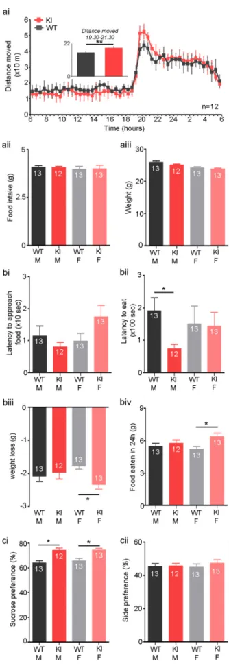
We began by investigating food seeking behaviour of C42S PKG1α KI mice (KI). There was no difference in locomotor activity between genotypes over a 24-h period (WT: 4.8 ± 0.5 m/h, KI: 4.8 ± 0.7; n = 12 per group; Fig. 1a). However, there was an increase in total distance covered at the beginning of the night, between 19:30 and 21:30, thefirst 2 h of their dark period (WT: 16 ± 2 m/h, KI: 20 ± 3; n = 12 per group; p = 0.005;Fig. 1a). There was no difference between males and females. Interestingly, there were no differences between KI mice and WTs of either sex in overall food consumption (n = 12 or 13 per group;Fig. 1aii), showing overall drive to feed is not altered by this mutation. Furthermore, there were no differences between KI and WTs of either sex in overall weight (n = 12 or 13 per group; Fig. 1aiii), meaning KI mice do not appear to have altered metabolism.
In a novelty-suppressed feeding test, there was no difference be-tween WT or KI mice of either sex in their approach to food (n = 12–13 per group,Fig. 1bi). However, male KI mice began to eat their food faster than male WTs, although there was no difference between fe-males of either genotype (male WT: 190.5 ± 40.6 s, male KI: 73.9 ± 13.9, female WT: 150.1 ± 53.6, female KI: 142.9 ± 43.0; n = 12 or 13 per group; p = 0.04;Fig. 1bii).
Though we did not observe differences between KI and WT female mice in latency to eat, KI female mice presented a higher weight loss than WT female following 24 h fasting prior to testing (male WT: 2.1 ± 0.2 g, male KI: 2 ± 0.2, female WT: 1.8 ± 0.1, female KI: 2.3 ± 0.2; n = 12 or 13 per group; p = 0.006;Fig. 1biii). Consistent with this, female KIs increased their food intake the day following testing compared to WT littermates, possibly to compensate for their weight loss (male WT: 5.5 ± 0.3 g, male KI: 5.7 ± 0.3, female WT: 5.2 ± 0.3, female KI: 6.4 ± 0.3; n = 12 or 13 per group; p = 0.02; Fig. 1biv). Interestingly, no difference were seen between KI males and their WT littermates in either weight loss or food consumption (Fig. 1biii,iv)
2.2. Specific effect on feeding behaviour in KI mice
Whilst alterations in food behaviour are prominent in PKG KI mice, this could be due to other confounding factors. However, there were no differences between KI or WT of either sex for tests of: Anxiety, i.e.,
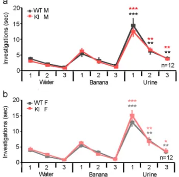
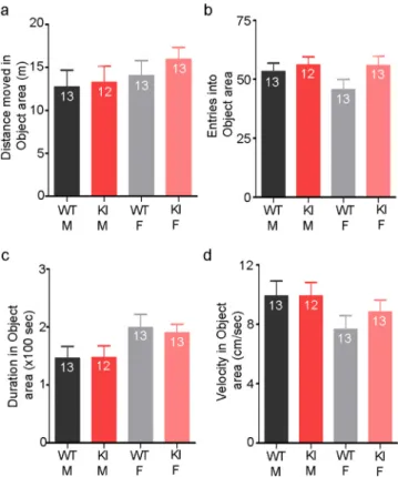
Openfield test and elevated-plus maze (Fig. 2); alterations in olfaction, i.e., olfaction habituation/dishabituation test (Fig. 3); Exploratory be-haviour, i.e., novel object recognition (Fig. 4); Memory, i.e., Y-maze (Fig. 5), and; general motor deficits, i.e., rotarod and grip and strength (Fig. 6). Therefore, changes in feeding behaviours were due to a change in the drive to feed or in the reward gained by eating, and not caused by any other, behavioural deficits.
2.3. Disulfide-PKG1α regulates rewarding behaviour
To distinguish between increased food seeking and reward we performed a sucrose preference test to compare hedonic sensitivity of KI or WT mice. Notably, KI mice had an increased preference for sucrose compared to WTs but no differences were observed between males and females (male WT: 63.9 ± 2%, male KI: 74.1 ± 2.1, female WT: 65.7 ± 2.2, female KI: 74.5 ± 1.5; n = 12 or 13 per group; p = 0.0002; Fig. 1ci). Furthermore, there was no difference in side
preference between genotypes of either sex, so excluding this as a confounding factor (n = 12 or 13 per group;Fig. 1cii). Therefore, our data indicate oxidation of PKG1α to the interprotein disulfide state Fig. 1. C42S PKG1α knock-in (KI) ‘redox dead’ mice display increased food
reward compared to wild-type littermates (WT). A) Home cage recordings show overall locomotor activity of KI mice is not different to WT. Insert: total distance moved is increased in KIs compared to WTs during thefirst 2 h of their awake cycle (19:30–21:30) when food intake is at its highest (ai). However, there was not an overall change in feeding as food intake was similar across groups (aii), or an overall change in metabolism as no differences in weight were found between groups (aiii). B) In a novelty-suppressed feeding test, after 24 h fasting, all mice approach food in a novel environment with the same delay (bi). However, male KI mice feed faster than male WTs (bii), whilst female KI mice lose more weight during food restriction (biii) and consume more food when the restriction is lifted (biv) than female WTs. Thus, feeding behaviour is altered in KI mice. C) In a sucrose preference test, KIs have a higher preference for 2% sucrose over water compared to WTs (ci), though they do not show any side preference under control conditions (cii). *- p < 0.05.
Fig. 2. KI mice did not show any alterations in anxiety levels. A) In an open-field paradigm, there were no differences between KI mice and WTs of either sex for the distance moved in the centre of the arena (ai), the number of entries in to the centre (aii), the amount of time spent in the centre (aiii), or the speed they moved through the centre of the openfield (aiv). B) In an elevated-plus maze paradigm, there were no differences between KI mice and WTs of either sex for the distance moved in the open arms (bi), the number of entries in to the open arms (bii), the amount of time spent in the open arms (biii), or the speed they moved whilst on the open arms of the maze (biv).
Fig. 3. KI mice did not show any alterations in olfaction. In an olfaction ha-bituation/dishabituation test, there was no difference between KI mice and WTs of either sex for time spent investigating water, banana odour, or urine from a mouse of the opposite sex with all groups displaying habituation/dishabituation to the different odours. Comparisons between KI and WT male mice (a) and KI and WT female mice (b).*- p < 0.05, ** - p < 0.01, *** p < 0.001.
decreases desire to consume rewarding substances.
2.4. L-DOPA or dopamine induces disulfide-PKG1α in the ventral tegmental area (VTA)
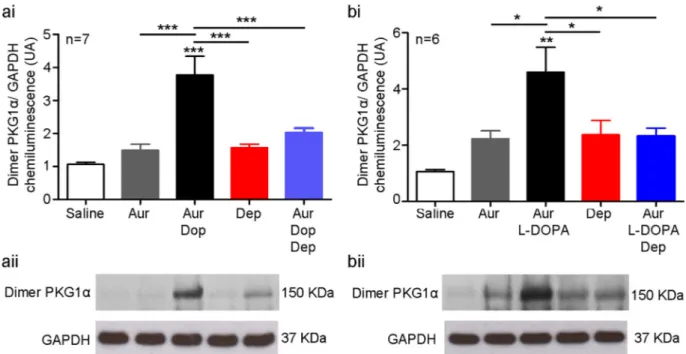
Using non-reducing western immunoblot analysis, we observed that disufide-PKG1α was increased in isolated VTA tissue blocks treated with dopamine (Cont: 1.1 ± 0.1 UA, dopamine: 3.8 ± 0.6; n = 7 per group; p = 0.01;Fig. 7ai,ii). Pre-treatment with deprenyl, an inhibitor of monoamine oxidase B (MAO-B), attenuated formation of disulfide-PKG1α dimers in response to dopamine in WTs (dopamine: 3.8 ± 0.6 UA, deprenyl: 2.0 ± 0.1; n = 7 per group; p = 0.001;Fig. 7ai,ii) to a level indistinguishable from controls (Cont: 1.1 ± 0.1 UA; n = 7; Fig. 7a). Similarly, increased PKG1α was observed in the brains of WT mice following an intraperitoneal injection of L-DOPA, a dopamine precursor (Cont: 1.1 ± 0.1 UA, L-DOPA: 4.6 ± 0.9; n = 6 per group; p = 0.0007;Fig. 7bi,ii). Pre-treatment with deprenyl also prevented this in vivo formation of disulfide-PKG1α in response to L-DOPA in WTs (L-DOPA: 4.6 ± 0.9 UA, deprenyl: 2.3 ± 0.3; n = 6 per group; p = 0.0007;Fig. 7b) to a level indistinguishable from controls (Cont: 1.1 ± 0.1 UA; n = 6 per group,Fig. 7b). Auranofin, which pharma-cologically inhibits thioredoxin reductase to attenuate reduction of disulfide bonds that form and therefore retain PKG1α in the oxidised state for detection using western immunoblotting, did not lead to a change in disulfide- PKG1α levels for either experiment (n = 7 per group,Fig. 7). Therefore, there is not a significant amount of PKG1α oxidation under basal conditions. Treatment with deprenyl or saline controls did not cause any change in disulfide-PKG1α formation in WTs in either experiment (n = 7,Fig. 7a; n = 6,Fig. 7b). Taken together, these results indicate dopamine increases disulfide-PKG1α in the brain of WT mice.
Fig. 4. KI mice did not show any alterations in exploratory behaviour. In a novel object recognition paradigm, there were no differences between KI mice and WTs of either sex for the distance moved in the object area (a), the number of entries into the object area (b), the amount of time spent in the object area (c), or the speed they moved through the object area (d).
Fig. 5. KI mice did not show any alterations in short-term memory. In a Y-maze paradigm, there were no differences between KI mice and WTs of either sex for the distance moved in the novel and unknown arms (a), the number of entries into the novel and unknown arms (b), the amount of time spent in the novel and unknown arms (c), or the speed they moved through the novel and unknown arms (d). *- p < 0.05, ** - p < 0.01, *** p < 0.001.
2.5. Inhibition of MAO-B reduces disulfide-PKG1α and increases sucrose preference in WT, but not KI mice
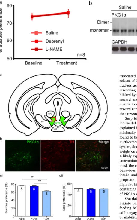
As one by-product of dopamine degradation by MAO-B is hydrogen peroxide [18], we investigated the effect of pharmacologically in-hibiting MAO-B on disulfide-PKG1α in WTs. Pre-treatment with 0.5 mg/kg deprenyl increased sucrose preference in WTs (64.0 ± 0.8–72 ± 1.0%; n = 4; p < 0.0001;Fig. 8ai), and reduced disulfide-PKG1α (1.0 ± 0.2–0.4 ± 0 UA; n = 4 per group; p = 0.04; Fig. 8aii,iii). Higher doses of deprenyl (2.5 or 5 mg/kg) did not increase sucrose preference beyond that of the lowest concentration (Fig. 8ai). KI mice unable to form disulfide-PKG1α (Fig. 9b) did not show any alteration in sucrose preference following deprenyl administration (63.0 ± 1.1–66.3 ± 1.2;Fig. 9a). Therefore, blocking degradation of dopamine that generates hydrogen peroxide as a by-product, increased disulfide-PKG1α dimers and sucrose preference of WTs.
2.6. Inhibition of NO synthase increases disulfide-PKG1α and decreases sucrose preference in WT, but not KI mice
Nitric oxide (NO) binds to guanylyl cyclase to stimulate production of cGMP [19]. As cGMP attenuates disulfide-PKG1α accumulation, elevating NO or stimulating guanylyl cyclase to increase cGMP should increase reward. Pre-treatment with an NO synthase inhibitor, L-NAME,
decreased sucrose preference of WTs (66.0 ± 1.2–59.2 ± 1.3%; n = 8 per group; P < 0.0001; Fig. 8bi), and increased disulfide-PKG1α (1.0 ± 0.3–7 ± 1.3 AU; n = 8 per group; p = 0.004; Fig. 8bii,iii). Higher doses of L-NAME (5 and 10 mg/kg) did not decrease sucrose preference beyond that of the lowest concentration (Fig. 8bi). KI mice, unable to form disulfide-PKG1α (Fig. 9b), did not show any alteration in sucrose preference following administration of L-NAME (Fig. 9a). Therefore, blocking the elevation of cGMP, decreased sucrose pre-ference in WTs.
2.7. VTA injection of the WT PKG1α rescues the behaviour phenotype of KI mice
As PKG levels within the VTA are directly associated with addiction [3,14], and we found a deficit in food reward in KI mice or WTs fol-lowing pharmacological inhibition of NOS, we hypothesized that de fi-ciency of disulfide-PKG1α in the VTA leads to enhanced food reward in KI mice. To test this we used an AAV vector to re-express the wild-type form of the PKG1α (WT PKG1α) in the VTA of KI mice (Fig. 10a,b).
Expression of WT PKG1α in the VTA of KI mice reduced their su-crose preference compared with KI mice injected with a control virus expressing solely GFP (GFP: 75.8 ± 1.4%, WT PKG: 62.4 ± 2.0; n = 8 per group; p = 0.0007;Fig. 10ci). Importantly, sucrose preference in KI mice injected with WT PKG1α was not different to that in WTs (64.8 ± 1.5%; n = 13). Transducing the VTA of KI mice to express C42S KI PKG1α confirmed that overexpressing PKG1α in the VTA per se does not downregulate food reward (75.2 ± 1.7%; n = 8 per group, Fig. 10ci). Overexpression of WT PKG1α, C42S KI PKG1α, or GFP did not lead to a change in side preference (Fig. 10cii). This indicates dis-ulfide-PKG1α in the VTA is a key component in food reward, acting as a negative regulator that switches off the rewarding properties of sucrose in mice.
3. Discussion
Feeding behaviour is a complex decision-making process involving where, when, what, and how much to eat. This isn't only a reflexive response to stimuli or change in energy level, but includes environ-mental factors and previous experience. Obesity is characterized by excess storage of body fat, which is often associated with other diseases, such as type 2 diabetes, hypertension, heart disease, stroke, cancer and non-alcoholic fatty liver disease. The cost of obesity to the National Health Service in the United Kingdom has increased from an estimated £ 479.3 in 1998 to £ 6.1 billion 2014. Thus, identification of mechan-isms underlying food preference behaviours, which lead to overeating, are now a priority.
PKG1α is a cytosolic kinase that is a downstream target of cGMP [20]. PKG has perhaps been most extensively studied in the cardio-vascular system[20], though it is found in many tissues including the brain[2]. PKG1α was found to undergo oxidant-induced targeting and activation, forming a catalytically active dimer [4,6,7,21,22]. Inter-estingly, increased cGMP activation of PKG produced by sildenafil, an inhibitor of phosphodiesterase 5-dependent cGMP degradation, in-creases reward[15]. Furthermore, reduction of PKG activity through administration of phosphodiesterase type 5 inhibitor or by pharmaco-logical inhibition or genetic knockout of NOS, which abrogates soluble guanylyl cyclase-dependent cGMP production, decreases reward [15,16]. As the NOS-cGMP-PKG1α pathway is highly conserved in both food seeking[1]and reward[14–16], and disulfide-PKG1α appears to negatively regulate this pathway[9], we hypothesized regulation of the cGMP/PKG1α pathway by disulfide-PKG1α would act as an off switch of food-reward.
We found C42S KI‘redox dead’ mice increased locomotor activity during thefirst 2 h of their dark period when feeding is at its highest [23], which may indicate an increase in foraging behaviour. Further-more, KI males began to eat faster than male WT littermates in the Fig. 6. KI mice did not show any alterations in general motor function. A) In a
grip strength test, there were no differences between KI mice and WTs of either sex for the force they could generate with their fore (ai) or hind limbs (aii). B) In a rotarod paradigm, there were no differences between KI mice and WTs of either sex for the time they took to fall from the apparatus. bi) Comparisons between male (bi) and female (bii).
novelty supressed feeding test, whilst female KIs ate more chow fol-lowing food restriction than female WT littermates, with no differences in any other behavioural tests performed. Therefore, changes in feeding behaviours were due to an altered drive to feed or in reward gained by eating, and not because of: a lack of foraging or exploratory behaviour
due to anxiety; general motor discoordination or fatigue; a disinterest in food caused by an inability to smell, or; overeating due to a deficit in learning and memory. Thus, conservation of the role of PKG1α in food seeking behaviour appears to extend to mammals. Interestingly in KI mice, food intake only increased in females following food deprivation Fig. 7. Increased dopaminergic signalling leads to increased disulfide-PKG1α through production of hydrogen peroxide during dopamine degradation. A) In isolated tissue slices of the VTA, dopamine in the presence of auranofin (added to stabilise dimer formation) led to an increased disulfide-PKG1α, which was decreased by the MAO-B inhibitor deprenyl. Saline, auranofin, and deprenyl controls did not lead to disulfide-PKG1α formation. ai) summary data, aii) representative western blots. B) In vivo L-DOPA in the presence of auranofin led to increased disulfide-PKG1α in the VTA, which was decreased by deprenyl. Saline, auranofin, and deprenyl controls did not lead to PKG1α formation. bi) summary data, bii) representative western blots. Therefore, increased dopaminergic activity stimulates disulfide-PKG1α in the VTA. Furthermore, pharmacological inhibition of dopamine breakdown by MAO-B, which otherwise generates hydrogen peroxide as a by-product, reduced disulfide-PKG1α in the VTA. *- p < 0.05, ** - p < 0.01, *** p < 0.001.
Fig. 8. Sucrose preference in WT mice is determined by a balance between NOS/cGMP- and disulfide-PKG1α. a) Sucrose preference is increased (ai) when disulfide-PKG1α is reduced by deprenyl (aii: representative western blots, aiii: summary data). b) Sucrose preference is decreased (bi) when disulfide-disulfide-PKG1α is increased by L-NAME, an inhibitor of NOS (bii: representative western blots, biii: summary data). All western blots were performed in the presence of auranofin to stabilise dimer formation. Inhibition of NOS with L-NAME, which lowers cGMP levels and so abrogates its ability to attenuate disulfide-PKG1α accumulation, increased oxidation of the kinase. *- p < 0.05, ** - p < 0.01.
which makes food more rewarding[24]or in both sexes when a re-warding stimulus, i.e., sucrose, was present, with neither sex increasing their food intake of non-palatable food when it was abundant.
Obesity may result from a form of addiction regulated through do-paminergic reward centres of the brain[25]. Eating palatable food is
associated with increased activity in the VTA, at least in part due to release of dopamine from its projections to the dorsal striatum[26]and nucleus accumbens[27], increasing pleasure sensation whilst eating rewarding food. Furthermore, when food is plentiful the VTA is in-hibited by satiating signals such as leptin or insulin[28], reducing food reward and feeding. Dopamine is so important to feeding that mice unable to synthesize it die of starvation[29]. Whilst PKG is found in reward centres of the brain[3,12–14], we show here for thefirst time that rewarding foods leads to increased disulfide-PKG1α in the VTA.
Surprisingly, the changes observed in foraging behaviour of the KI mouse did not lead to a change in body weight. However, this could be explained by the lack of rewarding value of standard chow, as this only minimally activates the mesolimbic dopaminergic system, which we found to be crucial to the change in foraging behaviour of the KI mice. Furthermore, a high sucrose diet which strongly engages the reward system, doesn't lead to weight gain in mice[30]. Whilst KI mice do gain weight on a high fat diet, they do not do so beyond that of controls[31]. A likely explanation is that high fat diets, whilst leading to weight gain, concomitantly reduces dopaminergic signalling [32–34], which may mask the effects that the C42S PKG1α KI mutation has on food seeking behaviour. Therefore, an obesogenic diet of high fat to increase caloric intake and high sucrose to increase reward would appear more ap-propriate than the use of either individually. This is because a diet of high fat high combined with sucrose is more rewarding than a diet containing either component alone[35], which may enhance the effects of PKG1α on feeding behaviour.
Our data also suggest that food restriction may also provide a way to initiate binge eating in the KI mice, which is a common feature of feeding behaviour in obese humans. Interestingly, restricting sucrose still engages the dopaminergic system [36,37], and restricting the availability of high sucrose [38] or high fat [39–41] causes binge eating, which could potentially be enhanced in KI mice. Therefore, further investigation into weight gain of the KI mice on a restricted obesogenic diet is warranted, which would more closely resemble a western diet and the feeding behaviour of obese people.
Inhibition of NOS, which diminishes cGMP-dependent activation of PKG1α, reduces consumption of rewarding food[42]and food intake in general[43]. Consistent with this and the fact that KI mice have po-tentiated food seeking, is the fact that inhibition of the NOS-cGMP pathway promotes accumulation of disulfide-PKG1 α[7,44]in the VTA and increased food reward. Interestingly, KI mice did not show reduced sucrose preference following pre-treatment with L-NAME. These mice have a basal heightened state of reward because they cannot form the disulfide-PKG1α ‘off switch’ that delimits sucrose preference. There-fore, inhibition of the NOS-cGMP-PKG1α reward pathway alone ap-pears insufficient to counteract the loss of negative regulation of reward by disulfide-PKG1α.
We found elevated disulfide-PKG1α in the VTA following stimula-tion of the dopaminergic system with L-DOPA, a dopamine precursor, as well as after administration of dopamine itself. Furthermore, Fig. 9. KI mice do not show alterations in su-crose preference following manipulations of the NOS/cGMP- and disulfide- activation pathways of PKG1α. A) Sucrose preference is unaltered by deprenyl or L-NAME. B) Representative western blots showing that C42S PKG1α KI mice cannot form disulfide bridges. Therefore, the balance between the NOS/cGMP- and - activation pathways of PKG1α cannot be altered. As such PKG1α is only activated through the NOS/cGMP pathway, leaving KI mice in a permanent state of increased reward.
Fig. 10. Adeno-associated virus-mediated transduction of VTA neurons of KI mice with WT PKG1α rescues food reward phenotype of WT mice. A) Cartoon of the location of transduced neurons shown in B (red box). B) Micrograph of transduction of dopaminergic neurons (red: marked by tyrosine hydroxylase) with WT PKG1α (Green: marked by GFP expression). c) In a sucrose preference test, KI transduced with WT PKG1α have reduced preference for 2% sucrose compared to WTs transduced with GFP (ci), though they do not show any side preference under control conditions (cii). Importantly, over expression of C42S PKG1α in KI mice did not show any change in sucrose preference (cii), showing an overall increase in PKG1α production/activity was not responsible for changes seen following transduction with WT PKG1α. * p < 0.05, ** -p < 0.01.
blockade of MAO-B, which will reduce cellular hydrogen peroxide, which is a by-product of dopaminergic breakdown [18], reduced dis-ufide-PKG1α in the VTA and increased sucrose preference in WT mice. This may provide some insight as to why antidepressants including MAO inhibitors can lead to weight gain[45,46]. That deprenyl did not alter food reward in KI mice, shows the effect of altering hydrogen peroxide on food reward was significantly through disulfide-PKG1α. As a WT phenotype was rescued by introducing WT PKG1α into the VTA of KI mice, this suggests this is a primary site of action of disulfide-PKG1α. Therefore, we conclude elevated neuronal activity in the VTA leads to an increase in dopamine production and recycling, and with it cellular hydrogen peroxide and disulfide PKG1α levels, which reduces reward via phosphorylation of a key (yet unknown) substrate, providing a negative feedback loop.
As MAO-B is a cytosolic enzyme found in dopaminergic neurons and not GABAergic neurons, this suggests dopaminergic neurons are a pri-mary site of action of PKG1α activity in food reward in reward centres [14]. This does not mean a role for PKG1α in GABAergic neurons should be excluded [3], but instead that the GABAergic and dopami-nergic PKG1α systems may work in tandem. Interestingly MAO-B in-hibition induces reward similar to amphetamines[47], and enhances nicotine [48] and cocaine [49] reward. Conversely, NOS inhibition attenuates reward to other stimulants, such as cocaine[50], metham-phetamine[51], MDMA[52], morphine[53], or alcohol[54], as it does for food reward. Taken together with the data presented here, it is possible PKG1α provides a target for reducing multiple types of ad-diction.
In summary, the NOS-cGMP-PKG1α pathway promotes food-seeking and -reward behaviour with oxidation of PKG1α to its inter-protein disulfide state acting as a negative regulator of this process. 4. Material and methods
4.1. Mice
All procedures were performed in accordance with the European Commission Directive 2010/63/EU (European Convention for the Protection of Vertebrate Animals used for Experimental and Other Scientific Purposes) and the United Kingdom Home Office (Scientific Procedures) Act (1986) with project approval from the Animal Welfare and Ethical Review Body of King's College London. Mice constitutively expressing C42S PKGIα were generated on a pure C57BL/6 background by Taconic Artemis. Age (~8 weeks) and body weight (~25 g) matched PKGIα C42S knock in (KI) mice or their WT littermates (WTs) of either sex were used in all studies. All animals had ad libitum access to standard chow (Rat and Mouse No. 1, Special Diet Services, Essex, UK) and water, unless otherwise stated.
4.2. AAV Vector particle production and purification
AAV transfer plasmids were derived from pAAV-MCS and scAAV-MCS (Biocat GmbH). The CMV promoter in pAAV-scAAV-MCS was excised using NcoI/SalI while retaining the CMV enhancer sequence.
Synapsin I proximal promoter (Syn) was amplified from pAAV-Syn-Venus (kindly provided by Rolf Sprengel, Institute for Pharmacy and Molecular Biotechnology, University of Heidelberg, Heidelberg, Germany). For insertion of Synapsin I proximal promoter in pAAV-MCS and to generate a GFP expression cassette as a positive control, Syn was amplified using specific primers (5′- ATCGCTATTACCATG G CTGCAG AGGGCCCTGCGTAT, 5′- ACG GTT CAC TAA ACG GCA GAC TGA GGC AGC GCT G) and another fragment comprising an intronic sequence along with GFP (5′-CGTTTAGTGAACCGTCAGATC, 5′-GCT TCT GCA GGT CGA CTT ACT TGT ACA GCT CGT CCA TG) from pscAAV-GFP (Cellbiolabs). Both fragments were inserted simultaneously into pAAV-MCS (digested with NcoI/SalI) using the InFusion HD Cloning Kit (Clontech) according to manufacturer recommendations to generate an
AAV transfer plasmid, pAAV-ESyn-GFP.
Subsequently, the pAAV-ESyn-GFP transfer plasmid was digested with EcoRI/SalI. GFP comprising a C-terminal 2A peptide (Thosea Asigna 2A peptide) was amplified (5′-CTTTGGAACTGAATT C ACC ATGGTGAGCAAGGGCGAG, 5′-GCT AGC CGG TGC AGG GCC GGG) and another fragment encoding PKGIα wildtype and mutant C42s (5′- CCT GCACCG GCTAGC ATGGACTACAAGGACGACGAC, 5′- GCT TCT GCA GGT CGA CTT AGA AGT CTA TAT CCC ATC CTG). Both fragments were inserted in a two-fragment cloning strategy into pAAV-ESyn-GFP (di-gested with EcoRI/SalI) using the InFusion HD Cloning Kit (Clontech) according to manufacturer recommendations to generate two AAV transfer plasmids pAAV-ESyn-GFP-2A-WT.PKGIα and pAAV-ESyn-GFP-2A- C42S.PKGIa. Integrity of AAV transfer plasmids was confirmed by restriction digestion using SmaI and sequencing.
AAV9 pseudotyped vectors were generated by co-transfection of 293AAV cells (Biocat GmbH) with the respective AAV transfer plasmid containing greenfluorescent protein gene expression cassette (eGFP) in frame with the gene of interest (PKGI) flanked by AAV2 inverted terminal repeats (ITR) and the AAV packaging plasmid pAAV2/9 (kindly provided by Julie Johnston, Pennsylvania University, Philadelphia, PA, USA) providing the AAV2 rep and AAV9 cap genes and an additional plasmid encoding adenoviral helper functions (pHelper, Biocat GmbH). Generation of recombinant AAV6 and AAV9 particles was carried out as described previously[55]with some var-iations: 293 cells were cultivated in DMEM (High Glucose) supple-mented with 10% (v/v) heat-inactivated fetal calf serum, 0.1 mM MEM Non-Essential Amino Acids (NEAA), 2 mML-glutamine, 100 IU/ml pe-nicillin, and 100 µg/ml streptomycin. Tissue culture reagents were obtained from Life technologies. Briefly, 1.5 × 10⁷ 293AAV cells were seeded to 15 cm plates each and transfected directly in suspension.
For production of AAV9, 4.5μg AAV transfer plasmid and 7.5 μg of pAAV2/9 and 10 µg pHelper (Biocat GmbH) with a molar plasmid-ratio 1:1:1 were diluted in 1.6 ml of 150 mM NaCl. 340 µl PEI (7.5 mM; Polysciences) were diluted with 1.3 ml 150 mM NaCl. PEI mix was added to the DNA mixture, incubated at room temperature for 15 min and added dropwise to cells. After 72 h cells were harvested, washed three times with phosphate-buffered saline (PBS) and resuspended in (50 mM Tris base, 150 M NaCl, 5 mM MgCl2, pH 8.5). After three freeze-thaw cycles, Benzonase (250 U/ml; Merk KGaA) was added and the lysates were incubated for 1 h at 37 °C. Cell debris was pelleted and vector containing lysates were purified using Iodixanol step gradients. The genomic titers of DNAse resistant recombinant AAV particles are determined by quantitative PCR using the SYBR Green qPCR Master MIX 2 (ThermoScientific) and a 7900 HT cycler (ABI). Vectors are quantified using GFP specific primers (5′-GCTGCTGCCCGACAACCACT ACCTGAGCAC and 5′-CGGCGGTCACGAACTCCAGCAGGACCATGT).
Realtime PCR was done in 10 µl with 0.3 µM for each primer. Fluorescence was measured at the end of each annealing phase. AAV transfer plasmid was employed as a copy number standard. A standard curve for quantification was generated by serial dilutions of the re-spective vector plasmid DNA. Cycling conditions were as follows: 50 °C for 2 min, 95 °C for 10 min, followed by 35 cycles of 95 °C for 15 s and 60 °C for 60 s. Calculations were done using SDS 2.4 software (ABI). 4.3. Stereotaxic brain injections
Mice were anaesthetized with isoflurane (primal healthcare) and anaesthesia maintained throughout the procedure. Animals were placed prone and the head wasfixed in a small animal stereotaxic frame (WPI). The skull was exposed via a 1 cm longitudinal midline skin incision. Mice were injected bilaterally using borosilicate glass pipettes con-nected to an air injector system (Narishige), with 200–300 nL of ESyn-GFP, ESyn-GFP-2A-WT.PKGIα, ESyn-GFP-2A-C42S.PKGIα, or synapsin-GFP containing AAV. VTA coordinates: 4.0 mm caudal and 480 µm ventral to lambda. The wound was sutured closed (6-0 mersilk; ethicon) and the animals were given postoperative buprenorphine (0.1 mg/kg;
intramuscular) and allowed to recover for 3 weeks before behavioural experiments began.
4.4. Behavioural testing
Mice were kept on a 12-h day/night cycle. All mice were allowed to habituate for 10 days before undergoing behavioural tests. Ambient temperature in all rooms was maintained at 21 ± 2 °C with 45% hu-midity. Sawdust and nesting materials (Datesand Ltd, Manchester) in each cage were changed once every two weeks by the experimenter, but never on the day before or the day of testing to minimize the disruptive effect of cage cleaning on behaviour. All mice undergoing behavioural tests were randomized and the experimenter blinded to genotype, with genotypes revealed only after analysis had been performed. Behaviours were recorded using a camera positioned above the test arenas and movement of each mouse tracked using EthoVision (Noldus Information Technologies bv, Wageningen, The Netherlands). After each individual test, boli and urine were removed from the test arena which was cleaned with 1% Anistel® solution ((high level surface dis-infectant, Trisel Solution Ltd, Cambridgeshire, UK) to remove any odours.
4.4.1. Home-cage locomotor activity
Mice were recorded in their home cage (20 × 36 × 14 cm). The test room lighting and cycle were adjusted to match their housing room. Red multi-LED cluster lights were used to track their movement in the dark cycle. Fresh sawdust was placed two days prior to the day of the test to allow better automated tracking of the mice by Ethovision software. Two hours prior to the end of their dark cycle (17:00) on the test day, their home-cage lid was removed and a Plexiglas lid, pierced with holes to allow normal air-exchanges was placed on top of each cage. Wet pellets of food were placed into one corner of the cage, and replenished every 8–10 h. Video recordings were analysed in detail using EthoVision, from which mean speed (cm/s), movement fre-quency, duration (s) and number of visits to the feeding/drinking area were calculated. Home-cage behaviour was recorded for 24 h.
4.4.2. Sucrose preference test
Mice were habituated to a two bottles paradigm in their home cage over 3 days with both bottles containing water, to verify that no side preference existed. On day 4, mice were given a free choice between two bottles, one with 2% (wt/vol) sucrose solution, and another with water for 5 days. In some cases, the sucrose preference test continued for a further 5 days with mice administered with various drugs or saline (0.9%) intraperitoneally (IP) once per day in the last hour of the light cycle for 5 consecutive days. IP drugs included: 50 mg/kg L-DOPA (a dopaminergic agonist); 0.5, 2.5, or 5 mg/kg deprenyl (a monoamine oxidase-B inhibitor) or; 2.5, 5, or 10 mg/kg NG-Nitro-L-arginine methyl ester (L-NAME), a NOS inhibitor. All drugs were purchase from Sigma-Aldrich and dissolved in saline.
Sucrose solutions were changed every 3 days to avoid bacterial growth and changes in taste of the solution. The side of sucrose and water bottles was switched daily to ensure results were not confounded by side preference. Extra care was taken to avoid liquid spillage. Bottles were filled in advance and kept in the housing room to reduce any effects of room temperature and pressure. Intake of water, 2% sucrose solution, and total intake was estimated by weighing bottles before starting the test and every 24 h, with re-weighing whenever solutions were changed. Side preference was calculated as the percentage of left side solution consumed out of the total amount of liquid consumed (left side solution intake/total intake [left and right side] × 100). Sucrose preference was calculated as the percentage of sucrose solution con-sumed out of the total amount of liquid concon-sumed (sucrose solution intake/total intake × 100).
4.4.3. Novelty-suppressed feeding
Mice were food restricted 24hrs prior to the novelty-suppressed feeding (NSF) test. On the day of testing, the mouse was weighed and placed wall-facing in a new cage containing only one pellet of food on the opposite side of the cage to the mouse. A red light was evenly distributed across the arena. Latency to approach chow and to begin eating was recorded within a 5-min period.
4.4.4. Openfield
The openfield arena was a clear circular custom-built light grey acrylic arena (40 cm diameter x 40 cm height). A maximum of 10 lx white light was evenly distributed across the arena during testing. Each mouse was taken from their home-cage and placed into the arena, wall-facing, for the beginning of the test session. The session was video-recorded for 5 mins and analysed using EthoVision. Number of faecal boli and presence of urination were recorded at the end of the test. In EthoVision, a virtual circle (20 cm diameter) of equal distance from the periphery was defined as the ‘central zone’ to determine centre loco-motor activity (cm), duration (s), speed (cm/s) and frequency of entries into the centre zone.
4.4.5. Elevated-plus maze
An elevated plus maze constructed from black opaque acrylic was used with 4 arms (30 cm × 5 cm) and a central platform (5 cm × 5 cm). One set of arms, opposing one another, were enclosed completely by 15 cm high walls of black acrylic, while the other 2 arms were open with an 0.5 cm ledge on either side. The maze was elevated 40 cm from the ground on an acrylic stand. Light intensity around the maze was set at 100 lx on open arms during testing. Mice were placed on the central platform, facing towards a closed arm, and allowed to explore the maze freely for 5 min. Number of faecal boli and presence of urination were recorded at the end of the test.
EPM was analysed using tracking and hand scoring functions in EthoVision, to extract: distances and speed travelled across closed and open arms; approximate time spent in closed and open arms; approx-imate total entries into closed or open arms; and approxapprox-imate latency of thefirst entry onto an open arm. The exact number of entries onto, time spent on, and latency to enter closed and open arms were manually scored by the experimenter. An arm entry was defined as all 4 paws in the arm; an exit was defined as 2 paws out of the arm.
4.4.6. Olfactory habituation/dishabituation test
The test was carried out in the mouse's home-cage with a light of 10 lx inside the cage. During acclimation, a fresh dry cotton-tipped wooden applicator is inserted through the lid and presented to mice for 2 min, prior to starting the test. Following this habituation, a series of different odours (water, banana, and opposite-sex urine) were psented each in three consecutive trials of 2 min each. Animal were re-corded during the entire test.
4.4.7. Novel object
This test was performed using a clean new home-cage. Green rough and black soft cylinders (7 cm height and 3 cm diameter) were in-troduced into the arena as novel objects. Low white lighting, at ap-proximately 30 lx, was evenly distributed across the arena during testing. The novel object was placed prior to introduction of mice in the cage. Each mouse was taken from their home cage and placed into a corner, wall-facing of the openfield arena at the start of the test. The third of the arena in which the object was placed was defined as the ‘exploration zone’ in EthoVision. Latency (s) to initial exploration of the object, as well as frequency and duration (s) to explore the object, and rearing behaviours during the 3 min were measured.
4.4.8. Y-maze
The spontaneous spatial novelty preference test was conducted using a perspex Ymaze. Each arm was 22 cm long, 7 cm wide, with
20-cm-high walls. For this test, one entry into an arm was defined as placement of 2 paws into that arm. For thefirst trial, one of the arms of the Y-maze was closed, therefore mice could either go left or right ac-cording to a pseudorandom sequence (equal numbers of left and right arms were blocked in total sessions), mice could also can move in the central arm for 5 min. In the second trial one hour later to assess short term memory, the test was repeated with access to both arms. An area was defining for each arm (right and left) and the main arm in EthoVision: speed (cm/s), distance moved (cm), frequency and duration (s) to explore each arm were measured.
4.4.9. Rotarod
Mice were tested over 3 days, with 24 h between sessions; on each day, mice underwent 2 trials, with 1 h intervals between them. Mice were placed on an accelerating rotarod (0–40 rpm over 5 min). Latency (s) to fall was the only measure recorded. Parameters analysed included latencies to fall at particular trials, as well as differences between trials. If mice fail to walk and simply grip onto the rotating drum, latency to fall was recorded after 2 complete revolutions without walking. 4.4.10. Grip strength
A Linton grip strength meter (MJS Technology) was used to mea-sured forelimb and hindlimb grip strength as an indicator of neuro-muscular function. The grip strength meter was positioned horizontally, and mice were held by the tail and lowered toward the apparatus. Mice were allowed to grasp the metal grid with their forelimbs and were then pulled backward in the horizontal plane. The force in grams applied to the bar at the moment the grasp is released was recorded. This was repeated three times, and average grip strength for each mouse was calculated. The test was repeated for hindlimbs. Mice were not trained prior to testing.
4.5. Ex vivo brain removal and dopamine treatment
For experiments testing dopamine, naïve mice were given an IP injection of either auranofin (25 mg/kg; to maintain disulfide bonds by inhibiting thioredoxin reductase; Sigma-Aldrich), or auranofin and de-prenyl. Brains were removed 30 mins following IP injections. The VTA was dissected out by eye with a razor blade and placed into artificial cerebrospinal fluid (aCSF: 124 mM NaCl, 3 mM KCl, 1 mM CaCl2, 26 mM NaHCO3, 1.25 mM NaH2PO4, 1 mM MgSO4, 10 mMD-glucose saturated with 95% O2/5% CO2; pH 7.5). Isolated VTAs were treated with 1 mM dopamine (Sigma-Aldrich) and either auranofin or aur-anofin and deprenyl (to match the previous IP injections), for 5 mins before being snap frozen in liquid nitrogen for subsequent analysis by western blotting.
4.6. In vivo L-DOPA treatment
Mice that had just completed sucrose preference testing, or naïve mice, were given either one or two IP injections separated by 30 mins. Single IP injections consisted of either saline, auranofin, or deprenyl. Experiments with two IP injections followed 2 protocols; 1) Initial in-jections consisted of auranofin followed by inin-jections of either L-DOPA, L-NAME, or deprenyl, or 2) Initial injections consisted of auranofin and deprenyl followed by an injection of L-DOPA. Carbidopa was added to all injections containing L-DOPA to stop breakdown of L-DOPA in the periphery so that a sufficient concentration may reach the brain. All drugs were dissolved in saline except auranofin, which was dissolved in DMSO. Brains were removed 30 mins after the final IP injection and snap frozen in liquid nitrogen for subsequent analysis by western blotting.
4.7. Western blot
Immunoblotting for PKG1α disulfide dimer was performed as
previously described [6] with maleimide (100 mM/L) used in pre-paration buffers to alkylate thiols and prevent thiol disulfide exchange. Antibodies used in these studies included cGKIα (Santa Cruz Bio-technology), and GAPDH (Cell Signalling Technology). Horseradish peroxidase-linked secondary antibody (Dako) and ECL reagent (GE Healthcare) were used. Digitized immunoblots were analysed quanti-tatively with a Gel-Pro Analyzer 3.1. The percentage of PKG1α disulfide dimer was normalized by the total GAPDH protein expression. 4.8. Statistics
Results are presented as mean ± SEM. Differences between groups were assessed using a 1-way ANOVA, post-hoc testing was performed as either a t-test for comparison of 2 groups or using a Bonferroni cor-rection for comparison of 3 or more groups. The only exception was home cage locomotor activity which was analysed with a 2-way re-peated measures ANOVA with Bonferroni correction for analysing dis-tance moved over 24 h in 30-min bins, and by t-test for total disdis-tance moved between 19.30 and 21.30. Differences were considered sig-nificant at a 95% confidence level.
Acknowledgements
This work was supported by the British Heart Foundation (RG/17/ 16/33294), the European Research Council (ERC Advanced award), the Medical Research Council (MR/L009684/1), and the Department of Health via the NIHR cBRC award to Guy's & St Thomas’ NHS Foundation Trust.
References
[1] K.R. Kaun, M.B. Sokolowski, cGMP-dependent protein kinase: linking foraging to energy homeostasis, Genome 52 (2008) 1–7.
[2] S. Feil, P. Zimmermann, A. Knorn, S. Brummer, J. Schlossmann, F. Hofmann, R. Feil, Distribution of cGMP-dependent protein kinase type I and its isoforms in the mouse brain and retina, Neuroscience 135 (2005) 863–868.
[3] F.S. Nugent, J.L. Niehaus, J.A. Kauer, PKG and PKA signaling in LTP at GABAergic synapses, Neuropsychopharmacology 34 (2009) 1829–1842.
[4] T. Nakamura, M.J. Ranek, D.I. Lee, V. Shalkey Hahn, C. Kim, P. Eaton, D.A. Kass, Prevention of PKG1α oxidation augments cardioprotection in the stressed heart, J. Clin. Investig. 125 (2015) 2468–2472.
[5] V. Alverdi, H. Mazon, C. Versluis, W. Hemrika, G. Esposito, R. van den Heuvel, A. Scholten, A.J.R. Heck, cGMP-binding prepares PKG for substrate binding by disclosing the C-terminal domain, J. Mol. Biol. 375 (2008) 1380–1393. [6] J.R. Burgoyne, M. Madhani, F. Cuello, R.L. Charles, J.P. Brennan, E. Schröder,
D.D. Browning, P. Eaton, Cysteine redox sensor in PKGIa enables oxidant-induced activation, Science 317 (2007) 1393–1397.
[7] J.R. Burgoyne, O. Prysyazhna, O. Rudyk, P. Eaton, cGMP-dependent activation of protein kinase G precludes disulfide activation: implications for blood pressure control, Hypertension 60 (2012) 1301–1308.
[8] W. Landgraf, S. Regulla, H.E. Meyer, F. Hofmann, Oxidation of cysteines activates cGMP-dependent protein kinase, J. Biol. Chem. 266 (1991) 16305–16311. [9] P.M. Müller, R. Gnügge, S. Dhayade, M. Thunemann, P. Krippeit-Drews, G. Drews,
R. Feil, H2O2 lowers the cytosolic Ca2+ concentration via activation of cGMP-dependent protein kinase Iα, Free Radic. Biol. Med 53 (2012) 1574–1583. [10] O. Prysyazhna, J.R. Burgoyne, J. Scotcher, S. Grover, D. Kass, P. Eaton,
Phosphodiesterase 5 inhibition limits doxorubicin-induced heart failure by at-tenuating protein kinase G Iα oxidation, J. Biol. Chem. 291 (2016) 17427–17436. [11] R.C. Pierce, V. Kumaresan, The mesolimbic dopamine system: thefinal common
pathway for the reinforcing effect of drugs of abuse? Neurosci. Biobehav. Rev. 30 (2006) 215–238.
[12] J.H. Oh, D.K. Lee, Y.-B. Shim, I.S. Ryu, S.Y. Seo, J. Kim, J.H. Yang, H.-W. Cho, E.S. Choe, Dopamine D4 receptors linked to protein kinase G are required for changes in dopamine release followed by locomotor activity after repeated cocaine administration, Exp. Brain Res. 233 (2015) 1511–1518.
[13] D.K. Lee, J.H. Oh, Y.-B. Shim, E.S. Choe, Protein kinase G regulates dopamine re-lease,ΔFosB expression, and locomotor activity after repeated cocaine adminis-tration: involvement of dopamine D2 receptors, Neurochem. Res. 38 (2013) 1424–1433.
[14] P. Jouvert, M.-O. Revel, A. Lazaris, D. Aunis, K. Langley, J. Zwiller, Activation of the cGMP pathway in dopaminergic structures reduces cocaine-induced egr-1 expres-sion and locomotor activity, J. Neurosci. 24 (2004) 10716–10725.
[15] P. Tahsili-Fahadan, N. Yahyavi-Firouz-Abadi, A. Orandi, B. Esmaeili, Z. Basseda, A. Dehpour, Rewarding properties of sildenafil citrate in mice: role of the nitric oxide-cyclic GMP pathway, Psychopharmacology 185 (2006) 201–207. [16] Y. Itzhak, C. Roger-Sánchez, K.L. Anderson, Role of the nNOS gene in
ethanol-induced conditioned place preference in mice, Alcohol 43 (2009) 285–291. [17] O. Prysyazhna, O. Rudyk, P. Eaton, Single atom substitution in mouse protein
ki-nase G eliminates oxidant sensing to cause hypertension, Nat. Med. 18 (2012) 286. [18] Á. Hermida-Ameijeiras, E. a. Méndez-Álvarez, S. a. Sánchez-Iglesias, C. Sanmartı́n-Suárez, R. Soto-Otero, Autoxidation and MAO-mediated metabolism of dopamine as a potential cause of oxidative stress: role of ferrous and ferric ions, Neurochem. Int. 45 (2004) 103–116.
[19] J.W. Denninger, M.A. Marletta, Guanylate cyclase and the⋅NO/cGMP signaling pathway, Biochim. Biophys. Acta (BBA) - Bioenerg. 1411 (1999) 334–350. [20] P.P. Rainer, D.A. Kass, Old dog, new tricks: novel cardiac targets and stress
reg-ulation by protein kinase G, Cardiovasc. Res. 111 (2016) 154–162.
[21] J.R. Burgoyne, S.-i. Oka, N. Ale-Agha, P. Eaton, Hydrogen peroxide sensing and signaling by protein kinases in the cardiovascular system, Antioxid. Redox Signal. 18 (2013) 1042–1052.
[22] O. Rudyk, O. Prysyazhna, J.R. Burgoyne, P. Eaton, Nitroglycerin fails to lower blood pressure in redox-dead Cys42Ser PKG1α knock-in mouse, Circulation 126 (2012) 287–295.
[23] M. Sanchez-Alavez, I. Klein, S.E. Brownell, I.V. Tabarean, C.N. Davis, B. Conti, T. Bartfai, Night eating and obesity in the EP3R-deficient mouse, PNAS 104 (2007) 3009–3014.
[24] J.D. Cameron, G.S. Goldfield, G. Finlayson, J.E. Blundell, É. Doucet, Fasting for 24h heightens reward from food and food-related cues, PloS One 9 (2014) e85970. [25] N.D. Volkow, R.A. Wise, R. Baler, The dopamine motive system: implications for
drug and food addiction, Nat. Rev. Neurosci. 18 (2017) 741.
[26] D.M. Small, M. Jones-Gotman, A. Dagher, Feeding-induced dopamine release in dorsal striatum correlates with meal pleasantness ratings in healthy human vo-lunteers, NeuroImage 19 (2003) 1709–1715.
[27] R. Norgren, A. Hajnal, S.S. Mungarndee, Gustatory reward and the nucleus ac-cumbens, Physiol. Behav. 89 (2006) 531–535.
[28] D.P. Figlewicz, S.B. Evans, J. Murphy, M. Hoen, D.G. Baskin, Expression of re-ceptors for insulin and leptin in the ventral tegmental area/substantia nigra (VTA/ SN) of the rat, Brain Res. 964 (2003) 107–115.
[29] M.S. Szczypka, K. Kwok, M.D. Brot, B.T. Marck, A.M. Matsumoto, B.A. Donahue, R.D. Palmiter, Dopamine production in the caudate putamen restores feeding in dopamine-deficient mice, Neuron 30 (2001) 819–828.
[30] N.M. Avena, P. Rada, B.G. Hoebel, Sugar and fat bingeing have notable differences in addictive-like behavior, J. Nutr. 139 (2009) 623–628.
[31] O. Rudyk, P. Eaton, Examining a role for PKG Iα oxidation in the pathogenesis of cardiovascular dysfunction during diet-induced obesity, Free Radic. Biol. Med. 110 (2017) 390–398.
[32] J. Carlin, T.E. Hill-Smith, I. Lucki, T.M. Reyes, Reversal of dopamine system dys-function in response to high-fat diet, Obesity 21 (2013) 2513–2521.
[33] N. Speed, C. Saunders, A.R. Davis, W.A. Owens, H.J.G. Matthies, S. Saadat, J.P. Kennedy, R.A. Vaughan, R.L. Neve, C.W. Lindsley, S.J. Russo, L.C. Daws, K.D. Niswender, A. Galli, Impaired striatal Akt signaling disrupts dopamine homeostasis and increases feeding e25169), PloS One 6 (2011) (e25169-e25169).
[34] P.M. Johnson, P.J. Kenny, Dopamine D2 receptors in addiction-like reward dys-function and compulsive eating in obese rats, Nat. Neurosci. 13 (2010) 635–641. [35] A.G. DiFeliceantonio, G. Coppin, L. Rigoux, S. Edwin Thanarajah, A. Dagher,
M. Tittgemeyer, D.M. Small, Supra-additive effects of combining fat and carbohy-drate on food reward, Cell Metab. 28 (2018) 33-44.e33.
[36] N.T. Bello, K.L. Sweigart, J.M. Lakoski, R. Norgren, A. Hajnal, Restricted feeding with scheduled sucrose access results in an upregulation of the rat dopamine transporter, Am J Physiol Regul Integr Comp Physiol 284 (2003) R1260–R1268.
[37] C. Colantuoni, J. Schwenker, J. McCarthy, P. Rada, B. Ladenheim, J.-L. Cadet, G.J. Schwartz, T.H. Moran, B.G. Hoebel, Excessive sugar intake alters binding to dopamine and mu-opioid receptors in the brain, Neuroreport 12 (2001) 3549–3552. [38] F.H.E. Wojnicki, J.G. Stine, R.L.W. Corwin, Liquid sucrose bingeing in rats depends
on the access schedule, concentration and delivery system, Physiol. Behav. 92 (2007) 566–574.
[39] R.L. Corwin, F.H.E. Wojnicki, J.O. Fisher, S.G. Dimitriou, H.B. Rice, M.A. Young, Limited Access to a Dietary fat option affects ingestive behavior But not body composition in male rats, Physiol. Behav. 65 (1998) 545–553.
[40] F.H.E. Wojnicki, G. Charny, R.L.W. Corwin, Binge-type behavior in rats consuming trans-fat-free shortening, Physiol. Behav. 94 (2008) 627–629.
[41] J.F. Davis, S.J. Melhorn, J.D. Shurdak, J.U. Heiman, M.H. Tschöp, D.J. Clegg, S.C. Benoit, Comparison of hydrogenated vegetable shortening and nutritionally complete high-fat diet on limited access-binge behavior in rats, Physiol. Behav. 92 (2007) 924–930.
[42] D.A. Czech, A nitric oxide synthase inhibitor, l-NAME, attenuates saccharin drinking in a two-choice test in water-deprived rats, Physiol. Behav. 67 (1999) 161–165.
[43] J.E. Morley, S.A. Farr, M.D. Suarez, J.F. Flood, Nitric oxide synthase inhibition and food intake: effects on motivation to eat and in female mice, Pharmacol. Biochem. Behav. 50 (1995) 369–373.
[44] P.M. Müller, R. Gnügge, S. Dhayade, M. Thunemann, P. Krippeit-Drews, G. Drews, R. Feil, H2O2 lowers the cytosolic Ca2+ concentration via activation of cGMP-dependent protein kinase Iα, Free Radic. Biol. Med. 53 (2012) 1574–1583. [45] T.G. Cantú, J.S. Korek, Monoamine oxidase inhibitors and weight gain, Drug Intell.
Clin. Pharm. 22 (1988) 755–759.
[46] S. Ranjbar, N.B. Pai, C. Deng, The Association of Antidepressant Medication and Body Weight Gain. In: Kakkilaya, D. S., ed.: Kakkilaya BS, 2013.
[47] W.-R. Wu, X.-Z. Zhu, The amphetamine-like reinforcing effect and mechanism of l-deprenyl on conditioned place preference in mice, Eur. J. Pharmacol. 364 (1999) 1–6.
[48] A.-S. Villégier, L. Salomon, S. Granon, P. Changeux, J.D. Belluzzi, F.M. Leslie, J.-P. Tassin, Monoamine oxidase inhibitors allow locomotor and rewarding responses to nicotine, Neuropsychopharmacology 31 (2005) 1704.
[49] K. Schiffer Wynne, M. Azmoodeh, M. Gerasimov, D. Volkow Nora, S. Fowler Joanna, L. Dewey Stephen, Selegiline potentiates cocaine‐induced increases in ro-dent nucleus accumbens dopamine, Synapse 48 (2003) 35–38.
[50] L. Pulvirenti, C. Balducci, G.F. Koob, Inhibition of nitric oxide synthesis reduces intravenous cocaine self-administration in the rat, Neuropharmacology 35 (1996) 1811–1814.
[51] S.-M. Li, L.-L. Yin, J. Shi, Z.-B. Lin, J.-W. Zheng, The effect of 7-nitroindazole on the acquisition and expression of d-methamphetamine-induced place preference in rats, Eur. J. Pharmacol. 435 (2002) 217–223.
[52] M.P. García-Pardo, M. Rodríguez-Arias, J. Miñarro, M.A. Aguilar, Role of nitric oxide pathway in the conditioned rewarding effects of MDMA in mice, Behav. Brain Res. 330 (2017) 75–77.
[53] A. Gholami, M.-R. Zarrindast, H. Sahraei, A. Haerri-Rohani, Nitric oxide within the ventral tegmental area is involved in mediating morphine reward, Eur. J. Pharmacol. 458 (2003) 119–128.
[54] Y. Itzhak, J.L. Martin, Blockade of alcohol-induced locomotor sensitization and conditioned place preference in DBA mice by 7-nitroindazole, Brain Res. 858 (2000) 402–407.
[55] J.C. Grieger, V.W. Choi, R.J. Samulski, Production and characterization of adeno-associated viral vectors, Nat. Protoc. 1 (2006) 1412–1428.