HAL Id: pastel-00987029
https://pastel.archives-ouvertes.fr/pastel-00987029
Submitted on 5 May 2014
HAL is a multi-disciplinary open access archive for the deposit and dissemination of sci-entific research documents, whether they are pub-lished or not. The documents may come from teaching and research institutions in France or abroad, or from public or private research centers.
L’archive ouverte pluridisciplinaire HAL, est destinée au dépôt et à la diffusion de documents scientifiques de niveau recherche, publiés ou non, émanant des établissements d’enseignement et de recherche français ou étrangers, des laboratoires publics ou privés.
Development of Top-Down Mass Spectrometry
Approaches for the Analysis of Type IV Pili
Joseph Gault
To cite this version:
Joseph Gault. Development of Top-Down Mass Spectrometry Approaches for the Analysis of Type IV Pili. Analytical chemistry. Ecole Polytechnique X, 2013. English. �pastel-00987029�
T
HESE
pour obtenir le grade de
D
OCTEUR DE
L’E
COLE
P
OLYTECHNIQUE
Spécialité : Chimie
par
J
OSEPH
F
REDERICK
G
AULT
Development of Top-Down Mass Spectrometry
Approaches for the Analysis of Type IV Pili
Directrice de thèse : Dr Julia Chamot-Rooke
Soutenue publiquement le 19 décembre 2013 à l’Institut Pasteur devant la commission d’examen composée de:
Prof. Yves Méchulam Président
Dr Daniel Lafitte Rapporteur
Prof. Jérôme Lemoine Rapporteur
Prof. Catherine E. Costello Examinateur
Dr Guillaume Duménil Examinateur
Acknowledgements
Wow, it’s over! The last four years have been an amazing experience over which I had had the joy to work with many groups in several labs both in and around Paris and Boston USA. These first couple of pages are devoted to the people I have met and friends I have made. You made the hard times more bearable and the happy ones truly fantastic. Thank you.
First of all I would like to thank the president Prof. Yves Méchulam and members of my jury Prof. Catherine Costello, Dr Guillaume Duménil, Dr Daniel Lafitte and Prof. Jérôme Lemoine for critical reading of my manuscript and for examination of my work.
It would not have been possible for me to undertake this thesis without the support of Gilles Ohanessian, director of my first lab, the DCMR at Polytechnique. Thank you for your support during my M2, my Monge application and throughout my time at l’X. Thank you also to all of the other members of the DCMR, and in particular the students with whom I shared my first couple of years, Magalie, Ophélie, Yasmine, Ahmad, Ashwani, Renjie, Jianqing and more recently Vanessa, Julien and Jana. To David and Amy for the late nights spent in the lab, for the raw vegetables, le vin rouge drunk from jam jars, French lessons and cheese. An introduction to French (Franco-American?) culture par excellence! David, I owe you much for the progress I made with my French, a special thanks for that. And to the other members of my group, to Edith for your patience and for helping me understand how to tame the Apex III, to Guillaume for the discussions, for sharing with me some of your vast knowledge and showing me that anything is possible with a bit of thought!
Much of the method development in the first part of my thesis relied experiments performed on mass spectrometers not present at the DCMR. Thanks to Philippe Maître for allowing me access to the ApeX Qe FT-ICR at Université Paris Sud (Orsay) and Vincent for your help making it work, and to Jean-Michel Camadro at the Institut Jacques Monod for access to the Orbitrap Velos and to Thibault and Manuel for all of your help.
My second home for much of this thesis has been the lab of Guillaume Duménil at the Paris Cardiovascular Research Centre, Hôpital Européen Georges-Pompidou. The sheer energy of the lab was a major driving force for me early on; I can’t thank you enough for that, and the resulting scientific story rests on many of the biological experiments which you have performed. Thanks to Magali and Anne-Flore who have been there from the beginning and to Silke with whom I enjoyed working with so much at the end. A special thanks to Corrine, the vast majority of the 700 or so pili preps would not have been possible without you! To all the other members of the group,
Audrey, Keira, Hebert, Valentina, Paula, Flore, Tiffany, Arthur and to Eric and his group, thanks for welcoming me and for all of the good times!
Eight months of the last four years were also spent in Boston, MA in the lab of Prof. Catherine Costello. Living and working in Boston was an eye opening experience for me in many ways and I would like to thank all of the members of Cathy’s lab for welcoming me so warmly. So far from France it was a joy to practise my French with Nancy, Roger and Sandrine. Thanks to Nancy for all the help with the Orbi, and for never making life dull, and to Sandrine for all your help with the FT. Thanks also to Cheng for spending so much time discussing with me and to Sean. A special thanks to Debora and Sabine for all of the fun times both in and out of the lab and particular those on the bike cycling around. Most of all thank you to Cathy, for accepting me into your lab and for your wise words, your guidance, support and for sharing your passion for science.
Back to France and at the beginning of my third year I moved to a new lab at the Institut Pasteur and a tiny apartment in the heart of Paris. Thanks to all of the brilliant scientists with whom I have had the privilege to collaborate (and talk about pili!): Deshmukh Gopaul (my tutor), Olivera Francetic, Gerard Pehau-Arnaudet, Rémi Fronzes, Michael Nilges, Mathias Ferber, Guillaume Bouvier and Raphaël Laurenceau. To my colleagues at Pasteur for introducing me to the strange new world of proteomics, especially those with whom I shared the last couple of months Sébastien, Mariette, Myriam, Cyril, Magalie (again!) Véronique, Thibault, Egor and Francis. To Debora for enriching the craziness of our office and being a great M2 student, and to “senior post-doc” Catherine, for being the best of friends and for all of our discussions.
Finally I would like to thank to thank the three people who have accompanied me on this journey from beginning to end. Guillaume, it has been a real pleasure working with you. Thank you for all of your time, for putting up with all of the questions and for helping me when things got tough. I will be forever grateful. To Julia, my supervisor. You have taught me a lot about science, about working with others and how to construct and communicate my ideas. It has been an incredible journey from Polytechnique to Pasteur and you have never stopped pushing forward, thank you. And to Christian, this thesis is in many ways as much yours as it is mine. You have shared the early mornings, the endless pili preps and have helped shape the ideas that have moulded both the work and me personally. You shall forever remain a friend.
Lastly I would like to thank Marianne, who has always been by my side, and the rest of my family. It is difficult to express how grateful I am to you all. You have always been there to support me, to guide me, and to offer sage advice. You have been my inspiration throughout and I have loved sharing my experiences with you.
Contents
Abbreviations ... i
General Introduction
... 1Chapter 1
Introduction ... 7Part I - Mass Spectrometry ... 9
1. Protein Structure ... 9
2. Diversity Imparted from the Genome ... 12
3. Posttranslational Modification ... 14
4. Identification of Proteins Using Bottom-Up Proteomics... 15
4.1. Beginnings of Bottom-Up Proteomics ... 15
4.2. Peptide Mass Fingerprinting ... 16
4.3. Peptide Sequencing By Tandem MS ... 19
4.4. LC-MS/MS for High-Throughput Bottom-Up Protein Identification ... 20
4.5. State-of-the-Art Bottom-Up Proteomics ... 22
5. Limitations of the Bottom-Up Approach for Proteoform Identification ... 25
6. Top-Down Mass Spectrometry ... 26
6.1. Efficient Sample Ionisation ... 28
6.2. Robust Sample Delivery Systems for Targeted TDMS ... 30
6.3. High Resolution Mass Measurement ... 32
6.4. Efficient Fragmentation Methods Adapted for Proteins ... 39
7. Performing Top-Down Mass Spectrometry ... 44
7.1. Targeted Mode Top-Down Mass Spectrometry for Deep Proteoform Characterisation 44 7.2. Examples of Targeted Mode Top-Down Mass Spectrometry ... 52
7.3. Discovery Mode Top-Down Proteomics ... 55
Part II - Neisseria meningitidis - A Deadly Human Pathogen ... 62
1. Neisseria meningitidis and Meningococcal Disease ... 62
1.1. Epidemiology ... 63
1.2. Meningococcal Disease ... 63
1.3. Vaccination ... 64
1.4. Escaping Immune Detection and Mechanisms of Disease ... 64
2. Type IV Pili ... 66
2.1. T4P Function... 66
2.2. Biogenesis & Structure ... 67
3. The Major Pilin - PilE ... 68
3.1. Structure of PilE ... 68
3.2. Sequence Variation ... 69
4. Posttranslational Modification of PilE ... 72
4.1. Glycosylation ... 72 4.2. Phosphoforms ... 75 5. Biological Role of PTM ... 76 5.1. Glycosylation ... 76 5.2. Phosphoforms ... 77 Bibliography ... 79
Chapter 2
Development of Bottom-Up Mass Spectrometry for the Analysis of PilE from Neisseria meningitidis ... 931. Development of a Bottom-Up MS Approach for the Characterisation of PilE from Neisseria meningitidis 8013 (PIlE-8013) ... 97
2. Published Article - “A combined mass spectrometry strategy for complete posttranslational modification mapping of Neisseria meningitidis major pilin” ... 99
3. Conclusions from Bottom-Up Characterisation of PilE-8013 ... 113
3.1. Mass Spectrometry ... 113
Bibliography ... 115
Chapter 3
Deciphering the Role of Phosphoglycerol Modification of Type IV Pili in Neisseria meningitidis... 1171. Application of the Bottom-Up Approach to Map Phosphoglycerol Sites in the pptBind+ Mutant 120 2. Understanding the Biological Role of Phosphoglycerol Modification ... 124
3. Published article - “Posttranslational Modification of Pili upon Cell Contact Triggers N. meningitidis Dissemination” ... 125
4. Additional Results for “Posttranslational Modification of Pili Upon Cell Contact Triggers N. meningitidis Dissemination” ... 155
5. Conclusions from the Investigation Into the Biological Function of PG ... 157
5.1. Biological Relevance ... 157
5.2. Mass Spectrometry ... 158
Bibliography ... 159
Chapter 4
Development of Top-Down Mass Spectrometry of PilE for Complete Posttranslational Modification Characterisation ... 1611. Top-Down Analysis of PilE-8013 on a 7 Tesla Bruker Apex III FT-ICR Mass Spectrometer164 1.1. Apex III Performance Overview ... 164
1.2. Ion Accumulation to Improve Signal Intensity ... 166
1.3. Initial Attempts at ECD MS/MS ... 166
1.4. An Automated Approach to Peak Picking, Deconvolution and Ion Assignment ... 167
1.5. Manual Peak Picking and Ion Assignment ... 171
1.6. Comparison of Manual and Automatic Data Analysis ... 171
1.7. Conclusions from ECD MS/MS of PilE-8013 using a 7T Bruker Apex III FT-ICR MS .... 173
2. Top-Down Analysis of PilE-8013 on a 7 Tesla Bruker Apex Qe FT-ICR Mass Spectrometer 173 2.1. Apex Qe Performance Overview ... 174
2.3. Effect of Experimental Parameters on Fragmentation ... 176
2.4. Conclusions from Fragmentation Performed on Apex Qe FT-ICR ... 179
3. Top-Down Analysis of PilE-8013 on a 12 Tesla Bruker solariX FT-ICR Mass Spectrometer 180 3.1. solariX Performance Overview ... 180
3.2. Creation of Software Tool for Ion Assignment and Fragment Map Generation ... 181
3.3. Effect of Precursor Ion Intensity on Sequence Coverage ... 182
3.4. Investigation of ECD Parameters and Charge State for Optimal MS/MS of PilE-8013 185 4. Top-Down Analysis of PilE-8013 on an Orbitrap Velos Mass Spectrometer ... 189
4.1. Orbitrap Velos Performance Overview... 190
5. Conclusions from the development of the top-down MS/MS Methodology ... 192
5.1. Fragmentation of PilE-8013 ... 192
5.2. Mass Spectrometry and Biological Relevance ... 194
Bibliography ... 197
Chapter 5
Investigation of PTM of PilE in Novel Clinical Isolates of Neisseria meningitidis 1. Selection of Strains ... 2012. Mass Profiling of PilE from Previously Uncharacterised Clinical Isolates ... 201
2.1. Initial Mass Profiling of Clinical isolates ... 202
2.2. Isolation and Mass Profiling of Clones of Clinical Isolates ... 204
3. Sequencing of the pilE Gene and Analysis of Mass Profiles from Clones ... 208
4. PTM of PilE from Clinical Isolates ... 210
5. Deep Characterisation of PilE-278534D ... 213
5.1. Accepted Article – “Complete Post-Translational Modification Mapping of Pathogenic N. meningitidis Pilins Requires Top-Down Mass Spectrometry” ... 213
6. Conclusions from Deep Characterisation of PilE-278534D ... 231
6.1. Mass Spectrometry ... 231
6.2. Biological Relevance ... 232
7.1. PTM of Intermediate Mass Form - Part I ... 234
7.2. Characterisation and Identification of the 434 Da PTM ... 240
7.3. PTM of Intermediate Mass Form – Part II ... 245
7.4. PTM of Low and High Mass Forms of PilE-427707C ... 248
7.5. Summary of PTM Assignment of PilE-427707C ... 251
8. Conclusions from Deep Characterisation of PilE-427707C ... 252
8.1. Mass Spectrometry ... 252
8.2. Biological Relevance ... 252
Bibliography ... 255
Chapter 6
Large Scale Analysis of the Glycosylation Pattern of PilE Expressed by Uncharacterised Clinical Isolates of Neisseria meningitidis ... 2571. Genomic Analysis of PilE Primary Structure ... 259
2. PilE from Class II Strains is Expressed in Multiple Proteoforms that Exhibit High Levels of Glycosylation ... 261
3. High Levels of Glycosylation are Directed by the Primary Structure of PilE ... 265
4. High Levels of Glycosylation Do Not Impact Pilus Fibre Morphology ... 268
5. Molecular Modelling Reveals that High Levels of Glycosylation Strongly Affect the Pilus Surface ... 270
6. Conclusions from Investigation into Glycosylation of Type IV Pili in Strains of N. meningitidis with Invariable Primary Sequences ... 271
7. Biological Role of Glycosylation in Strains of Neisseria meningitidis Expressing Invariable Pilin Sequences ... 272
Bibliography ... 275
Chapter 7
Top-down MS Characterisation of Pilins Expressed by Other Pathogens ... 2771. Identification and Characterisation of a Type IV Pilus in the Gram Positive Bacterium Streptococcus pneumoniae ... 279
2. Published Article – “A Type IV Pilus Mediates DNA Binding during Natural Transformation in Streptococcus pneumoniae” ... 281
Bibliography ... 301
General Conclusion
... 303Annex
Materials & Methods ... 3091. Pili Preparation ... 311
2. Mass Spectrometry ... 311
2.1. Basic Principles of ICR and Orbitrap Analysers ... 311
2.2. Fragmentation Modes ... 316
2.3. Strategies for Improving Coverage in TDMS ... 319
2.4. Application of Strategies to Improve Sequence Coverage on PilE-8013 ... 323
3. Electron Microscopy ... 332
i
Abbreviations
2DE – two dimensional electrophoresis
AA – amino acid
AGC – automatic gain control
AI-ECD – activated ion electron capture dissociation ASF – alternative splice form
BATDH – 2-acetamido 4-butyramido 2,4,6-trideoxy α-D-hexose BIRD –Blackbody infrared radiative dissociation
CAD – collision-activated dissociation CE – capillary electrophoresis
CFU – colony forming unit CI – chemical ionisation
CID – collision induced dissociation CLIO – “centre laser infrarouge d'Orsay” CSD – charge state distribution
CSF – cerebrospinal fluid
CSP – competence stimulating peptide
Da – Dalton
DATDH – 2,4-diacetamido 2,4,6-trideoxy α-D-hexose DMSO – dimethylsulfoxide
DNA – deoxyribonucleic acid DT – dynamic trapping DTT – dithiothreitol
ii EI – electron impact
ESI – electrospray ionisation ETD – electron transfer dissociation ETnD – electron transfer no dissociation ExD – electron activated dissociation eV – electron Volt
FAB – fast atom bombardment FD – Frequency domain FDR – false discovery rate FFT – Fast Fourier transform
FPLC – fast protein liquid chromatography
FT-ICR – Fourier transform ion cyclotron resonance
GATDH – 2-acetamido 4-glyceramido 2,4,6-trideoxy α-D-hexose GC-MS – gas chromatography mass spectrometry
HCD – high energy C-trap dissociation Hex – hexose
HILIC - hydrophilic interaction liquid chromatography HPLC – high pressure liquid chromatography
HPP - human proteome project
ICR – ion cyclotron resonance ID – identification
IEF – isoelectric focusing IgG – immunoglobulin
IPTG – isopropyl β-D-1-thiogalactopyranoside IR –infra-red
IRMPD –infra-red multi photon dissociation ISD – in source dissociation
iii LC – liquid chromatography
LESA – liquid extraction surface analysis LPS – lipopolysaccharide
LT – linear trap
LT Q – linear trap quadrupole
m – mass
MAIVI – matrix assisted ionisation vacuum ionisation MALDI –matrix assisted laser desorption ionisation Mav – average mass
m-NBA – meta-nitrobenzyl alcohol
m-RNA – messenger ribonucleic acid MLST – multi locus sequence typing Mmono – monoisotopic mass
MS – mass spectrometer / mass spectrometry MS/MS – tandem mass spectrometry
MW – molecular weight
Ng – Neisseria gonorrhoeae Nm – Neisseria meningitidis
NSD – nozzle skimmer dissociation
ORF – open reading frame
PC – phosphocholine PE – phosphoethanolamine PFD – prefolding dissociation PG – phosphoglycerol pgl – protein glycosylation pI – isoelectric point PI – precursor ion
iv PMF – peptide mass fingerprinting
ppm – parts per million
pptA/pptB – phosphoglyceroltrasferase A/B PTM – posttranslational modification PSD – post source decay
Q – quadrupole Qf – quality factor QqQ – triple quadrupole
Q-ToF – quadrupole time-of-flight
RF – radio frequency
RPLC – reverse phase liquid chromatography
S/N – signal-to-noise
SAP – single amino acid polymorphism SCX – strong cation exchange
SDS-PAGE – sodium dodecyl sulfate poly acrylamide gel electrophoresis SID – surface-induced dissociation
SEC – size exclusion chromatography sIEF – solution isoelectric focusing SIMS – secondary ion mass spectrometry SNP – single nucleotide polymorphism
SORI-CAD – sustained off resonance irradiation for collisional activation
T4P – type IV pili
TEM – transmission electron microscopy TCEP – tris(2-carboxyethyl)phosphine TD –time domain
TDMS – top-down mass spectrometry ToF – time of flight
v UVPD – ultraviolet photon dissociation
WCX – weak cation exchange WT – wild type
1
3
Mass spectrometry has developed into an incredibly powerful tool for the identification and characterisation of biological molecules. Within the space of forty years the technique has evolved from measuring the mass of small, volatile organic compounds to characterising the composition of huge macromolecular complexes containing multiple proteins, lipids and nucleotides. As biology is becoming more concerned with understanding cellular processes on a molecular level, mass spectrometry provides a unique tool to study the protagonists in the cell that are the proteins themselves.
The protein complement of a cell is exceptionally dynamic, constantly changing in response to the cellular environment to ensure homeostasis and adapting over time to regulate cell division and senescence. The term proteome was coined to define this set of proteins, and contemporary proteomics (the study of the proteome) strives to identify and quantify all protein forms expressed by a cell, tissue or organism at a given moment in response to precisely defined stimuli, including their interactions with other biomolecules and cellular localisation.
In the post-genomic age, proteomics has evolved in order to meet the challenges of analysing such large and dynamic systems. Sophisticated high-throughput, bottom-up proteomics methods have been developed where entire proteomes are digested by proteolytic enzymes into large numbers of peptides, which are then characterised by mass spectrometry. Particularly in the last few years, bottom-up proteomics has been coupled with robust, high resolution separation strategies and extremely fast scanning, sensitive mass spectrometers, to simultaneously identify and quantify large numbers of peptides and, through them, large numbers of proteins. Rigorous bioinformatics approaches have also been developed to validate this data statistically.
The recently proposed Human Proteome Project (HPP), which comprises two audacious programs to map the protein based molecular architecture of the human body, is testament to how far proteomics has advanced. However, despite the progress, bottom-up proteomics has failed to live up to its promise in the important field of disease biomarker discovery. The success of the human genome project probably meant that the bar was set too high, too early, but the return on the huge in investment in proteomics in this domain has been undeniably low.
More and more evidence is being presented linking posttranslational modification (PTM) to disease and this perhaps explains why proteomics has underperformed in this area. Indeed bottom-up proteomics is fundamentally unable to address the complexity of the proteome imparted by sequence variants and PTMs. PTM is unique among the processes leading to proteome variation in that complete characterisation of posttranslationally modified proteins can only be achieved at the protein level. Usual approaches based on proteolytic digestion are poorly suited to this task, as they destroy the connectivity between peptides and their parent
4
proteoforms. New approaches to proteome analysis are therefore required, particularly those that go beyond the capabilities of even state-of-the-art bottom-up proteomics and that are specially adapted for characterising posttranslationally modified proteoforms. In this regard top-down mass spectrometry holds great promise, despite still being in its adolescence.
Top-down mass spectrometry is a holistic approach to protein analysis that involves the identification and characterisation of intact proteins. It is therefore most often concerned with proteoforms - the many different molecular forms in which the protein product of a single gene can be found. The approach is technically challenging and only in the past few years have the technological advances been made to conceive application of this technique to large scale studies or entire proteomes.
Bacterial proteomes are ideal models for application of the top-down methodology. They are large enough in size for studies developing top-down proteomics (large-scale proteome analysis based on top-down mass spectrometry) and are rich in the small to medium sized proteins that are more amenable to top-down characterisation. They are also fertile hunting grounds for new and unusual posttranslationally modified proteins. In addition, many bacteria are pathogenic and their fundamental biology is both incompletely understood and relevant to human health. One such pathogen is the Gram negative bacterium Neisseria meningitidis (Nm), the etiological agent of cerebrospinal meningitis. Nm possesses a number of virulence factors, including type IV pili (T4P). T4P are extracellular organelles expressed by several bacterial species and are involved in multiple processes linked to colonisation and infection of the host. They are principally composed of a single protein, the major pilin, which is called PilE in Neisseria spp. In the small number of reference strains that have been partially characterised to date, this protein is found to be expressed in a number of heavily posttranslationally modified proteoforms. However, the biological function of many of these PTMs is currently unknown, as is the extent and variety of PTM present on PilE in the wider bacterial population.
In close collaboration with the group of Dr Guillaume Duménil, this thesis aimed to develop mass spectrometry methods and create a new top-down approach for the characterisation of proteoforms of PilE..
A reference strain of Neisseria meningitidis was chosen on which to set up the initial top-down experiment, with the aim of providing sufficient protein coverage for complete PTM localisation. The approach was then to be adapted for high-throughput analysis of PilE purified from more recent clinical isolates. Implementation of the methodology in a wider scale study, would allow the variety of PTM present on PilE in the wider bacterial population to be sampled. As an
5
additional but optional goal, it was desirable to transfer the developed technique from FT-ICR to Orbitrap mass spectrometers and from Nm to other pathogens for the characterisation of other pilins and different PTMs.
This manuscript is divided into eight chapters, covering the different aspects of the development and application of top-down mass spectrometry to pilin proteins. A materials and methods section is provided as an annex.
Chapter one is divided into two parts. The first provides an introduction to the methodology behind bottom-up proteomics and the fundamental limitations that render a top-down approach necessary for the complete characterisation mixtures of proteoforms and proteomes. The technological requirements of top-down mass spectrometry are outlined along with a brief discussion of how the top-down experiment is performed in both targeted and discovery modes, and adapted to the biological question at hand. This is illustrated by several examples. The second part of the chapter is dedicated to the bacterium Neisseria meningitidis and places the proteins under study in a biological context.
In chapter two, bottom-up mass spectrometry is used to completely characterise all proteoforms and map all PTMs expressed by PilE purified from the Nm 8013 reference strain. These results are presented in the form of a publication (Journal of Mass Spectrometry, 2013) and provide a basis for development of the top-down approach.
In chapter three, the refined bottom-up approach is applied to PilE expressed by a number of mutants of Nm 8013 in order to help elucidate the function of the phosphoglycerol modification present on PilE. The publication included in this chapter (Science, 2011) uses these results to detail a biological role for the phosphoglycerol modification. A molecular basis for our hypothesis is also outlined and the implications on the meningococcal lifecycle discussed.
Chapter four chronicles the development of a top-down MS strategy for complete PTM characterisation of PilE expressed by Nm 8013. The top-down experiment is constructed on both FT-ICR and Orbitrap platforms. The effect of several important experimental parameters is investigated using samples of PilE, and general trends in the fragmentation behaviour presented that can be used to guide future implementation of the top-down methodology. The technical challenges involved in implementing the top-down experiment on each instrument platform are reviewed.
Chapter five describes the characterisation of PilE expressed by previously uncharacterised clinical isolates of Neisseria meningitidis. These strains were isolated from cases of sepsis and meningitis treated at the Limoges university hospital during recent, sporadic outbreaks of
6
meningococcal disease in central France. Some of the issues dealing with clinical isolates are outlined and two case studies of complete top-down characterisation of all proteoforms of PilE are presented. The first, concerning the 278534D isolate is given in the form of an accepted invited manuscript (Proteomics, "Top-Down Proteomics" special issue scheduled 2014) and compares the bottom-up and top-down approaches for PilE proteoform characterisation. Analysis of this strain provides a concrete biological example of a case where the top-down methodology is required to completely characterise several proteoforms present in the same sample. The second study characterises multiple proteoforms of PilE from the 427707C strain which are expressed in a complex mixture and with similar masses. The suitability of the top-down methodology to handle such a situation is discussed and evidence is presented characterising a novel glycan expressed by this strain.
Chapter six places the characterisation of PilE from these clinical isolates of Nm in a wider context. All of the isolates belong to the class II group of Nm strains which express invariable pilin sequences. This is in contrast to the hypervariable primary structures of PilE expressed by class I strains. The results presented in this thesis map the PTMs of pilin from class II strains for the first time, and show that PilE from class II isolates consistently harbours an unprecedented level of glycosylation. Additional results presented in this chapter indicate that this elevated glycosylation level has little effect on the morphology of the pilus fibre but a substantial effect on its surface. It is also shown that the extent of glycosylation is not directed by the genetic background but rather by the pilin primary structure. For the first time a hypothesis is outlined detailing the role of glycosylation on PilE from class II strains as a means of antigenic variation.
Finally, in chapter seven, the top-down methodology developed during this thesis is used to characterise the major pilin from a novel transformation pilus expressed by the Streptococcus
pneumoniae. The results are presented in the form of a publication (PLOS Pathogens, 2013) and
represent both the first report of a bona fide type IV pilus in a Gram positive bacterium and the first characterisation of such a pilus by mass spectrometry.
A general conclusion will end this manuscript including perspectives for future development and application of the top-down mass spectrometry approach.
7
Chapter 1
9
Part I - Mass Spectrometry
1. Protein Structure
Proteins are biological polymers constructed from covalently linked building blocks called amino acids. Free amino acids (AAs) are small, chiral molecules containing a central α-carbon atom to which is attached a primary amine, carboxylic acid and a functionalised side chain (Figure 1A).
Figure 1 - A) Structure of a naturally occurring L-amino acid. Side chain represented by R B) Reaction of the amino acids phenylalanine and threonine to form the dipeptide Phe-Thr
A condensation reaction between the amine of one amino acid and the carboxylic acid of another results in the formation of an amide (or peptide) bond allowing several AAs to link together and form a non-branched covalently linked chain or peptide (Figure 1B). Peptides containing a few AAs (two to around twenty) are known as oligopeptides and longer continuous chains as polypeptides. Most proteins are long polypeptides greater than ≈ 50 amino acids in length whose number and the order in which they are arranged define the protein’s primary structure.
Proteins and large peptides do not often exist as extended chains, but fold into distinct structural motifs such as alpha helices, beta sheets and loop regions (Figure 2 A-C). These regions of secondary structure are held together by extended hydrogen bonding networks between the backbone NH and C=O groups and their formation is governed by the conformational flexibility of neighbouring amino acid side chains. Additional structural stability may be impacted by metal ions or other ligands, and cysteine residues may form covalent –S-S– bonds which act as chemical cross links, anchoring the secondary structure into place and providing conformational rigidity. Further folding of these local structural elements gives the mature protein a tertiary structure (Figure 2 C). Tertiary structure ultimately determines a protein’s function and even proteins with the same amino acid sequence may adopt different conformations that lead to drastically different activity. Misfolded proteins such as this are implicated in a number of important pathologies including cystic fibrosis, Alzheimers, Parkinsons and Gerstmann-Straussler-Scheinker disease[1]. The tertiary structure of a protein also determines its propensity to interact with other small
10
biomolecules and proteins. Indeed in many cases proteins do not function alone but interact with others in protein complexes. This defines a quaternary protein structure (Figure 2 D).
Figure 2 - A) & B) Schematic of protein secondary structure, α-helix and β-sheet respectively C) Protein tertiary structure - crystal structure of the PagA protective antigen, PDB 1ACC, exhibiting multiple secondary structural elements D) Protein quaternary structure showing interaction of 3 protein subunits
PagA, Cya (PDB 1K90) and Lef (PDB 1JKY)
Organisms have evolved to express a distinct set of proteins dependent on their specific adaptations and requirements. The individual set of instructions that ultimately determines the amino acid sequence of expressed proteins is locked within the genetic code of an organism. Protein Biogenesis
In both eukaryotes and prokaryotes (bacteria and archaea) genetic information is stored in the form of the polymeric biomolecule deoxyribonucleic acid (DNA). In the former, the DNA is housed in the nucleus of the cell as chromatin; tightly wound, linear, double helices of DNA coiled around protein complexes composed principally of histones. In the latter, DNA is mostly circular and double stranded, supercoiled around “histone like proteins” in the bacterial chromosome or as small, circular, individual sections known as plasmids. DNA is composed of 2’deoxyribonucleotide (or simply nucleotide) building blocks that are elaborated with four bases and joined together by
11
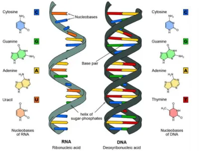
a phosphate di-ester linkage (Figure 3). Groups of three nucleotides code for a single amino acid and are referred to as codons. Since there are 43 = 64 possible codon combinations we should perhaps expect 64 amino acid variants. However it turns out that there is considerable genetic degeneracy and several codons may code for the same amino acid. In addition codons are required to define the beginning (start codon) and the end (stop codon) of a protein. This results in base pair combinations coding for a total of 23 proteogenic amino acids of which the last three are more recent additions[2-7].
Figure 3 - Representation the molecular structure of the DNA double helix and single stranded RNA
The DNA of an organism contains many thousands of nucleotides. The human genome for example contains 3 billion base pairs spread over 46 linear pieces or chromosomes. This vast array of information is further increased since there are three possible ways to read the DNA (three reading frames) and some organisms can decode DNA in both the 5’-3’ and 3’-5’ directions. However not all of the DNA sequence codes for proteins. Indeed human DNA is now only thought to contain just over 20,000-protein coding genes or exons[8]. Protein coding genes are characterised by specific start and stop codons that demark each section of DNA and denote an open reading frame (ORF). In eukaryotes there is only a single start codon, although several alternate start codons are known in prokaryotes. In all domains of life there are multiple stop codons. The start and stop codons allow the creation of lists of hypothetical protein candidates from sequenced genomic data. In combination with other motifs such as the Shine-Dalgarno sequence in prokaryotes they also serve to direct the cellular machinery to where protein-coding genetic material begins and ends[9].
12
The way an organism translates this genetic information into proteins is rather complex and differs substantially between eukaryotes and bacteria, however in both cases it essentially involves two steps. In the first, the double stranded DNA helix is separated and each base in the anti-sense strand is transcribed to a corresponding base on an mRNA intermediate (Figure 4). In eukaryotes considerable processing of the mRNA then occurs and it is shuttled out of the nucleus to the cytoplasm.
Figure 4 - Simplified description of the first stage in DNA transcription
In the second step, the mRNA is delivered to a very large set of protein complexes, the ribosome that translates the mRNA bases to their corresponding amino acids and polymerises them to form the nascent protein. Some detail of this process is shown in Figure 5.
2. Diversity Imparted from the Genome
Understanding the fundamental process of how stored genetic information is turned into an expressed protein allows one to appreciate the potential for gene product diversity. The situation described in the preceding section is an ideal one and in reality diversity may be imparted into the expressed proteome from a number of alternative outcomes of genetic processing (Figure 6). A common example of this is alternative splicing where combinations of exons from the same gene may be combined in different ways to form different gene products. These are known as alternative splice forms (ASFs). Errors in the copying process or other regulated processes can also give rise to single nucleotide polymorphisms (SNPs) in the gene which may (or may not) change the identity of an amino acid in the expressed gene product (single amino acid
13
polymorphism, SAP). Furthermore, there may be multiple possible start and stop codons for the same gene, which also result in distinct gene products.
Figure 5 - Detail of translation events in both eukaryotes and prokaryotes showing the role of important cofactors and the dissociation and association of the ribosome during translation. From Melnikov et al.[10]
14
3. Posttranslational Modification
Once the protein has been translated it may also become the target for further chemical elaboration by other proteins. This process is referred to as posttranslational modification (PTM) and encompasses a plethora of chemical changes including: proteolysis; modification of amino acid chirality (Pro cis→trans isomerism); modification of amino acid side chains or termini (deamidation, citrullination, pyroglutamate formation); the covalent addition of simple moieties such as hydroxyl, phosphate, methyl or acetyl groups and more complex molecules such as sugars (N-linked, O-linked); lipids and long aliphatic chains (myristolyation, palmitoylation); glycolipids (glypiation); polypeptides or even small proteins (SUMOlyation, ubiquitinination, neddylation, pupylation). The examples given here are by no means exhaustive but represent some of the most common modifications reported in the Swiss-Prot database[11]. Novel PTMs such as 3-phosphoglyceryl-lysine continue to be reported as understanding of cellular processes deepens[11]. Until relatively recently PTM was considered an exclusively eukaryotic process, however despite their smaller genomes many PTMs have since been discovered in bacteria and archaea, and perhaps because of their more promiscuous lifestyle they have become a rich fountain of new and interesting PTMs.
PTMs are increasingly being linked with regulation of protein function and as such are an essential piece of information to achieving total understanding of cellular processes. More and more evidence is being presented that many aspects of fundamental cell biology rest on pathways regulated by PTM; this includes transcription and gene expression directed by the histone code. The variety that PTM brings to gene products also helps explain the unexpectedly small number of proteins coded for by the human genome and others. Indeed it is being realised that the complexity that characterises biological processes occurring in higher organisms may not necessarily require a huge number of proteins but tight regulation and modulation of cell function may be imparted though PTM regulated process involving a much smaller set of modified proteins. Several expressions have been employed to describe the different posttranslationally modified forms of a protein including, isoform, protein species, protein variant, protein form etc. Isoform has a strict IUPAC definition that restricts its use to gene products having a high sequence identity and issuing from the same gene family or polymorphisms. The term proteoform has recently been coined to encompass all variants of the same gene product including changes due to genetic variations, alternatively spliced RNA transcripts and post-translational modifications. It is this term that will be used throughout this thesis[12].
15
4. Identification of Proteins Using Bottom-Up Proteomics
Identifying the protein content of a biological sample has been an enviable challenge ever since the first studies on protein rich substances such as egg white and blood plasma. Despite these samples being highly abundant in protein (albumin and fibrinogen respectively) identification of the protein molecules presented a considerable challenge because their high molecular mass and diverse chemical reactivity rendered them difficult to characterisation by classical chemical methods. Clearly the most definitive way to identify a protein would be to determine its complete amino acid sequence.
In 1951 Frederick Sanger & Hans Tuppy were the first to achieve this remarkable feat, sequencing the 30 amino acid beta chain of the protein insulin[13, 14]. This was quickly followed by sequencing of the alpha chain[15, 16] and localisation on of the intra-chain and inter-chain cysteine bonds[17]. Sanger received the Nobel Prize for this work in 1958 and his basic methodology persists to this day.
Sanger pioneered a “divide and conquer” strategy to protein sequencing where the protein, too complex to analyse intact, was cleaved into smaller pieces using a combination of acid hydrolysis and proteolytic enzymes. The resultant peptides were then separated and individually sequenced though highly laborious steps of chromatographic separation, chemical derivatisation and further chromatographic separation. Since different proteolysis methods resulted in different peptides, the small peptide fragments often overlapped and could thus be pieced back together to eventually reveal the sequence of the entire protein. This divide and conquer strategy has remained essentially the same and forms the cornerstone of contemporary “bottom-up” proteomics.
4.1. Beginnings of Bottom-Up Proteomics
The techniques used for protein identification have however developed substantially since the 1950s. The introduction of sodium dodecyl sulphate polyacrylamide gel electrophoresis (SDS-PAGE) enabled proteins to be easily separated as a function of their molecular weight and the introduction of two dimensional electrophoresis (2DE) allowed more complex mixtures of proteins to be separated by both molecular weight (MW) and isoelectric point (pI), permitting hundreds to several thousand proteins to be separated and on large format polyacrylamide gels, focused into spots and visualised using dyes and fluorophores[18].
Whilst gel based techniques revolutionised protein separation, correlating an observed spot to a known protein remained challenging. Estimates of only MW and pI are clearly insufficient to provide definitive protein identification (protein ID). Initially proteins were extracted from gels
16
onto membranes and N-terminal sequences determined using Edman degradation. Databases of genomic data provided lists of putative proteins against which the N-terminal sequence could be compared, with a positive match providing a protein ID. However, extraction of the intact protein from the gel could be difficult, especially for high MW proteins. N-terminal sequencing was limited to the first 30-50 N-terminal residues and was also problematic for proteins with a high MW or those with a blocked N-terminus. The process was cyclic, identifying one amino acid at a time and therefore slow, taking several hours per protein even on automatic sequencers. It was also not particularly sensitive, requiring several micrograms of pure sample and protein identification proved impossible if the examined protein was not already present in a database or if the N-terminus of the protein had been modified posttranslationally. Internal posttranslational modification could also arrest sequencing prematurely. Using this method the differentiation of proteins with homologous N-termini was often simply impossible although integration of MW and pI from the 2D gel as additional parameters could narrow down the possibilities.
An interesting improvement was reported where extracted proteins were chemically digested using cyanogen bromide or skatole (4-methyl-2,3-benzopyrrole), to give a small number of large peptides. The digestion products were then sequenced by Edman degradation and matched against databases in an approach called mixed peptide sequencing[19]. However, a method to digest proteins into peptides within the gel matrix was developed which abrogated the need for protein extraction so long as the resulting peptides could be efficiently analysed[20]. In this regard the arrival of biological mass spectrometry proved a game changer, opening up the possibility for higher throughput large scale protein identification by two principal methodologies.
4.2. Peptide Mass Fingerprinting
Mass spectrometry had been used for many years for the structural examination of organic molecules. The organic compound of interest was often ionised in vacuo using high energy electrons (electron impact, EI) forming unstable radical cations which would undergo fragmentation or rearrangement and produce a number of daughter ions (fragment ions). Accelerating these ions through a magnetic field causes a deflection of their path, proportionate to their mass (m) and inversely proportional to their charge (z). Scanning though a range of field strengths to separate the ions followed by detection in a photomultiplier can be used to calculate the absolute mass of each fragment ion produced and structural information can be obtained from careful interpretation of the resultant mass spectrum.
Applying this procedure to peptides or other fragile biomolecules proved difficult due to their poor ionisation and thermolability. New ionisation techniques such as secondary ion mass spectrometry (SIMS), fast atom bombardment (FAB) and chemical ionisation (CI) were developed
17
that enabled peptide ionisation for the first time. The implementation of experimentally simpler, softer ionisation techniques such as matrix assisted laser desorption (MALDI)[21, 22] and electrospray ionisation (ESI)[23] bought in a new era of sensitive and routine analysis of underivatised, intact peptide and protein ions (Figure 7).
Figure 7 - Soft ionisation methods A) MALDI A mixture of analyte and chemical matrix crystallised onto a metal support is ionised in vacuo by a UV laser. The mixture is vaporised and a cation (usually proton) transferred from the matrix to the analyte. Positive ions are transferred into the mass spectrometer B) ESI
Analytes dissolved in a volatile, usually acidic solution are sprayed from a positively charged needle or capillary Again the charge carrier is often a proton.
Refinement of ionisation techniques coupled with the development of new mass spectrometers equipped with time of flight (ToF) and quadrupole (Q) type mass analysers allowed the digest products of gel spots to be readily analysed in peptide profiling experiments. MALDI-time of flight mass spectrometry (MALDI ToF MS) became a popular choice since this type of analysis could be readily performed over a relatively large low mass range 400-4000 m/z and affords singly charged peptide ions. When proteins are digested with a proteolytic enzyme such as trypsin that cleaves at specific amino acid residues (Lys and Arg in the case of trypsin), mass analysis of the peptides formed gives a distinctive pattern for the protein of interest or peptide mass fingerprint (PMF). With the revolution occurring in informatics and gene sequencing occurring at the same time it was quickly realised that PMF, or rather the masses of peptide signature ions, could be used in conjunction with databases of genomic information for the identification of proteins. If one had a list of all of the expected proteins in an organism, a list of expected peptides could be generated by taking into account the specificity of the enzyme used for digestion. Protein databases derived from genomic data could then be annotated with the masses of expected peptide ions, and comparison of experimental ions from the PMF profile and these in silico digestions used to identify the proteins present in 2D gels.
Thus the classical proteomics workflow was born. Complex protein samples were separated by 2DE, spots stained with dyes then excised, digested and PMF performed. Matching of PMF data with protein database finally furnished protein identification (Figure 8).
18
Figure 8 - Classical proteomics workflow for protein ID by PMF
PMF can be quite a successful approach to achieve protein identification especially when the high-resolution mass measurement is performed. This has been made possible by substantial technical improvements in ToF instruments over the last decade or so. The PMF approach does however suffer several drawbacks.
Firstly, unexpected peaks in the peptide digest are particularly problematic as they increase the risk of false positive protein ID. Miscleavages from inefficient proteolysis and some posttranslationally modified peptides can now be handled by appropriate database matching algorithms. However, complex mixtures of peptides from multiple proteins are still not well supported due to a lack of dynamic range in the MS step and increased risk of false positive protein ID. Secondly, merely examining peptide masses does not provide any amino acid sequence information and thus several peptide fragments are required to produce a confident protein match. Missing peptides due to large size, poor ionisation efficiency and ion suppression mean that in some cases PMF is only sufficient to narrow down the list of possible proteins rather than provide definitive protein identification. In addition highly posttranslationally modified proteins
19
and those not already in a database simply cannot be identified. If the observed peptide ions could be sequenced inside the mass spectrometer an additional level of information could be provided that would greatly facilitate protein identification.
4.3. Peptide Sequencing By Tandem MS
Early work by Biemann had shown that the fragments produced during electron ionisation mass spectrometry of individual, derivatised peptides could be used for amino acid sequencing[24] and once intact peptides could be successfully ionised it was quickly demonstrated that they could be fragmented inside the mass spectrometer in tandem MS (MS/MS) experiments. Various fragmentation techniques were developed for different instrumental platforms such as post source decay (PSD) in the free field region of the flight tube in MALDI ToF instruments[25, 26], collisionally activated dissociation (CAD) in specially designed collision cells in triple quadrupole mass spectrometers[27] and resonant excitation of the parent ion, in several forms of ion trap. Fragmentation of peptides may either result in cleavage of the protein backbone or cleavage of the amino acid side chains. There are three principal places that cleavage can occur on the peptide backbone: between Cα and C=O of a particular amino acid; between C=O and NH (the amide bond) and between NH and Cα of the next amino acid. Roepstorff and Fohlman developed a nomenclature for peptide fragmentation[28] which was later extended by Biemann[29] that describes fragment ions issuing from cleavage of these three bonds. If the fragment ion contains the peptide N-terminus then it is termed a, b and c respectively. If the daughter ion is C-terminal then it is designated x, y and z. Ions are often followed by a subscript number indicating which peptide bond has been fragmented (counting from the respective terminus) (Figure 9).
Figure 9 - Roepstorff nomenclature for peptide ion fragmentation[28]
Both PSD and CAD impart vibrational energy to the parent ion though successive low energy collisions with inert gasses such as N2, He and Ar and produce characteristic b and y type fragment ions for which the activation barrier is generally the lowest. It was found that unlike Biemann’s EI spectra, fragmentation spectra of peptides issuing from PSD and CAD were rarely dominated by one or two stable product ions, rather they contained many intense peaks. These peaks
20
correspond to series (or ladders) of ions built up from the N-terminus (b type ions) and or C-terminus (y type ions) of the peptide. The mass difference between each pair of ions in the series (rung of the ladder) corresponds to that of an individual amino acid. Often the two ion series meet in the middle or even overlap allowing the sequence of the entire peptide to be derived. If complete sequencing is achieved, uncertainty only remains for the isobaric amino acids leucine/isoleucine and near isobaric amino acids lysine/glutamine when experiments are performed with low resolution instruments.
At around the same time techniques such as pre-flight tube ion gating in MALDI ToF instruments, modulation of the rod potentials in hybrid mass spectrometers such as QqQ or Q-ToF instruments and modulation of the trapping potentials in ion trap mass spectrometers quickly became available to isolate, or separate, individual ions from mixtures such as PMF profiles inside the mass spectrometer. These ions could then be fragmented to provide sequencing information. The advent of tandem mass spectrometry brought about two new methods for protein identification. The first was an augmentation of PMF identification where ions from a PMF profile would be selected and sequenced by MS/MS inside the mass spectrometer. The sequence tags produced helped to confirm protein identifications and brought added confidence to results. Instead of requiring many peptide fragments only one or two sequenced fragments were required to achieve a confident protein ID. Automation of this process would lead to high throughput identification of proteins from 2DE.
The second was the “direct sequence tag search” approach pioneered by Mann, where the PMF data could be discarded and the more specific sequence tags from MS/MS of peptide ions used directly to identify proteins from a database[30]. The amino acid sequences furnished though MS/MS could be searched directly against protein databases in order to identify the protein of interest.
This provoked a paradigm shift in protein identification and with the correct instrumental development one could now imagine peptides being constantly delivered to the mass spectrometer, selected and sequenced one after the other. Realisation of this idea relied on efficient ways to separate peptides and an efficient method to deliver them to the mass spectrometer.
4.4. LC-MS/MS for High-Throughput Bottom-Up Protein Identification
In contrast to MALDI, electrospray ion sources allow a free flowing liquid interface to be directly coupled to the mass spectrometer. To achieve efficient spray conditions several criteria must be met. In particular the analyte must be sprayed in a volatile solvent and be free of non-volatile salts
21
or particulates that may clog the electrospray needle. This requirement is even more stringent for smaller nanoESI interfaces that allow lower flow rates and thus greater sensitivity. Several such flow through methods such as capillary electrophoresis (CE) and liquid chromatography (LC) have been developed for the separation of both peptides and proteins from complex mixtures. CE involves separation as a function of differential mobility in an electric field and essentially depends on the charge carried by the analyte and its molecular mass. Whilst benefiting from incredibly high resolution, due to electroosmotic flow in the electrophoretic capillary, interfacing CE with mass spectrometry whilst maintaining high sensitivity has proved challenging and continues to be the subject of on-going work[31]. Liquid chromatography has enjoyed much more success.
LC involves separation of molecules based on their competing preference for stationary and mobile phases. Separation of peptides by this approach turns out to be much simpler than separation of proteins. For this reason the following description relates specifically to peptides, although the principles are essentially the same for proteins and the topic will be returned to later in this thesis. In the most commonly employed method, reverse phase LC (RPLC), a mixture of peptides is first adsorbed onto a hydrophobic solid phase. This usually is comprised of silica beads functionalised with long chain (often C18) alkyl groups tightly packed into a column. The bound analyte is then eluted in an increasing gradient of hydrophobic solvent, such as the organic solvents methanol and acetonitrile. Hydrophilic peptides with little affinity for the C18 phase are eluted first followed by those of increasing hydrophobicity as the percentage of organic solvent in the mobile phase is increased. Peptide separation is thus is achieved as a function of analyte hydrophobicity.
With the addition of a volatile acid (such as formic or acetic acid) to aid ionisation, relatively minor modifications to the electrospray source and an appropriate flow rate, LC systems can be easily coupled directly to a mass spectrometer. MS analysis can thus be performed on peptides as soon as they elute from the column. If chromatography has been performed correctly they arrive to the mass spectrometer in concentrated, well separated bunches. There is generally ample time for both mass measurement and tandem MS/MS to be performed.
In reality several peptides may co-elute from the LC column and arrive at the MS together. In such cases automated parent ion selection, then fragmentation, enables consecutive MS/MS experiments to be performed without the need to make repeat MS measurements. MS/MS spectra and thus sequence information is linked to a specific parent ion in the acquisition data file. (This of course required significant advances in acquisition software, automation of instrument control and improvements in mass analyser resolution.)
22
Identification can now be achieved by matching peptide masses (from m/z) obtained in the MS spectrum against an appropriately digested protein database (with a tolerance related to analyser resolution). This first-pass filter narrows down the possible number of candidate peptides. Then masses derived from the MS/MS experiment can be matched with the theoretical fragment ions expected from the peptide candidates. Confidence metrics or scores for these peptide-spectrum matches (PSMs) have been invented in order to enumerate the quality of the match and confidence metrics imposed to discard matches under a certain significance threshold.
In this “shotgun” approach to protein identification, it is the peptides which are examined and identified experimentally. Only when a certain number of appropriate peptides have been identified at a given significance or confidence level. is the related protein also identified. The presence of a protein is directly inferred from peptide identification. Robust statistical tests such as decoy database searching and various types of false discovery rate (FDR) have been introduced in order to reduce the number of false peptide and protein identifications. Additional methods for validation of peptide and protein ID may also be used to add a further level of confidence. This modern LC MS/MS bottom-up proteomics workflow is illustrated in Figure 10.
Figure 10 - Contemporary LC MS/MS bottom-up proteomics workflow
4.5. State-of-the-Art Bottom-Up Proteomics
Interestingly from its beginnings as a protein centric discipline, proteomics has become incredibly peptide centric. The arrival and adoption of robust LC-MS/MS methods represented a paradigm
23
shift for protein identification. No longer did proteins have to be painstakingly separated by 2DE then identified by MS but many proteins can be effectively identified after one pot digestion and LC-MS/MS. This idea is particularly compelling because it means that entire proteomes can be examined rather than isolated proteins and complete sample analysis achieved in much shorter time frame.
In order to divide very complex proteomes into smaller subsets or enrich for particular proteins or peptides of interest, many diverse fractionation techniques such as: strong and weak cation exchange (SCX, WCX); anion exchange; isoelectric focusing (IEF); hydrophilic interaction liquid chromatography (HILIC); size exclusion chromatography (SEC); gel based fractionation; and affinity purification strategies have been developed to complement separation by RPLC[32]. Specific protocols such as combined fractional diagonal chromatography (COFRADIC)[33] have also been developed for offline RPLC fractionation. MS workflows such as the MudPIT strategy pioneered by the Yates lab are then used for analysis of MS/MS data from these fractionated samples[34].
The quest to extend the dynamic range, to mine ever further into the proteome and to achieve higher and higher numbers of identified proteins has undoubtedly been an important driving factor in instrument development. Fast scanning, high resolution mass spectrometers with a large dynamic range and optimised peptide fragmentation such at the Thermo QExactive are producing very high quality, high confidence, high resolution MS/MS data. This means less and less is information is demanded before a positive peptide ID and therefore protein identification is accepted as fait accompli.
Instrument improvement and the introduction of ultra-high pressure LC systems is even allowing entire proteomes to be analysed in single LC runs on long nano-LC columns[35, 36]. As example of this performed during this thesis on the proteome of Neisseria meningitidis is shown in Figure 11. A recent report from the Mann group has identified over 4,000 proteins from Lys-C digested Saccharomyces cerevisiae lysates using this technique[37]. This is close to the total number of expected cellular proteins expressed under standard growth conditions. A very recent report from the Coon group has achieved similar results but with only a 1.3 hour acquisition time using a fast scanning Orbitrap Fusion mass spectrometer[38].
Increasingly accurate ways are being devised to quantify proteins from bottom up data including label-free and tag based techniques such as isotope-coded affinity tag (ICAT)[39], tandem mass tag (TMT)[40], isobaric tags for relative and absolute quantitation (iTRAQ)[41] and stable isotope labelling by/with amino acids in cell culture (SILAC)[42]. In the previous report by the Mann group
24
on the yeast proteome, over 3,500 proteins could be accurately quantified using a SILAC strategy with a similar method set up during a differential experiment.
A continually developing area of bottom-up proteomics is the identification of large numbers of post translational modifications at the proteome level. Since specific posttranslationally modified proteins often represent only a small fraction of the expressed proteome, enrichment of modified species at either the peptide or protein level is usually performed. Many PTM specific enrichment protocols have been devised such as immobilised metal affinity chromatography (IMAC) for phosphopeptides[43, 44], lectin enrichment in the case of glylcopeptides and immunoprecipitation of GG tagged peptides in the case of ubiquitination[45]. Specialised LC MS/MS strategies are then applied to increase coverage of modified peptides and identify PTM sites. For example a combination of SCX fractionation after protein digestion, IMAC phosphopeptide enrichment followed by RPLC MS/MS using CAD, ETD and a decision tree based MS method enabled the Coon group to identify 10,844 district phosphosites and 4,339 proteins in an analysis of the human embryonic cell phosphoproteome[46].
Figure 11 - Single run RPLC MS/MS on a cell lysate of Neisseria meningitidis using a 30cm nano-LC column and 4 hour chromatographic gradient. 1,115 proteins were identified at an FDR<1%. Data acquired by
Miss Debora Pasquali
This thesis does not deal explicitly with bottom up-proteomics and the state of the art of this field including various methods for quantification, PTM identification, applications to interactomic and


![Figure 16 - Representation of the [M] + ion of the protein ubiquitin with increasing resolution, R, from](https://thumb-eu.123doks.com/thumbv2/123doknet/2719734.64398/58.892.128.788.596.736/figure-representation-m-ion-protein-ubiquitin-increasing-resolution.webp)