HAL Id: tel-02931476
https://pastel.archives-ouvertes.fr/tel-02931476
Submitted on 7 Sep 2020HAL is a multi-disciplinary open access
archive for the deposit and dissemination of sci-entific research documents, whether they are pub-lished or not. The documents may come from teaching and research institutions in France or abroad, or from public or private research centers.
L’archive ouverte pluridisciplinaire HAL, est destinée au dépôt et à la diffusion de documents scientifiques de niveau recherche, publiés ou non, émanant des établissements d’enseignement et de recherche français ou étrangers, des laboratoires publics ou privés.
Regulation of the invasion suppressor Arpin by
Tankyrases
Angelina Chemeris
To cite this version:
Angelina Chemeris. Regulation of the invasion suppressor Arpin by Tankyrases. Cellular Biology. Université Paris Saclay (COmUE); Moskovskij gosudarstvennyj universitet imeni M. V. Lomonosova, 2018. English. �NNT : 2018SACLX073�. �tel-02931476�
NN
T: 2018S
ACLX073
Régulation du suppresseur
d'invasion Arpin par les Tankyrases
Thèse de doctorat de l'Université Paris-Saclay et de Lomonosov Moscow State Universitypréparée à l’Ecole Polytechnique
École doctorale n°573 : approches interdisciplinaires, fondements, applications et innovation (Interfaces) Spécialité de doctorat: biologie
Thèse présentée et soutenue à Palaiseau, le 21 septembre 2018, par
Angelina Chemeris
Composition du Jury :Pr. Philippe Minard
Professeur,Universite Paris-Sud XI Président
Mme. Aurélie Bertin
CR1, Institut Curie Rapporteur Mme. Maria-Carla Parrini
IR, Institut Curie Rapporteur M. Guillaume Romet-Lemonne
DR, Institut Jacques Monod Examinateur M. Nicolas David
CR, Ecole Normale Supérieure Examinateur Pr. Alexis Gautreau
Professeur, DR
Ecole Polytechnique, CNRS Directeur de thèse
Pr. Olga Sokolova Professeur, DR,
La migration cellulaire intervient dans des nombreux processus cruciaux comme l’embryogenèse, la réponse immunitaire, l’homéostasie des tissus et la cicatrisation des plaies. Les aberrations dans la migration cellulaire augmentent le risque de développement des processus pathologiques tels que le cancer, les maladies vasculaires, l'ostéoporose, les maladies inflammatoires chroniques et d’autres. Il existe généralement deux types de migration cellulaire : la migration cellulaire individuelle et collective. La migration cellulaire collective est typique pour les groupements multicellulaires cohésives. La migration individuelle est caractéristique aux cellules amiboïdes ou mésenchymateuses. Les cellules qui sont fortement attachées au substrat dont le cytosquelette est contractile migrent en mode mésenchymateux. A la première étape de leur migration, les cellules forment de grandes protrusions à leur bord appelées les lamellipodes et de petites protrusions en forme de doigt appelées les filopodes.
La migration qui implique la protrusion réunit plusieurs processus subcellulaires coordonnés qui peuvent être classés en cinq types : la protrusion du bord cellulaire, l’interaction de la cellule avec la matrice extracellulaire et la formation d’adhésions focales, la protéolyse focalisée, la contraction de l'arrière de la cellule par l'actomyosine et le détachement du bord arrière.
Les protrusions au bord devant sont faites par des structures appelées les lamellipodes et les filopodes. Les lamellipodes et les filopodes sont des structures à la base d'actine bien étudiées, assemblées au bord des cellules mobiles, ils sont d'une importance cruciale pour la mobilité des cellules. Les lamellipodes sont des structures tridimensionnelles temporelles qui génèrent de la force pour pousser la membrane cellulaire et provoquer ainsi la migration cellulaire. Au cours de la migration, le lamellipode reste stable suffisamment longtemps pour former de nouvelles adhésions focales avec la matrice extracellulaire et ainsi permettre à la cellule d’ancrer la protrusion. Le lamellipode est formé du réseau d'actine branchée qui est assemblé par le complexe Arp2/3.
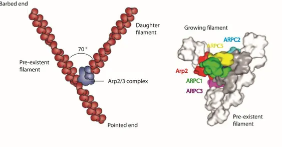
Le complexe Arp2/3 conservé au cours de l'évolution joue un rôle central dans la nucléation des réseaux de filaments d'actine branchée qui permet la migration cellulaire, l'endocytose et d'autres processus. Le complexe des protéines liées à l’actine 2 et 3 (Arp2/3) est l'un des principaux nucléateurs de l'actine. Le complexe Arp2/3 induit directement la formation d'un réseau d'actine branchée au bord des cellules mobiles en réponse à la signalisation extracellulaire, générant ainsi la force permettant la formation des lamellipodes et des invadopodes. Le complexe Arp2/3
comprend les protéines liées à l'actine Arp2 et Arp3 et cinq sous-unités supplémentaires ARPC1 (p40), ARPC2 (p35/p34), ARPC3 (p21/p18), ARPC4 (p20/p19) et ARPC5 (p16/p15). Les sous-unités Arp2 et Arp3 jouent un rôle essentiel dans la construction des réseaux de filaments d’actine. Grâce à leur homologie avec les monomères d'actine, les sous-unités Arp2 et Arp3 peuvent former un pseudo-dimère d'actine qui peut se lier à la première G-actine liée à l'ATP.
Ainsi, Arp2 et Arp3 forment le noyau pour l'élongation ultérieure du nouveau filament d'actine. Etant donné que les sous-unités Arp2 et Arp3 peuvent agir en tant que monomères d’actine et former un pseudo-dimère d’actine, les sous-unités Arp2 et Arp3 doivent être situées à proximité l’une de l’autre pour former le noyau pour l'élongation. La première structure cristalline du complexe Arp2/3 a révélé un gap entre les sous-unités Arp2 et Arp3, qui fait référence à un état ouvert et donc inactif du complexe Arp2/3. En soi, le complexe Arp2/3 est stabilisé dans l'état conformationnel ouvert. La liaison des facteurs favorisant la nucléation (NPF), portant les domaines VCA, induit des réarrangements conformationnels dans le complexe Arp2/3 : les unités Arp2 et Arp3 se déplacent vers le centre de la fente, le rapprochement ultérieur des sous-unités Arp2 et Arp3 forme un pseudo-dimère et met le complexe Arp2/3 dans son état fermé/actif. Ainsi, les NPFs déclenchent l'assemblage d'actine dirigé par le complexe Arp2/3.
Le complexe Arp2/3 génère le réseau de filaments d’actine branchée via la formation d’une jonction dite de branche entre deux filaments d’actine. On sait que les NPFs jouent un rôle crucial dans la formation de jonctions de branches. Les NPFs agissent à plusieurs étapes: ils activent le complexe Arp2/3, facilitent sa liaison au filament préexistant (mère) d'actine, augmentent le taux d'association du complexe Arp2/3 avec le filament mère et apportent le premier monomère d'actine lié à l'ATP à la jonction de branche. Par la suite, le nouveau filament (fille) s’allonge à un angle de 70 degrés par rapport au filament existant dans une orientation en Y. Les NPFs les mieux caractérisés sont les domaines VCA C-terminaux des protéines de la famille WASP (protéine du syndrome de Wiskott–Aldrich) : WASP, N-WASP (WASP neuronal), complexe WASH, complexes WHAMM et WAVE1-3. Le domaine VCA est constitué de trois motifs courts : motif d'homologie Verprolin également connu sous le nom de WASP homologie 2 (WH2) domaines (V), central (C), acide (A). Le motif V lie la G-actine et délivre ainsi une sous-unité initiale au filament fille. Le motif C lie à la fois le complexe G-actine et Arp2/3 au VCA. Le motif A ne lie que le complexe Arp2/3. Ainsi, l’incorporation de motifs CA dans le complexe Arp2/3 permet aux monomères d’actine de se lier à la jonction des branches. De plus, Arp2 fournit une hydrolyse ATP
rapide sur le monomère d’actine lié par VCA qui initie la polymérisation d’actine. Ainsi, les domaines VCA des protéines de la famille WASP activent le complexe Arp2/3 par formation du complexe Arp2-Arp3-noyau d’élongation sur le filament d'actine préexistante. Par la suite, l’attachement de domaines VCA entraîne des changements de conformation dans le complexe Arp2/3.
Etant donné la capacité de réorganisation des filaments d’actine, l’activité du complexe Arp2/3 doit être étroitement contrôlée. Un moyen qui permet de réguler l'activité du complexe Arp2/3 c’est la liaison des inhibiteurs. Les inhibiteurs sont les protéines qui convertissent le complexe Arp2/3 dans son état inactif et bloquent ainsi la nucléation de l'actine. A l'heure actuelle, il y a peu d’inhibiteurs connus du complexe Arp2/3. Il semble que dans chaque partie de la cellule où se produit la réorganisation de l'actine, il existe deux molécules antagonistes qui régulent l'activité du complexe Arp2/3.
Jusqu'à récemment, il n’était pas clairement défini comment le complexe Arp2/3 peut être directement inactivé dans le lamellipode. Arpin est un nouvel inhibiteur du complexe Arp2/3 au niveau du lamellipode qui a été identifié lors d’une recherche bioinformatique des protéines présentant l’homologie avec le motif VCA des NPFs. Arpin est une protéine relativement petite (25 kDa) à la queue C-terminale allongée. L'extrémité C-terminale d'Arpin contient un motif acide (mais il lui manque des motifs V et C) similaire au motif acide des NPFs qui interagissent avec le complexe Arp2/3. Il a été vérifié si le motif A seul est suffisant pour l'inactivation du complexe Arp2/3. En effet, le motif acide seul inactive le complexe Arp2/3, mais moins efficacement que Arpin dans son intégrité. En revanche, Arpin dépourvu de motif acide a perdu la capacité d'inhiber le complexe Arp2/3.
Il était prédit que Arpin pouvait se lier au complexe Arp2/3 via deux sites de liaison comme le domaine VCA des FNP. Nous avons utilisé la microscopie électronique à une seule particule (single particle electron microscopy) pour obtenir une reconstruction 3D du complexe Arp2/3 lié à Arpin à une résolution de 25 Å. Nous avons montré que la liaison de Arpin entraine la conformation ouverte du complexe Arp2/3. Nous avons confirmé qu'il existe deux sites de liaison sur le complexe Arp2/3 pour Arpin : l’un à l'arrière de la sous-unité Arp3 et le second est situé entre les sous-unités Arp2 et ARPC1. La distance entre le complexe Arp2/3 et Arpin (5 nm) permet de penser que Arpin interagit avec son partenaire via sa queue acide C-terminale non structurée.
Ensuite, grâce à l’analyse pull-down (pull-down assay), nous avons identifié des nouveaux partenaires de liaison de Arpin, Tankyrase1 et Tankyrase2. Les tankyrases sont des protéines de la famille des poly (ADP-ribose) polymérases (PARP) localisées dans les télomères qui se lient aux protéines de liaison à l'ADN télomériques, TRF1 et TRF2, tandis que TRF2 protège les extrémités des télomères et régule négativement la longueur des télomères. La surexpression de tankyrases provoque l’élongation des télomères dans les cellules cancéreuses grâce à la libération de TRF1 par les télomères. Tankyrase a deux homologues : 142 kDa Tankyrase1 et 130 kDa Tankyrase2 qui sont exprimés de manière omniprésente dans les lignées cellulaires de mammifères. Les tankyrases ont 85% d'acides aminés identiques et sont supposées d’avoir les mêmes fonctions. Cependant, contrairement à la tankyrase-1, la surexpression de Tankyrase-2 provoque l’apoptose précipitée.
Les tankyrases interagissent avec un large spectre de protéines et arbitre la poly (ADP-ribosyl) de ces protéines, régulant ainsi les processus cellulaires essentiels. Les tankyrases lient les télomères, les centrosomes, l'appareil de Golgi, la NuMA (protéine de l'appareil mitotique nucléaire), SH3BP2 et d'autres protéines. Une mutation qui supprime la liaison de SH3BP2 àaux tankyrases provoque un chérubisme chez l’homme. Les tankyrases sont également impliquées dans la régulation de la longueur des télomères, la séparation des télomères sœurs, la mitose et le métabolisme du glucose. Les tankyrases se sont avérées d’être les cibles de signalisation de la protéine kinase MAPK (mitogen-activated protein kinase) dans le Golgi.
Il est intéressant de noter que les Tankyrases et le complexe Arp2/3 possèdent des séquences d’acides aminés qui se chevauchent au niveau des sites de liaison de Arpin. Par conséquent, nous avons démontré une compétition entre le domaine ARC4 de Tankyrase1 et le complexe Arp2/3 d’une manière dose dépendante. Pour comprendre les principes de l'interaction Tankyrases-Arpin, nous avons créé une forme mutée d'Arpin (ArpinG218D) qui a perdu la capacité d'interagir avec les Tankyrases, mais pas avec le complexe Arp2/3 in vitro. Notons cépendant que ArpinG218D n'a pas été capable d'inhiber le complexe Arp2/3 in vivo, ce qui suggère que la Tankyrase pourrait être nécessaire pour l'interaction du complexe Arpin-Arp2/3.
Il était connu auparavant que l'efficacité de la migration cellulaire dépend de la vitesse de la cellule et de la capacité de conserver la direction lors de la migration. Arpin a été identifié
comme un facteur qui pousse les cellules mobiles à changer leur direction de migration : les microinjections d'Arpin dans les kératocytes de poisson dont la migration est très persistant et directionnel ont forcé ces cellules à tourner. Ainsi, la diminution de la persistance de migration des cellules a été identifiée comme une caractéristique clé d’Arpin. Nous avons présenté une analyse de la persistance de migration directionnelle de cellules exprimant Arpin sauvage (ArpinWT) ou Arpin muté (ArpinG218D) en parallèle avec la déplétion d'Arpin endogène. Les cellules présentant une surexpression de ArpinG218D ont montré une persistance de migration directionnelle supérieure à celle des cellules surexprimantes ArpinWT. Nous avons suggéré que Arpin muté (Arpin G218D) n'avait aucune activité inhibitrice in vivo, ce qui correspond à l’absence de l'interaction du complexe Arpin G218D - Arp2/3 in vivo.
Nous avons suggéré que le mutant ArpinG218D n’était pas actif et qu'il ne pouvait pas inactiver le complexe Arp2/3 puisqu'il n'était pas présent au lamellipode. Nous avons comparé la quantité de protéines ArpinWT et ArpinG218D dans la fraction membranaire des cellules en migration. Une différence significative (44%) de la quantité d'ArpinWT et d'Arpin G218D était cohérente avec notre hypothèse.
Les tankyrases sont des cibles thérapeutiques dans divers cancers, mais il n’existe actuellement aucun modèle structurel pour ces protéines volumineuses et flexibles. Dans le présent travail, nous avons obtenu pour la première fois deux reconstructions 3D de Tankyrase1 et Tankyrase1 liée à Arpin en utilisant la microscopie électronique à une seule particule. La résolution obtenue (25 Â) était suffisante pour détecter un changement de conformation spectaculaire dans les domaines SAM et PARP de Tankyrase lors de la liaison des molécules d'Arpin. Dans notre reconstruction, trois Arpins étaient liés aux domaines ARC1-2 et ARC4 de Tankyrase1. ARC5 s'est avéré d’être la partie la plus flexible du cluster ARC.
Toutes ces données réunies nous ont permis de proposer un modèle de régulation de l'activité d'Arpin par les Tankyrases. Selon notre modèle, les Tankyrases se lient à Arpin dans le cytoplasme, ce qui provoque des modifications dans l'état conformationnel des Tankyrases. De plus, les Tankyrases rapprochent Arpin à la membrane dans le lamellipode. En passant les signaux extracellulaires, Rac GTPase active Arpin, qui inactive séquentiellement le complexe Arp2/3, tandis que les Tankyrases sont libérés.
Étant donné que les complexes Tankyrases et Arp2/3 partagent le même motif de liaison et sont en compétition pour les interactions avec Arpin, nous supposons la présence de protéines
intermédiaires capables de sentir le changement de conformation des Tankyrases en présence d’Arpin et de réguler ensuite la délivrance d’Arpin à la membrane dans le lamellipode de la cellule.
Table of contents
Acronyms
... 4
Introduction
... 6
1.Cell migration ... 7
1.1. Cell polarity
... 7
1.2. Types of cell migration.
... 8
1.3. The basic machinery of actin-based protrusion
s ... 9
1.3.1. Membrane protrusions formation at the leading edge ... 9
1.3.2. Cell-extracellular matrix interactions ... 9
1.3.3. Focalized proteolysis ...10
1.3.4. Contraction of the cell rear ...11
1.3.5. Detachment of the trailing edge...12
1.4. Cell membrane protrusions
...12
1.4.1. Filopodia ...12
1.4.2. Lamellipodia ...13
1.4.3. Invadopodia and podosomes ...15
1.5. Cell migration signaling
...16
1.5.1. Rho family GTPases ...16
1.5.2. Rac GTPase ...18
1.5.3. Rho GTPase...18
1.5.4. Cdc42 ...18
1.6. Aberrations in cell motility that contribute to the metastasis of cancer cells
...19
2. Actin cytoskeleton dynamics provides a major driving force for cell motility ..21
2.1. Actin cytoskeleton
...21
2.2 Actin binding proteins regulating actin dynamics
...24
2.2.1. Elongators of actin filaments. ...25
2.2.2. Nucleators of actin filaments. ...26
2
3.1. Conformational states of Arp2/3 complex
...27
3.2. Mechanism of branch junction formation in lamellipodia via Arp2/3 complex
...30
3.2.1. Model of the branch junction ...32
3.2.2 Contribution of Arp2/3 complex’ subunits into the formation of branch junction ...32
3.3. Cortactin contributes to the regulation Arp2/3 complex
...34
3.4. Nucleation of new actin filaments for Arp2/3 complex activity
...34
4. Activation of the Arp2/3 complex via Nucleation Promoting Factors. ...38
4.1. Structure and activity of Nucleation Promoting Factors
...38
4.2. Model for Arp2/3 complex activation via NPFs
...39
4.3. Regulation of Nucleation Promoting Factors
...40
4.3.1. WASP and N-WASP...40
4.3.2. WASH ...41
4.3.3. WHAMM ...42
5. WAVE complex is an activator of Arp2/3 complex activity in lamellipodia ...44
5.1. Structure of WAVE complex
...44
5.2. Regulation of WAVE complex’ activity
...47
6. Inhibition of Arp2/3 complex ...48
6.1. Coronin
...48
6.2. GMF
...48
6.3. PICK1 and Gadkin
...49
7. Arpin is an inactivator of Arp2/3 complex in lamellipodia that counteracts the
WAVE complex ...50
7.1. Activity of Arpin in vivo
...51
7.2. Structure of Arpin
...53
Objectives
...55
Results
...58
Discussion
...107
3
2. First full-length three-dimensional structure of Tankyrase-1 and Tankyrase-2
bound to Arpin. ...112
3. Model of Arpin-Tankyrase interaction ...117
References
...118
4
Acronyms
ABP Arp2/3 AP ANK ARC ARD CC Crn1 DH Aip1 Ena/VASP ECM FAB FGF FAK FH1 FH2 GAB GFR GBD GAP GDP GTP IRSp5 JMY MMP NMMIIActin binding proteins Actin-related protein 2 and 3 Adaptor protein
Ankyrin
Ankyrin repeat cluster Ankyrin Repeats Domain Coiled-Coil
Coronin 1
Db1 homology domain Actin interacting protein 1
Enabled/vasodilator-stimulated phosphoprotein Extracellular matrix
Filamentous actin binding Fibroblast growth factor Focal adhesion kinase Formin Homology 1 Formin Homology 2 Globular actin binding Growth factor receptors GTPase binding domain GTPase-activating proteins Guanosine diphosphate Guanosine triphosphate
Insulin Receptor Substrate of 53 kDa Junction mediating and regulatory protein) Matrix metalloproteinase
5 NPFs GEF NuMA PI3K PI4P5K PIP2 PIP3 PH PARP SAXS TEM TNKS1 TNKS2 TNBC WASP WH2 WIP PAK
Nucleation Promoting Factors Guanine nucleotide exchange factors Nuclear mitotic apparatus protein Phosphatidyl inositol-3-kinase
Phosphatidyl inositol-4-phosphate 5-kinase Phosphatidyl inositol (4,5) bis phosphate Phosphatidyl inositol (3,4,5) tris phosphate Pleckstrin homology domain PH
Poly(ADP-ribose)polymerase Small-angle X-ray scattering Transmission Electron Microscopy Tankyrase-1
Tankyrase-2
Triple-negative breast cancer Wiskott-Aldrich syndrome protein WASP homology 2
WASP-interacting protein p21-activated kinase
6
7
1. Cell migration
Cell migration promotes a variety of crucial processes like embryogenesis, immune cell trafficking, tissue homeostasis and wound healing. Aberrations in cell migration facilitate the pathological processes like cancer, vascular disease, osteoporosis, chronic inflammatory diseases and others.
Cell migration is a cell polarity-based process. Polarized actomyosin-driven shape change of the cell body is a basic process for all types of migration (Lan et al. 2016). Central features of cell migration are also the formation of cell membrane protrusions and cell adhesions. Cell membrane protrusions are usually the result of intense actin polymerization at the leading edge of migrating cells in response to a variety of extracellular signals. Cell adhesion and attachment to extracellular matrix (ECM) is critical for the cell migration. These processes stabilize cell protrusions and form traction sites for cell migration. Cell adhesion is mediated by transmembrane glycoprotein adhesion receptors like integrins (Buck and Horwitz 1987).
1.1 Cell polarity
The ability of cells to dynamically polarize toward extracellular signals is critical to migration. To migrate, the cell has to polarize and define a leading edge and a cell rear.
Cells are getting spatially asymmetrical due to the chemical gradients (Wang 2009). Cells analyze the extracellular information coming from the all sides and identify the chemical gradient. Due to the gradient of extracellular signals, membrane receptors are getting occupied asymmetrically on the cell surface. This spatial asymmetry causes intracellular gradient of polarity effectors. For example, the front-rear polarity was identified in motile cells: a high concentration of actin at the cell leading edge and high concentration of myosin at the cell rear along with a
8
distinct localization of PI3K, PIP3 and small GTPases such as Cdc42, Rac and RhoA. In general, the polarity proteins include three groups of proteins: the Par complex which consist of Par proteins and protein kinase C, the Scrib complex and the Crb complex.
Cells stimulate the activity of actin nucleators and other proteins involved in the reorganization of cytoskeleton (Parent and Devreotes 1999). Extracellular signals inducing cell motility (or motility factors) include almost all growth factors including basic fibroblast growth factor (FGF), hepatocyte growth factor (scatter factor), vascular endothelial growth factor and epidermal growth factor.Motility factors bind to their receptors on the cell surface and induce stimulatory signals to reorganize the cytoskeleton at the leading edge resulting in the cell migration (Anand-apte et al. 1997).
1.2 Types of cell migration
Generally, two types of cell migration exist: collective and individual cell migration. Collective migration is common for cohesive multicellular units (Friedl and Gilmour 2004). Individual migration is typical for amoeboid or mesenchymal cells.
Amoeboid migration is rounded, blebby migration which is common for the cells that do not adhere to the substrate. These cell migrate by making blebs. Blebs are the protrusions of the cell membrane that are the result of the detachment of the cell membranes from the actin cortex or by the breaks in actin cortex due to the contractility of actin–myosin networks. Under these circumstances cytosol flows out and protrudes cell membrane (Charras and Paluch 2008).
Cells with the strong attachment to the substrate and cytoskeletal contractility migrate in a mesenchymal mode. At the first step of this migration cells make large sheet-like protrusions at its leading edge called lamellipodia and finger-like small protrusions called filopodia.
9
There are also several intermediate modes. Cells can migrate by pulling the membrane on and further move in a propulsive migration mode. More elongated amoeboid cells can form filopodia or pseudopodia at its leading edge and move slightly contacting substrate.
1.3 The basic machinery of actin-based protrusions
Protrusion-based migration involves several coordinated subcellular processes (Burridge and Wennerberg 2004; Lauffenburger and Horwitz 1996). These processes can be classified into five activities: Protrusion of the leading edge, interaction of cell with the extracellular matrix and formation of focal contacts, focalized proteolysis, cell rear contraction by actomyosin and detachment of the trailing edge (Friedl and Wolf 2003).
1.3.1 Membrane protrusions formation at the leading edge
In response to extracellular signals, branched actin networks start to polymerize at the leading edge of cells. The growing branched actin network provides a force to protrude the membrane. Formation of membrane protrusions called lamellipodia and filopodia at the cell leading edge enables a cell to protrude its membrane and establish new contacts with its environment (Condeelis 1993).
1.3.2 Cell-extracellular matrix interactions
If membrane protrusions are not able to adhere to the substratum, they fold back on themselves, forming membrane ruffles that do not support cell migration. Focal adhesions ensure the attachment of the cell to the ECM through an activation and clusterization of integral receptors at the cell membrane.
A large family of cell adhesion receptors, integrins, mediates cell contacts with many ECM molecules. The β subunit cytoplasmic domains of integrins activate signaling proteins and thus
10
provide extracellular signals inside the cell. The intracellular tail of integrins interacts with alpha-actinin, focal adhesion kinase, talin 1 and other proteins. These proteins further recruit regulatory molecules like GTPases and actin-binding proteins to focal contacts.
The dynamic assembly and disassembly of focal adhesions plays a central role in cell migration at this step. There are distinct types of cell-matrix adhesions: 1) classical focal adhesions located at the termini of stress fibers, that provide long-term cell anchorage,and colocalized with integrins (Hotulainen and Lappalainen 2006). Talin is a cytoplasmic protein that bind the intracellular part integrin β subunits and actin filaments. Thus talin acts as an integrin-cytoskeletal linker (Calderwood et al. 1999). Stress fibres are contractile actomyosin bundles. Actin bundle is a structure of parallel or antiparallel aligned actin filaments crosslinked by the actin-bundling protein alpha-actinin. Actin filaments crosslinking proteins are the proteins with multiple actin-binding domains, but mostly they contain two domains that are separated by a long flexible linker, which allows a perpendicular arrangement of actin filaments (Winder 2005). 2) Nascent focal complexes associated with lamellipodia and filopodia that support protrusion and traction at the leading edge. Focal complexes are signaled by Rac1 or Cdc42 and may transform into long-term focal adhesions (Kaverina, Krylyshkina, and Small 2002). It was found that focal complexes formation and Arp2/3 complex mediated actin polymerization at the leading edge are coupled through a integrin-associated protein vinculin (Galbraith, Yamada, and Sheetz 2002). Once coupled to adhesion complexes, the actin cytoskeleton generates the force to translocate the cell forward. 3) fibrillar adhesions located at the central area of the cell and colocalized with matrix fibrils like fibronectin.
1.3.3 Focalized proteolysis
Proteolysis of ECM serves to remove excess components and assembly of ECM. These processes play the key role in ECM synthesis. Focalized proteolysis is a process of ECM degradation, that involves several classes of proteolytic enzymes. The metalloproteinases like matrix metalloproteinase (MMPs) family of proteins are known to be the important regulators of ECM remodelling via proteolysis. MMPs like membrane-type 1 MMP and MMP 2 are able to
11
activate pro-MMPs and cleave ECM macromolecules like collagen into shorter pieces that undergo subsequent degradation.
MMPs are the zinc‐ and calcium-dependent proteases that share the conserved zinc-binding motif in their catalytic zinc-binding site. MMPs have a N-terminal signal peptide for secretion, a pro domain and a C-terminal catalytic domain. In inactive state pro-MMPs catalytic domain is sequestered by prodomain. Dissociation of the prodomain from the catalytic site activates the cleavage. The localization, activation and activity of MMPs are regulated by their interactions with other proteins, proteoglycan core proteins and/or their glycosaminoglycan chains, as well as other molecules (Hadler-Olsen et al. 2011).
1.3.4 Contraction of the cell rear
Actomyosin contraction induces tension through stress fibers and induces the detachment of contact points at the trailing edge of the cell and tightening the cell body toward the leading edge (W.-T. Chen 1981). Stress fibres are mostly consist of actin filaments and non-muscle myosin II. ATP-driven movement of myosin II motor domain supply the force for stress fibers formation at the cell rear. Small GTPase Rho contribute to the actomyosin-based contraction of the cell rear. Rho GTPases activate Rho kinase. Rho kinases consist of catalytic, Rho-binding, coil-coiled and plekstrin homology domains. Rho kinase is calcium and calmodulin-dependent kinases that phosphorylate light chain of myosin II at its Ser19 and/or Thr18 residues and thus promotes actomyosin contraction. Myosin phosphatase is a contraction regulating protein that dephosphorylates light chain of myosin II and thus ceases the actomyosin-based contraction.
It was found that Rho kinases contribute to the actomyosin contraction either by direct activation of myosin or by inactivation of myosin phosphatases. MLC is phosphorylated at its Ser19 and/or Thr18 residues by MLC kinase, which is a (Ca2+-calmodulin)-dependent kinase.
12
1.3.5 Detachment of the trailing edge
Focal complexes formed at the cell leading edge undergo several cycles of assembly and disassembly. It was shown that MMP-based proteolysis of ECM induces distinct integrin signals that leads to the calpain-mediated cleavage (Carragher et al. 1999). Calpain protease cleaves focal contact components like talin and cytoplasmic tail of β1 and β3 integrins. Calpain is essential for cytoskeletal organization during cell motility, apoptosis, cell proliferation and hemostasis. Calpain family of proteases are able to the regulate the dynamics of integrin-mediated focal adhesions, focal complexes and actin-based membrane protrusions. Calpain is a calcium-regulated cysteine protease that consist of large (80kDa) and small regulatory (28kDa) subunits. There are two types of calpains: μ-calpain and m-calpain that are activated by micromolar and millimolar concentrations of calcium respectively. Calpain colocalizes with focal adhesions and integrin clusters and cleaves many focal adhesions proteins including β3 integrin, spectrin and talin.
Calpains are also known to modulate signalling molecules like focal adhesion kinase (FAK), protein kinase C and Rho family GTPases (Franco 2005). FAKs cause the disassembly of focal adhesions. It was shown that calpains act downstream of microtubules to mediate adhesion complexes disassembly (Bhatt et al. 2002).
1.4 Cell membrane protrusions
Lamellipodia and filopodia are well investigated actin-based structures assembled at the leading edge of motile cells and crucially important for direct cell motility.
1.4.1 Filopodia
Filopodia consist of parallel bundles of actin filaments that are formed by the reorganization of lamellipodial actin filaments. Lamellipodial actin filaments first increase in length and then become aligned and cross-linked. This required elongation of actin filaments might be caused either by actin assembly acceleration or by barbed ends capping delay. Reorganization of lamellipodial branched actin filaments into filopodial parallel F-actins is crucial for processes like tumor metastasis and chemotaxis (Hansen and Mullins 2010; Svitkina et al. 2002).
13
1.4.2 Lamellipodia
Lamellipodia are the three dimensional temporal structures that generate force to protrude cell membrane and thus facilitate cell migration. During protrusion-based migration lamellipodium stays long enough to form new focal adhesions with extracellular matrix and thus cell anchors the protrusion (Lan et al. 2016; T. D. Pollard and Borisy 2003). The lamellipodium is formed by branched actin networks that are assembled by Arp2/3 complex (Fig.1).
14
Figure 1. Schematic illustration of the leading edge in motile cell. Modified from (Clainche
and Carlier 2008).
The leading edge of a migrating cell consists of membrane protrusions lamellipodium and filopodia. Lamellipodium is formed by Arp2/3 complex-dependent branched actin network.
15
In theory, cell membrane has to stretch a lot to make protrusions like lamellipodia. However, it was shown experimentally that cell membrane can physically stretch only for 2-3%. The expansion of the cell membrane can be explained by following models: 1) Fountain flow model says that membrane precursor vesicles fuse with the anterior cell membrane to supply membrane (exocytosis), and membrane is taken up at the rear (endocytosis). 2) The membrane unfolding model is the utilization of the membrane folds, folding and unfolding of the membranes. 3) Caterpillar flow model says that the cell membrane moves circularly in the order of the ventral, anterior, dorsal, and rear regions. In this case, the cell membrane may turn over everywhere (Tanaka et al. 2017).
1.4.3 Invadopodia and podosomes
Podosomes and invadopodia are special types of adhesion that mediate invasion of cancer cells. Both invadopodia and podosomes consist of actin-rich core,which distinguishes them from other matrix contacts, and are regulated by a multitude of signalling pathways including Rho GTPases, cortactin, N-WASP, adaptor proteins Tks4 and Tks5, tyrosine kinase Src, MT1-MMP and microtubule-dependent transport (Linder, Wiesner, and Himmel 2011). N-WASP as a regulator of Arp2/3 complex was found to be important in the formation of invadopodia and podosomes (Nürnberg, Kitzing, and Grosse 2011).
Though the similarity between invadopodia and podosomes, there are some differences in their functionality. Invadopodia are known to attach the ECM and form stable contacts for hours, while podosomes preferably form short protrusions that retract rapidly (Murphy and Courtneidge 2011).
16
1.5 Signalling of cell migration
Growth factors and insulin promote actin polymerization at the plasma membrane of different cell types and thus induce the formation of protrusions.
Chemokine receptors and growth-factors receptors activate PI4P5K (phosphatidylinositol-4-phosphate 5-kinase) that generate PIP2 (phosphatidyl inositol (4,5) bis phosphate) before PI3K
(phosphatidylinositol-3-kinase) can generate PIP3 (phosphatidyl inositol (3,4,5) tris phosphate).
PI4P5K generates PIP2 which is involved in regulation of actin assembly and cell growth.
PIP2 releases G-actin from profilin-GTP-bound actin complex that is a signal for actin assembly.
PIP2 also regulates the interaction of alpha-actinin and a number of actin-capping proteins with
actin filaments. PI3K is a family of intracellular lipid kinases associated with tyrosine-kinase receptors. PI3K family of kinases consists of three classes I, II and III. Classes I and II are known to generate both PIP2 and PIP3 (Jean and Kiger 2014). PIP2 and PIP3 are established regulators of
actin polymerization. PIP2 is responsible for the restriction of actin polymerization in the cortex
(Insall et al. 2001). PIPs are known to be the activators of Rho family of GTPases. PIP3 acts as a
second messenger that induces local actin polymerization. PIP3 controls the spatial sensing and
determination of place of actin polymerization in the cell (Haugh et al. 2000). Concentration of PIP3 is coupled to the actin polymerization signaling, increase of PIP3 level stimulation actin
assembly. Moreover, the spatial distribution of PIP3 overlaps the distribution of actin filaments
polymerization.
1.5.1 Rho family GTPases
Spreading and migration of the cells are mediated by Rho family GTPases, growth factors or integrin-dependent attachments of the cell to ECM. Rho family of small GTPases play a critical role in cell polarization, actin cytoskeleton assembly and its dynamics (Nobes et al. 1995). Later Rho family GTPases were found to be associated cell cycle progression, cell survival, neurogenesis
17
and immune response. All Rho family GTPases contain C-terminus hypervariable region that is isoprenylated for association with the membrane.
The most studied Rho GTPases are RhoA, Rac1 and Cdc42 proteins, while there are 22 mammalian Rho GTPases. All three proteins are found to be spatially asymmetric during the motility process. Rac1 and Cdc42 are activated at the leading edge of the cell, while Rho is active at the cell rear. Rho and Rac mediate signal transduction pathways between the transmembrane receptors like GFR (growth factor receptors) and control the polymerization of actin filaments. Rho GTPases are known to tune not only actin cytoskeleton dynamics and cell polarity, but also vesicle trafficking, endocytosis, oncogenesis, gene transcription and differentiation (Burridge and Wennerberg 2004).
GTPases cycle between GDP-bound inactive or GTP-bound active forms. This GTPase cycling between active and inactive states is crucial for cell growth and development. Rho GTPase activity is stimulated by guanine nucleotide exchange factors (GEF) that exchange GDP to GTP. GEFs facilitate dissociation of GDP from GTPases and consequent attachment of GTP. In contrast, GTPase-activating proteins (GAP) catalyze GTP hydrolysis and thereby inhibit Rho GTPases. Guanine nucleotide dissociation inhibitors (GDIs) remove inactive GTPases form the membrane and insulate them in the cytosol.
It is known that GEFs bear a catalytic Db1 homology domain (DH) that is adjoined to the Pleckstrin homology domain (PH) that can bind some phosphoinositol lipids and proteins. Interestingly, it was shown in fibroblasts that Rho GTPases may participate in a linear activating signalling cascade: Cdc42 activation causes activation of Rac and it subsequently stimulates Rho’s activity (Ridley et al. 1992). Moreover, Rac antagonizes Rho and induces cell spreading.
18
1.5.2 Rac GTPase
Rac-like subfamily of Rho family GTPases stimulate the formation of lamellipodia and ruffles (Ridley et al. 1992). Moreover, Rac induces focal complexes formation. Rac1 is important for the cell polarity identifying.
This subfamily includes GTPases Rac1, Rac2 and Rac3.. Rac1 is widespread, while location of Rac2 and Rac3 is confined by hematopoetic and neural tissues. As was mentioned already, Racis activated by guanine nucleotide exchange factors. It was shown that nucleotide exchange factor Tiam1 specifically activates Rac, but not the other Rho-like GTPases. Activation of Rac by Tiam1 induces an epithelial-like morphology with functional cadherin-based adhesions and inhibits migration of fibroblasts (Sander et al. 1999).
1.5.3 Rho GTPase
Rho-induced synthesis of PIP2 is necessary for focal adhesions and stress-fibres formation.
Rho undergoes post-translational modifications upon which methylation of Cys190 and proteolytic removal of the C-terminal three residues. These modifications are necessary for the Rho translocation to the cell membrane in response to extracellular signaling (Ren et al. 1996).
GTP-bound Rho activates downstream effectors like Rho-kinase and PIP5K. Rho-kinase plays important role in actomyosin contraction at the cell rear during cell migration.
1.5.4 Cdc42
Cdc42 is an essential regulator of cell polarity. The main function of Cdc42 is a direction sensing. Inhibition of Cdc42 excluded the chemotaxis of cells toward the extracellular signals. Without Cdc42 cell can be polarized, but it loses the ability to move toward the chemotactic gradient. Cdc42 initiates cell polarization via cell-polarity Par proteins. Cdc42 is also responsible for the centrosome and Golgi reorientation as well as microtubule network polarization (Etienne-Manneville 2004).
19
There are two ways to activate and recruit Cdc42 at the leading edge: PI3K pathway and G-protein signaling pathway. Epidermal growth factor may also activate Cdc42 through receptor tyrosine kinase. Cdc42 is dispensable for filopodia formation. Cdc42 binds and stimulates WASP activity and thus activates theArp2/3 complex.
1.6 Aberrations in cell motility that contribute to metastasis of cancer cells
Metastasis is an invasive migration of transformed cells into surrounding tissues via blood vessels or lymphatic system and formation of colonies at the secondary sites in a host organism. A critical step for a process of metastasis is the invasion of transformed cells. Actin assembly dynamics is necessary for cancer-cell invasion. It is known that Arp2/3 complex regulation in cancer cells is vastly modified. WAVE complex is involved in cancer cell invasion. For example, WAVE3 is necessary for lamellipodium formation in breast cancer cells. Moreover, subunits of WAVE complex are overexpressed in different types of cancer (Molinie and Gautreau 2018). However, it was established that CYFIP1 suppresses tumor invasion (Silva et al. 2009). In normal untransformed cells N-WASP contributes to endocytosis, while in cancer cells, N-WASP is involved in the formation of specific structures called invadopodia. It was shown that WAVE complex knockdown promotes N-WASP-dependent cancer cell invasion (Tang et al. 2013).
Cancer cell motility undergoes the same steps mentioned above as the non-transformed cells. Expression of ECM-degrading proteins is increased to stimulate the dissemination and metastasis.
Rho GTPases are known to be associated with development of cancer cells. Surprisingly, both loss- and gain-of-function mutations were found in genes encoding Rac1, GEFs that regulate Rac activity and Rac downstream p21-activated kinases PAK: PAK1, PAK4 and PAK5 (Bustelo 2018).
20
The basic machinery of cell motility in cancer cells seems to be unchanged in parallel with regulatory imbalance that is represented by the absence of stop-signals leading to the continuous migration. Cancer cells during the metastasis process migrate either by the ‘individual cell migration’ way which is more common for the early stages of cancer, or by the ‘collective migration’ way (Friedl and Gilmour 2004). Type of migration also depends on the type of cancer. Individual cell migration is represented by either mesenchymal or ameboid modes of migration. These modes of migration are interchangeable, cells can switch between ameboid and mesenchymal modes of migration due to the cooperation of Rac and Rho GTPases (Sanz-Moreno et al. 2008).
The basic migration machinery of cancer cells also requires the formation of actin-based protrusions and thus the Arp2/3 complex activity. Arp2/3 complex hence was found to be a crucial player in the cancer cell invasion and migration of different cancer cell types (Yang 2013). Arp2/3 complex is overexpressed in different types of cancer and this overexpression is associated with poor prognosis. In some cases, Arp2/3 complex overexpression was associated with WAVE overexpression, whereas the subunits of WAVE complex showed the inhibitory activity regarding the metastasis of epithelial cancer, prostate cancer and others. It was shown recently that the down regulation of the new inhibitor of Arp2/3 complex called Arpin was associated with poor prognosis (Lomakina et al. 2016).
21
2. Actin cytoskeleton dynamics provides a major driving force for cell motility
2.1 Actin cytoskeleton
Actin cytoskeleton has been well established to contribute to cell migration. Reorganization of actin filaments at the leading edge generates force for the formation of cell membrane protrusions like filopodia and lamellipodia (T. D. Pollard and Borisy 2003).
Actin cytoskeleton (also known as microfilaments) consists of actin and crosslinking proteins. Actin is one of the most spread and well-conserved (less than 5% difference in different species) proteins in many eukaryotic cells. Actin as a part of cytoskeleton plays important role in processes like muscle contraction, cytokinesis, cell adhesion and migration, apoptosis, endocytosis, immune response and other. There are 6 isoforms of actin, each encoded by its own gene. Four isoforms askeletal-actin, acardiac-actin, asmooth-actin, and csmooth-actin, are expressed
primarily in skeletal, cardiac, and smooth muscle. The remaining two isoforms, bcyto-actin and
ccyto-actin are ubiquitously expressed (Perrin and Ervasti 2010).
Actin is a protein with 42 kDa mass and has a globular shape called G-actin. At the critical concentration of G-actin, monomers of actin start to polymerize spontaneously into a thin 8 nm filament with a double helical structure called F-actin. Monomeric G-actin should be noncovalently bound to ATP at its ATP-binding cleft (Fig.2A). During actin filament polymerization ATP bound to G-actin is getting hydrolyzed into ADP and phosphate Pi. This
reaction goes through the intermediate step when F-actin is bound to ADP-Pi (Korn, Carlier, and
Pantaloni 1987). ATP-bound monomers of actin preferably associate with barbed end of actin filament. During the elongation of F-actin, ATP bound to previously embedded actin hydrolyzes, Pi releases and ADP-bound actin disassembles from the filament (Fig.2C). Released ADP-actin
undergoes nucleotide exchange and ATP-bound monomers of actin can be reused for polymerization of F-actin. Equilibrium where monomer disassembly from the minus end and polymerization at the plus end is balanced and sustained by a critical concentration of actin monomers in the cell is known as ‘actin treadmilling’.
22
Polymerization usually begins with a formation of G-actin trimer called nucleus and a subsequent attachment of G-actin to the growing filament. During spontaneous polymerization of actin, one end of F-actin grows faster than the opposite one. G-actin attaches to the plus end 10x faster than to the minus end (Fig.2B). Fast-growing end is called plus end, slow-growing end is called minus end. F-actin is also considered to be polarized due to the fact that all the microfilaments are located in the same direction: fast-growing ends toward the cell membrane (T. D. Pollard and Borisy 2003). When concentration of actin monomers is relatively low, F-actin starts to depolymerize. Thus F-actin microfilaments are the highly dynamic structures that can assemble and disassemble according to the concentration of G-actin.
23
Figure 2. Structure of actin filament. Modified from (T. D. Pollard 2016)
A) Space-filling model of actin monomer showing nucleotide-binding cleft and barbed-end
groove. B) Mechanism of nucleation and elongation of actin filament. Monomers of actin may spontaneously form a trimer. Trimer or nucleus enables the attachment of G-actins with high rate.C) Aging of actin filament. Over time ATP bound to actin is hydrolyzed randomly to ADP and Pi. Subsequently, Pi is slowly getting released and ADP-actin rapidly dissociates from the
24
The most important physiological function of actin filaments in cells is to produce force for the above-mentioned cellular processes. Polymerization of branched actin network provide the force to make membrane protrusions like lamellipodia. It also provides the force for formation of membrane invaginations during endocytosis (J. W. Pollard 2009; Tojkander, Gateva, and Lappalainen 2012).
Actin filaments dynamics is strongly regulated in vivo by distinct signaling pathways involving different regulatory proteins called actin binding proteins (ABP). Those are capping proteins, actin monomers sequestering proteins, F-actin crosslinking proteins, actin filaments severing proteins, nucleators of actin filaments assembly and others.
2.2 Actin binding proteins regulating actin dynamics
Rapid reorganization of actin filament network in response to extracellular and intracellular stimuli requires pool of available for incorporation ATP-bound G-actins. In vivo the availability of ATP-bound G-actins is tightly regulated by actin binding proteins.
Profilin is a small protein that bind ADP-bound monomeric actin, exchanges ADP on it to ATP and then delivers actin to the barbed end of F-actin. In its inactive state, profilin binds to membrane lipid PIP2. Profilin also binds the barbed end of actin filament. Profilin binds actin
monomers and contributes to barbed end elongation, but not the pointed end elongation. It was shown that low concentration of profilin blocks the actin assembly end elongation. However, profilin can promote the disassembly of aged actin filaments in a concentration dependent manner (Jégou et al. 2011). CAP protein also exchange ADP to ATP on G-actin.
Proteins, that are responsible for ATP-bound actin monomers delivery to the growing end of actin filament or to Arp2/3 complex, are: twinfilin, Srv2/CAP, profilin, verprolin/WIP and WASP family of proteins.
For the fast actin assembling it is critical to reserve actin monomers that can be released later for rapid F-actins growth. Thymosin β4 prevents incorporation of G-actin molecules into actin filaments. Thymosin β4 is the most abundant G-actin sequestering protein: it binds
25
monomeric actin and forms a G-actin/thymosin β4 complex thus blocking actin filaments assembly (Pantaloni 1993).
Capping proteins control the length of the actin filaments by blocking the addition of the new monomers to the barbed end. Capping protein gelsolin severs actin filaments and thus increases actin dynamics. There are also pointed end cappers that block actin filaments disassembly and thus contribute to the high-speed actin filament elongation. Thus cell motility requires capping proteins because they keep the steady-state pool of actin monomers and thereby control the rate of actin filaments growth (Cooper and Sept 2008).
2.2.1 Elongators of actin filaments.
There are also elongating proteins from formin and SPIRE family that contribute to the lamellipodium and filopodium formation at the leading edge. Formins both nucleate unbranched actin and act as the elongation factors that processively associate with growing barbed ends. Formins protect barbed ends of F-actin from capping thus contribute to the actin filaments elongation. Formins are characterized by conserved Formin Homology 1 (FH1) and Formin Homology 2 (FH2) domains. FH2 domain forms a unique stable and flexible tethered dimer that
de novo nucleates actin filaments assembly. FH2 domain continuously attaches to the barbed end
of actin thus protecting it from capping and accelerates unbranched actin filaments elongation. FH1 domain further stimulates the elongation binding profilin-G-actin and enables its delivery to the barbed end (Chesarone and Goode 2010). The source of energy for such processive association of formins via its FH2 domain with actin filaments remains controversial.
Cordon-Bleu (COBL) is a member of SPIRE family and has four V-motifs that bind G-actin form a novel single strand nucleus for ‘barbed end’ G-actin filament elongation.
Another group of proteins is an Enabled/vasodilator-stimulated phosphoprotein (Ena/VASP) family of proteins that promotes filopodial actin filaments assembly and contribute to its reorganisation into filopodial actin bundles. Ena/VASP is a capping protein that protects barbed end of actin filaments from capping (Hansen and Mullins 2010). Ena/VASP proteins
26
promote formation of both lamellipodial and filopodial actin networks. It was shown that deletion of VASP protein in fibroblasts suppressed filopodia formation (Bear et al. 2002).
Ena/VASP enable the binding of both profilin-G-actin complex and filamentous actin via its GAB (globular actin binding) and FAB (filamentous actin binding) domains. Profilin-G-actin complexes are not able for neither spontaneous actin assemly nor autonomous actin filament elongation. But profilin-actin complexes are known to be bound by both Ena/VASP and formins in a processive manner that enables the barbed ends elongation. JMY is another protein that has one Arp2/3 complex binding C-motif and thus enables Arp2/3 complex activation.
2.2.2 Nucleators of actin filaments
A critical part of actin filament assembly is the de novo nucleation of filament (E. D. Goley et al. 2010). De novo F-actin assembly is an energetically unfavourable, rate-limiting process that is characterized by a strong kinetic barrier. Formation of the nucleus from 3 actin monomers is needed for the subsequent elongation of the filament. ABPs are the proteins that overcome this kinetic barrier and ensure the rapid formation of the nucleus. Proteins that facilitate the de novo formation of actin filaments are called nucleators. Arp2/3 complex and formins are the best characterized nucleators that play prominent role in cell motility.
27
3. The Arp2/3 complex is a major nucleator of actin filaments at the leading
edge
As was mentioned before, Actin-related protein 2 and 3 (Arp2/3) complex is one of the major actin nucleators. The Arp2/3 complex directly induces the formation of branched actin network at the leading edge of motile cells in response to extracellular signalling, thus generating the force responsible for lamellipodia and invadopodia formation (T. D. Pollard 2007; Svitkina and Borisy 1999).
Crystal structure of bovine Arp2/3 complex was reported in 2001 at 2 Å resolution. The Arp2/3 complex consists of highly evolutionary conserved actin-related proteins Arp2 and Arp3 and five additional subunits ARPC1 (p40), ARPC2 (p35/p34), ARPC3 (p21/p18), ARPC4 (p20/p19) and ARPC5 (p16/p15) (Machesky et al. 1994). Subunits Arp2 and Arp3 play pivotal role in the Arp2/3 complex mediated actin filament assembly. Due to its homology with monomers of actin, Arp2 and Arp3 subunits may converge and form an actin pseudo-dimer that attracts the first ATP-bound G-actin. Thus Arp2 and Arp3 make the nucleus for subsequent elongation of the new actin filament.
3.1 Conformational states of the Arp2/3 complex
Since Arp2 and Arp3 subunits may act as actin monomers and form actin pseudo-dimer, Arp2 and Arp3 subunits should be located close to each other to form the nucleus. However, the crystal structure of the Arp2/3 complex showed a giant cleft between subunits Arp2 and Arp3 (Fig.3A). This cleft impedes the formation of actin pseudo-dimer and this conformational state of the Arp2/3 complex is called ‘inactive’.
Using Electron Microscopy and Single Particle Analysis, it was found that the Arp2/3 complex can exist in several conformational states (Rodal et al. 2005). By itself the Arp2/3 complex is stabilized in the open conformational state. Binding of Nucleation Promoting Factors (NPFs), bearing VCA-domains, induces conformational rearrangements in the Arp2/3 complex: subunits Arp2 and Arp3 move toward the center of the cleft, subsequent closure of subunits Arp2
28
and Arp3 forms an actin pseudo-dimer and shifts the Arp2/3 complex in its closed/active state (Fig.3C). Thus, NPFs trigger the Arp2/3 complex mediated actin assembly (Fig.3B).
29
Figure 3. Open and closed conformational states of the Arp2/3 complex. Modified from (Rodal
et al. 2005)
A) Crystal structure of the Arp2/3 complex at 2 Å resolution. Red arrow points the cleft between
subunits Arp2 and Arp3. B) 2D projections of the Arp2/3 complex in its open/inactive, closed/active and intermediate states. C) Scheme illustrating the activation of the Arp2/3 complex by Nucleation Promoting Factor WASP. Coronin as an inhibitor of the Arp2/3 complex stabilizes its inactive/open state. WASP attaches the complex and brings subunits Arp2 and Arp3 together. Red arrows show conformational changes in the Arp2/3 complex during its activation. Moreover, WASP stabilizes the closed conformational state of the Arp2/3 complex.
3.2 Mechanism of branched actin networks formation in lamellipodia via the Arp2/3 complex
30
Arp2/3 complex generates the branched actin filaments network via formation of so called branch junction between two actin filaments (Pfaendtner et al. 2011). NPFs are known to play a pivotal role in the branch junction formation. NPFs act at multiple steps: it activates Arp2/3 complex and facilitates its binding to the side of pre-existing (mother) actin filament, increases the association rate of Arp2/3 complex with the mother filament and brings the first ATP-bound monomer of actin to the branch junction (B. A. Smith et al. 2013). Subsequently, new (daughter) filament is elongating at an angle of 70 degrees from existing filament in a Y-branch orientation (Fig.4).
Arp2/3 complex requires tight regulation in vivo due to the importance of branched actin network assembly that is critical for complex cellular processes like lamellipodia and invadopodia formation as well as endocytosis involvement.
31
Figure 4. Activity of Arp2/3 complex at the leading edge of motile cell. Modified from (T. D.
Pollard 2007)
At the leading edge Arp2/3 complex is activated by WAVE complex in response to the extracellular stimuli. Activated Arp2/3 complex binds pre-existent actin filament and facilitates the growth of daughter filament. Daughter filament grows until it capped by capping proteins. ADF/cofilins cut maturated actin filament and thus reveal G-actin. Consequently, G-actins are getting recruited by profilins.
32
Electron microscopy three dimensional reconstruction of the branch junction was derived with a quite low resolution (2.6 nm), but crystal structures were well fitted into this reconstruction (Rouiller et al. 2008). This reconstruction helped to decipher the molecular mechanisms of the Arp2/3 complex-actin filament interaction. Model of the branch junction showed that all seven subunits of the Arp2/3 complex are involved in the branch junction assembly. Moreover, surprisingly, all seven subunits attach mother filament, that could be explained by the high significance of branch junction stability.
During the subsequent analysis of the branch junction it was established that Arp2/3 complex contact five monomers of actin from the pre-existing filament (E. D. Goley et al. 2010).
3.2.2 Contribution of Arp2/3 complex subunits into the formation of branch junction
The fact is that not only Arp2 and Arp3 subunits play pivotal role in the branched actin network assembly. It was found that loss of ARPC1 subunit dramatically decreases the ability of Arp2/3 complex to bind VCA-domain and nucleation activity of Arp2/3 complex (Pang 2004). Subunits ARPC1, ARPC2, ARPC4 and ARPC5 make a supportive platform for Arp2 and Arp3. Moreover, ARPC2 and ARPC4 form a heterodimer ARPC2/ARPC4 that is essential for the Arp2/3 complex functionality, and that is known to be a core structure for the attachment of Arp2/3 complex to the pre-existing filament. According to the yeast two-hybrid analysis ARPC3 can bind VCA-domain, thus ARPC3 is also significant for the Arp2/3 complex activity.
33
Figure 5. The actin branch junction.
Modified from (Erin D Goley and Welch 2006) and (Rouiller et al. 2008) consequently. Arp2/3 complex binds pre-existent (mother) actin filament, forms so-called branch junction and contributes to the growth of daughter F-actin in a Y-shape manner at a 70° angle. On the 3D reconstruction we observe that all subunits of Arp2/3 complex are involved in branch junction formation.
34
3.3 Cortactin contributes to the regulation Arp2/3 complex
Not only NPFs regulate Arp2/3 complex activity. Cortactin promotes and stabilizes Arp2/3-induced branched actin filaments formation.
Cortactin like NPFs has A-motif that attracts Arp2/3 complex, but instead of a G-actin binding V-motif, cortactin has an actin filament binding motif. Interestingly the A-motif of cortactin is functionally and biochemically different from the NPF A-motif. Cortactin is relatively weak activator of Arp2/3 complex, because it lacks actin monomer binding site. However, in the presence of active N-WASP, cortactin dramatically stimulates Arp2/3-induced branched actin formation compared to branching with domain or cortactin alone. Thus cortactin and VCA-domains synergistically promote actin branching. Moreover, cortactin potentially inhibits the debranching of actin filaments (Weaver et al. 2001). Cortactin is known to bind preferably ATP- or ADP-Pi- bound actin thus protecting new added monomers from Pi release. In contrast,
cofilin/ADP has higher affinity to ADP-bound actin monomers and stimulates Pi detachment.
Cooperated activity of cortactin and cofilin balances the actin dynamics at the leading edge of migrating cells.
3.4 Nucleation of new actin filaments for the Arp2/3 complex activity
As was mentioned before, the Arp2/3 complex requires the presence of pre-existing actin filaments. It was found so far that new actin filaments can be generated either by severing the existing filaments via ADF/Cofilins or can be assembled de novo.
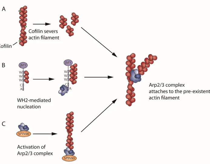
The Arp2/3 complex requires the presence of already existing actin filaments. There are three potential mechanisms that can explain the origin of the initial actin filament for the Arp2/3
35
complex activity in lamellipodia. The first possible mechanism is a severing by cofilins that gives new actin filaments for Arp2/3 complex priming (Fig.4A).
The second way is a JMY protein activity. JMY (junction mediating and regulatory protein) has three V-motifs and one C-motif. As was mentioned before several V-motifs are also found in SPIRE family of proteins. JMY can bind actin monomers via its V-motifs, form an actin nucleus and thus contribute to the elongation of F-actin (Zuchero et al. 2009) (Fig.4B). However it is very unlikely that JMY could be a universal provider of primary actin filaments for Arp2/3 complex. JMY is participated in cell migration of only some cell lines, decreasing E-cadherin protein stability and cell–cell adhesion, thereby indirectly increasing cell migration.
The last possible way is to promote Arp2/3 complex activity without any pre-existing filaments. Dip1 protein (also known as NCK interacting protein with SH3 domain, SPIN90 or WISH) can activate Arp2/3 complex in non-NPF-like mode. Without any pre-existing filament, Dip1 brings Arp2 and Arp3 subunits together, thus Arp2 and Arp3 mimic to the actin dimer and can initiate the polymerization of actin filament (Krause and Gautreau 2014; Wagner et al. 2014) (Fig.4C).
ADF/cofilin group of proteins includes cofilin and actin depolymerizing factor protein ADF. Cofilin was originally found as a protein that enables actin filaments formation (COFILamentous structures of actIN). Cofilin is highly similar to ADF and they share mostly the same biochemical properties, thus these proteins were considered to compose single group of proteins called ADF/cofilin. All eukaryotes express proteins from the ADF/cofilin family. In mammals three forms are expressed: ADF or destrin, cofilin-1 and cofilin-2. Cofilin-1 is expressed in higher concentration and it has been studied more profoundly (Goroncy et al. 2009).
Cofilin is known for its actin depolymerizing and severing abilities. In its inactive form cofilin is bound to the membrane via PIP2 and due to the extracellular signals, PIP2 is getting
hydrolyzed, cofilin gets released and attach to actin filaments. Cofilin has its highest affinity for ADP-actin monomers that are accumulated in the aged region of the actin filament. Cofilin binds to the two neighboring actin monomers along the whole aged filament and causes significant twist
36
of actin filament. Due to this twist actin filament becomes fragile and finally it falls apart into short fragments. Moreover, cofilin accelerates the dissociation of Pi from ADP-Pi-bound actin in
filament and thus promotes debranching of actin filament (Blanchoin and Pollard 1999).
Thereby, Cofilin severs actin filaments making more ends and thus accelerating actin filament dissociation. Pointed ends of new short fragments rapidly disassemble and G-actin are released. This turnover of actin is highly important for actin dynamics and thus to cell locomotion and other processes.
37
Figure 6. Generation of pre-existent mother filament de novo for priming of the Arp2/3 complex. Modified from (Krause and Gautreau 2014)
A) Cofilin may severs actin filament and provide multiple number of actin filaments that will
encounter Arp2/3 complex. B) JMY protein linear actin filament via its WH2 domains and also activates Arp2/3 complex. C) SPIN90 brings Arp2 and Arp3 subunits together without pre-existent mother filament.
38
4. Activation of the Arp2/3 complex via Nucleation Promoting Factors
As was mentioned above, tertiary structures of Arp2 and Arp3 subunits were homologous to the actin monomers and these subunits could be possible sites for G-actins during the nucleation. However, the crystal structure couldn’t explain how nucleation of actin filaments happens because of the big cleft between subunits Arp2 and Arp3. Afterwards with the help of Electron Microscopy it was shown that Arp2/3 complex fluctuates between open (inactive) and closed (active) conformational states (Rodal et al. 2005).
Without NPFs Arp2 and Arp3 are spatially separated and cannot nucleate the daughter filament. To form the branch junction Arp2/3 complex has to be activated by so called Nucleation Promoting Factors (NPFs). Further Arp2 and Arp3 subunits of the activated Arp2/3 complex bind ATP-bound actin monomer. It is known that the slowest step in a spontaneous actin polymerization is a formation of actin dimer. It is believed that in the presence of NPFs, subunits Arp2 and Arp3 overcome kinetic barrier to nucleation by mimicking to the actin dimer and binding the first actin monomer of new actin filament.
This theory was confirmed by Electron Microscopy and Single Particle Analysis showed that NPFs induce conformational changes of originally inactive Arp2/3 complex so that Arp2 and Arp3 close in and form a nucleus for ATP-bound actin connection (Rodal et al. 2005).
4.1 Structure and activity of Nucleation Promoting Factors
Arp2/3 complex activity is regulated by NPFs, also called activators of Arp2/3 complex, and inactivators. The WASP family proteins are the biggest class of NPFs, which in mammals includes WASP, N-WASP, WASH, WHAMM, WAVE 1 to 3 and already mentioned JMY. Distinct NPFs activate Arp2/3 complex regulating different processes and respectively have different structural features. In general, WASP-family proteins share the well-conserved C-termini and dissimilar N-termini. Unique N-terminus domains regulate the activity of WASP family proteins, intracellular localization and interaction with other proteins.