U
U
n
n
i
i
v
v
e
e
r
r
s
s
i
i
t
t
é
é
d
d
e
e
L
L
i
i
è
è
g
g
e
e
F
F
a
a
c
c
u
u
l
l
t
t
é
é
d
d
e
e
M
M
é
é
d
d
e
e
c
c
i
i
n
n
e
e
D
D
é
é
p
p
a
a
r
r
t
t
e
e
m
m
e
e
n
n
t
t
d
d
e
e
c
c
h
h
i
i
r
r
u
u
r
r
g
g
i
i
e
e
c
c
a
a
r
r
d
d
i
i
o
o
-
-
v
v
a
a
s
s
c
c
u
u
l
l
a
a
i
i
r
r
e
e
P
P
r
r
o
o
f
f
e
e
s
s
s
s
e
e
u
u
r
r
R
R
a
a
y
y
m
m
o
o
n
n
d
d
L
L
I
I
M
M
E
E
T
T
F
F
r
r
o
o
m
m
c
c
l
l
i
i
n
n
i
i
c
c
a
a
l
l
o
o
b
b
s
s
e
e
r
r
v
v
a
a
t
t
i
i
o
o
n
n
t
t
o
o
g
g
e
e
n
n
o
o
m
m
i
i
c
c
s
s
t
t
u
u
d
d
y
y
:
:
c
c
o
o
n
n
t
t
r
r
i
i
b
b
u
u
t
t
i
i
o
o
n
n
t
t
o
o
t
t
h
h
e
e
k
k
n
n
o
o
w
w
l
l
e
e
d
d
g
g
e
e
o
o
f
f
t
t
h
h
e
e
m
m
e
e
c
c
h
h
a
a
n
n
i
i
s
s
m
m
s
s
o
o
f
f
g
g
r
r
o
o
w
w
t
t
h
h
a
a
n
n
d
d
r
r
u
u
p
p
t
t
u
u
r
r
e
e
o
o
f
f
a
a
b
b
d
d
o
o
m
m
i
i
n
n
a
a
l
l
a
a
o
o
r
r
t
t
i
i
c
c
a
a
n
n
e
e
u
u
r
r
y
y
s
s
m
m
s
s
N
N
a
a
t
t
z
z
i
i
S
S
A
A
K
K
A
A
L
L
I
I
H
H
A
A
S
S
A
A
N
N
D Doocctteeuurr eenn mmééddeecciinnee D Doocctteeuurr eenn sscciieenncceess cclliinniiqquueess M Méémmooiirree pprréésseennttéé eenn vvuuee ddee ll’’oobbtteennttiioonn dduu ggrraaddee dd’’aaggrrééggéé ddee l l’’eennsseeiiggnneemmeenntt ssuuppéérriieeuurr.. 2 2000055UNIVERSITE DE LIEGE
FACULTE DE MEDECINE
Le présent mémoire peut être livré à l’impression Liège, le 15 février 2005,
Le Secrétaire de la Faculté, Le Doyen de la Faculté de Médecine,
(s) Ph. BOXHO (s) G. MOONEN
Le Secrétaire du Jury (s) J.O. DEFRAIGNE
Article 6 de l’Arrêté Royal du 10 mai 1931 appliquant la loi du 21 mai 1929 sur la collation des grades académiques et le programme des examens universitaires : « En aucun cas, les opinions de l’auteur ne peuvent être considérées, par le fait de l’autorisation d’impression de la
De l'observation clinique à l'étude
génomique : contribution à la
connaissance des mécanismes de
formation et de rupture des anévrysmes
Surgical research, like other clinical research, is essential. There is almost certainly not a single living surgeon or other clinician who is satisfied with all aspects of contemporary surgical and clinical management. It follows that surgeons and other clinicians must carry out research.
A Lysiane,
A Sarah et Elif.
REMERCIEMENTS
"Ne dis jamais 'merci' à celui qui te propose de l'aide avant qu'il te l'ait vraiment donnée", m'enseignait dans ma jeunesse un vieux paysan qui, pour prix de la leçon, avait bien
veillé à ne pas me fournir l'assistance qu'il venait pourtant de me promettre un peu plus tôt. Eh bien, toutes celles et ceux que je vais remercier ici en revanche le méritent véritablement, tant l'aide qu'ils m'ont apportée fut ample, précieuse et, si souvent, tellement chaleureuse.
Avant de parler des arbres, regardons la forêt : c'est à Liège et à son Université que je veux d'abord adresser de vifs sentiments de gratitude. Certains disent que les étrangers ne sont pas toujours bienvenus ? J'ai vécu le contraire. Les Liégeois, et plus encore les Liégeois de cette Université, méritent une mention très spéciale : je leur adresse un "merci" longtemps tenu discret mais tout droit sorti du coeur.
C'est un plaisir supplémentaire que de détailler les arbres de cette forêt. Leur frondaison m'a abrité quand il le fallait. Et j'ai pu m'appuyer sur leurs troncs vigoureux pour affermir mes projets. Ce sont des arbres forts auxquels je dois plus que je ne pourrai jamais leur rendre, et sans lesquels cette thèse n'aurait pu exister.
Je veux parler d'abord du Professeur Raymond Limet, qui m'a accepté dans son service de chirurgie et sans lequel rien n'aurait même commencé. Sa confiance, son expérience, sa pédagogie et ses conseils si précieux furent de tous les instants. C'est lui aussi, dont la force emporte tout, qui m'a lancé dans cette thèse.
Quant au Docteur Jacques Fourny, je ne l'oublierai pas davantage. Il fut mon premier contact à mon arrivée en Belgique, le premier à me faire partager sa table. Impossible à oublier car, au-delà du doigté et de la qualité du travail, il faisait preuve d'une grande patience pour me guider et m’enseigner les techniques opératoires .J'ai eu de la chance : c'est un chirurgien de première force et j'ai reconnu en lui un véritable exemple.
Le Professeur Thierry Grenade, autre excellent chirurgien, m'a soutenu si vivement, dans ces moments difficiles des débuts que je vivais non au CHU, encore à venir, mais à
l'hôpital de Bavière, lui aussi est gravé dans ma mémoire. Un homme sur lequel on peut toujours compter.
De forts sentiments m'habitent également quand je pense au Docteur Etienne Creemers – un autre brillant chirurgien – qui m'a fait autrefois l'amitié de conforter mes connaissances et techniques chirurgicales.
Le Docteur Guy Dekoster fut l'un de mes premiers contacts en salle d'opération et qu’il ait guidé mes premières expériences en chirurgie mérite ma gratitude.
Rick Van Damme, mon vieil ami... chirurgien, mérite également ma reconnaissance. Il est de ceux dont je puis dire qu'ils m'ont donné le temps de rédiger ce travail. Le remplacement dans les gardes malvenues, cela ne l'effraie pas. La disponibilité tout-terrain, c'est sa gentillesse à lui. Une amitié indéfectible.
Je dois aussi beaucoup, et même davantage, au Professeur Jean-Olivier Defraigne. Ses connaissances en physiologie sont une source. Son soutien est celui d'un tout grand ami. Il m'en fait présent depuis 20 ans déjà avec un humour et une présence indéfectibles.
Aux Professeurs Betty Nusgens et Charles Lapiere aussi, je suis redevable. Et combien ! Ils m'ont insufflé leur goût si vivace de la recherche. Leur science des tissus conjonctifs a beaucoupcompté, en 15 ans, pour soutenir les résultats présentés dans cette thèse...
Au tour d'Olivier Defawe, un biologiste remarquable avec qui j'ai eu la chance de travailler voilà 4 années, de recevoir ma reconnaissance.
Chaque ouvrage a ses piliers plus matériels. Le mien doit beaucoup à la mémoire de Jacqueline Dehousse, à celle qui lui a succédé, Geneviève Peters, ainsi qu'à Micheline Delcour. Des secrétaires comme on en souhaite pour toujours.
Un "merci" de plus à mes jeunes collègues, à d'autres secrétaires ainsi qu'à nombre d'infirmières pour leur patience, quand l'humeur l’emporte sur l'humour...
Soulignant la qualité humaine, la compétence, l'esprit critique et la rigueur de ceux que je viens de citer pour leur rendre hommage, j'en arrive enfin à celles que je chéris le plus. La tendresse de mon épouse, Lysiane, son amour primordial et sa présence essentielle ont été et sont, chaque jour, plus qu'un don mais véritablement un fondement, une nécessité. Ils trouvent cependant leur égal dans les yeux de Sarah et d'Elif, nos filles qui, malgré des absences rimant avec urgences, sont les merveilles d'une vraie famille et d'un père comblé.
Résumé
L’anévrysme de l’aorte abdominale (AAA) est une cause importante, et pourtant évitable, de décès chez les personnes âgées. Dans le monde occidental, le taux de mortalité dû à un AAA avoisine 1,3 % de tous les décès des personnes âgées de 65 à 85 ans. Le risque de rupture s’accroît avec l’augmentation du diamètre de l’AAA. C’est la raison pour laquelle nous nous sommes intéressés aux mécanismes impliqués dans la croissance des AAA et aux facteurs qui la déterminent. Les travaux repris dans notre mémoire représentent la suite logique de notre travail de doctorat défendu en 1994.
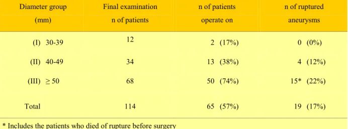
L’histoire naturelle des AAA (leur vitesse de croissance et leur incidence de rupture) a été retrouvée dans un groupe de 114 patients observés sur une période moyenne de 26 mois. A la fin du suivi, 65 des 114 patients avaient été opérés. Si nous mettons en relation l’incidence d’une chirurgie d’urgence pour rupture, en fonction du diamètre initial de l’anévrysme, nous voyons que, en-dessous de 40 mm, l’incidence de rupture est nulle et qu’elle est de 12 et 22 % dans les groupes de 40-50 et supérieur à 50 mm. Nos observations concordent avec les résultats de différents auteurs : le risque de rupture s'accroît avec la taille de l’AAA. La croissance de l'AAA est exponentielle, mais différente chez chaque individu.
Un groupe de 110 patients hospitalisés pour chirurgie coronaire ou vasculaire périphérique (79 M, 31 F) a été soumis à une mesure systématique du diamètre de l’aorte abdominale infrarénale. Nous avons pu identifier 8 anévrysmes, dont 7 chez des sujets masculins. Dans ce petit groupe de patients, l’incidence d’anévrysme était plus importante chez les sujets admis pour chirurgie coronaire que chez les patients admis pour chirurgie vasculaire périphérique. Une corrélation positive existait entre la présence d’un AAA et le taux de cholestérol sérique, de même que la réduction de l’alpha 1 antitrypsine plasmatique. Ces divers éléments nous ont permis de recommander un dépistage de l’AAA par ultrasonographie chez les sujets masculins de plus de 55 ans présentant une maladie coronarienne ou vasculaire périphérique.
En continuité avec ces travaux, nous avons réalisé un dépistage systématique des AAA dans une population masculine de 65 et 75 ans de la ville de Liège. Durant la période de 1995 à 1996, 1764 hommes nés en 1920 ou 1930 ont été invités à subir un examen ultrasonographique abdominal. Dans cette étude, nous avons constaté que la prévalence de l’AAA (3,8 %) était similaire à celle relevée dans d’autres études où elle se trouve aux alentours de 3 à 4 % dans les populations âgées de 60 ans et plus. En nous basant sur cette étude, il y aurait, actuellement, 14.000 belges âgés de 80 ans ou plus porteurs d’AAA. L’incidence de l’hypertension et de la consommation de tabac était significativement plus importante dans le groupe des AAA (Chapter 2).
Les résultats de la chirurgie prophylactique de l’AAA par mise à plat n’ont cessé de s’améliorer depuis quarante ans. Ils restent cependant grevés d’une lourde morbidité-mortalité opératoire chez les patients âgés. L'étude rétrospective de 138 patients octogénaires, admis entre 1984 et 1996 pour AAA mettent nos résultats de la chirurgie élective dans un classement très favorable. Au total, 52 patients ont bénéficié d’une intervention de mise à plat d’AAA dans des conditions électives résultant en un taux de mortalité opératoire de 5,7% ; 21 patients ont été opérés en urgence pour un AAA douloureux avec une mortalité de 28%, et 41 patients ont été opérés en urgence pour une rupture. Dans le groupe des ruptures, la mortalité opératoire a été de 68%. La mortalité de la chirurgie élective pour AAA chez les octogénaires reste plus élevée par rapport à celle de patients plus jeunes (2,7%). Elle peut, toutefois, être recommandée aux octogénaires porteurs d’AAA importants et dotés d’un bon état général. Notre option est la surveillance armée, chez les patients présentant un risque opératoire majeur (Chapter 3).
La pathogenèse de l’AAA avait déjà largement retenu notre attention au cours du Doctorat. L’élastine et le collagène sont les principales protéines de la matrice extracellulaire qui assurent les propriétés rhéologiques de l’aorte abdominale. Des prélèvements chirurgicaux ont été soumis à des analyses chimiques permettant d'établir une relation entre le diamètre de l’anévrysme et la concentration d'élastine, de même qu'avec l'extractibilité du collagène. L'évolution de ces paramètres est parallèle à l'accroissement de la taille de l’anévrysme pour atteindre un maximum dans les anévrysmes rompus. La perte d’élastine débute dès la phase
précoce de la transformation anévrysmale, tandis que l’altération du collagène, évoquée par l'accroissement de son extractibilité, atteint son paroxysme dans les AAA rompus.
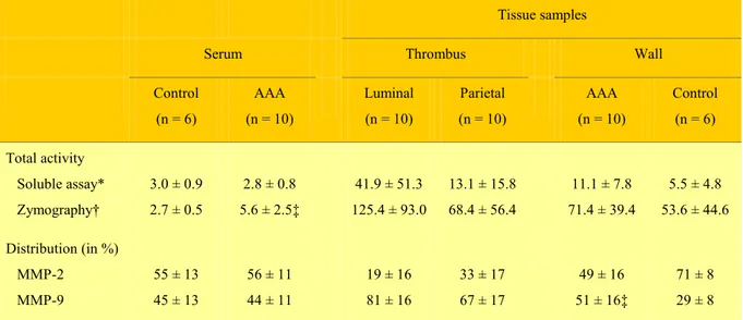
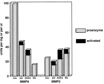
Les mécanismes responsables de ces altérations ont été précisés par l'analyse du rôle des métalloprotéases MMP-2 et MMP-9 (72 kDa et 92 kDa) douées d'activité élastasique. Nous en avons mesuré l'activité dans le sérum, le thrombus pariétal, ou la paroi des anévrysmes. Par comparaison, nous avons également déterminé l’activité de ces élastases dans des parois d’aorte athéromateuse ainsi que dans le sérum d’individus normaux. La paroi aortique anévrysmale contient une proportion plus importante de la gélatinase 92 kDa que celle des contrôles non anévrysmaux. Les formes actives aussi bien des 72 kDa que des 92 kDa sont significativement accrues dans la paroi anévrysmale par rapport aux contrôles et deux fois supérieures à celle observée dans le thrombus pariétal. Ces résultats plaident pour une production in situ des gélatinases plutôt qu'une diffusion des élastases sériques (Chapter 4).
La collaboration de O. Defawe (Laboratoire de Biologie des Tissus Conjonctifs, Prof. B.V. Nusgens) nous a permis de préciser le rôle des différentes métalloprotéinases et de leurs inhibiteurs dans le développement des lésions anévrysmales (AAA) et athérosclérotiques (AOD). Le profil d'expression (ARN-messagers) des gènes des MMPs, des sérines protéinases et de leurs inhibiteurs ont été mesurés par RT-PCR quantitative. Des échantillons provenant de 6 aortes anévrysmales (AAA), de 6 aortes occlusives (AOD) prélevés lors d’interventions, mais aussi de fragments de parois normales d'aortes abdominales et des prélèvements d'aorte thoracique ascendante réalisés lors de pontages aorto-coronaires. En parallèle avec l'expression très accrue des protéases par rapport au tissus normaux dans les deux pathologies, deux de leurs inhibiteurs, TIMP2 et PAI-1, sont réduits dans les AAA mais pas dans les AOD, ce qui introduit une différence significative entre les deux pathologies. Ces données suggèrent qu’un déséquilibre entre les métalloprotéinases, leurs activateurs et leurs inhibiteurs peut expliquer le développement d’une dilatation anévrysmale au niveau de l’aorte abdominale athéromateuse. Le profil d'expression comparatif (AAA, AOD, aorte normale) de multiples gènes potentiellement impliqués dans le développement de l'AAA a dévoilé une importante hétérogénéité des taux d'expression de diverses enzymes considérées comme essentielles par
divers auteurs. Il ne nous a pas été possible, malgré les données de profil d'expression des gènes opérant dans le remodelage de la paroi de l'AAA, de clairement démontrer le mécanisme d'accroissement d'extractibilité du collagène. Il pourrait résulter de l'activation de sa synthèse, le collagène néoformé étant plus extractible, ou d'une dégradation des liaisons intermoléculaires notamment par l'excès de MMP-13 (Chapitre 5).
Nous nous sommes interrogés sur le caractère représentatif des prélèvements tissulaires ponctuels réalisé pendant une intervention pour rupture. Lors de l’intervention, un prélèvement des tissus, juste au niveau du site de rupture, est difficile, à cause de l’hématome dans la paroi, ainsi qu’au niveau de l’espace rétropéritonéal ; en outre, un tel prélèvement est dangereux, en raison d’autres priorités liées à la situation précaire du patient. Un examen postmortem précoce a permis de réaliser des prélèvements de parois aortiques au niveau du site de rupture, et tous les 10mm jusqu'à une distance de 50 mm. Dans ces échantillons, au site de rupture, les MMP-2, -9, -12, -13 sont fortement accrues de même que leurs inhibiteurs. A distance de la rupture, l'hétérogénéité d’expression des MMP-3, -8 et -11 est manifeste. L’expression de TIMP2 ainsi que PAI-1, plus importante au niveau du site de rupture se réduit de façon irrégulière à distance. L'étude histologique du site de rupture et à distance démontre également un infiltrat inflammatoire diffus qui devient focal à distance, et une forte hétérogénéité de destruction de la média (Chapter 6).
Pour démontrer, in vivo, la topographie de ces activités enzymatiques présentes au sein des macrophages de l'infiltrat inflammatoire dans la paroi aortique anévrysmale, nous avons utilisé le PET-scan. Le 18FDG, analogue du glucose marqué par un isotope du fluor visible en résonance magnétique, permet l’évaluation du métabolisme glucidique régional. Cette technique est largement utilisée pour démontrer, notamment, la présence d'un infiltrat néoplasique mais également les réactions inflammatoires granulomateuses et les tissus infiltrés par des macrophages. Nous avons réalisé un PET-scan utilisant le 18FDG chez 26 patients, porteurs d’un AAA. Les 10 patients dont le PET-scan était positif, étaient caractérisés par une augmentation récente de la taille de l’AAA, des signes de fissuration, voire de rupture imminente. La positivité de la paroi aortique lors de l’examen PET- scan chez
un patient référé pour anévrysme de l’aorte abdominale représente dès lors pour nous, un facteur renforçant l’indication opératoire. (Chapter 7).
En outre, nous avons étudié la corrélation entre la captation de FDG et les données histologiques dans une paroi aortique anévrysmale. Les analyses histologiques de tous les échantillons montrent une réaction inflammatoire surtout importante au niveau de l’adventice, et beaucoup moins marquée au niveau de la média. La zone luminale du thrombus contient également des cellules inflammatoires. L'immunohistologie confirme la présence principalement des macrophages. Leur localisation préférentielle est cependant l’adventice de la paroi aortique anévrysmale. Nous avons constaté une corrélation significative entre la positivité du PET-scan et la présence des leucocytes en surface du thrombus et au niveau de la paroi aortique, plus intense au niveau de l’adventice (Chapter 8).
Comme les métalloprotéinases sont libérées par les leucocytes activés, notamment par stress oxydatif, nous avons mesuré le taux sanguin de la vitamine E comme un reflet indirect de l’intensité de ce stress. La vitamine E plasmatique des patients anévrysmaux est significativement réduite, de même que son rapport avec les lipides totaux. Cette observation suggère l’intérêt potentiel d’un monitorage de la vitamine E sérique chez les patients à risque de développement d’un AAA de taille critique (Chapter 9).
Comme les AAA surviennent tard dans la vie, le caractère familial de l'affection a été, en général, sous-estimé. Lors d'analyse de ségrégation, la transmission génétique de l’AAA se ferait sur un modèle autosomal récessif. En 1995, sur un échantillon plus large nous avons ainsi retrouvé 276 cas sporadiques et 81 cas dans un contexte d'atteinte familiale (76 sujets masculins et 5 sujets féminins). Nous avons également constaté quelques différences entre cas familiaux et sporadiques, notamment l’âge au moment de la rupture qui est inférieur dans le groupe familial (65,4 ±6,6) à celui du groupe sporadique (75,2 ±1,6). Le pourcentage de rupture est, par ailleurs, de 32,4 % pour les cas familiaux et 8,7 % pour les cas sporadiques. Nous avons pu déterminer que le risque relatif d’AAA pour les frères d’un sujet mâle porteur d’un AAA est de 18. En testant l’hypothèse d’un modèle mixte, l’explication la plus vraisemblable pour la survenue d’anévrysmes familiaux est la présence d’un gène unique, présentant un caractère dominant (Chapter 10).
Après la publication de nos travaux concernant l’aspect familial et génétique des anévrysmes, nous avons été invités par le groupe de Helena Kuivaniemi (Center for Molecular Medicine and Genetics, Wayne States University Detroit, USA) à participer à une étude multicentrique. Dès leur admission pour chirurgie de l’AAA, les patients sont interrogés sur leurs antécédents familiaux. Les membres des familles qui ne présentent pas d’AAA connu sont invités à subir une échographie et un prélèvement sanguin pour analyse d'ADN. Notre contribution à cette étude a été de pourvoir 59 familles sur les 234 qu'elle comporte. Par la suite, 420 nouveaux patients ont été découverts porteurs d’un anévrysme de l’aorte abdominale. Contrairement aux hypothèses antérieures, l'étude génétique a montré que dans 72 % des familles le mode de transmission est autosomal récessif, et que chez 25 % il est autosomal dominant.
Nous avons participé à l'analyse génomique des patients souffrant d'AAA comparés à des sujets indemmes de l'affection. Cette étude a permis de montrer que les inhibiteurs des métalloprotéinases (TIMP I et 2) présentent un polymorphisme au sein d'une séquence codante chez les patients porteurs d’un AAA, mais également chez les sujets sains. Il s’agit donc d’un polymorphisme neutre.
La poursuite de cette étude multicentrique a mis en évidence une hétérogénéité génétique et la présence de deux régions susceptibles pour l’AAA au niveau des chromosomes 19 et 4. Ces deux régions contiennent plusieurs gènes candidats plausibles. Il s'agit des gènes codant pour l’interleukine 15, la GRB2 Associated binding protein I, le récepteur de type 1 à l’Endothéline, la LDL receptor related protéin III, la trans-membrane protease serin I, le programmed cell -dead 5 et les gènes de peptidase D (Chapter11).
Au terme d’un travail de plus de 15 ans qui s’est intéressé successivement à l’observation clinique des patients porteurs d'AAA, parfois jusqu'à leur rupture, aux modifications de la matrice extracellulaire de la paroi anévrysmale et aux mécanismes physiopathologiques qui en sont responsables, à l’utilisation de nouvelles méthodes d’imagerie (PET-scan), nous avons également coopéré à l’étude multicentrique du génome du patient porteur d’AAA, ou menacé de l’être.
Depuis l’opération princeps de Dubost en 1953, 50 années ont été nécessaires au raffinement des techniques et des indications opératoires, de la réanimation, et des modes de diagnostic. Le but jusqu'à présent a été de prévenir le décès du patient par rupture de son AAA grâce à une mise à plat chirurgicale ou prothèse endovasculaire prophylactiques. Le travail continue. Les prochaines étapes consisteront en l’utilisation de moyens pharmacologiques susceptibles de réduire ou supprimer le développement des AAA chez l’individu génétiquement enclin à cette affection. La tâche de la communauté scientifique sera de prévenir, non plus seulement la rupture de l’anévrysme, mais plus fondamentalement sa survenue et sa croissance.
TABLE OF CONTENTS
REMERCIEMENTS...5
CHAPTER I...22
GENERAL INTRODUCTION AND AIM OF THE WORK ...22
1. Generalities ...22
2. Summary of doctoral thesis ...24
2.1.Study of prevalence in high-risks patients...24
2.2.Study of the growth rate and risk of rupture...27
2.3. Initial study on molecular mechanisms ...30
CHAPTER 2 ...36
EPIDEMIOLOGY OF AAA: A POPULATION-BASED STUDY (Appendix 1) .36 INTRODUCTION...36
MATERIAL AND MEDHODS...37
RESULTS...37
CONCLUSION ...42
CHAPTER 3 ...44
ABDOMINAL AORTIC ANEURYSMS REPAIR IN ELDERLY (appendix 2) ...44
INTRODUCTION...44
MATERIAL AND METHODS ...44
RESULTS...45
DISCUSSION ...48
CONCLUSIONS ...50
INVOLVEMENT Of MATRIXMETALLOPROTEINASES (MMPS ) IN ThE
DEVELOPMENT OF AAA
(Appendix 3) 51
INTRODUCTION...51
MATERIAL AND METHODS ...53
RESULTS...54
DISCUSSION ...57
CONCLUSION ...59
CHAPTER 5 ...60
THE ROLE OF THE MATRIX METALLOPROTEINASES (MMPS ) AND THEIR INHIBITORS (TIMPS) ON THE REMODELLING OF THE ABDOMINAL AORTA IN AOTIC ATHEROSCLEROTIC AND ANEURYSMAL LESIONS. (APPENDIX 4) ...60
INTRODUCTION...60
MATERIAL AND METHODS ...63
Patients characteristics ...63
RNA isolation and quantitative RT-PCR procedure ...63
Zymographic analysis of the gelatinases MMP-2 and MMP-9 ...65
Statistics ...65
RESULTS...66
DISCUSSION ...66
CONCLUSIONS ...69
CHAPTER 6 ...70
HISTOLOGICAL AND BIOCHEMICAL HETEROGENEITY IN THE WALL OF RUPTURED AAA ( appendix 5)...70
INTRODUCTION...70
CASE REPORT ...70
MATERIAL AND MEDHODS...71
RESULTS...71
DISCUSSION ...73
CONCLUSION ...75
CONTRIBUTION OF POSITRON EMISSION TOMOGRAPHY (PET) TO THE
EVALUATION OF AAA (appendix 6&7)...76
INTRODUCTION...76
MATERIAL AND METHODS ...77
Patients ...77 Radiopharmaceutical ...78 PET protocol ...78 Image interpretation...78 RESULTS...79 DISCUSSION ...80 CONCLUSION ...81 CHAPTER 8 ...82
CORRELATION THE FONCTIONAL IMAGING WITH THE HISTOLOGICAL FINDINGS IN THE WALL OF ANEURYSMAL ABDOMINAL AORTA ( appendix 8 ) ...82
INTRODUCTION...82
MATERIAL AND METHODS ...82
RESULTS and DISCUSSION ...83
DISCUSSION ...83
CONCLUSION ...84
CHAPTER 9 ...85
VITAMIN E (αααα-tocopherol) LEVEL IN PATIENTs WITH ABDOMINAL AORTIC ANEURYSM ( appendix 9 ) ...85
INTRODUCTION...85
MATERIAL AND METHODS ...86
RESULTS...86
DISCUSSION ...87
CONCLUSION ...88
CHAPTER 10 ...89 FAMILIAL OCCURENCE OF AAA (LOCAL EXPERIENCES) ( appendix 10 )89
MATERIAL AND METHODS ...89
RESULTS...90
DISCUSSION ...91
Familial Aspect ...91
CHAPTER 11 ...97
MULTICENTRIC RESEARCH ON FAMILIAL AND GENETIC ASPECT OF AAA (appendix 11,12 &13) ...97
INTRODUCTION...97
Familial Screening ...99
MATERIAL AND METHODS ...99
RESULTS...100
DISCUSSION ...103
CONCLUSION ...104
Analysis of coding sequences for tissue inhibitors metalloproteinases (TIMPs) genes in patients with AAA ...104
INTRODUCTION...104
MATERIAL AND METHODS ...105
RESULTS...105
DISCUSSION AND CONCLUSION ...106
Linkage of familial abdominal aortic aneurysm to chromosome 19 ...107
INTRODUCTION...107
METHODS ...107
Subjects and phenotyping...107
Genotyping ...108
Statistical analyses...109
RESULTS...109
Whole Genome Scan with 36 AAA Families ...109
Follow-up Studies with 119 AAA Families...109
Chromosome 19 ...110
DISCUSSION ...110
CONCLUSION ...112
General conclusions and perspectives ...113
Pertinent publications posterior to the “Thèse de doctorat en sciences cliniques à
l’université de Liège” 1994 ...157 Appendix 1 ...158
Routine ultrasound screening for abdominal aortic aneurysm among 65- and 75-year-old men in a city of 200,000 inhabitants. C. Vazquez, N. Sakalihasan, J.B. D'Harcour, R. Limet. Ann Vasc Surg 1998;12:544-549 ...158 Appendix 2 ...165
Abdominal aortic aneurysms in octogenarians. H. Van Damme, N. Sakalihasan, C. Vazquez, Q. Desiron, R. Limet. Acta Chir Belg 1998, 98 : 76-84 ...165 Appendix 3 ...175
Activated forms of MMP-2 and MMP-9 in abdominal aortic aneurysms. Natzi Sakalihasan, Philippe Delvenne, Betty V. Nusgens,Raymond Limet, Charles M. Lapière. J Vasc Surg 1996;24:127-133...175 Appendix 4 ...183
TIMP-2 and PAI-I mRNA levels are lower in aneurysmal as compared to athero-occlusive abdominal aortas. Olivier Defawe, Alain Colige, Charles Lambert, Carine Munaut,Philippe Delvenne, Betty Nusgens, Charles Lapière, Raymond Limet, Natzi Sakalihasan.Cardiovasc Res 2003;60:205-213...183 Appendix 5 ...193 Gradient of proteolytic enzymes, their inhibitors and matrix proteins expression in a ruptured abdominal aortic aneurysm. O. Defawe, A. Colige, C.A. Lambert,
P.Delvenne, C.M. Lapière, R. Limet, B. Nusgens, N. Sakalihasan. Eur J Clin
Invest,2004;34.(7) :513-4...193
Appendix 6 ...196 Positron emission tomography (PET) evaluation of abdominal aortic aneurysm (AAA). N. Sakalihasan, H. Van Damme, P. Gomez, P. Rigo, C.M. Lapière, B. Nusgens, R. Limet. Eur J Vasc Endovac Surg 2002;23:431-436...196 Appendix 7 ...203
Contribution of PET scanning to the evaluation of abdominal aortic aneurysm. Natzi Sakalihasan, Roland Hustinx, Raymond Limet. Sem Vasc
Surg,2004;17:144-153 ...203
Appendix 8 ...213 Distribution of F-fluorodeoxyglucose in Abdominal Aortic Aneurysm : High
Defawe, M.S.,R. Hustinx, JO.Defraigne, R. Limet, N.Sakalihasan. Clin Nucl Med (in press)...213 Appendix 9 ...220
Decrease of plasma vitamin E (α-Tocopherol) levels in patients with abdominal aortic aneurysm. N. Sakalihasan, J. Pincemail, J.O. Defraigne, B.Nusgens, C.M. Lapière, R. Limet.Ann NY Acad Sci 1996;800:278-282 ...220
Appendix 10 ...226 Aneurysms of the abdominal aortic aorta : familial and genetic aspects in three hundred thirteen patients A. Verloes, N. Sakalihasan, L. Koulischer, R. Limet. J
Vasc Surg 1995;21:646-655...226
Appendix 11 ...238 Familial abdominal aortic aneurysms: collection of 233 multiplex families. Helena Kuivaniemi, Hidenori Shibamura, Claudette Arthur, Ramon Berguer, C. William Cole, Tatu Juvonen, Ronald A. Kline, Raymond Limet, Gerry McKean, Orjan Norrgard, Gerard Pals, Janet T. Powell, Pekka Rainio, Natzi Sakalihasan, Clarissa van Vlijmen-van Keulen, Alain Verloes, Gerard Tromp.
J Vasc Surg 2003;37:340-345...238
Appendix 12 ...247 Analysis of coding sequences for tissue inhibitor of metalloproteinases 1 (TIMP1) and 2 (TIMP2) in patients with aneurysms. Xiaoju Wang, Gerard Tromp, C.William Cole, Alain Verloes, Natzi Sakalihasan, Sungpil Yoon, Helena Kuivaniemi. Matrix Biology 1999;18:121-124...247 Appendix 13 ...255 Genome scan for familial abdominal aortic aneurysm using sex and family history as covariates suggests genetic heterogeneity and identifies linkage to chromosome 19q13. H. Shibamura, J.M. Olson, C. van Vlijmen-van Keulen, S.G. Buxbaum, D.M. Dudek, G. Tromp, T. Ogata, M. Skunca, N. Sakalihasan, G. Pals, R. Limet, G.L. McKean, O. Defawe, A. Verloes, C. Arthur, A.G. Lossing, M. Burnett, T. Sueda, H. Kuivaniemi. Circulation 2004;109:2103-21...255
TABLE OF ABBREVIATIONS
AAA ... Abdominal aortic aneurysm AOD ... Aorto-occlusive disease APMA ... Aminophenylmercuric acetate ApoE... Apolipoprotein E
ARP ... Affected relative pair ASP... Affected sib pair
CAA ... Control abdominal aorta
COPD ... Chronic obstructive pulmonary disease CT... Computerized tomography
CTA... Control thoracic aorta
EDTA ... Ethylene-diamine-tetra acetic acid FDG... Fluorodeoxyglucose
HPN... Transmembrane protease serine 1 HSMC... Human smooth muscle cells ICAM-1 ... Intercellular adhesion molecule-1 IL-1... Interleukine-1
kDa ... KiloDalton
LDL ... Low density lipoprotein LOD... Logarithm of odds
MMPs ... Matrix metalloproteinase MRI ... Magnetic resonance imaging mRNA ... Messenger ribonucleic acid PAI-1 ... Plasminogen activator inhibitor PCR ... Polymerase chain reaction PEPD3 ... Peptidase D3
PET... Positron Emission Tomography PMNs... Polymorphonuclear neutrophil rAAA... Ruptured abdominal aortic aneurysm RNA ... Ribonucleic acid
RT-PCR... Reverse transcriptase-polymerase chain reaction SDS... Sodium dodecylsulfate
SMC ... Smooth muscle cells
SPIO ... Supra paramagnetic iron oxide sRNA... Synthetic ribonucleic acid
TIMP ... Tissue inhibitor of metalloproteinases t-PA ... Tissue plasminogen activator
u-PA ... Urokinase-type plasminogen activator US... Ultrasound
CHAPTER I
GENERAL INTRODUCTION AND AIM OF THE WORK
1. Generalities
The abdominal aorta originates from the thoracic aorta at the level of the diaphragmatic hiatus. It courses on the left anterior flank of the spine in the retroperitoneal space and ends at the aorto-iliac bifurcation at the level of the lower lumbar rachis facing the left anterior side of L4. The infrarenal abdominal aorta is limited to the distal aortic segment starting below the origin of the renal arteries, situated at the level of the lower third of L1 or of the upper side of L2. The skin projection of the bifurcation corresponds to a point located on the abdominal wall, one or two centimeters above the navel. Thus, on average, the infrarenal abdominal aorta has a length of 10 cm, a dimension that can vary according to the exact position of the bifurcation. Its diameter varies according to the age, sex and constitution of the subject. In normal Man, there is a progressive narrowing of the aortic diameter from the supravalvular origin to the bifurcation. For example, it is generally observed that the diameter of the infrarenal aorta is 2 mm less than that of the suprarenal aorta (Steinberg et al., 1965).
An aneurysm can be defined as a permanent irreversible localized dilatation of a vessel. This “abnormal” dilatation involves the three layers of the vascular tunic: the intima, the media, and the adventitia. This definition differentiates an aneurysm sensu stricto from a false aneurysm, which corresponds to a perivascular pulsatile hematoma secondary to a vessel rupture. In this latter instance, however, the capsule is devoid of any residual vascular structure: the external limit of this pulsatile dilatation is made of an amorphous fibrous material. Similarly, the infiltration of blood within the vascular wall, associated to
enlargement of the diameter of the artery (like in aortic dissection) is not an aneurysm in the strict meaning of the term.
In terms of morphology, reviewed in details by Slaney (Slaney, 1990), two types of dilatation involving all the layers of the vessel wall can be recognized. The “fusiform” one has been described since the beginning of medical science. In this type of lesion, the parietal weakening concerns the whole circumference of the artery and participates in the aneurysmal dilatation. In contrast, an aneurysm is designed “saccular” if it involves only a part of the circumference.
If these definitions are straightforward for large aneurysms, there are ambiguous for smaller aneurysms. In fact, the term of “abnormal dilatation” used in the definition is purely qualitative. However, the definition should be initially precise, since it is important to decide what is considered or rejected as an abdominal aortic aneurysm (AAA) in the infrarenal position. In fact, this aspect may have unsuspected effects on epidemiological studies and segregation analyses where an infrarenal dilatation is always considered with respect to the diameter of the normal aorta, that is variable for the gender and the height of the patient, as mentioned previously.
Our interest has been centered on the clinical and experimental investigations of AAA. The abdominal aortic aneurysm (AAA) is indeed important cause of preventable deaths in old patients. In fact, the mortality rate due to AAA is about 1,3% of all deaths among men aged between 65 and 85 years in the western world (Law et al., 1994). Among these, many lives could be saved if rupture of AAA can be prevented. On the other hand, the increasing number of patients presenting an aneurysm of the abdominal aorta represents a burden for the population and a source of expenditure for the Health Care System. Better knowledges of the pathogenesis and of the natural history aimed at a better treatment of aortic aneurysm should be an important goal for physicians. In this approach, our initial goals were to analyze the prevalence of AAA in high-risk patients and also to determine both rate of growing and risk of rupture. It also rapidly became evident that specific molecular mechanisms (matrix elaboration and remodeling) were implicated in the development of AAA. Therefore, our ultimate goal is to identify molecular markers that would be useful in the diagnosis and
treatment of aortic aneurysm, so that patients at risk for developing AAA could be identified before the rupture occurs and operated on electively.
2. Summary of doctoral thesis
Our investigations started in 1989 and have been conducted in the departement of the Cardio vascular Surgery (Professor Limet) and in the Laboratory of Connective Tissu Biology (Professor Lapiere and Nusgens) to result in the publication of our doctoral thesis in 1994. At the beginning of our clinical and experimental study, the majority of studies examining the epidemiology of AAA were based on selected groups of individuals often poorly defined in terms of age, sex, and other risk factors. In addition, most of them have been carried out retrospectively.
At this time, the results of most screening studies, which intended to determine the prevalence in high risk populations differed substantially (Allardice et al., 1988, Allen et al., 1987, Allen, Tudway & Goldman, 1987, Bengtsson et al., 1991, Bengtsson, et al., 1989, Bengtsson, Norrgard et al., 1989, Collin et al.1988, Nevelsteen et al., 1991, Lederle et al., 1988, Lerderleet al., 1994, Scott et al., 1988). The prevalence observed in these groups was usually much higher than that found in the general population according to ultrasound investigation. A question raised up concerning the benefit and the cost-effectiveness of screening by ultrasonograph, to detect unknown AAA in high-risk population.
2.1.Study of prevalence in high-risks patients
Thus we decided to set up a screening program in order to detect AAA in 110 consecutive patients admitted to our department (Sakalihasan et al., 1992), for coronary artery (n = 72) and/or peripheral vascular (n = 38) disease (Table I).
Coronary artery disease Peripheral vascular disease
n Mean age ± SD (years) n Mean age ± SD (years) Total
Male 53 62.7 ± 4.7 26 66.9 ± 4.9 79
Female 19 64.1 ± 4.3 12 61.3 ± 4.4 31
Total 72 38 110
Table I. Characteristics of study population screened for AAA.
Except for those presenting disease, age ranging from 55 to 74 years was the only other criterion used for patients' inclusion. On admission all patients were submitted to physical examination, standard laboratory testing, routine lung function test and abdominal ultrasound examination. We defined AAA as a local infrarenal aortic dilatation with diameter equal to 30 mm or larger. Eight aortic dilatations equal to or greater than 30 mm were evidenced, 7 in males (12.3%), and 1 in female (3.1%) (Table II). All patients with AAA were aged 60 or older and the observed prevalence of AAA was higher in groups of patients suffering from coronary artery disease (15% male patients) versus 5.9% in peripheral vascular disease.
Coronary artery disease Peripheral vascular disease
n AAA % n AAA %
Male 40 6 15.0 25 1 4.4
Female 17 1 5.9 7 0 0.0
Total 57 7 12.3 32 1 3.1
Table II. Prevalence of AAA according to risk groups patients aged equal or more than 60 years.
We also observed a positive relation between abnormally elevated cholesterol levels, decreased α1-antitrypsin plasmatic levels and the occurrence of AAA (TableIII). Therefore we suggested monitorizing α1-antitrypsin, an inhibitor of leukocyte elastase, in patients at risk of
developing AAA. According to our results, we recommended the screening of AAA by ultrasound examination in selected high-risk patients aged 60 or more.
Patients without AAA Patients with AAA Patients with incipient AAA
Male Female Total Male Female Total Male Female Total
n patients 66 29 95 7 1 8 6 1 7 Cholesterol * 17 4 21 4 1 5 3 1 4 α1-antitrypsine ** 5 2 7 3 - 3 2 - 2 Hypertension 17 14 31 4 1 5 2 1 3 Diabetes mellitus 4 8 12 2 - 2 1 - 1 COPD 16 4 20 3 1 4 2 - 2 Current smoker 48 17 65 6 - 6 6 1 7 Mean age (years) 63.9 ± 5.7 63.3 ± 4.5 64.9 ± 4.8 61 63.8 ± 5.6 55 Mean weight (kg) 74.9 ± 9.8 67.3 ± 11. 3 77.1 ± 7.7 75 74.7 ± 6.4 80 Mean height (cm) 172.1 ± 6. 2 161.9 ± 6 168.9 ± 7. 4 170 174.7 ± 6. 5 167 * χ2 . p < 0.005 odds ratio = 5.286 ** χ2 . p < 0.01 odds ratio = 6.286
Table III. Characteristics of patients screened for AAA.
The discovery of non ruptured AAA, notably by screening examination, led us to the problem of treatment choice: is it necessary to operate on all patients prophylactically, or must
we reserve prophylactic surgery only for a determined subgroup where factors indicative of a probable rupture could be identified? On the other hand, until early 1980s, the expansion rate and incidence of rupture of AAAs with respect to their size was a source of controversies. Most studies have shown that the speed of change in size increases (slope) as the aneurysm gets larger, indicating a non-linear relationship (Bernstein et al., 1984, Bernstein et al., 1976, Cronenwett et al., 1985, Cronenwett et al., 1990). We therefore investigated the possibility of modeling the speed of expansion of AAA by an exponential rather than by a linear function. This would have the additional advantage of predicting the future evolution of the aneurysm from two single ultrasound examinations performed at any moment during the time course of the disease.
2.2.Study of the growth rate and risk of rupture
We focused therefore on aneurysm growth in a population of 114 patients who had not been operated on initially, because of various reasons (patients’ refusal, high surgical risk, or small diameter as assessed by CT-scan and ultrasonography) (Limet et al., 1991). At the end of the follow-up period of 26,8 months (range 3-132), we studied the risk of rupture according to the size of AAA (TableIV and V).
Diameter group (mm) First examination n of patients (%) Final examination n of patients (%) Average observation period (± SD) (I) 30-39 49 (43%) 12 (10%) 31.8 ± 3.62 (II) 40-49 41 (36%) 34 (30%) 26.8 ± 2.41 (III) ≥ 50 24 (21%) 68 (60%) 16.7 ± 2.33 Total 114 (100%) 114 (100%) 26.8 ± 2.10
Table IV. Distribution of 114 patients with AAA based on their initial and final diameter values (mm) ; average observation periods (months) are also given.
Diameter group (mm) Final examination n of patients n of patients operate on n of ruptured aneurysms (I) 30-39 12 2 (17%) 0 (0%) (II) 40-49 34 13 (38%) 4 (12%) (III) ≥ 50 68 50 (74%) 15* (22%) Total 114 65 (57%) 19 (17%)
* Includes the patients who died of rupture before surgery
Table V. Distribution of 114 patients with AAA based on their initial and final diameter (mm) ; average observation periods (months) are also given.
Using individual serial measurements, we determined the linear growth rate (λ) and the exponential growth rate (α) for each of our patients (Tables VI and VII).
Total patient population (n = 114) Restricted set (n = 101)* Initial diameter group
(mm)
n Mean (± SEM) n Mean (± SEM)
(I) 30-39 49 5.30 ± 0.588 46 5.65 ± 0.592 (II) 40-49 41 6.87 ± 0.905 36 7.82 ± 0.924 (III) ≥ 50 24 7.45 ± 1.254 19 9.41 ± 1.230 F = 1.72 (2 and 111 df) p = 0.1838 F = 4.58 (2 and 98 df) p = 0.0125 * 13 patients showed no increase in size (λ = 0)
Table VI. Mean (± SEM) linear expansion rate λ (mm/year) of AAA according to initial diameter size in total patient population and in patients with positive λ values.
Total patient population (n = 114) Restricted set (n = 101)* Initial diameter group
(mm)
n Mean (± SEM) n Mean (± SEM)
(I) 30-39 49 0.133 ± 0.0138 46 0.142 ± 0.0138 (II) 40-49 41 0.134 ± 0.0176 36 0.152 ± 0.0180 (III) ≥ 50 24 0.114 ± 0.0185 19 0.144 ± 0.0177 F = 0.35 (2 and 111 df) p = 0.7055 F = 0.14 (2 and 98 df) p = 0.8695 * 13 patients showed no increase in size (α = 0)
Table VII. Mean (± SEM) exponential expansion rate α (year-1) of AAA according to initial diameter size in total patient population and in patients with strictly positive α values.
Our data provided sufficient and statistically significant evidence that the enlargement rate of AAA was exponential. We concluded that :
1. Change in size of an aneurysm increases with time as a function of the initially observed size, thus suggesting a non-linear evolution of the diameter.
2. The exponential expansion rate model was dependent on the initial diameter size. 3. In patients who underwent three or more consecutive examinations, the use of an
exponential model was significantly superior that of a linear model, as measured by the percentage of variance applied to the regression line.
4. Our study clearly indicated that the evolution of the disease process could be adequately described by an exponential model, once it strongly suggested that “exponential” rather than the classical “linear” expansion rate should be calculated to assess relative change in size of an aneurysm.
5. Finally our data revealed that rupture of aneurysms is related not only to their size, but also to their expansion rate.
2.3. Initial study on molecular mechanisms
One of the most consistent observations in AAA is the disorganization and distribution of elastin lamellae, collagen and other matrix components of the vessel wall by blood born cells (macrophages and lymphocytes) (Ghorpade & Baxter, 1996, Sakalihasan et al., 1993). Matrix metalloproteinases (MMPs) displaying an elastase activity have been involved in aortic wall degeneration, both in human and experimental animals. The MMPs’ activity is further controlled by physiological inhibitors, the tissue inhibitors metalloproteinases (TIMPs) ( Sternlicht & Werb, 2001, Wojowicz et al., 1997).
Despite several recent publications, the mechanism underlying the development of aortic aneurysm remained unclear. A consistent finding was a substantial loss of elastin, demonstrated both mechanically and histochemically (Campa et al., 1987, Rizzo et al., 1989, Sumner et al., 1970), but the collagen content has been reported to be reduced, unaltered or increased (Dubick et al., 1988, Menashi et al., 1987, Sumner et al., 1970). A relation between the size of the aneurysm, index of its evolution and changes in the composition of the main extracellular matrix proteins of the aortic wall had never been described. Therefore we have studied a potential relationship between the aortic diameter and the collagen and elastin concentration in the wall of resected aneurysmal aortas (Sakalihasan et al., 1993) (Table VIII).
Group Collagen (%) Elastin (%)
Control 28.4 ± 6.1 (n = 8) 15.3 ± 6.3 (n = 8)
I (< 50 mm) 25.5 ± 7.8 (n = 4) 6.8 ± 3.9* (n = 6)
II (50-75 mm) 34.8 ± 10.0 (n = 6) 4.4 ± 3.5* (n = 10)
III (> 75 mm) 34,8 ± 6.9 (n = 7) 4.6 ± 1.5* (n = 7)
IV (ruptured) 32.7 ± 6.6 (n = 6) 3.4 ± 1.6* (n = 6)
Significantly different from control group with p < 0.05*
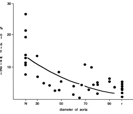
We observed that, when individual values of elastin concentration were plotted against the respective diameter of aortas, the elastin loss occured mainly during the early phases of the aneurysmal development (Figure1). However, the extractable collagene content was unchanged in small aneurysms (< 50 mm) and was higher in large and ruptured A (Figure 2).
Figure 1. Significant quadratic relationship between the elastin concentration ( in % of
defatted dry weight ) and the individual values of the diameter of the normal aortas (N), AAA of increasing size ( mm ) and ruptured AAA (r).
According to our findings, we concluded that early degradation of elastin, followed by modification of the collagen polymers, supported the suggestions of Dobrin (Dobrin et al., 1984) that elastin plays a role in dilatation, while subsequent collagen alteration could lead to rupture.
Figure 2. Significant positive linear correlation between the collagen extractability (dry wright ng/mg ) and the individual values of the diameter of the normal aortas (N), AAA of increasing
size ( mm ) and ruptured AAA (r).
These results led us to develop some hypotheses, that will be tested by new investigations followed our doctoral thesis (1994).
Aim of the study
Whatever the safety of procedures to treat AAAs before rupture, none of them is performed without a certain mortality (at best less than 2%), morbity, or consequences on health costs. A better knowdlege of the prevalence of AAA in an ageing occidental population, the demonstration of a genetic mecanism and the elucidation of the biochemical pathways depending on heredity and risk factors will provide a gobal approach of AAAs, thus considered as a pathological continuum from birth until death. These knowledges will perhaps allow, for the present, a better therapeutic classification for surgery, but also in the future, will perhaps lead to true prophylactic measures (i.e. chemotherapy along with other interventions). Such are the general goals of our study.
Our work plan answers to the following questions:
1) Is the prevalence of AAA in Belgium similar to the prevalence observed in other countries? Is routine screening with ultrasound examination a cost effective method? Are there any correlations between AAA disease and classical risk factors for atherosclerosis?
Abdominal aortic aneurysm is a common pathology, with an estimate incidence of 20 to 40 cases /100,000 persons per year, and its prevalence lies between 3 and 4% in men aged 60 years or older. All population-based screening studies performed at the beginning of the last decade were performed in American, British, and Scandinavian countries. Therefore, we started a population-based study in Liège among men aged between 65 and 75 years old. (Chapter 2)
2) Based on previous results, the incidence of AAAs in octogenerians is far to be negligeable. So, what are the results of surgical treatment of AAA in octogenarians? (Chapter 3)
3) As shown in our doctoral thesis, the presence of an inflammatory infiltrate in the wall of AAA and degradations of elastin and collagen are early features in the development of AAA. So do metalloproteases play a significant role in the development of AAA? Thus, specific metalloproteinases displaying elastolytic contents (as the 72 and 92 kDa gelatinases) were
identified and quantified in the serum, in the thrombus and in the wall of the blood vessels. (Chapter 4)
4) The involvement of proteases/antiproteases in AAA and AOD (aortic occlusive disease) already documented in aneurysmal progression and plaque instability (Knox et al., 1997) is most often restricted to one or a few potential factors. The following question arise from this conflicts: are the respective implications of MMPs, TIMPs and repair process similar in aneurysmal aorta and AOD? (Chapter 5)
5) Imbalance between MMPs and their inhibitors (TIMPs) in the wall of aneurysmal abdominal aorta is implicated in the expansion of ruptured AAA (Baxter et al., 2002, Knox et al., 1997). Can informations gathered from the samples collected during surgery for ruptured AAA reflect a local activity at the precise site of rupture. (Chapter 6)
6) Although the size of the aneurysm still remains the most accepted predictor of rupture, small AAA may also rupture. So, are there any marker or functional imaging techniques such PET scan able to monitor the development and evolutivity of AAA? (Chapter 7)
7) Since we found a positive relationships between the clinical status of AAA and the functional imaging (chapter 7), a potential correlation was searched between FDG uptake and histological findings in the wall of AAA. (Chapter 8)
8) If our findings favored the local production and/or activation of the MMP-2 and MMP-9 in aortic wall by polymorphonuclear leucocytes (PMNs), does the activation of PMNs result in a systemic oxidative stress ? In order to answer this question, the concentration of a major antioxidant (vitamin E) was determined in the plasma of the patients with AAA. (Chapter 9) 9) Published observations suggest a familial incidence of AAA. (Bengtsson et al., 1996, Powel & Greenhalgh, 1987, Tilson & Seashore, 1984, Webster, St Jean et al., 1991). If AAA is really a familial disease, what is the genetic basis? In order to disclose a possible familial incidence and genetic predisposition, we started a familial enquiry of AAA carriers in the family of AAA patients recruited between 1986 and 1991. (Chapter 10)
10) Some genetic pattern associated to development of AAA in the belgian population were identified (chapter 10). So, may these pattern be transposed to other countries and populations and is it possible to identify a specific genetic locus? Are these genetic predisposition correlated to changes in activities of MMPs and TIMPS? To answer these questions, a multicentric European and North-American study was initiated. (Chapter 11)
CHAPTER 2
EPIDEMIOLOGY OF AAA: A POPULATION-BASED STUDY (APPENDIX 1)
INTRODUCTION
Epidemiology deals with the frequency and distribution of disease within the population. Epidemiological methodology may be helpful in identifying possible causes of disease. The basic estimates of the presence of a disease in the population are the prevalence and the incidence. The prevalence describes the number of patients having a disease at a certain time. The incidence describes how many new individuals will develop the disease during a certain period of time.
According to the definition set up by the World Health Organization, screening means medical investigation that does not arise from patient's request for advice because of specific complaints. Certain prerequisites are necessary before to start screening procedures. The disease must be potentially dangerous and should be asymptomatic. It must be possible to detect the disease before symptoms occur and finally some form of treatment must be available. In the case of AAA all these premises are at hand. Abdominal aortic aneurysm (AAA) is a serious medical problem that affects a significant proportion (± 4%) of the more than 60 year-old (± 3500 men in Liège). It can be foreseen that the prevention of AAA will further be needed in the next decades with the life expectancy progress.
Epidemiology of AAA has been poorly studied in the past. The disease is mostly asymptomatic (until rupture) and previous diagnostic methods, such as conventional X-rays and abdominal palpation, present obvious limitations in detecting aneurysms (Collin et al., 1988, Lederle et al., 1988). With the introduction of new non-invasive diagnostic methods such as computerized tomography (CT) and ultrasonography (US), it has become possible to
AAA (Erikson et al., 1980, Gomes et al., 1977) and screening possibilities for AAA diagnosis (Collin, 1985) have led to increase interest for epidemiological knowledge.
The prevalence of AAA seems to vary in the different geographical regions. Fowkes compared information from the United States (Lilienfeld et al., 1987) with data from England and Wales (Fowkes et al., 1989), and found six times higher mortality in Great Britain. Population-based studies from Sweden and Great Britain for ruptured AAA have shown incidence rates from 6 to14 per 100,000 inhabitants (Ingoldby et al., 1986, Johanson & Swedenborg, 1986, Thomas & Stewart, 1988). These differences could result from geographical and racial characteristicsbut there are also possible methodological explanations.
Until 1995 the majority of published ultrasound screening studies has been carried out in Great Britain and in Scandinavian countries (Bengtsson et al., 1996, Chichester Aneurysm Screening Group et al., 2001, Chosky et al., 1999, Collin et al., 1988, Lindholt, 2001, Scott et al., 1988). Therefore we decided to perform the first Belgian population-based study in Liège1.
MATERIAL AND MEDHODS
Between December 14, 1995 and November 24, 1996 a personalized letter offering the opportunity to undergo free ultrasound screening for AAA was sent to 1764 men born in 1920 and 1930. Selection was based on birth records for the city of Liege. Ultrasound examinations were performed by a radiologist and a surgeon from the Medical Imaging and Cardiovascular Departments of the University of Liège (for more details, see Material and Methods in Appendix 1). The incidence of AAA and the cost of the diagnosis (calculated by dividing the overall cost by the number of AAA discovered) were determined.
RESULTS
As comparison of number of participants with previous AAA screening studies performed in Sweden and Great Britain, compliance in our study was lower (only 41%).
Ultrasound examination was feasible in 98.5% of cases. Anteroposterior diameter was > 29 mm in 33 subjects (4.5 %). The prevalence of AAA defined as largest aortic diameter ≥ 30 mm was 3.8% (Table IX). On the basis of epidemiological data collected, a high-risk population for AAA was identified. The incidence of hypertension and smoking habits were significantly higher in AAA group as compared with non-AAA group (Table X).
Reference Compliance (%) Number of
subjects
Age (years) Incidence of AAA (%) * Smith FCT et al. 76,3 2664 65-75 8,4 Collin J. 51.7 497 65-74 42 Bebgtsson H et al. 75/0 364 74 8.5
This study 41.0 727 65 and 75 4.53
* Defined as maximum transverse diameter >29 mm Table IX : Data from previous screening studies for AAA
Without AAA With AAA
n % n % p Dyslipidemia 172 24.92 13 35.13 NS Diabetes 78 11.30 7 18.91 NS Hypertension 188 27.24 17 45.94 < 0.05 Smoking 156 21.73 13 35.13 < 0.06 NS, non-significant
Table X : Risk factors for cardiovascular disease in the study population (n = 727) without (n = 694) or with (n = 33) AAA
The overall cost of screening was 18,175 €. The cost of diagnosis of each AAA was 551,00 €.
DISCUSSION
Ultrasound screening is considered the method of choice for AAA screening (Bengtsson et al., 1996, Akkersdijk et al., 1991). In addition to being feasible in 98.5% of cases, the sensitivity of ultrasound is over 95% and its specificity is 100%. Other advantages of ultrasound include non-invasiveness, good reproducibility, and cost-effectiveness (seven-fold less expensive than an abdominal CT scan).
The definition of AAA is of great importance in epidemiological studies since the prevalence of the disease will vary substantially simply because of variations in the criteria for the diagnosis. There is as yet no definite consensus on the definition of abdominal aortic aneurysm. However for practical reasons, McGregor’s (McGregor et al., 1975) definition is still the most widely used in clinical practice, as well as in research.
In 1965, Steinberg (Steinberg et al., 1965) established normal standards for the aortic diameter by using angiography. Based on the findings of Steinberg, in 1975 McGregor (McGregor et al., 1975) proposed the definition of an AAA as the largest measured infrarenal diameter being ≥ 30 mm. Twelve years later, Sterpetti (Sterpetti et al., 1987) suggested that abdominal aorta is considered as aneurysmal when infrarenal aortic diameter exceeds suprarenal diameter of, at least, 50 %. A few months later, Collin (Collin et al., 1988), who designed one of the first large population screening program in Oxford, proposed that the infrarenal diameter should be ≥ 40 mm or exceed suprarenal diameter by at least 0.5 cm. In 1991, the SVS/ISCVS Ad Hoc Committee (Johnston et al., 1991) proposed the definition of infrarenal diameter being 1,5 times the expected normal diameter.
AAA occurs more commonly among men. Melton (Melton et al., 1984) in Rochester, Minnesota, showed that the incidence rate is at least two times higher in men and that the prevalence of AAA increases markedly with age in both sexes. The prevalence of AAA is between 2 and 3% of males over the age of 65 (Collin et al., 1988, Scott et al., 1988). In an
autopsy study performed by Bengtsson (Bengtsson et al., 1992), based on 45,838 autopsies between 1958 and 1986, the frequency of AAA was 4.3% among men and 2.1% among women. The age-specific frequency of AAA increased markedly after 55 years in men and 70 years in women. The male frequency reached a peak of 5.9% at 80 years and the female 4.5% at 90 years and above. Therefore, although recent English population-based studies show an increasing prevalence of AAA in females (Brown & Powell, 1999), all screening studies unsurprisingly show a higher prevalence of AAA in men than in women. One of the most consistent risks factors for AAA is smoking. According to Janet Powell (Powell, 2003) « the epidemic of aortic aneurysms follows some 40 years after an epidemic of smoking. Since 1950 there have been an increasing proportion of women who smoke, and this may alter future sex ratios for the prevalence of AAA ».
In our study, anteroposterior aortic diameter > 30 mm was observed in 28 male subjects (mean diameter 34.7 mm) and the incidence of AAA was 3.8%. Anteroposterior aortic diameter > 29 mm was observed in 33 cases (mean diameter 30.9 mm) and the incidence of AAA was 4.5%. This incidence of AAA is comparable with previous reports (Collin, 1985, Bengtsson et al., 1996, Smith et al., 1993, Akkersdijk et al., 1991).
In the present study, we targeted 65- and 75-year-old men for routine ultrasound screening. The highest incidence of AAA is in subject over 80 years, the optimal effect on mortality in function of age at diagnosis is observed between 70 and 75 years, and the lowest operative mortality is observed between the ages of 55 and 65 years. Thus, since the detection rate is lower and the cost of screening per case diagnosed is higher (Smith et al., 1993, Collin et al., 1990) in these cases, the best age group for screening in terms of cost effectiveness is that between 65 and 75 years (Bengtsson et al., 1996, Smith et al., 1993, Collin, 1990). In the city of Liège, there were 9344 men between the ages of 65 and 75 years. We choose to study 65-years old men (born in 1930) and 75-years-old men (born in 1920) to obtain an easily useable sample for statistical analysis.
The reliability of population-based studies depends on compliance. The percent of compliance in previous AAA screening studies in English-speaking and Scandinavian
study, compliance is only 41.1%. It can be considered too low to allow comparison. The effectiveness of screening studies could be enhanced by specific targeting of high-risk groups. The incidence of AAA in subjects over the age of 65 with hypertension was 12% in the study of Allen et al. (Allen et al., 1987) and 9% in the study of Lederle et al. (Lederle et al., 1988). In our department, screening for AAA was carried out on 110 patients scheduled to undergo surgery for either coronary artery disease (n = 72) or peripheral vascular disease (n = 38). The incidence of AAA was 9.7% in patients with coronary artery disease and 2.6% in patients with peripheral vascular disease (Sakalihasan et al., 1992, Sakalihasan & Limet, 1994).
On the basis of epidemiological data obtained, we attempted to define a high-risk population for AAA. The incidence of hypertension was 45.9% in the AAA group as compared with 27.2% in the general population (p < 0.05) and the incidence of smoking was 35.1% in the AAA group as compared with 21.7% in the general population (p < 0.06). The higher incidence of CABG [18.1% in the AAA group versus 55% in the general population (p < 0.01)] could be the result of a bias as patients who have undergone previous surgery may be more aware of the risks of cardiovascular disease and thus more likely to take part in screening.
The cost of each screening examination for AAA was calculated on the basis the officially established rate for ultrasound study : $25.00 /procedure. The cost of diagnosis of each AAA was $551.00, calculated by dividing the overall cost by the number of AAA discovered.
The effects of screening of the incidence of ruptured AAA were recently investigated with a stepped wedge study design in the screening program ongoing in the Huntingdon district (Chosky et al., 2001). This study concluded that screening 65-80 year-old males for AAA could reduce the incidence rate of ruptured AAA by 49%.Similarly randomized clinical trial involving 65-75 year-old men in Chichester (Chichester Aneurysm Group et al., 2001) showed a 55% reduction in the incidence of ruptured AAA by screening as well as the reduction of Health Care burden. The Cost evaluation for elective and emergent AAA repair in our institution is represented in Table XI. For the 20 electively operated patients the hospital stay averaged 14 days (extremes 2 and 31) versus 30 days (extremes 1 and 65) for