AVIS
Ce document a été numérisé par la Division de la gestion des documents et des archives de l’Université de Montréal.
L’auteur a autorisé l’Université de Montréal à reproduire et diffuser, en totalité ou en partie, par quelque moyen que ce soit et sur quelque support que ce soit, et exclusivement à des fins non lucratives d’enseignement et de recherche, des copies de ce mémoire ou de cette thèse.
L’auteur et les coauteurs le cas échéant conservent la propriété du droit d’auteur et des droits moraux qui protègent ce document. Ni la thèse ou le mémoire, ni des extraits substantiels de ce document, ne doivent être imprimés ou autrement reproduits sans l’autorisation de l’auteur.
Afin de se conformer à la Loi canadienne sur la protection des renseignements personnels, quelques formulaires secondaires, coordonnées ou signatures intégrées au texte ont pu être enlevés de ce document. Bien que cela ait pu affecter la pagination, il n’y a aucun contenu manquant.
NOTICE
This document was digitized by the Records Management & Archives Division of Université de Montréal.
The author of this thesis or dissertation has granted a nonexclusive license allowing Université de Montréal to reproduce and publish the document, in part or in whole, and in any format, solely for noncommercial educational and research purposes.
The author and co-authors if applicable retain copyright ownership and moral rights in this document. Neither the whole thesis or dissertation, nor substantial extracts from it, may be printed or otherwise reproduced without the author’s permission.
In compliance with the Canadian Privacy Act some supporting forms, contact information or signatures may have been removed from the document. While this may affect the document page count, it does not represent any loss of content from the document.
Characterization of the AP Endonuclease Enzyme APN-l from C. elegans
par Devang Patel
Programme de sciences biomédicales Faculté de médecine
Mémoire présenté à la Faculté des études supérieures en vue de l'obtention du grade de maîtrise (M.Sc.)
en sciences biomédicales
Décembre 2007
© Devang Patel, 2007
Faculté des études supérieures
Ce mémoire intitulé:
Characterization of the AP Endonuclease Enzyme APN-l from C. elegans
présenté par: Devang Patel
a été évalué par un jury corl'mosé des personnes suivantes:
Dr. Puttaswamy Manjunath président-rapporteur Dr. Dindial Ramotar directeur de recherche Dr. Ashok Srivastava membre du jury
apuriniques/apyrimidiques (AP), sont des enzymes multifonctionnelles impliquées dans la réparation de l'ADN. En effet, les enzymes endo IV chez E. coli et apurinic/apyrimidinic endonuclease 1 (Apnl) chez S. cerevisiae sont capables de traiter des sites abasiques et des extrémités 3' bloquées. Des études récentes ont révelé que les AP endonucléases peuvent non seulement agir en tant que 3' ~ 5' exonucléases, mais participent aussi à l'incision de certaines bases oxidées. Jusqu'à présent, peu de connaissances existent au sujet d'APN-l de C. elegans (CeAPN-l); le premier homologue retrouvé chez un organisme multicellulaire. Nous avons développé un système d'expression chez la levure permettant de produire CeAPN -1 fusionnée à l'étiquette GST, et de la purifier à l'homogénéité pour une caractérisation approfondie.
À l'aide d'essais in vitro, nous avons confirmé que, tout comme ses homologues, CeAPN -1 possède les activités AP endonuc1éase et 3' -diestérase. Pourtant, on a pu démontrer par la complémentation inter-espèces que les variantes E97G et E143G de CeAPN-l ne confèrent aucune résistance à une souche de levure déficiente en AP endonucleases lors d'un traitement aux différents agents d'endommagement à l'ADN, tels que le MMS, le H202 et la Bléomycine. Ces résultats confirment des observations antérieures lors de la substitution de résidus chargés dans un des quatre domaines conservés d'Endo IV liée à l'activité. Cependant, on a révélé que l'activité enzymatique de CeAPN-l semble être stimulée en présence d'une composante thermostable. Cela peut suggérer l'implication d'un inducteur ce qui différencie CeAPN-l de ses homologues.
Mots Clés: CeAPN-l, Endo IV, AP endonucléases, site AP, réparation par excision de bases, réparation de l'ADN, C. elegans.
ABSTRACT:
Proteins of the Endonuclease IV (Endo IV) family of AP endonucleases have been weIl documented for their multifunctional capacities in DNA repair. Studies with the E. coli endo IV and S. cerevisiae apurinic/apyrimidinic endonuclease 1 (Apnl) have demonstrated that these enzymes have the ability to process abasic sites and 3' -blocked ends. Additional investigations have revealed a capability to act as 3' -7 5' -exonucleases and, furthermore, have exposed a participation in the incision of certain oxidatively damaged bases. To date, very little is known of APN-l (CeAPN-l), the corresponding
C. elegans Endo IV prote in and the first structural homologue to be found in a multicellular animal model. In this study, we have engineered an expression system in yeast to produce a CeAPN-l fusion protein and subsequently, purified the polypeptide to homogeneity for characterization. We confirmed, through in-vitro assays, that CeAPN-l also possesses AP endonuclease and 3'-diesterase activities as its counterparts found in unicellular models. Moreover, domain function analysis has revealed that the introduction of the CeAPN-l variants, E97G and E143G, in a yeast strain lacking AP endonuclease activity is unable to restore parental resistance when exposed to the DNA damaging agents MMS, H202 and Bleomycin. These results coincide with previous observations made following the substitution of charged residues contained in one of the four conserved domains of Endo IV affecting activity. However, unlike its homologues, we showed that the enzymatic activity is enhanced by a heat stable component. This suggests a likely key feature which differentiates this protein from other counterparts, despite structural and functional homology.
Key Words: CeAPN-l, Endo IV, AP endonuclease, AP site, base excision repair, DNA repair, C. elegans.
TABLE OF CONTENTS
Resumé ... i
Abstract ... ii
Table of Contents ... iii
List of Tables ... vii
List of Figures ... , ... viii
Abbreviations ... ix
DedicatioD ... xiii
Acknowledgements ... . xiv
1. INTRODUCTION ... 1
1.1. Reactive Oxygen Species ... 3
1.2. DNA Repair Mechanisms ... 4
1.2.1. Mismatch Repair ... , ... 5
1.2.2. Direct ReversaI. ... 5
1.2.3. Double Strand Break Repair. ... 6
1.2.4. Nucleotide Excision Repair ... 7
1.2.5. Base Excision Repair. ... 8
1.3. BER in Mammalian Cells ... 13
1.3.1. Short Patch Repair. ... .13
1.3.2. Long Patch Repair. ... 13
1.4. BER in Saccharomyces cerevisiae ... 14
1.5. BER in Schizosaccharomyces pombe ... 15
1.6. BER Key Machinery ... 16
1.6.1. Glycosylases ... 16
1.6.2. Monofunctional Glycosylases ... 16
1.6.3. Bifunctional Glycosylases ... .17
1.6.4. AP Endonuclease Action in the Processsing of AP Sites ... 18
1.7. Endo IV Family Members ... 20
1. 7.1. Escherichia coli endo IV ... 20
1.7.3. Schizosaccharomyces pombe (SpApn1) ... 27
1.7.4. Caenorhabditis elegans CeAPN1 ... .28
1.8. Research Project ... "' ... 29
1.81. Background and Objectives ... 29
2. METHOD AND 'MA TERIALS ... "' ... 32
2.1. Yeast and Bacterial Strains ... 32
2.2. Plasrnid Construction ... _ ... 32
2.3. Isolation of Plasmid DNA and Transformation ... 34
2.4. Expression System for Recombinant Proteins ... 34
2.5. Gradient Plate Assay ... "' ... 35
2.6. Extraction of Proteins for Expression Analysis ... 35
2.7. Extraction of Proteins for Purification ... 35
2.8. Site Directed Mutagenesis ... 36
2.9. Protein Purification Using His Affinity (Talon) Column ... 37
2.10. Protein Purification Using Glutathione Sepharose Column: ... 37
2.11. SDS PAGE Staining and Western Blot ... 38
2.11.1. Sypro Staining ... 38
2.11.2. Western Blot. ... 38
2.12. Preparation of AP Endonuclease Substrate ... ., .•. .,38
2.13. Preparation of 3' Diesterase Substrate ... 39
2.14. Preparation of YW778 Extracts for Activity Assays ... 39
2.15. Enzyme Assays ... ' ... 40
2.16. Homology Modelling of CeAPN-l ... 40
3. RESULTS ... 41
3.1. Plasmid Construction ... 41
3.2. Cross Species Complementation ... 44
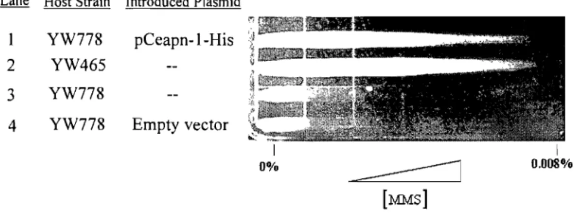
3.3 Sampling of Adopted Purification Schemes ... : ... 45
3.4. Purification Attempts of His Tagged CeAPN·1 Fusion Protein ... 49
3.4.1. Gravit y-Flow Column purification of HIS-CeAPN-l recombinant protein ... .49
3.4.2. Evaluation of Detected Proteins Following Purification ... 52
3.4.3. New NLS-Ceapn-l-His Construct. ... 53
3.4.4. Single Step Protein Purification ... 54
3.5. Characterization of Partially Purified Extracts Containing CeAPN .. 1-HIS ... 56
3.5.1 Protein Concentration Dependence of AP Site Processing ... 56
3.5.2 Time Dependence of AP Site Processing ... 57
3.5.3 Concentration Dependence in the Processing of 3' a,~ Unsaturated Aldehyde 3.5.4 Substrate (3'-Blocked Ends) ... 58
3.6. Complete Purification of the CeAPN·l Recombinant Protein •••••.•.••.••.•• 60 3.6.1. GST-His-Ceapn-l Construct. ... 60
3.6.2. Purification of GST Tagged Protein ... 60
3.7. AP Endonuclease Activity Assay of GST recombinant Protein •...•.•.•.••..• 62
3.8. Addition of YW778 Protein Extracts Stimulate CeAPN-l Activity ....•...•.. 64
3.8.1 Addition of Heated Extracts ... 66
3.8.2 Addition of Specifie Metal Factors ... 67
3.8.3 Addition of Dialyzed Extracts ... 69
3.8.4 Summary ofObserved Results ... 71
3.9. Domain Function Analysis ... 71
3.9.1. Expression of Variant Proteins Following Copper Induction in the YW778 Background ... 74
3.9.2. Gradient Plate Analysis of Variant and Native Proteins ... 76
4. DISCUSSION ... 79
4.1.0 Utilization of a His Affinity Tag ... 79
4.1.1 Utilization of a GST Affinity Tag ... 82
4.2. Activity Assays of GST Tagged Protein (GST -HIS-CeAPN-l) ...•... 83
4.3. Conclusions Stemming from Activity Assays ... 85
4.4. Domain Function Analysis ... 88
4.5. Conclusion and Future Perspectives ... 91
4.5.2 Future Work ... , ... 93
LIST OF TABLES:
Table 1-1: Homologous BER Genes in Different Organisms ... 12 Table 2-1: Oligonucleotides Utilized in Study ... 32
LIST OF FIGURES:
Figure 1-1: Examples of Lesions Stemming from Altered Bases .•..•... 2
Figure 1-2: Different Forms of AP Sites ...•...•.•••.••.••.••••••••.•...•. 3
Figure 1-3: Base Excision Repair Pathway ... 10
Figure 1-4: AP Site and Resulting Repair ... 18
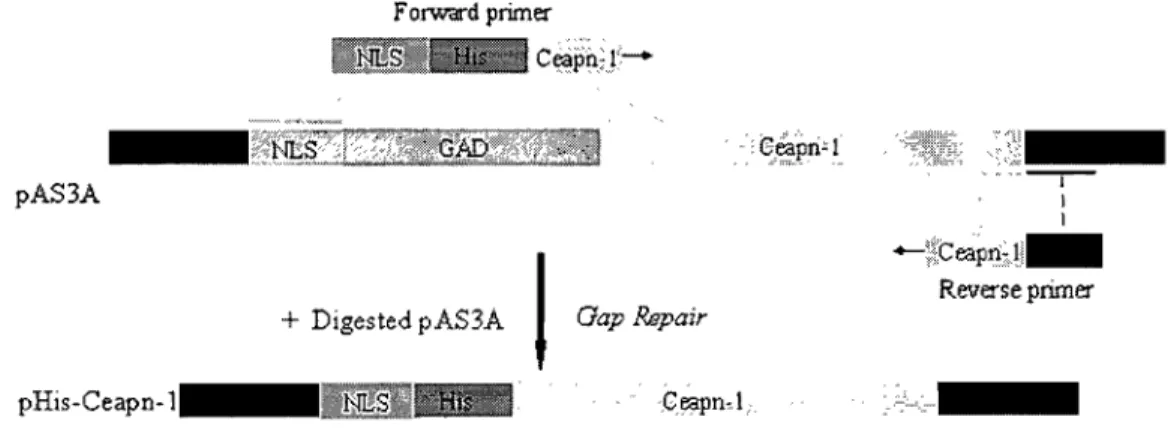
Figure 3-1: Construction of Ceapnl Expression Plasmid with N-terminal His Tag ... .41
Figure 3-2: Construction of Ceapnl Expression Plasmid with C-terminal His Tag ... .42
Figure 3-3: Construction of Ceapn-l Expression Plasmid with Dual N-terminal GST and His Tags ... 43
Figure 3-4: Construction of Ceapn-l Expression Plasmid with dual tagged N-terminal GST and C-terminal His ...•... 43
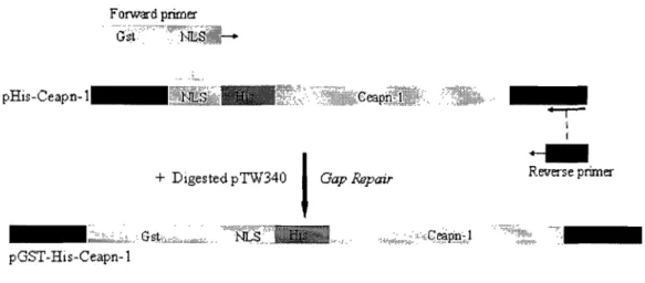
Figure 3-5: Cross Species Complementation Analysis ... 45
Figure 3-6: Purification of HIS-CeAPN-l ... 47
Figure 3-7: Purification of CeAPN-I-HIS ... 48
Figure 3-8: Purification of Dual Tagged Protein ... 48
Figure 3-9: Evaluation of single step His Affinity (Talon) column purification •••••••••••.•.••••.•••.•••...•...•...••... 51
Figure 3-10: Purification Comparison Using Extracts from YW778 and YW778 Expressing HIS·CeAPN-l ... 53
Figure 3-11: Talon Column Purification of CeAPN-I-HIS Protein ... 55
Figure 3-12: Protein Concentration Dependence of AP Site Processing ...••...•..•..•.••.•••••••••••••••••••• 57
Figure 3-13: Time Dependence of AP Site Processing ... 58
Figure 3-14: Concentration Dependence of 3'
a,p
Unsaturated Aldehyde ...•.•••••.••..• 59.
"
Figure 3-15: Analysis of Eluted Fractions from Glutathione Sepharose Resin ... 62Figure 3-17: Addition ofYW778 (apnlA apn2A) Protein
Extracts Stimulate CeAPN-1 Activity ... 64
Figure 3-18: Treatment of Protein Extracts ... 65
Figure 3-19: Addition of Heated Protein Extracts ... 67
Figure 3-20: Evaluation of Metal Ion Dependence of GST -HIS-CeAPN -1 ... 68
Figure 3-21: Addition of Dialyzed Extracts ... 70
Figure 3-22: Summary of Observed Results Following the Addition of YW778 Protein Extracts ... 71
Figure 3-23: Model of CeAPN-1 Based on Known endo IV Structure ... 72
Figure 3-24: Chosen Sites for Mutagenesis ... 73
Figure 3-25: Visualization of Variant Proteins ... 74
Figure 3-26: Protein Expression Following Copper Induction ... 75
Figure 3-27: Cross Species Complementation Analysis Using MMS ... 77
LIST OF ABBREVIATIONS
8-0H-Gua 8-hydroxyguanine
AP apyrimidic/ Apurinic
Apel apurinic/apyrimidinic endonuclease 1 (hum an)
Apnl and 2 apurinic/apyrimidinic endonuclease land 2 (yeast)
ATP adenine triphosphate
BER base excision repair
BLM bleomycin
C. elegans Caenorhabditis elegans
CDC9 cell division cycle 9 (DNA ligase)
DHT 5,6-dihydrothymidine
DHU 5,6-dihydro-2' -deoxyuridine
DNA deoxyribonucleic acid
hDR direct reversaI
dRPase deoxyribophosphodiesterase
DSB double strand breaks
E.coli Escherichia coli
EDTA ethylenediaminetetraacetate.
Endo III endonuclease III
Endo VIII endonuclease VIII
ExoIV exonuclease IV
fapy-ade 4, 6-diamino-5-formamideadenine
fapy-gua 1 4, 6-diamino-5 -formamideguanine
FENl flap endonuclease 1
Fpg Formamidopyrimidine-DNA Glycosylase
GAD GAL4 activation domain
GFP green fluorescent protein
IGG global genomic
GST glutathi one-S-transferase
HhH hairpin helix domain
His histidine
HR homologous recombination repair
IPTG isopropy 1 ~-D-thigalactopyranoside
kb Kilobase
kDa Kilodalton
LIGI ligase 1
LB Luria Broth
Mg2+ magne sium ion
MGMT 06-methylguanine DNA methyltransferase
MMR mismatch repair
MMS methyl methane sulfate
NER nucleotide excision repair
NHEJ non homologous end joining repair
NIR nucleotide incision repair
NLS nuclear localization sequence
Oggl 8-oxo-deoxyguanosine DNA glycosylase 1
PBS Phosphate buffer saline
PCNA proliferating cell nuclear antigen
PCR polymerase chain reaction
Pol~,(5,E polymerase beta, delta, epsilon
RFC replication factor C
RNA ribonucleic Acid
RNAi RNA interference
ROS reactive oxygen species
S. cerevisiae Saccharomyces cerevisiae
S. pombe Schizosaccharomyces pombe
siRNA small interfering RNA
SSB single strand breaks
tBH tert-Butyl hydroperoxide
TC transcription coupled
TIM triosephosphateisomerase
UDG uracil DNA glycosylase
UV ultraviolet
UVDE ultaviolet damage endonuc1ease
yeast extract, peptone, dextrose
XRCCI x-ray cross-species complementing 1
• adA a-anomeric 2-deoxyadenine
This thesis is dedicated in Ioving memory of my eIder sister, aIl that 1 am or ever hope to be, 1 owe to her.
ACKNOWLEDGEMENTS:
First and foremost, 1 would like to express a big thank you to my research director, Dr. Dindial Ramotar, who allowed me to work in his laboratory and to whom 1 owe a great deal. His passion and zeal for discovery is, undoubtedly, most admirable and unmatched.
A special thank you to Dr. Xiao Ming Yang, who took the time to teach me about conducting science and, moreover, about life in general. His wisdom, sense of humour and kindness brought a unique levity to the working environment.
During the past years, 1 have had an exception al opportunity to meet and surround myself with individuals of great intellect and experience: A. Leduc, Dr. A. Shatilla, Dr. A. Jilani, Dr. 1. David, Dr. J. Fan, Dr. J.Y. Masson, & Dr. S. Azam. 1 am profoundly appreciative for the insightful counsel, support and encouragement that they provided.
ln addition, 1 would like to commend my HMR comrades Ali K, Chadi Z, Jasmine L., Jeremie P., Julie D., Lisa B. Nath J., Mus A., Rad R., Ratsa V. to whom 1 had the pleasure to work and exchange with on a daily basis.
Finally, 1 would also like to articulate my immense gratitude to my family for their encouragement, patience and teaching me to aim for the highest of goals.
1. INTRODUCTION AND LITERA TURE REVIEW
DNA is victim to continuous attack. Mutations, being products of DNA damage and subsequent genomic instability, have serious consequences to a given cell. From a human perspective, the inability to counter various insults affecting DNA integrity has been correlated to cancer, aging, and multiple disorders [1]. Interestingly, DNA damage can stem from both exogenous and endogenous sources. Thus, exogenous sources (environment and mutagenic chemicals) include ultraviolet and ionizing radiations as weIl as a plethora of toxins and chemicals. In contrast, endogenous sources of DNA damage primarily include reactive oxygen species (ROS) which arise from metabolic byproducts [2]. Each source manifests itself by causing specific types of modifications to ultimately harm the normal state of the nucleic acid macromolecule. Thus, common insults affecting the double helix can be observed via base/sugar damage (hydrolysis, deamination, alkylation, oxidation, strand breaks, abasic sites), odd base mismatches or pairing (pyrimidine dimers) and strand cross linking [2].
1. Alkylated bases 2. Deaminated bases
3 -Methyladenine 7 -Methyladenine Uracil
3. Oxidized Bases o
o~<Jt~H'
8-0xoguanine (8oxoG) oOH~l
"lN~
H 5 -hydroxyuracil4. Other examples of Lesions
N 1
<XN:l
R 1,NtS-Etheno-adenine 2,5-Amino-5-F onnamidopyrimidine (fapy) Thymine glycol 5'3 ,N4-Etheno-cyto sine a-deoxyadenine
Figure 1-1: Examples of Lesions Stemming from Altered Bases (adapted from [2,3])
A particular interest in this study would be devoted to the formation and repair of apurinic 1 apyrimidinic (AP) sites (shown below). These lesions (abasic) are characterized by a 10ss of the base component from the sugar phosphate moiety (se en in figures 1-2, 1-3, 1-4) [4]. These sitescan occur spontaneously (hydrolysis) and also via enzymatic means to remove damaged bases [4, 5].
~ ~
O~o_:;,---==
...
=OyOH,
Lo
o
0/'-\
~ ~ Regular AP site 5' 5'O)~OH
1r
OHO~OH
Ho
3' Reduced AP siteo
3' l' -oxidized AP site 5' 0 =O~H
o 3' 4' -oxidized AP siteFigure 1-2: Different Forms of AP Sites (adàpted from [3])
1.1. Reactive Oxygen Species
A primary source of endogenous cellular damage arises from oxidative stress. This results from an increased prevalence of oxidant molecules in the cell. Known as reactive oxygen species (ROS), these oxidant molecules are highly unstable due to an inherited unpaired electron [6]. Naturally, these reactive intermediates have an uncanny ability to react with electron rich components; notably, lipids, macromolecules and most importantly, DNA [7]. In fact, Fentonoxidants (i.e. Fe2+ + H202 -> Fe3+ + 'OH + ·OH)
are believed to be the most significant type of ROS causing DNA damage [2]. It should
be stressed that changes made to the double helix structure require significant attention; hence, unlike other components, DNA cannot be tumed over or replaced with normal type molecules. Lesions formed due to oxidative stress are associated with base alterations which can result in 80xoG lesions, ring saturated pyrimidines, and also contribute to the formation of AP sites. ROS could also cause sugar fragmentation which can result in the formation of DNA strand breaks (with phosphoglycolate and phosphate blocked ends) [8].
ROS are produced through processes occurring during aerobic metabolism in the mitochondrion [6]. They can also be formed via a cell's interaction with exogenous sources, which may include ionizing radiation, UV radiation, carcinogenic compounds and anti-cancer drugs (Bleomycin, Neocarzinostatin) [9]. A cell can defend itself against ROS by exploiting antioxidant molecules and specific enzymes. A reduced capacity, if not an absolute failure, to counter DNA damage caused by ROS can easily hinder efficient replication and subsequently be highly mutagenic or otherwise, lethal [8].
1.2. DNA Repair Mechanisms
In order to counter DNA damage, the cell has multiple DNA repair mechanisms which ensure survival. Thus, these include mismatch repair (MMR), direct reversaI (DR), double strand break repair (DSBR), nucleotide excision repair (NER), and also base excision repair (BER). Each repair pathway is responsible in countering specific types of lesions [2]. Undoubtedly, overlapping of the individual pathways is inevitable; thus, this phenomenon ensures substitute mechanisms in the repair ofDNA lesions [10].
1.2.1. Mismatch Repair (MMR)
The fundamental role of the MMR system lies in the removal of mismatched bases and the insertionldeletion loops that form during DNA synthesis [11]. It is a
process that is highly conserved from prokaryotes to eukaryotes. MMR, in essence, involves the removal of a patch of nucleotides (similar seen in excision repair), followed by repair synthesis and subsequent ligation [12]. It should be noted that two
forms of mismatch repair systems have so far been characterized: long patch mismatch repair and short patch mismatch repair. As a result, long patch MMR is capable in repairing a vast array of mismatche~ by excising segments up to 1 Kb long. On the other
hand, short patches are characterized by an ability to handle only specific types of mismatches due to the fact that excision is restricted to a length of 1-10 nucleotides [12]. Recognition of mistmatches is carried through several enzymes which include MSH2 with MSH3 or MSH6. The complex allows the recruitment of a different group of proteins comprising of MLH1, MLH3, PMS 1 or PMS2. These prote in heterodimers allow the recruitment of other proteins implicated in the repair of the damaged strand [13]. Interestingly, defects in mismatch repair machinery in humans have been linked to the onset ofhereditary nonpolyposis colorectal cancer [12].
1.2.2. Direct ReversaI (DR)
The direct reversaI system (DR) is thought to be the least complex of the DNA repair mechanisms. Unlike other pathways composed of multiple steps and subsequently, of multiple enzymes, the DR system involves a single step to remove the lesion in question. Hence, this type of quick reversaI is observed primarily when thymidine di mers (caused by UV) or methylated guanines are formed [12]. Several
moleeules have been eharaeterized as DR enzymes; thus, these include photolyases and MGMT (06-methylguanine DNA methyltransferase) among others [14]. The funetional role of photolyases in humans has not yet been clearly elueidated. However, the corresponding homologues in yeast and bacteria respectively, have shown a capacity to proeess damage caused by UV and cisplatin treatment. MGMT, on the other hand, is believed to be vital in processing lesions caused by alkylation [14]. Briefly, the alkyl group stemming from the les ion is removed and simultaneously shifted to the cysteine residue found in the active site of the protein. AIthough methyl transferases have shown to possess the ability to repair alkylated damage, the activity is known to be very energy costly. In fact, these DNA repair proteins are irreversibly inactivated following the enzymatic reaction and, subsequently, degraded by an ATP-dependent ubiquitin pathway [12].
1.2.3. Double Strand Break Repair (DSBR)
Double strand breaks (DSB), as the name implies, refers to the fragmenting of both opposing strands making up the DNA helix. These particular lesions are considered to be fairly dangerous and, consequently, may lead to harmful genome rearrangements or translocations, if not properly processed [15]. As a result, double strand breaks are countered via two principle systems known as non-homologous end joining repair (NHEJ) and homologous recombination repair (HR) [16]
Thus, in NHEJ, a specifie complex of proteins joins the two fragmented ends direetly without the presence of a homologous template. This process is thought to be most valu able in a pre-replication setting where no template is available [17]. NHEJ, in mammals, is initiated by a eomplex formed of MRE Il, RAD50 and MRN amongst
other proteins. This complex enables the activation of DNA dependent prote in kinase (DNA-PK) which phosphorylates several key proteins (histones). In the step, a particular DNA ligase, known as DNA ligase IV, associates with XRCC4 and XLF in order to seal the gap [18]. In contrast, homologous recombination does require the presence of a template (sharing homology to the damaged sequence) which is found on a sister chromatid (S or G2 phase)' and allows the strand to be restored [15]. Undoubtedly, the HR process is known to be very efficient in preserving the integrity
, lu'
and identity of the sequence [19]. In yeast, HR repair involves an initial complex of Mrel1, Rad50, Xrs2 as weil as specifie exonuclease enzymes; thus, the pathway is further mediated by Rad51 paralogs, Rad55 and Rad57. The final step (following resolution of the Holliday junction) requires, most notably, DNA polymerase and DNA Iigase [20].
1.2.4. Nucleotide Excision Repair (NER)
The NER pathway is the major system utilized to counteract bulky DNA adducts or helix distorting modifications (i.e. 6-4 photoproducts and pyrimidine dimers) affecting long portions (2-20 bases) of a particular,'strand. Such lesions are formed via UV or specifie chemical agents (aromatic hydrocarbons, cisplatin) [21]. The repair process is composed of 4 general steps starting with damage recognition, pre-incision, damage excision, gap filling and ligation [22]. Nonetheless, NER is categorized into two sub-pathways: Global genomic (GG) NER and Transcription coupled (TC) NER. These sub-pathways are differentiated by the fact that GG NER repairs ail appropriate les ions in the genome; thus, including both transcribed and untranscribed segments of DNA. TC NER, on the other hand, processes damages encountered during transcription
where RNA polymerase becomes stalled at a helix distorting lesion (i.e. thymine dimer) [23].
1.2.5. Base Excision Repair (BER)
The BER pathway is a vital mechanism composed of multiple enzymatic steps that encompass the repairing of damaged DNA bases and AP sites [2, 24]. Briefly, the pathway is initiated via a DNA glycosylase enzyme which recognizes and removes the damaged base in question (Fig 1-3). The excision of the modified base from the deoxyribose-phosphate backbone forms an abasic site (AP site), which is itself a form of DNA damage that requires prompt attention to prevent further mutagenic consequences [25]. It should be noted that the resulting base loss (AP site) can also
occur through spontaneous hydrolysis. In any case, the AP site is processed by either an AP endonuclease or apurinic/apyrimidinic (AP) lyase activity (processes 3' side of AP site via cleavage between
c-o
bond within deoxyribose) [2]. Thereafter, the single nucleotide gap is filled and sealed via the sequential action of DNA polymerase and DNA ligase [25].The identity of the glycosylase acting on a particular base is crucial in determining subsequent steps. In essence, glycosylases are classified under two subsets: monofunctional and bifunctional. The former subset (monofunctional glycosylase) possesses a single activity which allows it to excisè' the damaged base [26]. The single function glycosylase primes the AP site for AP endonuclease to act on the lesion. Thereafter, AP endonuclease incises 5' to the AP site and forms a 3'-OH and a 5'-abasic sugar phosphate (deoxyribose-phosphate) [2]. In comparison, the bifunctional glycosylase is able to further process the AP site (AP lyase activity) itself by causing an
incision 3' to the lesion and forming a 3'-fragmented deoxyribose and a 5'-phosphate
Monofunctional
!
DN A glycosylase S "J j • • • 1 • • • •.
,
...
,
y AP endonucleasel
5'ni<k .~" S jg .... "J 5' -dRP • • • • • • • •... ..
,
...
Displacement of 1 ~Vu 5'-dRP ..'y-1
S Darnaged b ase "J•••• E ••••
...
....
,.
1l
DN A Bifunctional glycosylasesi
i i
i
1i .
ii"J
• ••••••••
l
AP Lyase o 3'nick. ..--.i
,
..
,
...
~'ii'
1 3' Phosphodiesterase • (AP endonuclease)...
~,
f ••••••••
S °lg r "J .•••• i. ••
Sm~e•••••• t ••
nucleotide gapRemoval of fup 1 ~ Gap fllling
structure ..
0111 S 011' "J
S • • • • • • •
...
,ii"J
•••• ij •••
..t ... .
2-13nt~
. _~atch
patch \ DNA ligation ( ' 1 nt p'
•••••••••
•••••••••
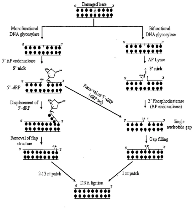
Figure 1-3: Base Excision Repair (BER) Pathway (adapted from
[28])
Diagram highlights important steps in BER, including the sub-pathways (short and long patch) invo1ved in mammalian cells. Damaged base is removed via either monofunctional or bifunctional glycosylase enzymes. The resulting AP site may be processed via AP endonuclease, forming a 3'-OH and a 5'-deoxyribose-phosphate (5'dRP) via a 5' nick. The AP
site may also be countered through an AP lyase activity, which results in a 3' -unsaturated aldehyde. The short patch repair pathway involves formation of a single nucleotide gap via the removal of the 5' dRP (following AP endonuclease activity through 5'dRPase) and in addition, the resulting repair of the unsaturated aldehyde (via 3'diesterase activity of AP endonuclease). The long patch repair pathway is associated with the formation of a flap structure which is later enzymatically removed. Both short and long patch pathways involve sealing the single nucleotide gap via DNA Ligase l or the action of DNA ligase III and XRCC 1.
Activity E. coli S. cerevisiae Homo S. pombe C. elegans
sapiens
ung UNG UDG ungl ung-l
alkA MAG MPG/AAG magl
-nth NTGl, NTG2 NTHl nthl nth-l
jjJg OGGl,OGG2 OGGl -
-Glycosylase nei - NEILl,
NEIL2 -
-mutY - MYH myhl R13A5.9
(wormdatabase)
mutt - MTH pcdl ndx-l, ndx-2
TDG
-AP xth APN2 APEl, APE2 apn2 exo-3
Endonuc1ease nfo APNl not found apnl apn-1
yet
polI RAD27 FENl rad2 crn-l
- POL4 POLf3 pol4
-holC F33H2.5,
Post-incision, (X subunit) POL2,3 POLNE: cdc2O/cdc6 FIOC2.4
Gap Filling & (wormdatabase)
Sealing
MIPl POLG mipl Y57AI0A.15
-(wormbase)
polIII (fJ) POL30 PCNA pcnl pcn-l
polIII (y) CDC44 RFC rfcl rfc-l
(RFCl)
ligA CDC9 LIGl,3 cdcl7 lig-l
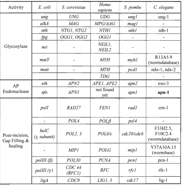
Table 1-1: Homologous BER Genes in Different Organisms
Sequences were retrieved from gene databases (i.e. NCBI, wormbase, etc). http://www .nc bi.nlm.nih. gOY /
http://www. wormbase.org/
1.3. BER in Mammalian Cells
The completion of the BER pathway in mammalian cells, following AP site processing, can either diverge into short patch repair or long patch repair (as shown in Fig 1-3) [28]. As a result, the short patch repair path is characterized by the replacement of a single nucleotide while its counterpart, the long patch repair system, is distinguished by the replacement of 2-13 nucleotides [29].
1.3.1 Short Patch Repair
The short patch mechanism can be completed following either AP endonuclease or AP lyase action upon the AP site. In essence, the 5' deoxyribose-phosphate terminus created by AP endonuclease is processed via the deoxyribophosphodiesterase (dRPase) component of polp (DNA polymerase P) [29]. Thereupon, the 3' -blocked end created by the bifunctional glycosylase (AP lyase activity) is removed through the 3' -diesterase component of AP endonuclease (also bifunctional). The single nucleotide gap, formed in both situations, is filled in by polp and the resulting nick is ligated via DNA Ligase 1 or the concerted action ofDNA ligase III and XRCCI [27]
1.3.2. Long Patch Repair
ln contrast to short patch repair, the long patch mechanism is only initiated following the action of monofunctional glycosylase and AP endonuclease. Long patch repair, in fact, has been usually associated with the processing of oxidized or reduced bases [30]. Hence, the 3' -OH group that results from AP endonuclease is extended via polymerase beta or delta/epsilon (polP or Blf..), and the joint action of proliferating cell nuc1ear antigen (PCNA) and replication factor C (RFC) [27]. The synergistic action of
these enzymes allows strand displacement to replace the missing nucleotides and forms an extensive flap structure (Fig 1-3). As a result, the formation of this structure primes the entry of flap endonuclease (FENI) which acts to cleave the flap [30]. FEN1 action causes a strand break which is in tum sealed by DNA ligase 1 (LIG1) or a complex of DNA ligase III and x-ray cross-species complementing ] (XRCC 1) [31]. Evidently, the long patch mechanism is differentiated from its counterpart (short patch repair) by the fact that it requires more wide-ranging machinery [27, 32].
1.4. BER in Saccharomyces cerevisiae
Although the components of the BER pathway are fairly conserved, there are notable divergences that should be accounted in S. cerevisiae. In fact, contrary to what is observed in mammalian cells, pol~ homologues in S. cerevisiae have not been clearly documented in the literature. However, pol~-like nucleotidyltransferase domains are present in several yeast proteins (PoI4, Trf4, Trf5) [33]. It has been discovered that homologues of long patch repair proteins in mammalian cells such as PoIs (DNA polymerase s) (DNA repair synthesis) and Fenl (5'-flap endonuclease) are found in yeast as Pol2 and Rad27 respectively [10]. These findings indicate the presence of the long patch base excision repair pathway in yeast [33]. Recently, the discovery of Trf4 in S. cerevisiae, a potential homologue of pol~ in mammals, possessing the 5' dRP lyase activity suggests that the short patch repair pathway may also co-exist to complement long patch repair in yeast [33].
In addition, coordination between the various proteins in yeast and mammals also appears to differ. This phenomenon can be illustrated, for example, via Ape land its reported direct interactions with the BER proteins, UDG and MYH (glycosyslases)
[34]. The human AP endonuclease (Apel) has also been revealed to play a role in regulating the function of several enzymes participating in the latter stages of the pathway; thus, these include FEN1, pol~, RPA, XRCC, PCNA and LIG1[34]. Studies have weIl exposed the complexities encompassing the Ape 1 prote in which go beyond its weIl characterized role in DNA repair. Thus, the mammalian protein has shown to be activated by thioredoxin which in turn, enables it to act as a redox factor. The redox component of Apel allows the protein to activate transcription factors such as activator protein 1 (AP-l) and nuclear factor KB (NF -KB) by maintaining specific cysteine residues in a reduced state [35]. Interestingly, Ape! has also been reported to be a strong activator of p53. These distinctive features associated with the mammalian AP endonuclease have not been clearly shown to exist in the corresponding yeast homologous proteins. It should be noted, nonetheless, that the 3-phosphodiesterase and
3'-75' exonuclease activities of Apn2 have been revealed to be enhanced in the presence of PCNA [36]. Undoubtedly, differences associated between the yeast and mammalian homologues, become evident in the differing range of activities that they are composed of. Notably, Apn2, unlike its mammalian homologue Ape!, is not endowed with the ability to incise altered bases in a glycosylase independent manner (Nucleotide Incision Repair) [37].
1.5. BER in Schizosaccharomyces pombe
Fission yeast distinguishes itself from its evolutionary counterpart, budding yeast, in the processing of AP sites. Most notably, the classical AP endonuclease members, Apnl and Apn2, have not been reported to being efficient in countering such lesions in fission yeast [38, 39]. In fact, it was found that the AP lyase activity of Nth
addresses this situation by producing blocked 3' -(l,~ unsaturated aldehydes [40]. The resulting 3'blocked ends finally promote the entry of Apn2 (major AP endonuclease) which repairs the blocked end via 3' -phosphodiesterase activity. It should be noted that
Apnl and Uvel (minor AP endonuclease) act as a 'back-up' to Apn2 [41, 42]. Subsequent downstream events follow the traditional process of nucleotide gap filling and re-establishment of the phosphodiester bond [41].
1.6. BER Key Machinery 1.6.1 Glycosylases
As described earlier, glycosylases initiate the BER pathway by removing the modified base via cleavage of the N-glycosylic bond (linking base to sugar phosphate moiety). Glycosylases are known to be small monomeric proteins that, in general, function without the necessity of a particular cofactor [26]. The glycosylase farnily of enzymes is divided into two groups: monofunctional and bifunctional [10, 27].
1.6.2 Monofunctional Glycosylases
MonofunctÎonal glycosylases are known to possess a single activity, which allows for the cleavage of the modified base. UDG, from E. coli, is reported to be the first DNA glycosylase to be characterized [26, 43]. Evidently, corresponding homologues can be found in S.
cerevisiae
and humans. The enzyme in question is fairly restricted in its specificity; thus, it has a strong ability to recognize uracil bases and closely related products (5-hydroxy-uracil) on DNA that may be formed as a result of the deamination of cytosine or misincorporation. [ 44].Another example of a weIl characterized monofunctional family is the Alkylbase DNA glycosylases, which excise alkylated bases resulting from both exogenous and endogenous sources [45]. These enzymes are known to be of wider specificity than its UDG counterparts. Members have been found in E. coli (AlkA), S. cerevisiae (MAG) and in human (MPG) [46-49]
1.6.3. Bifunctional Glycosylases
Bifunctional glycosylases possess an additional AP lyase activity (Figure 1-4) that is responsible for the hydrolysis of the 3' phosphodiester bond of the AP site by a
!3
or
!3b
elimination process generating a 3'a ~ unsaturated aldehyde [50]. This particular group of enzymes could be further divided on their ability to remove either oxidized pyrimidines or oxidized purines respectively [51l The former sub-group (excising pyrimidine bases) is represented by the Endo III-like glycosylases and could be found inE. coli (endo III or nth) , S. cerevisiae (Ntgl, Ntg2), S. pombe (Nth), and H sapiens (hEndoIII) [26].
An additional bifunctional glycosylase known as endo VIII has also been documented in bacteria. Thus, despite being functionally overlapping, endo VIII shares no sequence similarities with endo III. However, sequence similarities are observed when comparing endo VIII to Fpg [26, 52]. The latter sub-group, known as the Fpg family, characterized by the ability to excise oxidized purines, also has homologues in
E. coli (Fpg), S. cerevisiae (Oggl) and hum an (a-hOggl and ~-hOggl). Fpg has been demonstrated to excise fapy-ade, fapy-gua and 8-oxoG (refer to Fig 1-1 for representation of lesions) [53]. Interestingly, despite having common substrate
specificity, the eukaryotic Ogg 1 is not closely structurally related to the bacterial Fpg [54] [26]
1.6.4. AP Endonuclease Action in the Processing of AP Sites
AP sites are known to be one of the most common spontaneous lesions that occur in DNA. 10 000 bases a day are 10st per mammalian cell [10]. In addition to spontaneous circumstances, the lesion is also formed by the enzymatic removal of a base which may have been damaged or misincorporated. Undoubtedly, AP sites are highly mutagenic and cytotoxic if not properly repaired [5]. In essence, the presence of AP sites can promote the blocking of transcription and replication [2].
+
Figure 1-4: AP Site and Resulting Repair (adapted from [55])
Diagram illustrating an AP site (represented by star notation) and the immediate processing which occurs to counter the lesion; thus, this may
be achieved Via a 5' cleavage stemming from an AP endonuclease
activity or through a 3' cleavage provided by an AP lyase activity.
Repair of chromosomal AP sites in bacteria has been indicated to be performed by BER and to a smaller degree by a series of additional mechanisms: NER, translesion DNA synthesis and recombination [56]. It must be stressed that the BER pathway
serves as the most predominant and effective route for processing AP sites [10]. The key enzymes in countering AP sites are AP endonucleases. This particular group of enzymes, as mentioned earlier, is multifunctional; thus, it is composed of AP endonuclease and 3' -phosphodiesterase components [57]. Specifically, the AP endonuclease activity is responsible for the nicking of the sugar phosphate bond at the 5' end of the AP site, producing 3' -hydroxyl group and 5' -deoxyribose phosphate. The 3' -phosphodiesterase component, in contrast, is responsible for countering 3' blocked ends produced by free radicals. It may equally come into play to counter the end
products of lyase activity (3'
-ap
unsaturated aldehyde) to subsequently enable the formation of the polymerase responsive 3' -OH [4, 58, 59].AP Endonucleases have been traditionally categorized into two families, the Exo III and the Endo IV, which are structurally unrelated but generally perform the same function. The classic elements differentiating both families stem from the fact that Exo III homologues are Mg2+ dependent enzymes, readily inactivated via the addition of EDTA while, conversely, the Endo IV family is characterized by being Mg2+ independent [60]. Exo III members have been found in E. coli, S. cerevisiae, S. pombe, drosophila, C. elegans, mouse and human. The corresponding Exo III members in
drosophila, mouse and human are thus far the sole AP endonuclease type found in these
[55]. It should be emphasized that despite being associated in large part with the same
function, both families of AP endonucleases have subtle variations in their effectiveness to counter certain les ions and differ in their overall activity within a given organism (highlighted in the next section) [61]. In particular, Endo IV enzymes, unlike the Exo III family, have an ability to repair lesions induced by specific oxidants (Bleomycin and tBR) [55, 61]. As a result, although not yet found, an Endo IV member in mammalian cells may exist due to its efficiency to process the se specific lesions refractory to Exo III. The existence of a mammalian Endo IV member may stern from a functional homology as oppose to a strictly structural one. Thereupon, examples of this type of relationship may be illustrated when investigating Fpg in bacteria versus Oggl in yeast. Although both enzymes in the corresponding organisms are not closely related in terms of structure, they do, in fact, share functional homology [26]. This thesis will focus primarily on the Endo IV family.
1.7. Endo IV Family Members
1.7.1 Escherichia coli en do IV
Known to be a heat stable monomeric protein, endo IV is approximately 32 kDa
ln size [60],[62]. It is a multifunctional enzyme composed of, among others, weIl
established AP endonuclease and 3-diesterase activities [53]. Thus, endo IV can readily process DNA lesions consisting of phosphoglycoaldehyde, 3' -phosphate, 3'-deoxyrobose-5-phosphate and 3' -hydroxy-2-pentanal [2]. Interestingly, it has also been comprised of endonuclease activity against DNA composed of urea residues [63]. However, endo IV is considered to serve as a 'back up' or the minor AP endonuclease to exo III in E. coli. Data has, indeed, showed that endonuclease IV was responsible for
only 10% of AP endonuc1ease activity in a bacterial cell. NaturaIly, the remaining proportion is attributed to exonuc1ease III [61]. Its expression was shown to be strongly induced in the presence of superoxide anions. In fact, it was reported that paraquat, plumbagen, menadione and phenazine metho sulfate , aIl known to form superoxide radical anions, could induce endo IV by 10-20- fold and, thus, enable the protein to attain a similar level as that of its counterpart exo III [64]. Hence, this induction effect is under the regulation of an intricate system involving the SoxR and SoxS proteins. Elevated levels of superoxide anions are reported to stimulate SoxR to later induce the expression of the SoxS protein, which in turn causes an increase in endo IV expression. It should be noted that the SoxRJSoxS proteins stimulate, in addition to endo IV, a significant array of components capable in restoring potential damage caused by unstable radicals [65].
Studies have showed that endo IV is endowed with additional activities to go along with the traditionally recognized components highlighted above. Although initially not detected, it was discovered that this enzyme comprises of a 3'75' exonuc1ease activity acting predominantly towards 3' recessed ends [66]. In essence, it serves as a proofreading activity allowing the removal of matching/mismatching nuc1eotides and altered bases (80xoG) at c1eaved ends [67]. Contrary to AP endonuc1ease activity, the exonuc1ease component appeared to be readily inhibited in the presence of EDT A and reducing conditions (ev en in the presence of DN A substrate) [60,66].
Structural analyses have revealed the presence of a tri-nuc1ear ZInC c1uster
forming the active site which was demonstrated to play a crucial role in its biological activities. Two of the three zinc atoms are weIl buried and inaccessible to solvent. The
metal factors are coordinated to key amino acids which appear to be conserved across the Endo IV family [55, 68]. The presence of the zinc atoms was reminiscent of other similar c1usters involved in phosphodiester c1eavage. In fact, Pl nuc1ease in Penicillum
citrinum and phospholipase C in Bacil/us cereus have revealed an ability to exploit the
corresponding zinc c1uster to achieve hydrolysis of the phosphate bonds [68]. Structural studies involving endo IV have also disc10sed the existence of a TIM (triosephosphateisomerase) barrel fold which was identified to play a significant role in DNA binding and the recognition of AP sites. The TIM barrel has been characterized by a parallel configuration of eight ~ strands surrounded by eight
a
helices. Thedynamics involved in catalytic action are thought to arise from the enzyme to bend DNA 90° and in the process, flip the abasic (AP) site [69]. The underlying structural features of the bacterial AP endonuc1ease parallels many characteristics observed with UVDE (UV damage endonuc1ease) [70]. Interestingly, UVDE plays a significant role in DNA repair by its ability to process UV -induced lesions but also acts as an AP endonuc1ease in certain organisms (N crassa, S. pambe) [70]
Phenotype characterization has shown that a mutant lacking
nia
(gene encoding endo IV) is hypersensitive to the antibiotic Bleomycin (BLM). This particular anti tumor drug is an established DNA damaging agent which has been displayed to produce a series of single strand breaks forming 3' blocked ends (i.e. 3' phosphoglycolate DNA terminus), double strand breaks and the release of free bases from the sugar phosphate moiety [71-73]. Cunningham et al (1986) reported that the single mutant showed aslight increase in sensitivity to methyl methanesulfonate (MMS), an agent readily able to alkylate DNA bases (i.e. N3 position of adenine or N7 of guanine) and indirectly cause the formation of AP sites [5, 61, 74]. The partial sensitivity may be indicative of
the fact that exo III was still being expressed and, furthermore, en do IV is not the major
AP endonuclease in bacteria. Interestingly, the strain was generally unaffected, in comparison to the wild type, to hydrogen peroxide, a potent oxidant known to cause single strand breaks with 3' phosphate blocked ends. The sum of these results contrasts to the situation where a single mutant lacking xth (gene encoding major bacterial AP endonuclease exonuclease III), in fact, displayed greater sensitivity towards MMS, hydrogen peroxide, near UV light and X-rays. However, sensitivity of the xth mutant was less dramatic in the presence of the oxidizing agent tert-Butyl hydroperoxide (tBH) and, furthermore, not reflected at aIl during BLM treatment [61]. The discrepancy in survival between both strains following BLM and tert-Butyl hydroperoxide (tBH) exposure illustrates that endo IV and exo III are not products of redundancy.
Accordingly, a double mutant lacking nfo/xth demonstrated a synergistic susceptibility while under presence of alkylating agents (MMS) and oxidative agents (H202, BLM, tBH) [61]. As expected from the reported data, the overexpression of en do IV re-established drug resistance (BLM, tBH) in an xth single mutant bacterial strain. Conversely, the overexpression of exo III in a strain void of an Endo IV member has yet to demonstrate a similar consequence in the presence of identical agents [75]. Undoubtedly, endo IV is endowed with greater versatility in countering specific types of lesions (revealed by BLM and tBH exposure) that exo III is unable to match.
Additional studies have shed significant light to further the divergences that exist between endo IV and its exo III counterpart. For example, endo IV, unlike exo III, has been associated to a newly characterized mechanism complementing BER now known as Nucleotide Incision Repair (NIR). This pathway is differentiated by the fact that it is a glycosylase independent process, whereby the protein in question incises the
altered base and the deoxyribose component to subsequently promote eventual DNA synthesis. The NIR activity of endo IV allows the enzyme to process a diverse array of les ions stemming from oxidative stress: dihydro-2'-deoxyuridine (DHU), 5,6-dihydrothymidine (DHT), 5-hydroxy-2' -deoxyuridine, 2 ,6-diamino-4-hydroxy-5-N-methylformamidopyrimidine and a-anomeric 2' -deoxynuc1eotides (adA and adT) [67]. Moreover, en do IV has also been associated with providing a protective effect to circumvent potential oxidative damage caused by UVB [76]. Unlike its counterpart exo III, endo IV is unable to directly repair the lesions caused by UVB. It should be noted
that oxidative damage formed as a result of UVB irradiation is believed to be associated with AT to GC transitions and GC to CG transversions [76].
1.7.2. Saccharomyces cerevisiae Apnl (ScApnl)
ScApnl has been reported to be the major AP endonuc1ease enzyme in S.
cerevisiae. Data has revealed that it accounts for 97% of the AP endonuc1ease and 3'-diesterase activity in the corresponding eukaryotic cell. The 42 kDa protein shares 41 % amino acid identity with the bacterial endo IV and composed of a similar zinc c1uster (3.3 atoms) found in the active site [77]. It should be stressed that like the bacterial
endonuc1ease IV, ScApnl does not necessitate an additional metal co-factor for its reported activities. Moreover, ScApnl is able to act on a similar range of substrates as its counterpart in bacteria; thus, these inc1ude 3' -phosphoglycoaldehyde, 3' -phosphoryl groups and 3' -a,p-unsaturated aldehydes [2]. However, unlike en do IV, ScApnl is endowed with a longer C-terminal end which is in fact shown to be composed of 3 important c1usters (c1uster 1, 2, and 3). These series of domains, rich in basic amino acids, have been elucidated to serve as an NLS. Cluster 1, in fact, was reported to being
essential for nuclear localization. Thus, the removal or replacement of cluster 1 may not affect enzyme activity in-vitro; however, it abolished the localization process to the nucleus. The substitution of cluster 1 with the NLS of SV40 (simian virus 40) did not appear to restore proper nuclear targeting. Furthermore, it was observed that neither cluster 1 or 2, on its own, was able to aIlow ScApnl to be signaled to the nucleus and moreover, the concurrent removal of both produced the same effect. Thus, it was postulated that cluster 1 and 2 behave as a bipartite NLS [78]. Experiments have shown that the bipartite cluster also plays a role in targeting the protein to the mitochondrion. In fact, this phenomenon appears to be mediated via an interaction with the protein Pirl [79]. In the absence of Pirl, ScApnl accumulates in the nucleus and cytoplasm. In contrast, deletion of a significant portion of the C-terminal domain contributes to subsequent accretion of the prote in in the cytoplasm only. [79].
Classical phenotypic analysis has revealed that ScApnl single mutants are hypersensitive to MMS (alkylating agent) as weIl as to the oxidants tBH and hydrogen peroxide. As expected, DNA coIlected foIlowing treatment with the particular alkylants and oxidants has showed a striking accumulation of DNA lesions. Spontaneous mutation rates of Apn 1 null mutants increased from 6 to 12 fold in comparison to the wild type [80]. Moreover, mutants have been correlated with a 59 foid increase in A.T to C. G transversion events [81]. The sum of these results displays the importance of Apnl in protecting the genome against a wide array of lesions. Nonetheless, the single Apnl mutant failed to show sensitivity following Bleomycin and Phleomycin treatment; thus, this piece of data contributed to the suggestion of a possible existence of an additional 3' -diesterase in Saccharomyces cerevisiae [80, 82]. Hence, Apn2 (Exo III family) was subsequently discovered and demonstrated to possess the same bifunctional
nature as its Endo IV counterpart. However, it should be noted that Apn2 remains the minor AP endonuclease in yeast [5]. As such, this conclusion rests on the evidence that an apn2 single mutant fails to demonstrate a dramatic hypersensitivity towards the same agents (MMS, H202) as was observed with the apnl mutant. The increased mutagenesis
associated with single apnl null mutants reveal that Apn2 lacks the ability to optimally substitute for its AP endonuclease counterpart [5]. Double mutants void of both APNl and APN2 did, however, reveal an extensive sensitivity to MMS, H202 and Phleomycin
that transcends what is observed with the respective single mutants [82] [5].
Recent investigations concerning Apnl have found that akin to its bacterial homologue, it also is composed of a 3'-75' exonuclease component which complements the well established AP Endonuclease/3-diesterase activities. Thus, this newly associated function is characterized by a strong preference for duplex DNA with 3' recessed ends [67]. The exonuclease activity has been, interestingly, showed to permit the elimination of 80xoG lesions (formed at the 3' end of gap) resulting from the direct oxidation of guanine or a misincorporation by DNA polymerase. The ability of Apnl to process 80xoG residues prevents G.C to T.A transversions to become prevalent in oggl single mutants. As a result, it is believed that the Endo IV member in
Saccharomyces cerevisiae may act as an alternative pathway to Oggl in the removal of 80xoG from DNA. Apnl has also shown to reflect its bacterial homologue in its ability to take part in NIR and process a-anomeric 2-deoxynucleotides (adA and adT) and other oxidative lesions [67]
ScApnl has recently garnered important attention in its potential benefit to humans. Thus, the expression of Apnl has showed an ability to counter damage instigated by ROS in the development of certain neurodegenerative diseases [83]. In
fact, the production of Apnl in mammalian ceUs revealed a decrease in sensitivity following oxidative stress [83].
1.7.3 Schizosaccharomyces pombe Apnl (SpApnl)
Contrary to the situation in 8 cerevisiae, SpApnl is not considered the major AP endonuclease in S. pombe; a title which is rather held by Apn2. Contrary to the apn2 mutant, data has revealed that the apnl mutant failed to demonstrate sensitivity to the alkylating agent MMS [39]. An additional minor AP endonuclease activity is found in 8 pombe stemming from the Uvel enzyme, a previously established component of an alternate UV damage repair pathway to NER [42]. It has been reported by Fraser at al.
that spontaneous mutations which occur in uvel null cells (8 pombe) are similar to the
transversion mutations induced by 80xoG in S. cerevisiae. This evidence first suggested the role of Uvel in oxidative DNA damage repair in fission yeast [42]. However, SpApnl and SpUvel proteins were reported to possess AP endonuclease activity
in-vitro but failed to initiate AP site repair in-vivo [41]. As discussed earlier (section 1.6), the direct processing of AP sites in 8 pombe has been predominantly reported to be the domain of the Nth enzyme (bifunctional glycosylase) via its AP lyase activity (producing 3'C1.,~-unsaturated aldehyde ends) and not a member of either the Exo III or
Endo IV families. The implication of such proteins, belonging to the respective AP endonuclease families, has thus far been restricted to the repair of 3' blocked ends (processed predominantly by Apn2, with Apnl and Uvel serving as back up components) induced in large part by Nth [41]. Undoubtedly, there are profound differences which exist between 8 pombe and 8 cerevisiae in the identity of the machinery having access to the lesions in question. These divergences are further
revealed through the expression and resulting complementation of ScApnl in the repair of both AP sites and blocked ends in mutant fission yeast strains (nthJ!j and apn2J!j
single mutants) when exposed to MMS [41]. Interestingly, localization studies using GFP have revealed that SpApnl is distributed in the nucleus and, in addition, the cytoplasm (not observed with SpApn2); thus, suggesting that it may possibly play additional roles than simply being involved in maintaining genomic stability [41].
1.7.4. Caenorhabditis elegans CeAPN-l
The gene encoding the C. elegans Endo IV AP endonuclease, CeAPN-I, was
first identified following the utilization of a radiolabelled oligonucleotide (representing the sequence of yeast APN1) which was probed against a cDNA library of the multicellular organism [84]. The expressed prote in was initially reported to be composed of 396 amino acids but subsequent findings have reported that it is ev en larger (discussed below). Nonetheless, CeAPN-l shares considerable homology with Endo IV members found in the unicellular organisms highlighted. Hence, it shares 40.4% identity with the yeast APNl and 44.9% identity with the bacterial nlo [84].
Much of the homology is concentrated in 5 distinct domains, thought to represent, notably, portions of the DNA binding domain and active site. Interestingly, C. elegans
is the first higher eukaryote, representing an animal model, which has been reported to have both Endo IV (CeAPN-l) and Exo III (CeEXO-3) homologues. The latter member (CeEXO-3) has been weB characterized to being Mg2+ dependent and endowed with AP endonuclease/3' diesterase activities. However, it has also been found that the metalloenzyme is composed of a number of unique features which distinguishes itself from its Exo III counterparts; notably, a GST fusion CeEXO-3 protein failed to display
an important 3' -7 5' exonuclease activity following AP sites (opposite observed with bacterial and yeast homologues) and furthermore, it revealed to lack the ability to participate in Nucleotide Incision Repair (as observed with human Exo III, Apel) [85]. However, contrary to CeEXO-3, very little is known about CeAPN-l. Numerous attempts have been made to express and purify the full length protein for characterization; however, these past efforts have not been as fruitful [55].
1.8. Research Project
1.8.1. Background and Objectives
Attempts have been made in developing an expression system in E. coli to allow for the isolation of CeAPN-l. This was performed by constructing a plasmid containing the Ceapn-l gene under the control of an isopropyl P-D-l-thiogalactopyranoside (IPTG) inducible promoter. Unfortunately, the expressed product detected via immunoblot appeared to be smaller (10 kDa) than the expected size. Hence, a possible proteolytic process in bacteria may have been hindering the detection of the fulliength prote in [55]. Due to the inability to detect the full length CeAPN-l in bacteria and the fact that a sufficient yield (for characterization experiments) could not be obtained via C. elegans, an expression system was engineered in yeast [86]. This particular eukaryotic model has multiple advantages stemming from the fact that it is easy to manipulate and grow. Moreover, it should be recognized that a significant benefit arises from the fact that a mutant strain void of AP endonuclease, YW778 (tppl 1'1: :METl5 apnl A: :HIS3
apn2A::KanMX4), was readily available. Through the subsequent transformation ofthis strain, one could easily ascertain whether the introduction and expression of Ceapn-l can functionally complement in the presence ofDNA damaging agents [86].
It was shown that the yeast plasmid containing the Ceapn-1 gene, pNLS-OAD-Ceapn-J (recovered from a C. elegans cDNA library), was able to substitute for yApn1 in the presence of MMS when introduced in the YW778 strain. As a result, a new construct was designed in order to equip the expressed product with a OST affinity tag for purification purposes [86]. Unfortunately, unlike the previous attempt, the introduction of pOST -Ceapn-1 failed to restore MMS resistance to the AP endonuclease deficient strain. An identical approach exploiting a OST tagged Ceexo-3 construct introduced into YW778 (tppl tl..::METl5 apnll1::HIS3 apn211::KanMX4) (void of AP endonuclease activity) did, however, display an ab il it y to complement in the presence of
MMS. Thus, it was hypothesized, at the time, that the failure of OST -Ceapn-l to restore resistance was likely due to the OST tag that may have been hindering the activity domain of the protein [86]. It was, however, later discovered (during the course of the
CUITent project) that the Ceapn-l sequence was more extensive than originally believed to be. Updated and better annotated sequences submitted to gene databases revealed that Ceapn-1 is composed of an additional 118 amino acids at the N-terminal which was not previously accounted for. Upon close inspection, the additional residues have been pointed out to being highly rich in basic residues (characteristic of NLS) which may be indicative that it may play a role in protein localization [87].
As a result, this project set out firstly to develop an effective expression system for CeAPN-I, secondly, to isolate a homogeneous fraction of the prote in and, thirdly, characterize its precise biological function in an in-vitro setting. The goal in characterizing CeAPN-I rests primarily on determining the types of les ions (AP sites, 3' -blocked ends, oxidized bases) it can process, evaluate its range of activities, assess whether additional co-factors are needed for its function and to differentiate it from its
![Figure 1-1: Examples of Lesions Stemming from Altered Bases (adapted from [2,3])](https://thumb-eu.123doks.com/thumbv2/123doknet/2080145.7033/19.919.179.794.102.954/figure-examples-lesions-stemming-altered-bases-adapted.webp)
![Figure 1-2: Different Forms of AP Sites (adàpted from [3])](https://thumb-eu.123doks.com/thumbv2/123doknet/2080145.7033/20.919.211.751.348.648/figure-different-forms-ap-sites-adapted.webp)
![Figure 1-4: AP Site and Resulting Repair (adapted from [55])](https://thumb-eu.123doks.com/thumbv2/123doknet/2080145.7033/35.921.194.790.524.977/figure-ap-site-resulting-repair-adapted.webp)