RNA Sequencing-Based Genome
Reannotation of the Dermatophyte
Arthroderma benhamiae and
Characterization of Its Secretome and
Whole Gene Expression Profile during
Infection
Van Du T. Tran,aNiccolò De Coi,bMarc Feuermann,cEmanuel Schmid-Siegert,a
Elena-Tatiana Ba˘gut¸,dBernard Mignon,dPatrice Waridel,eCorinne Peter,f
Sylvain Pradervand,fMarco Pagni,aMichel Monodb
Vital-IT group, SIB Swiss Institute of Bioinformatics, Lausanne, Switzerlanda; Department of Dermatology, Centre Hospitalier Universitaire Vaudois, Lausanne, Switzerlandb; Swiss-Prot Group, SIB Swiss Institute of Bioinformatics, Geneva, Switzerlandc; Fundamental and Applied Research for Animals & Health (FARAH), Department of Infectious and Parasitic Diseases, Faculty of Veterinary Medicine, University of Liège, Liège, Belgiumd; Protein Analysis Facility, Center for Integrative Genomics, University of Lausanne, Lausanne, Switzerlande; Genomic Technologies Facility, Center for Integrative Genomics, University of Lausanne, Lausanne, Switzerlandf
ABSTRACT Dermatophytes are the most common agents of superficial mycoses in humans and animals. The aim of the present investigation was to systematically identify the extracellular, possibly secreted, proteins that are putative virulence fac-tors and antigenic molecules of dermatophytes. A complete gene expression profile of Arthroderma benhamiae was obtained during infection of its natural host (guinea pig) using RNA sequencing (RNA-seq) technology. This profile was completed with those of the fungus cultivated in vitro in two media containing either keratin or soy meal protein as the sole source of nitrogen and in Sabouraud medium. More than 60% of transcripts deduced from RNA-seq data differ from those previously depos-ited for A. benhamiae. Using these RNA-seq data along with an automatic gene an-notation procedure, followed by manual curation, we produced a new anan-notation of the A. benhamiae genome. This annotation comprised 7,405 coding sequences (CDSs), among which only 2,662 were identical to the currently available annotation, 383 were newly identified, and 15 secreted proteins were manually corrected. The expression profile of genes encoding proteins with a signal peptide in infected guinea pigs was found to be very different from that during in vitro growth when using keratin as the substrate. Especially, the sets of the 12 most highly expressed genes encoding proteases with a signal sequence had only the putative vacuolar as-partic protease gene PEP2 in common, during infection and in keratin medium. The most upregulated gene encoding a secreted protease during infection was that en-coding subtilisin SUB6, which is a known major allergen in the related dermatophyte
Trichophyton rubrum.
IMPORTANCE Dermatophytoses (ringworm, jock itch, athlete’s foot, and nail in-fections) are the most common fungal infections, but their virulence mechanisms are poorly understood. Combining transcriptomic data obtained from growth under various culture conditions with data obtained during infection led to a significantly improved genome annotation. About 65% of the protein-encoding genes predicted with our protocol did not match the existing annotation for A. benhamiae. Compar-ing gene expression durCompar-ing infection on guinea pigs with keratin degradation in
Received 23 March 2016 Accepted 27 June
2016 Published 2 August 2016
Citation Tran VDT, De Coi N, Feuermann M,
Schmid-Siegert E, Ba˘gut¸ E-T, Mignon B, Waridel P, Peter C, Pradervand S, Pagni M, Monod M. 2016. RNA sequencing-based genome reannotation of the dermatophyte
Arthroderma benhamiae and characterization of its secretome and whole gene expression profile during infection. mSystems 1(4):00036-16. doi:10.1128/mSystems.00036-16.
Editor Barbara Methe, University of Pittsburgh Copyright © 2016 Tran et al. This is an
open-access article distributed under the terms of theCreative Commons Attribution 4.0 International license.
Address correspondence to Marco Pagni, Marco.Pagni@isb-sib.ch, or Michel Monod, Michel.Monod@chuv.ch.
V.D.T.T. and N.D.C. contributed equally to this article, and M.P. and M.M. are joint senior authors of this work.
Host-Microbe Biology
vitro, which is supposed to mimic the host environment, revealed the critical
impor-tance of using real in vivo conditions for investigating virulence mechanisms. The analysis of genes expressed in vivo, encoding cell surface and secreted proteins, par-ticularly proteases, led to the identification of new allergen and virulence factor can-didates.
KEYWORDS: Arthroderma benhamiae, RNA-seq, Trichophyton, annotation,
dermatophytes, infection, proteases, secreted proteins
P
athogenic dermatophytes are the most common agents of superficial mycoses, almost exclusively infecting the stratum corneum, nails, and hair (1, 2). The ge-nomes of these fungi, smaller than those of Aspergillus spp., range from 22.5 to 24 Mb and are highly collinear. The number of predicted protein-encoding genes varies from 7,980 in Arthroderma benhamiae to 8,915 in Microsporum canis (3, 4). A large number of orthologs were found to be shared by all dermatophytes (6,158 groups, including paralog duplications) (4). Dermatophyte genomes were found to be enriched in genes encoding secreted proteases and depleted in genes encoding enzymes involved in sugar metabolism, as for example those typically involved in plant cell wall breakdown. These differences from other fungi attest to the high specialization of dermatophytes and their adaptation to particular proteinaceous substrates other than vegetal debris. The molecular mechanisms involved in the establishment of dermatophyte infec-tions are poorly understood and remain an open field of investigation. Host-fungus interactions involve pathogen offense, host defense, and pathogen counterattack. In these processes, fungal and host cell-associated and secreted proteins play a major role. For instance, secreted aspartic proteases are now considered important virulence factors of Candida albicans, being associated with adhesion, invasion, and tissue damage (5). Secreted enzymes referred to as “effectors” are also of major importance for host attack by plant pathogens (6). Likewise, proteins secreted in vivo, in particular proteases, are clearly the best candidates for virulence factors of dermatophytes.Current knowledge regarding dermatophyte gene expression during infection was acquired using a cDNA microarray based on transcripts of A. benhamiae, grown in a protein medium, covering approximately 20 to 25% of its genome and on a few selected protease-encoding genes (7). As a striking result, the genes encoding most major proteases secreted by the fungus in vitro (8–11) were found to be not expressed
in vivo, and therefore, these proteases appeared not to be involved during the
establishment of infection. In contrast, the gene encoding the subtilisin SUB6 was found to be highly expressed during skin infection but not when the fungus grew in any culture medium. Of particular importance, SUB6 is the ortholog of the major allergen Tri r2 in Trichophyton rubrum (12). Tri r2 was found to induce dual immune responses and elicit either immediate or delayed-type hypersensitivity skin test reac-tions in different individuals. Numerous antigenic molecules eliciting host immune responses still remain to be discovered. In view of the importance of secreted proteins, both as antigens and as possible virulence factors, the goals of this work were the following: (i) to obtain a complete gene expression profile of A. benhamiae during infection using state-of-the-art RNA sequencing (RNA-seq) technology, (ii) to compare it with the expression profiles of the fungus grown in vitro in different media, and (iii) to identify which proteins, and in particular individual proteases, are secreted in vivo during infection as possible new virulence factors. By exploiting RNA-seq data for
A. benhamiae growing under different culture conditions and during infection in guinea
pigs, we first established a new annotation of the genome, with 7,405 protein-encoding genes. The previously available genome annotation of A. benhamiae showed its limits, as many discrepancies were found after comparison with new experimental data.
RESULTS
Arthroderma benhamiae experimental infections in guinea pigs. Skin samples from
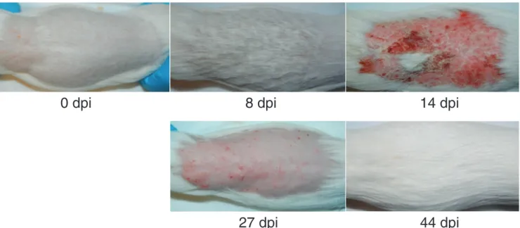
during infection. As shown in Fig. 1, at day 8 after infection, the animals showed no or minimal skin symptoms. The direct mycological examination showed numerous fila-ments present on the hair and skin samples with the presence of a low number of conidia (data not shown). At 14 days, the guinea pigs exhibited macroscopic skin lesions, but direct mycological examination showed fewer fungal filaments on the infected skin samples with thicker septa than at 8 days. We considered day 8 as the time point for the peak of infection and day 14 as the time point for the peak of inflam-mation. After 27 days, the skin lesions were still present but regressing, while very few fungal elements were observed by direct mycological examination. At day 44, the guinea pigs had fully recovered from infection, and no A. benhamiae filaments were observable. At this time, three animals that had recovered from primary infection were reinfected by A. benhamiae but did not develop a new infection.
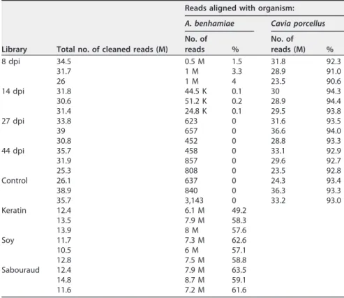
RNA sequencing.RNA was extracted in triplicate from the fungus grown in keratin medium, soy protein medium, and Sabouraud medium and from each infected animal. Approximately 13 million strand-specific reads were obtained for each RNA sample extracted from the fungus growing in the three tested culture media (Table 1). Approximately 30 million strand-specific reads were acquired from each RNA sample extracted from infected skin samples, consisting of a mixture of reads from the fungus and from its mammalian host. As a result, roughly 1 million fungal reads (2.8%) were obtained with RNA extracted from skin samples of guinea pigs at day 8 of infection, while 91.3% of the reads could be aligned with the guinea pig genome.
New gene annotation of the Arthroderma benhamiae genome.A preliminary
investigation of the RNA-seq reads mapped onto the A. benhamiae genome revealed that many gene and intron locations from the original genome annotations were not supported by our experimental data. Hence, reannotating the coding sequence (CDS) of the genome appeared to be a prerequisite before further analyzing the transcrip-tome expression. Particular attention was paid to the location of the start codons because of our interest in secreted proteins, which should be endowed with a signal peptide at the N terminus.
We used Augustus (13), a program for gene prediction in eukaryotic organisms that relies on a statistical model of an organism’s gene structure. The correctness of Augustus predictions is, however, highly dependent on this model, and great care must be used at the time of training this model (i.e., establishing the model using a training data set). Practically, we mapped all RNA-seq reads onto the genome, deduced
full-0 dpi
8 dpi
14 dpi
27 dpi
44 dpi
FIG 1 Experimental infection of the natural host of Arthroderma benhamiae. Cutaneously infected guinea pigs developed skin symptoms that were the most severe at 14 days postinfection (dpi) due to inflammation, while 8 dpi was the time point for the peak of infection.
length gene transcripts, and retained only those with sufficient coverage. Then, we translated the filtered transcripts into their three possible coding frames. Full-length CDSs were detected by aligning the transcripts against a set of high-quality protein sequences, namely, the protein sequences reviewed by Swiss-Prot of the model organ-isms Saccharomyces cerevisiae and Aspergillus nidulans. The CDS annotations were back-propagated onto the genome, introducing intron descriptions, and supplied as a training set to Augustus to generate a new gene model. With the latter, the A.
benha-miae genome was reannotated and yielded 7,405 protein-encoding genes.
Table 2 compares our 7,405 newly predicted genes with the original set of 7,979 and shows that about 65% of the genes have been affected one way or another: for example, the intron boundaries within 1,246 genes were corrected and 383 new genes
TABLE 1 RNA-seq data summarya
Library Total no. of cleaned reads (M)
Reads aligned with organism: A. benhamiae Cavia porcellus No. of reads % No. of reads (M) % 8 dpi 34.5 0.5 M 1.5 31.8 92.3 31.7 1 M 3.3 28.9 91.0 26 1 M 4 23.5 90.6 14 dpi 31.8 44.5 K 0.1 30 94.3 30.6 51.2 K 0.2 28.9 94.4 31.4 24.8 K 0.1 29.5 93.8 27 dpi 33.8 623 0 31.6 93.5 39 657 0 36.6 94.0 30.8 452 0 28.8 93.3 44 dpi 35.7 458 0 33.1 92.9 31.9 857 0 29.6 92.7 25.3 808 0 23.5 92.8 Control 26.1 637 0 24.3 93.4 38.9 840 0 36.3 93.3 35.7 3,143 0 33.2 93.0 Keratin 12.4 6.1 M 49.2 13.5 7.9 M 58.3 13.9 8 M 57.6 Soy 11.7 7.3 M 62.6 10.5 6 M 57.1 12.8 7.5 M 58.8 Sabouraud 12.4 7.9 M 63.5 14.8 8.7 M 59.1 11.6 7.2 M 61.6
aM, million; K, thousand; dpi, days postinfection.
TABLE 2 Comparison of new gene set and original onea
New versus old gene prediction Gene count in complete genome
Gene count in secretome only
With GPI Without GPI
Auto Manual Auto Manual
Matched 2,662 47 (13) 2 (2) 155 (55) 0 Alternative 1,246 19 (6) 0 49 (19) 1 Different 2,752 31 (6) 1 83 (19) 10 (4) Merged 286 5 (2) 0 7 (2) 1 Split 76 1 (1) 0 5 (2) 0 New 383 6 0 34 (8) 0 Total 7,405 109 (28) 3 (2) 333 (105) 12 (4)
aMatched, identical old and new gene annotations; alternative, conserved start and stop codons but
different splicing; different, different start or stop codons, possibly different splicing; merged, more than one old gene merged into a single new one; split, old gene split into several new ones; new, genes found only in the new predictions (708 original genes were lost); auto, gene annotations as produced by Augustus; manual, manual correction of the start codon. The number of genes whose products were confirmed by mass spectrometry in culture supernatants is given in parentheses. GPI,
were recorded. In addition, 39 genes in the existing annotation were split into two genes, and, in contrast, 286 genes in the new annotation corresponded to fusions of previously annotated genes.
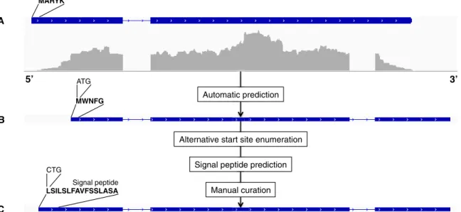
In silico definition of the secretome.We defined the secretome as the set of all secreted proteins, which is made of all proteins with a signal peptide, excluding transmembrane proteins. In practice, this set is not trivial to define. The presence/ absence of a signal peptide depends on the tools used to predict it, on the strength of the signal itself, and on its presence at the N terminus, which ultimately relies on the correct detection of the start codon. Hence, all genes predicted by Augustus were further subjected to prediction refinements as follows. For every predicted CDS, vari-ants were enumerated by considering every AUG or CUG (14) as an alternative start codon, when found within 30 amino acids from the AUG given by Augustus. Signal peptides were then searched for in all CDS variants. The retained CDS was finally selected manually by comparing the results of the different predictions and by con-sidering additional evidence, such as prior biological knowledge or the presence of a glycosylphosphatidylinositol (GPI) anchor at the C terminus. GPI anchors affect the localization of these proteins in the plasma membrane or the cell wall, but removal of the GPI lipid moiety by phospholipases can generate soluble secreted forms of the protein (15). The overall procedure of gene prediction followed by manual correction is summarized with an example in Fig. 2.
A total of 634 proteins with a signal peptide, including 112 probable GPI-anchored proteins, has been predicted. Using transmembrane predictors, we removed all pro-teins that contained one or more transmembrane spans in addition to the signal peptide and that were probably targeted to membranes. This refinement led to a final
A. benhamiae predicted secretome, made of 457 proteins that are listed and
charac-terized in Table S1 in the supplemental material.
A few A. benhamiae proteins have been experimentally characterized, in particular secreted proteases (16) and hydrophobin HypA (17). In order to associate functional information with predicted proteins, we searched for homologs using Blast against UniProtKB (18), paying particular attention to the matches against S. cerevisiae, the best-characterized fungus; C. albicans, the best-characterized yeast pathogen; and filamentous fungi, such as Aspergillus spp. We completed functional predictions by checking for the presence of specific domains or protein family signatures by scanning
A B C MWNFG MARYK Signal peptide LSILSLFAVFSSLASA Automatic prediction Manual curation 5’
Alternative start site enumeration Signal peptide prediction
3’
CTG ATG
FIG 2 Prediction and manual correction of the gene coding for the autophagy protein Atg27 (ARB_01857; a transmembrane protein). (A) Original gene prediction; (B) automatic prediction from Augustus (signal peptide is missing); (C) final (new) gene prediction after manual correction. The reannotation of this particular gene is remarkable, as it produced a new intron, an alternative stop codon, and a manually corrected start codon.
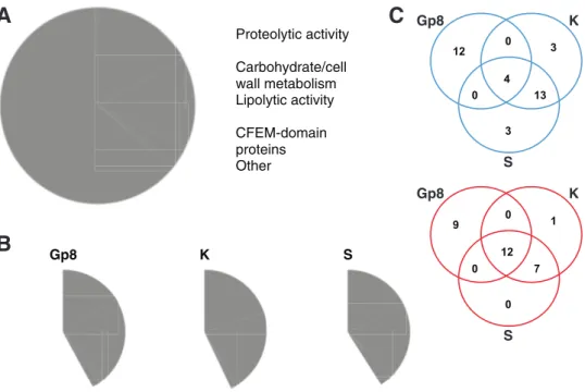
the InterPro database (19, 20). We were able to associate putative functions with 316 out of our 457 predicted cell surface/secreted proteins, including main functional groups such as proteases, carbohydrate/cell wall metabolism proteins, or proteins with lipolytic activities (Fig. 3A; see also Data Set S1 in the supplemental material for details). In addition to thaumatin-like proteins, we identified 46 gene products showing homol-ogies to known allergens (see Table S2 in the supplemental material), of which 21 were predicted to be secreted. Among the 141 uncharacterized secreted proteins, 25 had homologs in other dermatophytes, suggesting that they are involved in dermatophyte-specific functions/processes.
Validation of the new gene predictions of the secretome.The secretome can be relatively easily subjected to investigation by mass spectrometry (MS) because it represents a small fraction of all proteins, and those found in the supernatant of in
vitro-grown cultures can be recovered easily. We conducted a new analysis of the MS
data that we previously published (16) regarding proteins secreted by cells grown in soy protein liquid medium, using the new secretome definition. The presence of 139 proteins in the supernatant at either pH 4 or 7 was confirmed (see Table S1 in the supplemental material), including 8 of the newly predicted ones. Moreover, among the 708 proteins from the original annotation that were lost in our new prediction, 31 were supposed to be secreted, but none of them could be detected in our MS data.
Similarity search is another way to test the quality of gene prediction. As an example, ARB_07403 encodes a putative A1 peptidase. In our prediction, ARB_07403 was shortened at the N terminus by 68 residues. This correction not only allows for the identification of a strong signal peptide at the new N terminus but also aligns better with the sequences of orthologs in closely related species, including TRV_06366 of
Trichophyton verrucosum (UniProt accession no.D4DGR1) and MCYG_07979 of
Arthro-derma otae (UniProt accession no.C5FZ57).
However, it happens that neither prediction fitted with related proteins, requiring a further step of manual sequence correction. ARB_06467 (SUB10) and ARB_04678 (SED3)
Proteolytic activity Carbohydrate/cell wall metabolism Lipolytic activity CFEM-domain proteins Other Gp8 K S 12 0 4 3 13 0 3 9 0 1 12 0 7 0
A
B
C
Gp8 Gp8 K K S SFIG 3 Characterization of the secretome. (A) Pie chart showing the main functional groups identified within the 457 proteins of the secretome. See detailed description in Data Set S1 in the supplemental material. (B) Pie charts showing the same functional groups as in panel A but within the 100 most expressed genes in Gp8 (in vivo 8 days postinfection), K (in vitro in keratin medium), and S (in vitro in soy medium). (C) Venn diagram of proteases (top) and carbohydrate/cell wall metabolism proteins (bottom) present in the 100 most expressed secreted proteins under the 3 conditions described for panel B. Proteases represent about 20% of the 100 most expressed proteins under the 3 conditions; however, the batch of proteins in Gp8 is clearly different from those in K and S. This trend is not as significant when comparing carbohydrate/cell wall metabolism proteins.
were found by similarity search to belong to the S8 and S53 families of serine proteases, respectively, but the predicted proteins missed the N-terminal signal peptide and propeptide. A reanalysis of the nucleotide sequence of SUB10 revealed a probable genome assembly error in a poly(T) stretch localized just behind the actual initiator codon, leading to a frameshift at position 4 (accession no.KX519317) (see Fig. S1A in the supplemental material). An error was also identified within the coding sequence of ARB_04677, upstream of ARB_04678. Correcting this error removed a frameshift at residue 109 of ARB_04677 and led to the fusion of the two open reading frames (ORFs) (accession no.KX519316) (see Fig. S1B). Sanger resequencing of the regions surround-ing the two predicted errors confirmed our predictions and allowed us to restore both protease sequences with clear signals and propeptides. The actual protein sequences of SUB10 and SED3 have been updated in the UniProtKB database (accession numbers D4AQG0andD4AK75, respectively).
Finally, it is interesting that, within the 40 new predicted ORFs, sequence alignments with other fungal proteomes revealed that two have homologs in filamentous fungi, such as Aspergillus species, and 22 are conserved in other dermatophyte species (see Table S1 in the supplemental material).
Arthroderma benhamiae gene expression under different growth condi-tions.Gene expression levels were computed by mapping the reads onto the newly predicted gene set and are expressed as TMM-normalized Voom-transformed counts (see Table S3 in the supplemental material). The nomenclature used for the samples and the corresponding growth conditions are given in Table 3. Figure 4 presents an overview of the gene expression in the different samples, considering either the complete genome or the secretome subset. Both hierarchical clustering and principal component analysis indicate that the biological replicates are closer to each other than to other conditions, even for the in vivo samples at 14 days postinfection, where the number of obtained fungal reads (about 50,000) is possibly too low to perform a statistically significant analysis. However, the small distinction between the Gp8 and Gp14 in vivo conditions, which is on the order of intra-Gp variations, seems to indicate the consistency of Gp14 samples.
The expression differences are strongly dominated by the contrast between in vivo Gp8⫹Gp14 and in vitro S⫹Sa⫹K conditions. This result confirms and generalizes the observations made previously on a much smaller gene set (7). The analysis of the expression data from the complete genome (including the secretome) and of the secretome yielded the same strong contrast, possibly even slightly reinforced for the secretome.
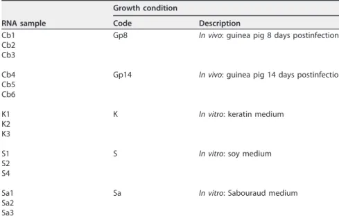
TABLE 3 Designation of samples and growth conditions
RNA sample
Growth condition
Code Description
Cb1 Gp8 In vivo: guinea pig 8 days postinfection Cb2
Cb3
Cb4 Gp14 In vivo: guinea pig 14 days postinfection Cb5
Cb6
K1 K In vitro: keratin medium
K2 K3
S1 S In vitro: soy medium
S2 S4
Sa1 Sa In vitro: Sabouraud medium Sa2
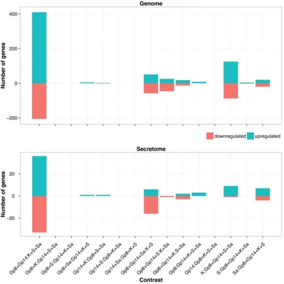
Among the in vitro conditions, the gene expression levels in the soy and Sabouraud media appeared closer to each other in the complete gene set, while soy and keratin appeared closer in the secretome subset. None of the three in vitro conditions tested is a good proxy for in vivo growth conditions, despite the keratin medium being supposed to mimic the host environment. To address this question in more depth, we enumerated all possible partitions of growth conditions into two subsets, to contrast a subset of conditions with the remaining ones. The list of all possible contrasts is given in Fig. 5, with the corresponding amounts of differentially expressed genes. This confirms that the in vivo-in vitro contrast is dominant and that not much information can be expected to be gathered by separating Gp8 from Gp14. Interestingly, two other contrasts seem to carry additional signals:
K:Gp8⫹Gp14⫹Sa⫹S in the genome complete gene set and Gp8⫹Gp14⫹Sa:S⫹K in
the secretome subset.
We utilized a different statistical approach, namely, weighted gene correlation network analysis (WGCNA) and gene ontology (GO) enrichment analysis, to further explore these additional contrasts. The unsupervised clustering algorithm of WGCNA subdivided the input gene set (genome) into 35 different modules, which are disjoint subsets of genes. Then, these modules were individually correlated with the 15 possible different contrasts to detect optimal correlations. As shown in Fig. S2 in the supple-mental material, the in vivo-in vitro contrast again dominated the results, with 2,122 genes found in the turquoise and blue modules. Figure S3A in the supplemental
FIG 4 Hierarchical clustering (A and C) and principal component (PC) analysis (B and D) of RNA sequencing samples considering the genes from the complete genome (A and B) or only the secretome subset (C and D). The sample names reflect the growth conditions: Cb, in vivo in guinea pig; S, in vitro in soy medium; Sa, in vitro in Sabouraud medium; K, in vitro in keratin medium. The in vivo samples cluster together.
material presents the gene expression heat map for the turquoise module as an example. The blue module also showed a high correlation with the in vivo-in vitro contrast, although the expression in Sabouraud medium was intermediate (see Fig. S3B). A few other smaller modules appeared to be correlated with different contrasts, such as the tan module with 209 genes that strongly correlates with K:Gp8⫹Gp14⫹Sa⫹S (see Fig. S3C) and the midnight blue module with 177 genes, highly correlating with Gp8⫹Gp14⫹S:K⫹Sa (see Fig. S3D). The 323 genes from the yellow module also correlate with Gp8⫹Gp14⫹S:K⫹Sa, despite an intermediate ex-pression in Sabouraud medium (see Fig. S3E).
We mapped about 40% of the predicted proteins of A. benhamiae to their ortholo-gous counterparts in S. cerevisiae using Inparanoid and propagated the latter gene ontology (GO) annotations onto the dermatophyte genes. Table S4 in the supplemental material presents the modules for which the most significant GO term enrichment was detected, especially the yellow, midnight blue (correlated with K⫹Sa: Gp8⫹Gp14⫹S), and tan (correlated with K:Gp8⫹Gp14⫹Sa⫹S) modules. The results are, however, very general, revealing changes in translational and RNA-related activities but also indicating that some proteasome-related activities might be specifically altered during growth on keratin. These somewhat modest results are certainly more related to the lack of specific gene annotation for A. benhamiae than to a lack of well-formed gene modules.
FIG 5 Number of differentially expressed genes versus the enumeration of all possible contrasting conditions in the genome and the secretome, using a cutoff of 1eⴚ3 for FDR and 2 for the fold change.
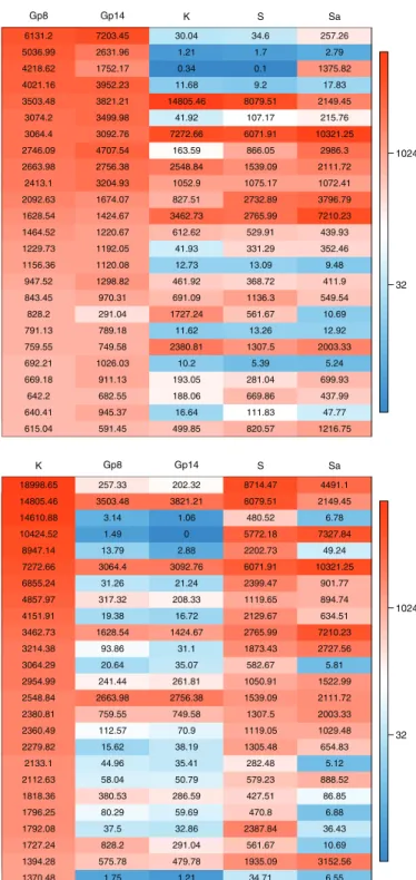
Gene expression profile of Arthroderma benhamiae cell surface/secreted proteins during inflammatory cutaneous infection highly differs from the profile obtained during growth on keratin.Figure 6A lists the 25 secretome genes most highly expressed in vivo, including five putative protease genes. The first gene, ARB_01183, encodes a protein which contains a thaumatin domain. The second gene, ARB_05307, encodes the subtilisin SUB6. Four genes encode proteins for which we did not find any functional data. These include ARBNEW_231, a newly predicted gene and the third most highly expressed gene in vivo. Remarkably, the secretome expression
A
B
Antigenic thaumatin−like protein : ARB_01183 Subtilisin−like protease SUB6 (peptidase S8 family) : ARB_05307 Uncharacterized protein conserved in filamentous fungi : ARBNEW_231 Uncharacterized protein : ARB_03496 GPI−anchored CFEM domain protein : ARB_02741 Uncharacterized protein : ARB_05215_05217 Uncharacterized protein : ARB_02803 Glycoside hydrolase : ARB_07954 GPI−anchored cell wall protein : ARB_01627 GPI−anchored cell wall protein : ARB_02697 Ribosomal protein−like : ARB_06463 GPI anchored serine−threonine rich protein : ARB_07696 1,3−beta−glucanosyltransferase (glycosyl hydrolase 72 family) : ARB_07487 GPI−anchored CFEM domain protein : ARB_01545 Neutral protease 2 homolog (peptidase M35 family) : ARB_04336 1,3−beta−glucanosyltransferase (glycosyl hydrolase 72 family) : ARB_05770 Aspartic−type endopeptidase PEP2 (peptidase A1 family) : ARB_02919 Secreted lipase (type−B carboxylesterase family) : ARB_02369 Subtilisin−like protease SUB10 (peptidase S8 family) : ARB_06467 PGA52−like protein (Asp f 4 homolog) : ARB_06390 Sialidase : ARB_02206 Extracellular matrix protein : ARB_06538 Subtilisin−like protease SUB8 (peptidase S8 family) : ARB_00777 Putative stress−responsive protein : ARB_05496 NAD−dependent malate dehydrogenase : ARB_00653
32 1024 Gp8 Gp14 K S Sa 6131.2 7203.45 30.04 34.6 257.26 5036.99 2631.96 1.21 1.7 2.79 4218.62 1752.17 0.34 0.1 1375.82 4021.16 3952.23 11.68 9.2 17.83 3503.48 3821.21 14805.46 8079.51 2149.45 3074.2 3499.98 41.92 107.17 215.76 3064.4 3092.76 7272.66 6071.91 10321.25 2746.09 4707.54 163.59 866.05 2986.3 2663.98 2756.38 2548.84 1539.09 2111.72 2413.1 3204.93 1052.9 1075.17 1072.41 2092.63 1674.07 827.51 2732.89 3796.79 1628.54 1424.67 3462.73 2765.99 7210.23 1464.52 1220.67 612.62 529.91 439.93 1229.73 1192.05 41.93 331.29 352.46 1156.36 1120.08 12.73 13.09 9.48 947.52 1298.82 461.92 368.72 411.9 843.45 970.31 691.09 1136.3 549.54 828.2 291.04 1727.24 561.67 10.69 791.13 789.18 11.62 13.26 12.92 759.55 749.58 2380.81 1307.5 2003.33 692.21 1026.03 10.2 5.39 5.24 669.18 911.13 193.05 281.04 699.93 642.2 682.55 188.06 669.86 437.99 640.41 945.37 16.64 111.83 47.77 615.04 591.45 499.85 820.57 1216.75
Extracellular serine−threonine rich protein : ARB_04464 GPI−anchored CFEM domain−containing protein : ARB_02741 Subtilisin−like protease SUB3 (peptidase S8 family) : ARB_00701 GPI−anchored cupredoxin : ARB_05732−1 Subtilisin−like protease SUB4 (peptidase S8 family) : ARB_01032 Uncharacterized protein : ARB_02803 Uncharacterized protein also found in T. rubrum : ARBNEW_164 Uncharacterized protein : ARB_06477 Extracellular proline−rich protein : ARB_00287 GPI anchored serine−threonine rich protein : ARB_07696 Cell wall serine−threonine−rich galactomannoprotein : ARB_04561 Leucine aminopeptidase 1 LAP1 (peptidase M28 family) : ARB_03568 Probable extracellular glycosidase : ARB_05253 GPI−anchored cell wall protein : ARB_01627 PGA52−like protein (Asp f 4 homolog) : ARB_06390 Uncharacterized protein : ARB_00449 Uncharacterized protein : ARB_06937 Metallocarboxypeptidase MCPA (peptidase M14 family) : ARB_07026_07027 Aspartic−type endopeptidase OPSB (peptidase A1 family) : ARB_04170 Exo−beta−1,3−glucanase (glycosyl hydrolase 5 family) : ARB_04467 Extracellular metalloprotease (peptidase M43B family) : ARB_05317 Leucine aminopeptidase 2 LAP2 (peptidase M28 family) : ARB_00494 Secreted lipase (type−B carboxylesterase family) : ARB_02369 GPI anchored serine−rich protein : ARB_05667 Extracellular metalloprotease/fungalysin MEP3 (peptidase M36 family) : ARB_05085
32 1024 K Gp8 Gp14 S Sa 18998.65 257.33 202.32 8714.47 4491.1 14805.46 3503.48 3821.21 8079.51 2149.45 14610.88 3.14 1.06 480.52 6.78 10424.52 1.49 0 5772.18 7327.84 8947.14 13.79 2.88 2202.73 49.24 7272.66 3064.4 3092.76 6071.91 10321.25 6855.24 31.26 21.24 2399.47 901.77 4857.97 317.32 208.33 1119.65 894.74 4151.91 19.38 16.72 2129.67 634.51 3462.73 1628.54 1424.67 2765.99 7210.23 3214.38 93.86 31.1 1873.43 2727.56 3064.29 20.64 35.07 582.67 5.81 2954.99 241.44 261.81 1050.91 1522.99 2548.84 2663.98 2756.38 1539.09 2111.72 2380.81 759.55 749.58 1307.5 2003.33 2360.49 112.57 70.9 1119.05 1029.48 2279.82 15.62 38.19 1305.48 654.83 2133.1 44.96 35.41 282.48 5.12 2112.63 58.04 50.79 579.23 888.52 1818.36 380.53 286.59 427.51 86.85 1796.25 80.29 59.69 470.8 6.88 1792.08 37.5 32.86 2387.84 36.43 1727.24 828.2 291.04 561.67 10.69 1394.28 575.78 479.78 1935.09 3152.56 1370.48 1.75 1.21 34.71 6.55
FIG 6 (A) The twenty-five most highly expressed genes encoding secreted proteins during infection compared to in vitro expression. (B) The twenty-five most highly expressed genes encoding secreted proteins in vitro (keratin medium) compared to in vivo expression. Abbreviations are as defined in the Fig. 4 legend.
pattern was completely different during growth on keratin, an in vitro condition that was supposed to mimic the host environment (Fig. 3B and C and 6B). Only five genes were found to be common to Fig. 6A and B: two encoding putative GPI-anchored proteins (ARB_01627 and ARB_07696), ARB_02741 encoding a CFEM domain protein, ARB_06390 encoding a putative cell wall protein, and ARB_02369 encoding a carboxy-lesterase domain-containing protein. This difference is even more striking when we focus our analysis on secreted proteases. Even if about 20% of the 100 most expressed secreted proteins are proteases both in vivo and in keratin (Fig. 3B), the batch of proteins expressed under these different conditions is clearly different (Fig. 3C). This is in accordance with our above-mentioned WGCNA in which relevant correlation groups were found only when in vivo and keratin conditions were contrasted (Gp8⫹Gp14:
Sa⫹S⫹K, Gp8⫹Gp14⫹Sa:S⫹K, Gp8⫹Gp14⫹S:Sa⫹K, or Gp8⫹Gp14⫹Sa⫹S:K).
Expres-sion patterns in soy and Sabouraud media are closer to that in keratin, and yet they are distinct from each other (Fig. 4), which explains their relatively neutral impact in the WGCNA.
Figure S4 in the supplemental material lists the 12 most highly expressed genes encoding proteases during infection and those expressed on keratin. The genes encoding SUB6 (ARB_05307), SUB10 (ARB_06467), and the deuterolysin (ARB_04336) are highly and specifically upregulated during the infection phase with changes of 2,000-fold, 60-fold, and 100-fold, respectively. The gene encoding SUB8 (ARB_00777) was relatively downregulated in keratin. PEP2 (ARB_02919), which is a putative ortholog of the vacuolar aspartic protease of S. cerevisiae PrA and has been subsequently identified in other filamentous fungi, was found to be highly expressed under all the in
vivo and in vitro conditions. On the other hand, the protease genes upregulated in
keratin include subtilisins SUB3 (encoded by ARB_00701) and SUB4 (ARB_01032), the metallocarboxypeptidase MCPA of the M14 family (ARB_07026_07027), the leucine aminopeptidases LAP1 (ARB_03568) and LAP2 (ARB_00494), the aspartic protease OPSB (ARB_04170), and two extracellular metalloproteases (ARB_05085 and ARB_05317). Likewise, in the soy culture, only four protease genes were highly expressed: those for SUB4 (encoded by ARB_01032), LAP2 (ARB_00494), PEP2 (ARB_02919), and DPPV (ARB_06651) (see Table S1 in the supplemental material). With the Sabouraud culture, in addition to PEP2, SUB8 (ARB_00777), OPSB (ARB_04170), DPPIV (ARB_06110), and a gene encoding an uncharacterized S10 family protease (ARB_01491) showed relatively high expression (see Table S1).
DISCUSSION
Most previously available dermatophyte ORFs had been deduced by cDNA analysis and by expressed sequence tag sequencing using RNA extracted from dermatophytes grown in vitro. RNA-seq data obtained from A. benhamiae grown under various liquid culture conditions and, most importantly, during infection in guinea pigs led us to an improved gene prediction and annotation of its genome. A complete gene expression profile of A. benhamiae was obtained during infection of its natural host.
New Arthroderma benhamiae gene annotation. About 65% difference and,
particularly, 383 new protein-encoding genes were detected compared to the existing gene prediction. We used previously acquired MS data to validate a posteriori the presence of the predicted ORFs in culture supernatant. A comparable approach with emphasis on proteogenomics has been recently used to review the genome and proteome of T. rubrum (21). In this study, the identification of 323 new peptides by MS in culture supernatant led to the refinement of 161 genes and the prediction of nine new genes. However, the RNA-seq analysis to validate the whole-genome proteomics was performed only with RNA extracted from T. rubrum cultured in vitro on potato glucose agar but not during infection. This previous study and our results have in common the combination of experimental data with bioinformatics software and manual curation to generate an improved gene annotation. Our study focuses, further-more, on the biology of infection.
In silico analysis of our predicted proteome led to the identification of 457 putative
cell surface and secreted proteins. Our list of probable secreted proteins is likely to also contain proteins targeted to intracellular organelles, such as the endoplasmic reticulum or vacuole, since the exploited prediction tools cannot distinguish between such proteins and secreted ones. The Fungal Secretome and Subcellular Proteome Knowl-edgeBase (http://bioinformatics.ysu.edu/secretomes/fungi2/index.php) tries to address this concern by providing the prediction of secreted and organellar localization of proteins. It basically utilizes the same tools as those that we used in our strategy and reveals the same functional groups (22). In addition, they use WoLF PSORT (http:// www.genscript.com/wolf-psort.html), which converts protein sequences into numerical localization features, based on sorting signals, amino acid composition, and functional motifs. Nevertheless, this tool can produce a high number of false positives. Moreover, homologs of well-known intracellular proteins have been found in the secretome proteomic data. As an example, ARB_02919 is the closest A. benhamiae homolog of the
A. fumigatus vacuolar aspartic peptidase (PEP2) and S. cerevisiae vacuolar proteinase A
(PEP4). The latter is a vacuolar enzyme required for the processing of vacuolar precur-sors (23), whereas the former plays an additional role linked to the cell wall (24). ARB_02919 was found as a secreted protein by MS (16) and is one of the most expressed proteins under all of the five studied conditions. Contaminations cannot be ruled out, but our strategy ensures the best coverage of cell surface and secreted proteins, even if some false positives are probably still present.
Reprogramming of gene expression from a saprophyte to a parasite life-style. Striking differences were revealed between transcriptomes of A. benhamiae during growth under various conditions in vitro and during infection of its natural host. Such differences emphasize the importance of performing transcriptional analysis directly during infection, instead of using in vitro conditions that are expected to mimic the host environment. We also identified several newly predicted genes, as well as genes with unknown functions, that were differentially expressed in the contrast of in
vivo and in vitro and, thus, might have a relevant role in infection. To sum up, the ability
of dermatophytes to switch from a saprophyte to a parasite lifestyle is attested by an important reprogramming of gene expression.
Several comparative RNA-seq analyses were performed for other species of human-pathogenic fungi (25–28), but as these studies rely on infection-mimicking conditions and not on the real in vivo situation, we think that they should be considered with caution. Only a few studies were performed under real infection conditions. Gene expression profiles of C. albicans were obtained during infection in both the mouse kidney and the insect Galleria mellonella (29). Interestingly, gene expression values in these very distinct hosts were much closer to each other than in the in vitro liquid cultures used as controls. More recently, transcriptional profiling of Blastomyces was performed in cocultures with human bone marrow-derived macrophages and during in
vivo pulmonary infection in a mouse model (30). The authors identified a number of
functional categories upregulated exclusively in vivo, including secreted proteins and zinc acquisition proteins, as well as cysteine and tryptophan metabolism. Nine secreted proteins were identified, including products of five of the 10 most upregulated genes during infection. One of these genes, BDFG_00717, encodes a CFEM domain-containing protein, highlighting the importance of those proteins in virulence.
Potential nonprotease virulence factors of Arthroderma benhamiae. Numer-ous genes that were highly expressed during infection encode uncharacterized pro-teins. Highly expressed protein-encoding genes with a putative function other than proteolysis included ARB_01183, encoding a putative antigenic thaumatin domain protein, and two genes encoding 1,3-beta-glucanosyltransferases (ARB_07487 and ARB_05770). ARB_01183 was the most highly expressed secreted protein-encoding gene in vivo. Thaumatin-like proteins (TLPs) are found in many eukaryotes and have been particularly studied in plants, in which they are involved in defense against fungal pathogens. Plant TLPs also have been shown to act as important allergens (31). TLPs are also found in fungi, such as Moniliophthora perniciosa, and may be involved in the
inhibition of growth of fungal competitors and pathogenicity (32). The 1,3-beta-glucanosyltransferases play an important role in fungal cell wall morphology and pathogenicity. Deletion of the gene GEL2 encoding a 1,3-beta-glucanosyltransferase in
A. fumigatus leads to altered cell wall composition as well as to reduced virulence in a
murine model of invasive aspergillosis (33). GAS1 of the entomopathogenic fungus
Beauveria bassiana contributes similarly to its mycoinsecticide activity (34).
ARB_02741, like Blastomyces BDFG_00717, encodes a GPI-anchored CFEM domain protein which is highly expressed under in vivo and in vitro conditions. Its function has not been characterized yet, but it is interesting that the closest homologs of ARB_02741 in the human fungal pathogen Coccidioides posadasii are the proline-rich antigens Ag2/PRA and Prp2, which have been reported to be leading vaccine candidates (35, 36). CFEM domain proteins have been shown to be important for heme uptake and virulence in C. albicans (37). The ability to acquire iron from host tissues is a major virulence factor of pathogenic microorganisms. However, the exact involvement of these proteins in infection processes is still unclear. As an example, the three A.
fu-migatus CFEM domain proteins have been shown to be important for cell wall stability,
not for virulence (38). Other proteins may also be involved in immune escape, such as ARB_06975, whose A. fumigatus hydrophobin homolog was shown to prevent immune recognition by forming a hydrophobic layer on the cell surface (39).
Arthroderma benhamiae secreted proteases during infection.SUB6 was the
most highly expressed gene encoding a secreted protease during infection in guinea pigs. In addition to SUB6, other A. benhamiae protease genes, encoding the subtilisins SUB7, SUB8, and SUB10 as well as a neutral protease of the deuterolysin family (M35), were also specifically upregulated. RNA-seq analysis results also confirmed that genes encoding major proteases secreted by the fungus during growth in a protein medium (i.e., SUB3, SUB4, MEP3, MEP4, LAP1, and DPPIV) were expressed at a relatively low level during infection as well as in Sabouraud medium and were not upregulated. These results are in accordance with recent findings by proteomic analysis (liquid chromatography-tandem MS [LC-MS/MS]) in T. rubrum-infected nails that revealed SUB6 as the major protein secreted by the fungus in onychomycosis (40). The closely related SUB7 (subtilisin-like protease 7, Q8NID9) and DPPV (dipeptidyl-peptidase 5, Q9UW98) were also detected. Likewise, most major proteases secreted by the fungus during its growth in vitro in a protein medium (11, 41) were not detected and, therefore, appeared not to be involved during the establishment of onychomycosis. As a general conclusion, the proteases secreted in vitro during protein degradation and in vivo during infection are different, regardless of the dermatophyte species and the tinea. The view that the proteases isolated from dermatophytes grown in vitro in a protein medium are virulence attributes and exert a major role during infection appears to be too naive and can no longer be accepted. Dermatophytes evolved from soil saprophytic fungi that are able to efficiently degrade hard keratin into amino acids and into short peptides in the process of recycling nitrogen, and the pathogenic phase of dermato-phytes has to be dissociated from their saprophytic phase. Some of the multiple members of protease gene families in dermatophytes are dedicated exclusively to protein degradation, while others, such as SUB6, likely fulfill specific roles during infection. The notion that proteases secreted in proteinaceous media correspond to virulence attributes has also been discarded for other pathogenic fungi. Two different
A. fumigatus mutants unable to secrete proteolytic activity in a protein growth medium
did not show attenuated virulence when tested in a leukopenic mouse model. In the first mutant, the genes encoding the two major secreted proteases ALP and MEP (42) were deleted. In the other mutant, the gene encoding a transcriptional activator (PRTT) which regulates transcription of genes encoding the major proteases secreted in a protein medium was deleted. Noteworthily, no homolog of PRTT in Aspergillus spp. (43, 44) has been identified in A. benhamiae.
Genes encoding major proteases secreted by dermatophytes during in vitro growth in a protein medium are tightly controlled by DNR1, the ortholog of AREA in Aspergillus
nidulans (45). In the absence of ammonium and glutamine, this transcription factor was
found to be required for the expression of genes involved in nitrogen metabolism. Although dermatophytes infect keratinized tissues, our results suggest that the panel of proteases secreted during infection depends on other transcription factors that remain to be discovered.
Arthroderma benhamiae secreted proteins as allergens.Secreted proteins are allergens that play a key role in the pathogenic process. SUB6, DPPV, and the beta-glucosidase ARB_05770 (encoded by three of the most highly expressed genes of
A. benhamiae during infection) are orthologs of the three known major dermatophyte
allergens Tri t1, Tri r2, and Tri r4, which are involved in bronchial sensitization and symptomatic asthma (12, 46, 47). Dermatophyte antigens are also involved in eczem-atous skin reactions at a location distant from the area of dermatophyte infection (dermatophytids). The etiology of common dyshidrotic and vesicular eczema on the hands (palms and fingers) is rarely investigated and may remain elusive because no commercially standardized antigens are available to perform routine skin tests and antibody detection. Trichophytin, a fungal extract that greatly varies in its preparation and composition, was used to diagnose dermatophytids (48, 49). The secreted proteins encoded by genes highly expressed during infection are the best candidates for the detection of dermatophyte allergic diseases. At a time when quality in laboratory techniques is a key issue, it would be relevant to perform skin test reactions using standardized antigens in cases of eczematous skin reactions of unknown origin. A positive reaction could be indicative of a nondetected dermatophyte infection and could suggest possible antifungal treatment.
Conclusion.Comparing gene expression during infection phase with keratin deg-radation in vitro shows the importance of using real in vivo conditions to further investigate the virulence mechanisms of dermatophytes, instead of using some in vitro conditions supposed to mimic the host environment. Focusing our analysis on genes encoding cell-associated and secreted proteins, in particular proteases, led to the identification of strong candidates as allergens and putative virulence factors. The new genome annotation provided in this study might serve as a reference for annotation or reannotation of other dermatophyte species and evolutionarily related filamentous fungi.
MATERIALS AND METHODS
Strains and growth media. Arthroderma benhamiae Lau2354-2 (CBS 112371) (3, 50) was used in this study. This strain, deposited in the Belgian Coordinated Collections of Microorganisms (BCCM/IHEM) under IHEM20161, is the reference strain that was chosen for A. benhamiae genome sequencing (3). It was isolated from a patient suffering from a highly inflammatory dermatophytosis in the Centre Hospitalier Universitaire Vaudois (CHUV). The A. benhamiae strain was maintained at 28°C on Sabouraud dextrose agar medium.
Arthroderma benhamiae was grown in vitro in Sabouraud liquid medium, soy protein liquid medium,
and keratin liquid medium as previously described (7). Soy medium was prepared by dissolving 2 g of soy protein (Supro 1711; Protein Technologies International) in 1 liter of distilled water. Aliquots of 100 ml of keratin medium were prepared by adding 0.2 g of keratin (Merck, Darmstadt, Germany; keratin is derived from animal hooves and horns) and 5 ml of soy medium to 95 ml of distilled water. A small amount of soy protein in keratin liquid medium was found to be necessary to initiate the growth of dermatophytes with keratin as the sole substrate (7). A plug of fresh A. benhamiae mycelium grown on Sabouraud agar was inoculated in 100 ml of liquid Sabouraud, soy, and keratin medium and incubated for 5, 10, and 24 days, respectively, at 30°C without shaking. At the indicated time points, growth in protein medium was accompanied by substantial proteolytic activity along with clarification of the medium and, in the case of keratin medium cultures, also by visible dissolution of the water-insoluble keratin granules.
Animal infection. Specific-pathogen-free, 3-month-old female guinea pigs (cross-bred white albinos, Dunkin Hartley strain; Charles River Laboratories International, Wilmington, MA, USA) were infected with
A. benhamiae Lau2354-2. Arthroderma benhamiae mycelium scraped from freshly grown 18-day-old
Sabouraud plates and suspended in 5% (wt/wt) poloxamer 407 (BASF, Germany) was applied to a 16-cm2 back skin surface that had been clipped and scarified previously. Each guinea pig was infected with 6⫻ 109to 2⫻ 1010CFU. Noninfected control guinea pigs were subjected to the same procedure, except that the poloxamer 407 mixture did not contain any fungal elements. Three guinea pigs were sacrificed after 8, 14, 27, and 44 days and at 14 days after reinfection once they had healed. The infected skin from sacrificed animals was frozen at⫺80°C for subsequent total RNA isolation. Both the hair and stratum
corneum were examined for the presence of fungal elements by direct mycological examination. Animal experiments were approved by the local ethics committee (University of Liège, ethics protocol no. 1052). RNA extraction. RNA extraction from A. benhamiae cultures and infected guinea pig skin was performed using a specific procedure to yield sufficient amounts of quality RNA (see Data Set S1 in the supplemental material).
RNA sequencing. In close collaboration with the Lausanne Genomic Technologies Facility and using the Illumina technology (HiSeq 2000 sequencer), we performed a TruSeq stranded single read total RNA analysis, using one lane with a multiplex level of 15, acquiring approximately 30 million “strand-specific” reads with a length of 100 bp for each sample. Reads were aligned against the A. benhamiae and guinea pig genomes using tophat2 (version 2.0.9) (51).
Strain. The genome assembly GCA_000151125.2 ASM15112v2 of A. benhamiae Lau2354-2 was used throughout this study.
Gene prediction and annotation. Gene prediction was made with Augustus (version 3.0.2) (13) using a specific gene model obtained as follows. Gene transcripts and intron locations were obtained using Cufflinks (version 2.2.1) (52). The transcripts were three-frame translated into potential amino acid sequences using Transeq from EMBOSS (version 6.5.7) (53). The complete proteomes of Saccharomyces
cerevisiae and Aspergillus nidulans (reviewed by Swiss-Prot) were mapped onto the potential amino acid
sequences with Glsearch36, from the FASTA alignment tools (version 3.6) (54), to identify coding phase and CDS location within transcripts. Based on the alignment quality and on the presence of start and stop codons near alignment extremities (⫾10 amino acids), a set of confidently predicted CDSs was gathered and converted into gene annotations using intron locations previously given by Cufflinks. These annotations were used as a training set to build a gene model (available upon request) with the scripts supplied in the Augustus distribution.
In silico identification of putative cell surface and secreted proteases. To identify putative secreted proteins, we checked for the presence of an N-terminal signal sequence using both Phobius (version 1.01) (55) and SignalP (version 4.1) (56). Signal peptides have been confirmed by the prediction of N-terminal transmembrane spans using TMHMM (version 2.0) (57, 58). The presence of a potential glycosylphosphatidylinositol (GPI) anchor has been checked by using PredGPI (version 1.0) (59). Using the transmembrane span predictors TMHMM (version 2.0), ESKW (version 1.0) (60), and MEMSAT (version 1.8) (61), we refined the secretome prediction by removing the proteins that contain one or more transmembrane spans in addition to the signal peptide and that are probably targeted to membranes. All the secreted proteins have been subjected to Blast analysis against the UniProtKB database (18) as well as to InterPro scanning (19, 20) to associate and reveal some putative functions.
Mass spectrometry and experimental validation of new secreted proteins. Precipitation and separation of proteins from A. benhamiae cultures at pH 4 and pH 7 along with shotgun mass spectrometry (MS) experiments have been described by Sriranganadane et al. (16). A new search of MS/MS spectra against the sequences of our new predicted proteome was performed.
Transcriptome analysis. The number of reads mapped onto each newly predicted gene locus was obtained with Htseq-count (version 0.5.4p3) (62). Genes with counts of fewer than one per million in all samples were removed from the statistical analyses (i.e., 81 genes). Gene expression was normalized using the TMM-normalized Voom transformation (63); hierarchical clustering and principal component analysis were done using R (version 3.1.1). Differential gene expression analysis was performed with the R Bioconductor package Limma (64). The cutoffs of 1e⫺3 for false discovery rate (FDR) (Benjamini-Yekutieli-adjusted P value) (65) and 2 for fold change were applied to identify genes relevant to each contrast. The R software package WGCNA (66) was used for correlation network analysis, using the Pearson correlation.
Pathway enrichment. The predicted A. benhamiae proteins were aligned against Saccharomyces
cerevisiae proteins from Swiss-Prot with Inparanoid (version 4.1) (67) to identify the orthologs from which
the gene ontology (GO) terms were extracted and applied to A. benhamiae. We then performed the GO enrichment analysis on the weighted gene correlation network analysis (WGCNA) gene modules.
Accession number(s). The raw RNA-seq data investigated here are accessible under the SRA accession numberSRP064455. The annotation has been deposited as a Whole Genome Shotgun project at DDBJ/ENA/GenBank under the accession numberDAAX00000000. The version described in this paper is version DAAX01000000. The CavPor3 draft assembly of the guinea pig genome was used.
SUPPLEMENTAL MATERIAL
Supplemental material for this article may be found at http://dx.doi.org/10.1128/ mSystems.00036-16. Table S1, XLSX file, 0.1 MB. Table S2, XLSX file, 0.1 MB. Table S3, XLSX file, 1.4 MB. Table S4, XLSX file, 0.1 MB. Figure S1, PDF file, 0.1 MB. Figure S2, PDF file, 0.1 MB. Figure S3, PDF file, 1.1 MB. Figure S4, PDF file, 0.1 MB.
Figure S5, PDF file, 0.4 MB. Data Set S1, PDF file, 0.2 MB.
ACKNOWLEDGMENTS
This work was supported by the Swiss National Science Foundation grant 31003A_140514 to Michel Monod and Marco Pagni. It was also partly supported by grant 3.4558.10 from the Fonds de la Recherche scientifique Médicale (FRSM, Belgium) and 594/2012 from the Executive Unit for Higher Education, Research Development and Innovation Funding (UEFISCDI, Romania) to Bernard Mignon. E.-T.B. was the recipient of a research grant provided by the University of Liège and supported by the agreement that binds Wallonia-Brussels (WBI) and Romania. The computations were performed at the Vital-IT Center for high-performance computing ( http://www.vital-it.ch) of the SIB Swiss Institute of Bioinformatics. SIB receives financial supports from the Swiss Federal Government through the State Secretariat for Education and Research.
We thank Sandra Calderon for support in the RNA-seq analysis and Edouard de Castro for support in the signal peptide prediction.
FUNDING INFORMATION
This work, including the efforts of Van Du Thuong Tran, Niccolò De Coi, Marc Feuer-mann, Marco Pagni, and Michel Monod, was funded by Schweizerischer Nationalfonds zur Förderung der Wissenschaftlichen (SNF) (31003A_140514). This work, including the efforts of Elena-Tatiana Ba˘gut¸ and Bernard Mignon, was funded by Fonds De La Recherche Scientifique Médicale - FRSM - Belgium (3.4558.10). This work, including the efforts of Elena-Tatiana Ba˘gut¸, was funded by Executive Unit for Higher Education, Research, Development and Innovation Funding - UEFISCDI - Romania (594/2012).
REFERENCES
1. Weitzman I, Summerbell RC. 1995. The dermatophytes. Clin Microbiol Rev 8:240 –259.
2. Degreef H. 2008. Clinical forms of dermatophytosis (ringworm infec-tion). Mycopathologia 166:257–265. http://dx.doi.org/10.1007/s11046 -008-9101-8.
3. Burmester A, Shelest E, Glöckner G, Heddergott C, Schindler S, Staib P, Heidel A, Felder M, Petzold A, Szafranski K, Feuermann M, Pe-druzzi I, Priebe S, Groth M, Winkler R, Li W, Kniemeyer O, Schroeckh V, Hertweck C, Hube B, White TC, Platzer M, Guthke R, Heitman J, Wöstemeyer J, Zipfel PF, Monod M, Brakhage AA. 2011. Comparative and functional genomics provide insights into the pathogenicity of dermatophytic fungi. Genome Biol 12:R7.http://dx.doi.org/10.1186/gb -2011-12-1-r7.
4. Martinez DA, Oliver BG, Gräser Y, Goldberg JM, Li W, Martinez-Rossi NM, Monod M, Shelest E, Barton RC, Birch E, Brakhage AA, Chen Z, Gurr SJ, Heiman D, Heitman J, Kosti I, Rossi A, Saif S, Samalova M, Saunders CW, Shea T, Summerbell RC, Xu J, Young S, Zeng Q, Birren BW, Cuomo CA, White TC. 2012. Comparative genome analysis of
Trichophyton rubrum and related dermatophytes reveals candidate
genes involved in infection. mBio 3:e00259-12. http://dx.doi.org/ 10.1128/mBio.00259-12.
5. Naglik JR, Challacombe SJ, Hube B. 2003. Candida albicans secreted aspartyl proteinases in virulence and pathogenesis. Microbiol Mol Biol Rev 67:400 – 428.http://dx.doi.org/10.1128/MMBR.67.3.400-428.2003. 6. Lo Presti L, Lanver D, Schweizer G, Tanaka S, Liang L, Tollot M,
Zuccaro A, Reissmann S, Kahmann R. 2015. Fungal effectors and plant susceptibility. Annu Rev Plant Biol 66:513–545. http://dx.doi.org/ 10.1146/annurev-arplant-043014-114623.
7. Staib P, Zaugg C, Mignon B, Weber J, Grumbt M, Pradervand S, Harshman K, Monod M. 2010. Differential gene expression in the pathogenic dermatophyte Arthroderma benhamiae in vitro versus dur-ing infection. Microbiology 156:884 – 895. http://dx.doi.org/10.1099/ mic.0.033464-0.
8. Jousson O, Léchenne B, Bontems O, Capoccia S, Mignon B, Barblan J, Quadroni M, Monod M. 2004. Multiplication of an ancestral gene encoding secreted fungalysin preceded species differentiation in the dermatophytes Trichophyton and Microsporum. Microbiology 150: 301–310.http://dx.doi.org/10.1099/mic.0.26690-0.
9. Jousson O, Léchenne B, Bontems O, Mignon B, Reichard U, Barblan
J, Quadroni M, Monod M. 2004. Secreted subtilisin gene family in Trichophyton rubrum. Gene 339:79 – 88. http://dx.doi.org/10.1016/ j.gene.2004.06.024.
10. Monod M, Léchenne B, Jousson O, Grand D, Zaugg C, Stöcklin R, Grouzmann E. 2005. Aminopeptidases and dipeptidyl-peptidases se-creted by the dermatophyte Trichophyton rubrum. Microbiology 151: 145–155.http://dx.doi.org/10.1099/mic.0.27484-0.
11. Zaugg C, Jousson O, Léchenne B, Staib P, Monod M. 2008. Tricho-phyton rubrum secreted and membrane-associated carboxypeptidases. Int J Med Microbiol 298:669 – 682. http://dx.doi.org/10.1016/ j.ijmm.2007.11.005.
12. Woodfolk JA, Wheatley LM, Piyasena RV, Benjamin DC, Platts-Mills TA. 1998. Trichophyton antigens associated with IgE antibodies and delayed type hypersensitivity. Sequence homology to two families of serine proteinases. J Biol Chem 273:29489 –29496. http://dx.doi.org/ 10.1074/jbc.273.45.29489.
13. Stanke M, Steinkamp R, Waack S, Morgenstern B. 2004. Gene pre-diction with a hidden Markov model. University of Göttingen, Göttingen, Germany.
14. Starck SR, Jiang V, Pavon-Eternod M, Prasad S, McCarthy B, Pan T, Shastri N. 2012. Leucine-tRNA initiates at CUG start codons for protein synthesis and presentation by MHC class I. Science 336:1719 –1723.
http://dx.doi.org/10.1126/science.1220270.
15. Ehlers MR, Riordan JF. 1991. Membrane proteins with soluble counterparts: role of proteolysis in the release of transmembrane pro-teins. Biochemistry 30:10065–10074.
16. Sriranganadane D, Waridel P, Salamin K, Feuermann M, Mignon B, Staib P, Neuhaus J-M, Quadroni M, Monod M. 2011. Identification of novel secreted proteases during extracellular proteolysis by dermato-phytes at acidic pH. Proteomics 11:4422– 4433. http://dx.doi.org/ 10.1002/pmic.201100234.
17. Heddergott C, Bruns S, Nietzsche S, Leonhardt I, Kurzai O, Kni-emeyer O, Brakhage AA. 2012. The Arthroderma benhamiae hydropho-bin HypA mediates hydrophobicity and influences recognition by hu-man immune effector cells. Eukaryot Cell 11:673– 682.http://dx.doi.org/ 10.1128/EC.00037-12.
18. UniProt Consortium. 2015. UniProt: a hub for protein information. Nucleic Acids Res 43:D204 –D212.http://dx.doi.org/10.1093/nar/gku989. 19. Quevillon E, Silventoinen V, Pillai S, Harte N, Mulder N, Apweiler R,
Lopez R. 2005. InterProScan: protein domains identifier. Nucleic Acids Res 33:W116 –W120.http://dx.doi.org/10.1093/nar/gki442.
20. Zdobnov EM, Apweiler R. 2001. InterProScan—an integration platform for the signature-recognition methods in InterPro. Bioinformatics 17: 847– 848.http://dx.doi.org/10.1093/bioinformatics/17.9.847.
21. Xu X, Liu T, Ren X, Liu B, Yang J, Chen L, Wei C, Zheng J, Dong J, Sun L, Zhu Y, Jin Q. 2015. Proteogenomic analysis of Trichophyton rubrum aided by RNA sequencing. J Proteome Res 14:2207–2218. http:// dx.doi.org/10.1021/acs.jproteome.5b00009.
22. Meinken J, Asch DK, Neizer-Ashun KA, Chang G-H, Cooper CR, Jr, Min XJ. 2014. FunSecKB2: a fungal protein subcellular location knowl-edgebase. Comput Mol Biol 4(6):1–17.
23. Ammerer G, Hunter CP, Rothman JH, Saari GC, Valls LA, Stevens TH. 1986. Pep4 gene of Saccharomyces cerevisiae encodes proteinase A, a vacuolar enzyme required for processing of vacuolar precursors. Mol Cell Biol 6:2490 –2499.http://dx.doi.org/10.1128/MCB.6.7.2490.
24. Reichard U, Monod M, Odds F, Rüchel R. 1997. Virulence of an aspergillopepsin-deficient mutant of Aspergillus fumigatus and evi-dence for another aspartic proteinase linked to the fungal cell wall. J Med Vet Mycol 35:189 –196.
25. Irmer H, Tarazona S, Sasse C, Olbermann P, Loeffler J, Krappmann S, Conesa A, Braus GH. 2015. RNAseq analysis of Aspergillus fumigatus in blood reveals a just wait and see resting stage behavior. BMC Genomics 16:640.http://dx.doi.org/10.1186/s12864-015-1853-1.
26. Chen F, Zhang C, Jia X, Wang S, Wang J, Chen Y, Zhao J, Tian S, Han X, Han L. 2015. Transcriptome profiles of human lung epithelial cells A549 interacting with Aspergillus fumigatus by RNA-Seq. PLoS One 10:e0135720.http://dx.doi.org/10.1371/journal.pone.0135720. 27. Muszkieta L, Beauvais A, Pähtz V, Gibbons JG, Anton Leberre V,
Beau R, Shibuya K, Rokas A, Francois JM, Kniemeyer O, Brakhage AA, Latgé JP. 2013. Investigation of Aspergillus fumigatus biofilm for-mation by various “omics” approaches. Front Microbiol 4:13.http:// dx.doi.org/10.3389/fmicb.2013.00013.
28. Edwards JA, Chen C, Kemski MM, Hu J, Mitchell TK, Rappleye CA. 2013. Histoplasma yeast and mycelial transcriptomes reveal pathogenic-phase and lineage-specific gene expression profiles. BMC Genomics 14:695.http://dx.doi.org/10.1186/1471-2164-14-695.
29. Amorim-Vaz S, Tran VDT, Pradervand S, Pagni M, Coste AT, San-glard D. 2015. RNA enrichment method for quantitative transcriptional analysis of pathogens in vivo applied to the fungus Candida albicans. mBio 6:e00942-15.http://dx.doi.org/10.1128/mBio.00942-15.
30. Muñoz JF, Gauthier GM, Desjardins CA, Gallo JE, Holder J, Sullivan TD, Marty AJ, Carmen JC, Chen Z, Ding L, Gujja S, Magrini V, Misas E, Mitreva M, Priest M, Saif S, Whiston EA, Young S, Zeng Q, Goldman WE, Mardis ER, Taylor JW, McEwen JG, Clay OK, Klein BS, Cuomo CA. 2015. The dynamic genome and transcriptome of the human fungal pathogen Blastomyces and close relative Emmonsia. PLoS Genet 11:e1005493.http://dx.doi.org/10.1371/journal.pgen.1005493. 31. Palacín A, Rivas LA, Gómez-Casado C, Aguirre J, Tordesillas L, Bartra
J, Blanco C, Carrillo T, Cuesta-Herranz J, Bonny JA, Flores E, García-Alvarez-Eire MG, García-Nuñez I, Fernández FJ, Gamboa P, Muñoz R, Sánchez-Monge R, Torres M, Losada SV, Villalba M, Parro V, Blanca M, Salcedo G, Díaz-Perales A. 2012. The involvement of thaumatin-like proteins in plant food cross-reactivity: a multicenter study using a specific protein microarray. PLoS One 7:e44088. http://dx.doi.org/ 10.1371/journal.pone.0044088.
32. Franco SDF, Baroni RM, Carazzolle MF, Teixeira PJPL, Reis O, Pereira G, Mondego JMC. 2015. Genomic analyses and expression evaluation of thaumatin-like gene family in the cacao fungal pathogen Monilioph-thora perniciosa. Biochem Biophys Res Commun 466:629 – 636.http:// dx.doi.org/10.1016/j.bbrc.2015.09.054.
33. Mouyna I, Morelle W, Vai M, Monod M, Léchenne B, Fontaine T, Beauvais A, Sarfati J, Prévost M-C, Henry C, Latgé J-P. 2005. Deletion of GEL2 encoding for a beta(1-3)glucanosyltransferase affects morpho-genesis and virulence in Aspergillus fumigatus. Mol Microbiol 56: 1675–1688.http://dx.doi.org/10.1111/j.1365-2958.2005.04654.x. 34. Zhang S, Xia Y, Keyhani NO. 2011. Contribution of the gas1 gene of the
entomopathogenic fungus Beauveria bassiana, encoding a putative glycosylphosphatidylinositol-anchored beta-1,3-glucanosyltransferase, to conidial thermotolerance and virulence. Appl Environ Microbiol 77: 2676 –2684.http://dx.doi.org/10.1128/AEM.02747-10.
35. Cox RA, Magee DM. 2004. Coccidioidomycosis: host response and vaccine development. Clin Microbiol Rev 17:804 – 839.http://dx.doi.org/ 10.1128/CMR.17.4.804-839.2004.
36. Herr RA, Hung C-Y, Cole GT. 2007. Evaluation of two homologous proline-rich proteins of Coccidioides posadasii as candidate vaccines against coccidioidomycosis. Infect Immun 75:5777–5787. http:// dx.doi.org/10.1128/IAI.00807-07.
37. Weissman Z, Kornitzer D. 2004. A family of Candida cell surface haem-binding proteins involved in haemin and haemoglobin-iron utilization. Mol Microbiol 53:1209 –1220. http://dx.doi.org/10.1111/j.1365 -2958.2004.04199.x.
38. Vaknin Y, Shadkchan Y, Levdansky E, Morozov M, Romano J, Os-herov N. 2014. The three Aspergillus fumigatus CFEM-domain GPI-anchored proteins (CfmA-C) affect cell-wall stability but do not play a role in fungal virulence. Fungal Genet Biol 63:55– 64.http://dx.doi.org/ 10.1016/j.fgb.2013.12.005.
39. Aimanianda V, Bayry J, Bozza S, Kniemeyer O, Perruccio K, Elluru SR, Clavaud C, Paris S, Brakhage AA, Kaveri SV, Romani L, Latgé J-P. 2009. Surface hydrophobin prevents immune recognition of airborne fungal spores. Nature 460:1117–1121. http://dx.doi.org/10.1038/ nature08264.
40. Méhul B, Gu Z, Jomard A, Laffet G, Feuilhade M, Monod M. 2016. Sub6 (Tri r 2), an onychomycosis marker revealed by proteomics analysis of Trichophyton rubrum secreted proteins in patient nail samples. J Invest Dermatol 136:3105–3114.http://dx.doi.org/10.1038/jid.2015.367. 41. Giddey K, Monod M, Barblan J, Potts A, Waridel P, Zaugg C, Quad-roni M. 2007. Comprehensive analysis of proteins secreted by Tricho-phyton rubrum and TrichoTricho-phyton violaceum under in vitro conditions. J Proteome Res 6:3081–3092.http://dx.doi.org/10.1021/pr070153m. 42. Jaton-Ogay K, Paris S, Huerre M, Quadroni M, Falchetto R, Togni G,
Latgé JP, Monod M. 1994. Cloning and disruption of the gene encoding an extracellular metalloprotease of Aspergillus fumigatus. Mol Microbiol 14:917–928.http://dx.doi.org/10.1111/j.1365-2958.1994.tb01327.x. 43. Punt PJ, Schuren FH, Lehmbeck J, Christensen T, Hjort C, van den
Hondel CA. 2008. Characterization of the Aspergillus niger prtT, a unique regulator of extracellular protease encoding genes. Fungal Genet Biol 45:1591–1599.http://dx.doi.org/10.1016/j.fgb.2008.09.007. 44. Bergmann A, Hartmann T, Cairns T, Bignell EM, Krappmann S. 2009.
A regulator of Aspergillus fumigatus extracellular proteolytic activity is dispensable for virulence. Infect Immun 77:4041– 4050. http:// dx.doi.org/10.1128/IAI.00425-09.
45. Yamada T, Makimura K, Abe S. 2006. Isolation, characterization, and disruption of dnr1, the areA/nit-2-like nitrogen regulatory gene of the zoophilic dermatophyte, Microsporum canis. Med Mycol 44:243–252.
http://dx.doi.org/10.1080/13693780500410909.
46. Deuell B, Arruda LK, Hayden ML, Chapman MD, Platts-Mills TA. 1991. Trichophyton tonsurans allergen. I. Characterization of a protein that causes immediate but not delayed hypersensitivity. J Immunol 147: 96 –101.
47. Woodfolk JA. 2005. Allergy and dermatophytes. Clin Microbiol Rev 18:30 – 43.http://dx.doi.org/10.1128/CMR.18.1.30-43.2005.
48. Grappel SF, Bishop CT, Blank F. 1974. Immunology of dermatophytes and dermatophytosis. Bacteriol Rev 38:222–250.
49. Ilkit M, Durdu M, Karakas¸ M. 2012. Cutaneous id reactions: a compre-hensive review of clinical manifestations, epidemiology, etiology, and management. Crit Rev Microbiol 38:191–202.http://dx.doi.org/10.3109/ 1040841X.2011.645520.
50. Fumeaux J, Mock M, Ninet B, Jan I, Bontems O, Léchenne B, Lew D, Panizzon RG, Jousson O, Monod M. 2004. First report of Arthroderma benhamiae in Switzerland. Dermatology 208:244 –250.http://dx.doi.org/ 10.1159/000077311.
51. Kim D, Pertea G, Trapnell C, Pimentel H, Kelley R, Salzberg SL. 2013. TopHat2: accurate alignment of transcriptomes in the presence of in-sertions, deletions and gene fusions. Genome Biol 14:R36. http:// dx.doi.org/10.1186/gb-2013-14-4-r36.
52. Trapnell C, Roberts A, Goff L, Pertea G, Kim D, Kelley DR, Pimentel H, Salzberg SL, Rinn JL, Pachter L. 2012. Differential gene and tran-script expression analysis of RNA-seq experiments with TopHat and cufflinks. Nat Protoc 7:562–578. http://dx.doi.org/10.1038/ nprot.2012.016.
53. Rice P, Longden I, Bleasby A. 2000. EMBOSS: the European Molecular Biology Open Software Suite. Trends Genet 16:276 –277. http:// dx.doi.org/10.1016/S0168-9525(00)02024-2.
54. Pearson WR. 1991. Searching protein sequence libraries: comparison of the sensitivity and selectivity of the Smith-Waterman and FASTA algo-rithms. Genomics 11:635– 650. http://dx.doi.org/10.1016/0888 -7543(91)90071-L.