Papers
Cattle enterotoxaemia and Clostridium
perfringens: description, diagnosis and
prophylaxis
M. Lebrun, J. G. Mainil, A. Linden
Cattle enterotoxaemia is one of numerous pathologies caused by Clostridium perfringens.
These anaerobic Gram-positive bacteria are naturally present in the intestinal flora of
mammals, but their uncontrolled multiplication under certain conditions results in the
overproduction of toxins in the intestinal tract. Major clinical signs are induced by the
systemic spread of these toxins in the blood and tissues. Enterotoxaemia may be acute or
peracute, and sudden death is often reported in rapidly growing, apparently healthy cattle.
Enterotoxaemia can be prevented only with better understanding of its risk factors and
pathogenesis. This paper provides an up-to-date overview of knowledge concerning the
aetiology of the syndrome, its epidemiological context, pathogenesis, clinical signs and
lesions, the diagnostic procedures and prophylactic tools, with specific attention to field
aspects that are directly relevant to practitioners and clinical researchers.
THE word ‘enterotoxaemia’ denotes a toxaemia of intestinal origin that occurs when toxins produced in the intestines are absorbed into the bloodstream. Enterotoxaemia can be caused by various microor-ganisms, but the term is most frequently used in relation to the absorp-tion of toxins produced by species of the genus Clostridium (Manteca and Daube 1994, Daube and others 1996a, Popoff 1998).
The genus Clostridium comprises more than 100 species, several of which are pathogenic, producing exotoxins that act locally or sys-temically on the host’s cells and tissues. Although several species of
Clostridium can cause enterotoxaemia in different hosts, the syndrome
has come to be associated with the species Clostridium perfringens in ruminants (Popoff 1998).
Sheep enterotoxaemia caused by C perfringens is well documented. Different strains of C perfringens are responsible for several clinical syn-dromes, including lamb dysentery, pulpy kidney and struck (entero-toxaemia in adult sheep that causes sudden death) (Buxton and others 1978, Kadra and others 1999, Gkiourtzidis and others 2001, Soler-Jover and others 2004, Uzal and others 2004). Prevention is straight-forward, through vaccination of ewes during pregnancy and of the lambs (Clarkson and others 1985, Green and others 1987, Odendaal and others 1988, 1989, de la Rosa and others 1997, Uzal and Kelly 1998, Uzal and others 1999, Rosskopf-Streicher and others 2004). The situation is less clear in cattle.
This article reviews aspects of the aetiology, epidemiology and pathogenesis of cattle enterotoxaemia, with special reference to beef cattle. Its intention is to improve the accuracy of field and routine lab-oratory diagnosis, and to improve prophylaxis through management practices and vaccination.
C perfringens
Description
C perfringens is a Gram-positive, anaerobic, oxygen-tolerant,
rod-shaped bacterium, successively named Bacillus aerogenes capsulatus (Welch and Nuttall 1892, Lucey and Hutchins 2004), Bacterium
welchii and Clostridium welchii in honour of William Henry Welch,
who first described it in 1892. The species name perfringens (‘break-ing through’ in Latin), proposed by Veillon and Zuber (1898) (Bacillus
perfringens), replaced welchii in the fifth edition of Bergey’s Manual of
Determinative Bacteriology (Bergey and others 1939). C perfringens is probably the best-known and most widespread anaerobic pathogen throughout the world. It is a normal component of the intestinal flora of healthy warm-blooded animals and human beings (Popoff 1989, Rood and Cole 1991, Songer 1996).
Like all bacterial species, C perfringens can be subdivided into strains according to the results of different typing methods. Although sub-division by serotyping was proposed in the past, sub-division into strains according to the combinations of toxins produced (or toxinotypes) is still the most widespread and routinely useful method today. Genotyping is generally used only in PCR analysis of toxin genotype (Daube and others 1994, Meer and Songer 1997, Kanakaraj and oth-ers 1998, Baums and othoth-ers 2004).
C perfringens can produce up to 30 potential toxins, and strains
are traditionally classified into five categories (A, B, C, D and E) according to the combination of the four major toxins (a, b, ι and ε) they produce (Hatheway 1990, Songer 1996, Petit and others 1999) (Table 1). These five typescan be further subdivided according to the production of two additional toxins: the enterotoxin (encoded by the
cpe gene) and the b2 toxin (encoded by the cbp2 gene) and numerous
so-called minor toxins (Niilo 1987, Daube and others 1996a, b, Ghazi and others 1997, Gibert and others 1997, O’Brien and Melville 2004) (Table 1).
M. Lebrun, DVM, MSc,
Association de Santé et d’Identification Animale, Allée des Artisans 2, 5590 Ciney, Belgium
J. G. Mainil, DVM, MSc, PhD, A. Linden, DVM, MSc, PhD,
Bacteriology Section, Department of Infectious Diseases, Faculty of Veterinary Medicine, University of Liège, B43A Boulevard de Colonster 20, 4000 Liège, Belgium
E-mail for correspondence: maude.lebrun@arsia.be Provenance: not commissioned; externally peer reviewed
Associated pathologies
Type A strains cause most pathologies associated with C perfringens in human beings: gas gangrene (type A, non-enterotoxigenic), sporadic or antibiotic-associated diarrhoea (type A, ±enterotoxigenic, ±cpb2) and food poisoning (type A or D, enterotoxigenic) (Vogel 1995, Meer and Songer 1997, Sparks and others 2001, Carney and others 2002, Fisher and others 2005, Vaishnavi and others 2005). Necrotising enteritis (type C) is also seen in human beings (Kreft and others 2000).
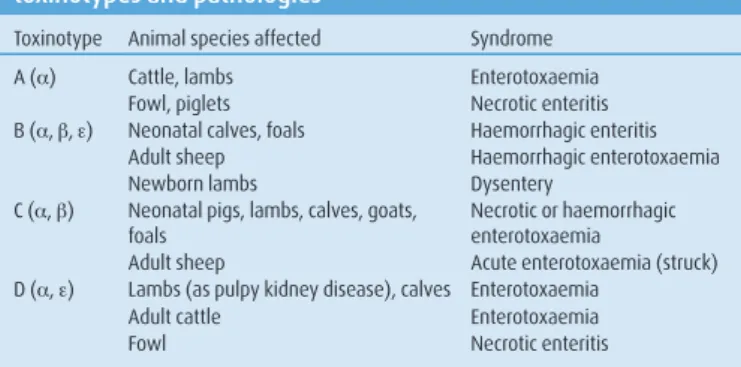
In animals, the five toxinotypes cause numerous forms of enteri-tis and enterotoxaemia (Popoff 1989, Songer 1996, Greene and Jang 2003, Marks and Kather 2003, Coetzer and Tustin 2005, Radostits and others 2007a) (Table 2).
Type A strains are associated with diarrhoea, dysentery and entero toxaemia in ruminants, pigs, horses and poultry (Manteca and Daube 1994, Songer 1996, Daube and others 1996b, Glock and DeGroot 1998, Herholz and others 1999, Manteca and others 2001, Bacciarini and others 2003, Bueschel and others 2003, Songer and Uzal 2005, Radostits and others 2007b). The role of the a toxin in enteritis and enterotoxaemia awaits full experimental confirmation: the a toxin has not yet been shown to be present in the bloodstream of mammals in non-experimental infections, experimental trials to reproduce enterotoxaemia in mammals using the a toxin have failed, and although clinical signs of necrotic enteritis can be reproduced in poultry (Fukata and others 1988), more recent work with mutant strains has questioned the supposed role of the a toxin (Keyburn and others 2006) and implicated the more recently discoveredNetB toxin in this pathology (Keyburn and others 2008).
Type B and C strains cause most cases of dysentery and entero-toxaemia with necrotic or haemorrhagic enteritis, and type D strains cause the pulpy kidney enterotoxaemic syndrome in sheep, and per-haps in calves in which no necrotic or haemorrhagic intestinal lesions are present (Griner and Bracken 1953, Niilo 1986, Leary and Titball 1997, Popoff 1998, Radostits and others 2007c, d). The major b and ε toxins have been shown to have a role in these syndromes, and pre-vention by vaccination with toxoids is effective, at least in small rumi-nants (Uzal and Kelly 1998, Springer and Selbitz 1999, Uzal and oth-ers 2002, Rosskopf-Streicher and othoth-ers 2004, Fernandez-Miyakawa and others 2007).
The role and importance of C perfringens type E (ι toxin) is still unclear, but type E strains appear to cause enteritis in sheep and goats in the Middle East and in calves in North America (Billington and oth-ers 1998, Bueschel and othoth-ers 2003, Sipos and othoth-ers 2003, Songer and Miskimmins 2004, Greco and others 2005, Radostits and others 2007c).
Specific toxins
The enterotoxin (cpe gene) is responsible for enteritis, diarrhoea and food poisoning in human beings (Schalch and others 1997, 1999, Lin and Labbe 2003, Miyamoto and others 2004, 2006, Fisher and others 2005) and could also be involved in cases of diarrhoea in ani-mals (Tschirdewahn and others 1991, Kokai-Kun and McClane 1997, Bueschel and others 1998, 2003, Netherwood and others 1998).
The b2 toxin (cpb2 gene) described in the late 1990s (Gibert and others 1997) has been associated with enteritis in piglets (Klaasen and others 1999, Bueschel and others 2003), typhlocolitis in horses (Herholz and others 1999, Bacciarini and others 2003) and diarrhoea in dogs (Thiede and others 2001). However, its role remains to be determined in human beings and in some animal species in which b2-positive strains have been isolated. The role of the b2 toxin will be further discussed in this paper in the specific context of cattle entero-toxaemia (see ‘Strain typing’ section).
The last C perfringens toxin discovered is NetB. It has been identified as a definitive virulence factor of the poultry necrotic enteritis syndrome (Keyburn and others 2008). This toxin is found in type A strains from poultry with or without necrotic enteritis and was also detected in one strain from a cow with liver abscesses (Martin and Smyth 2009).
Cattle enterotoxaemia
Cattle enterotoxaemia is an acute or peracute syndrome with a case fatality rate close to 100 per cent, associated with an uncontrolled mul-tiplication of C perfringens in the small intestine with an over production
of toxins (Manteca 2004). These toxins act both locally and systemically and may cause death within a few min-utes to a few hours (Barker and others 1993, Manteca and others 2001). Recent cattle enterotoxae-mia literature is sum-marised in Table 3.
History
The first case of cattle enterotoxaemia was reported in Australia in young calves grazing on lush spring grass (Rose and Edgar 1936). Several cases were reported in the following years, mainly in suckling calves of beef cattle breeds (Griner and Bracken 1953, Niilo and others 1974, Manteca and Daube 1994, Manteca and others 2000). Unlike in sheep, enterotoxaemia in cattle has remained a poorly defined syndrome. Today, clostridial enterotoxaemia in cattle is characterised by sudden death, most often with lesions of haemorrhagic and segmental enteritis at postmortem examination and high colony counts of C perfringens in the small intestine (Daube and others 1996a, Songer 1996, Manteca and others 2001). Field cases of enterotoxaemia remain difficult to confirm, and the differential diagnosis of sudden death needs to be considered.
Moreover, the role and identity of the strains was uncertain until recent developments in molecular biology techniques (Manteca and Daube 1994, Songer 1996, Manteca and others 2001, Sawires and Songer 2005, 2006).
Most of the description of cattle enterotoxaemia hereafter is based on specific studies carried out on beef cattle breeds in Belgium (Daube and others 1996a, Manteca and others 2000, 2001, Manteca 2004), complemented with published data from other beef cattle breeds in other countries (Griner and Bracken 1953, Fleming 1985, Efuntoye and Adetosoye 2003). At the end of the 20th century a pathology closely related to enterotoxaemia was described in North America: the haemorrhagic bowel syndrome (HBS) or jejunal haemorrhage syn-drome (JHS) (Dennison and others 2002, 2005, Abutarbush and oth-ers 2004, Abutarbush and Radostits 2005, Ceci and othoth-ers 2006). This syndrome, affecting mainly high-producing dairy cattle, is so closely related that it will be discussed commonly with ‘beef cattle’ enterotox-aemia in this paper. Both syndromes (enterotoxenterotox-aemia and HBS) share some common features: intensive rearing conditions, sudden onset, haemorrhagic enteritis (jejunitis), high C perfringens counts, identical
C perfringens toxinotypes (mainly A) and stressful breeding conditions.
The differences between enterotoxaemia and HBS may be linked to a differential evolution of C perfringens strains on both sides of Atlantic, as suggested by Johansson and others (2006). However, some cases have been described in Europe (Miyamoto and others 2006).
Epidemiology
Target population
Cattle enterotoxaemia affects mainly suckling and veal calves in good to excellent body condition up to four months of age living in farms with high-quality management (Fleming 1985, Manteca and Daube 1994, Manteca and others 2000). Although beef cattle breeds (Belgian blue, Blonde d’Aquitaine, Charolais, Hereford) are more frequently affected, the rearing and breeding conditions have a greater effect than breed on whether an animal is at risk from enterotoxaemia. Therefore, cattle enterotoxaemia is tightly linked to intensive rearing conditions, both in beef and in veal production (Griner and Bracken 1953, Songer 1996, Popoff 1998, Manteca and others 2000). In Belgian blue cattle in Belgium, losses due to enterotoxaemia cause up to 10,000 deaths (4 to 5 per cent of the total annual registered mortalities) of suckling calves per year (Manteca and others 2000). These intensive breeding conditions seem to be present in the context of HBS too (Abutarbush and Radostits 2005, Berghaus and others 2005), with high-producing dairy cows most commonly affected (Dennison and others 2002, Berghaus and others 2005).
TABLE 1: Clostridium perfringens conventional toxinotypes
Toxin Type A Type B Type C Type D Type E
a X X X X X b X X ε X X ι X Enterotoxin (x) (x) b2 (x) (x) (x) (x) (x) X Classic (x) Potential
Triggering factors
Cattle enterotoxaemia is a non-contagious sporadic disease, even if several fatal cases may be observed in a same herd (Shirley 1958, Sudaric and Nadazdin 1983, Katitch 1987, Popoff 1998, Schotte and others 2004). The disease is often associated with stressful conditions observed 24 to 36 hours before sudden death (Manteca and Daube 1994, Manteca and others 2000). Stress may be caused by an abrupt change in diet (for example, introducing high carbohydrate and protein substrates such as heavy grain, or grazing on rich pasture without a transition phase) or by a water supply defect. Stressful environmental conditions such as animals being regrouped or transported, hierarchi-cal troubles or rough handling during dehorning or dipping should
also be considered. Medical treatment (antibiotherapy, antiparasitic prophylaxis) and some physiological states such as calving, lactation peak and oestrus in the dam are also considered as triggering factors of enterotoxaemia.
In a recent study on the risk factors associated with HBS in dairy cattle, the conclusion was that management practices implemented to achieve high milk production may increase the risk of HBS development. The increased consumption of a high-energy diet is the most plausible common pathway for all of the risk factors (Berghaus and others 2005).
Clinical data
Clinical signs
Cattle enterotoxaemia is an acute or peracute syndrome classically described by the farmer or the veterinarian as ‘sudden death’ or ‘found dead’. Very few animals show premonitory clinical signs such as abdominal pain or nervous disorders. In Europe, there is no associ-ated diarrhoea and only rarely tympanism (Fleming 1985, Manteca and Daube 1994). This is in contrast with the observations made in the USA in high-producing dairy cows or beef cattle suffering from HBS (Clarkson and others 1985, Dennison and others 2002, 2005, Abutarbush and others 2004, Abutarbush and Radostits 2005, Ceci and others 2006, Muskens and others 2007). If the animal does not die suddenly, some agonic signs may be observed (respiratory distress, lateral recumbency and convulsions before coma).
Postmortem lesions
Rapid putrefaction of the abdomen with gas production and a putrid smell is characteristic of clostridial infection. The most typical local TABLE 2: Relationship between Clostridium perfringens
toxinotypes and pathologies
Toxinotype Animal species affected Syndrome
A (a) Cattle, lambs Enterotoxaemia
Fowl, piglets Necrotic enteritis
B (a, b, ε) Neonatal calves, foals Haemorrhagic enteritis
Adult sheep Haemorrhagic enterotoxaemia
Newborn lambs Dysentery
C (a, b) Neonatal pigs, lambs, calves, goats, foals
Necrotic or haemorrhagic enterotoxaemia
Adult sheep Acute enterotoxaemia (struck)
D (a, ε) Lambs (as pulpy kidney disease), calves Enterotoxaemia
Adult cattle Enterotoxaemia
Fowl Necrotic enteritis
TABLE 3: Publications on cattle enterotoxaemia since 1980 (previous references were listed in Manteca and others 1994) showing the pathology, toxinotype, type of animal and country reported
Year Authors Pathology Toxinotype b2* Animals Country
1985 Fleming Enterotoxaemia A, B, C, D Calves USA
Sakurai Necrotic enteritis A Dairy cows Japan
Sato and others Necrotic enteritis C Beef cattle Japan
1987 Katitch Enteritis A, C Cattle France
Niilo Enterotoxaemia C Calves Canada
Worrall and others Enterotoxaemia A Buffaloes Indonesia
1989 Popoff Enterotoxaemia B, C, D, E Calves and adult cattle France
1990 Hatheway Enterotoxaemia A, E Calves and adult cattle USA
1993 Barker and others Enteric infection C, D Feedlot cattle USA
1994 Manteca and others Enterotoxaemia A Cattle Belgium
1996 Daube and others Enterotoxaemia A Beef cattle Belgium
Songer Enteritis, enterotoxaemia A, B, C, D, E Calves and adult cattle USA
1998 Billington and others Enteritis E Calves USA
Glock and Degroot Sudden death A, C, D Feedlot cattle USA
1999 Petit and others Enteritis, enterotoxaemia A, B, C ,D, E Yes Calves France
2000 Garmory and others Diarrhoea A, E Yes Calves USA
Manteca and others Enterotoxaemia A Yes Cattle Belgium
2001 Manteca and others Enterotoxaemia A Yes Calves Belgium
2002 Dennison and others Haemorrhagic bowel syndrome A Yes Dairy cattle USA
Manteca and others Enterotoxaemia A Yes Calves Belgium
2003 Bueschel and others Sudden death, enterotoxaemia, enteritis A, C, EE Yes Calves and adult cattle USA
Efuntoye and Adetosoye Diarrhoea A, C, D Cattle Nigeria
Manteca Enterotoxaemia A Yes Cattle Belgium
Philippeau and others Enterotoxaemia Not specified Cattle France
Sipos and others Enteritis, enterotoxaemia A, E Domestic and exotic ruminants Austria
2004 Abutarbush and others Jejunal haemorrhage syndrome A Beef cows Canada
Manteca and others Enterotoxaemia A Yes Cattle Belgium
Schotte and others Enterotoxaemia A Calves and adult cattle Germany
Songer† Enteritis, enterotoxaemia A, B, C, D, E Calves and adult cattle USA
Songer and Miskimmins Enteritis E Calves USA
2005 Abutarbush and Radostits Jejunal haemorrhage syndrome A Dairy and beef cattle Canada
Dennison and others Haemorrhagic bowel syndrome A Yes Dairy cattle USA
Jost and others Enteric diseases A, C, E Yes Cattle USA
2006 Johansson and others Enteritis A Yes Roe deer Sweden
Rooney and others Haemorrhagic enteritis Not specified Cattle USA
Sawires and Songer Enteritis, sudden death A, C, E Cattle USA
2007 Ceci and others Haemorrhagic bowel syndrome A Yes Dairy cattle Italy
Lebrun and others Enterotoxaemia A Yes Cattle Belgium
Muskens and others Jejunal haemorrhage syndrome A suspected Dairy cows Netherlands
* Some b2-positive strains were detected in these studies
† Personal communication
lesion is acute necrohaemorrhagic jejunoileitis, with a thickened haem-orrhagic and necrotic intestinal wall and abundant liquid haemhaem-orrhagic contents (Manteca and others 2001). The affected intestinal loops may extend from only 10 cm to the entire length of the small intestine. In some cases, the lesions can involve more than one intestinal segment, with successive uninjured and injured loops providing a picture of seg-mentary enteritis (Worrall and others 1987). However, in a few peracute cases, the intestinal wall is only congested. There is also gas accumula-tion and a reactive secondary satellite adenitis of the mesenteric lymph nodes (Manteca and Daube 1994, Glock and DeGroot 1998). Similar lesions were described in the past in North American Hereford calves from high-quality dairy cows (Griner and Bracken 1953). The postmor-tem lesions of HBS cases (type A) differ to some extent, since blood clots sticking to the intestinal wall and sometimes fibrinous peritonitis are described (Dennison and others 2002, Abutarbush and Radostits 2005).
Microscopic lesions
Under the microscope, the intestinal lesions (haemorrhages, cell necro-sis) extend from the tip of the villi to the base of the crypts. There is progressive erosion of the villi reaching the conjunctive axis and caus-ing haemorrhages. White blood cell infiltration is present with mas-sive margination of neutrophils and accumulation of lymphocytes in small blood vessels in the underlying mucosa (Arbuckle 1972, Sakurai 1985, Worrall and others 1987, Manteca and others 2001, 2002). In ortho- and parasympathetic nodes of the muscular and mucosal layers of the gut, hydropic degeneration is observed (Manteca and others 2000, Manteca 2004).
Internal organs can present lesions typical of toxaemia. The liver parenchyma can present centrilobular vacuolar degeneration or necro-sis and the serous membranes may show petechiae. The kidneys may be congested with parenchymatous degeneration of convolut-ed tubules and sometimes haemorrhagic areas lining the cortex and medulla (Worrall and others 1987).
The local and systemic microscopic lesions of bovine enterotox-aemia are reminiscent of cases of necrotic enteritis in poultry caused by type A of C perfringens (Vissiennon and others 1996). In cattle, clus-ters of bacteria similar in appearance to C perfringens can be observed sticking to the intestinal cells. These bacteria remain localised in the necrotic areas (Manteca and others 2001) and do not cross the lamina propria or spread deeply through the intestinal wall.
Bacteriological findings
Clostridial population
In the past, bacteriological data were scarce, since diagnosis was usu-ally limited to the growth of the bacteria in the intestinal contents on blood agar plates in anaerobic conditions followed by identification and typing of one or two colonies of the predominant colony type, most often C perfringens (Manteca and Daube 1994). More recently, quantitative bacteriological analysis of the intestinal contents of typi-cal enterotoxaemia cases, and comparison with other cases of ‘sudden death’ for which the diagnosis of enterotoxaemia could be excluded, confirmed the fact that bovine enterotoxaemia is characterised by a drift of the commensal flora of the small intestine and by uncontrolled overgrowth of C perfringens at the location of the necrohaemorrhagic lesions (Popoff 1998, Manteca and others 2001). C perfringens is nor-mally present at a level of 102 to 105 cfu/ml of intestinal contents but
can reach counts higher than 106 to 108 cfu/ml of intestinal contents
in cases of enterotoxaemia (Manteca and others 2001, Philippeau and others 2003).
In cattle, it is questionable whether the overgrowth involves the whole C perfringens population or is limited to one toxinotype (Manteca and others 2001, Manteca 2004) or even to one clone as described in poultry (Engström and others 2003, Nauerby and oth-ers 2003, Gholamiandekhordi and othoth-ers 2006, Rooney and othoth-ers 2006). Avian strains isolated from clinical outbreaks of necrotic enteri-tis (mainly netB-positive) present an interesting property. They are able to inhibit the growth of other C perfringens strains more efficiently than strains isolated from healthy chickens (Timbermont and others 2009). This intraspecies interstrain growth inhibition could be an advantage in the competition for the supply of essential amino acids, allowing exponential multiplication of those strains.
Strain typing
In Europe, type A non-enterotoxigenic C perfringens is mainly isolated from enterotoxaemic cattle (Daube and others 1994, 1996b, Garmory and others 2000, Manteca and others 2001, Johansson and others 2006). HBS, encountered in feedlot and dairy cattle in the USA, is also associated with type A non-enterotoxigenic C perfringens strains (Dennison and others 2002, 2005, Abutarbush and Radostits 2005, Ceci and others 2006).
The situation could nevertheless be different in some cases. Glock and Degroot (1998) identified types C and D as responsible for entero-toxaemia in feedlot cattle in the USA. Moreover, the presence of the
cpe gene (with or without cpe expression) in type A strains or other
types complicates the interpretation of the results. Bueschel and oth-ers (2003) demonstrated that 51 out of 91 bovine strains were of type A cpe-negative, 37 were of type EE (type E, cpe-positive) and the other five were of type C cpe-negative. But the cpe-positive strains seemed to be unable to produce the corresponding toxin (in vitro). Silent cpe gene carriage in type A C perfringens was also described in healthy and diar-rhoeic human beings (Fisher and others 2005, Heikinheimo and oth-ers 2006). In a Nigerian study, six of the 11 type A cattle strains were
cpe-positive and able to produce enterotoxin (Efuntoye and Adetosoye
2003). However, the origin of the strains was not clearly described in those studies; the information was limited to ‘calves with enteritis or abomasitis’ or ‘diarrhoeic cattle’.
If type A represents the main toxinotype isolated in cattle (Yoo and others 1997), strains of this toxinotype present a high degree of genetic diversity (Ginter and others 1996, Manteca and others 2002, Waters and others 2003). Johansson and others (2006) observed that epidemiologically related isolates were of the same pulsed-field gel electrophoresis type, which could partially explain some differences linked to geography and breeding usage (Johansson and others 2006). In the same way, the association between a similar clinical condition and the toxinotype was also described by Rooney and others (2006). The most recent study on C perfringens clonal relationships was per-formed by multilocus sequence typing and allowed regrouping of strains in clusters (Jost and others 2006). Type A strains represent the majority of one cluster and are found in another of the three major clusters. This technique helps to identify host-species relationships with respect to these C perfringens isolates.
There is thus strong evidence that type A strains are the main strains involved in cattle enterotoxaemia. But, within type A, it is likely that toxins other than a toxin contribute to the pathophysio-logical process. In this context, some studies related to the b2 toxin provide new insights on the subject.
The b2 toxin
The b2 toxin is the fifth major toxin of C perfringens. The major toxins are lethal for mice after intravenous administration. The b2 toxin-producing strains (type C) were originally isolated from necrotic enteritis in young piglets (Gibert and others 1997). The b2 toxin has comparable biological activities to the first described b toxin (lethality to mice, haemorrhagic necrosis of intestinal wall in the guinea pig ligated loop model and cytotoxic properties) but the two toxins differ in molecular size and lack a significant amino acid identity (Gibert and others 1997).
Different variants of the b2 toxin have been described in ani-mals and in human beings, with some being more toxic than others (Fisher and others 2005) and some encoding genes or alleles that are expressed more than others due to differences in the gene sequence promoting or impairing expression (Jost and others 2005). The variant present in the porcine strains associated with haemorrhagic diarrhoea (type C) seems to be toxic and highly expressed (Waters and others 2003). This good correlation between the presence of the gene and toxin expression has also been observed in horses with typhlocolitis (type A) (Bacciarini and others 2003). The situation is less clear in cattle. Bueschel and others (2003) reported that less than 50 per cent of C perfringens bovine strains (from enteritis and enterotoxaemia) produced the b2 toxin. In a study performed on ligated intestinal loops, Manteca and others (2002) suggested that a and b2 toxins act in a synergistic way to induce intestinal lesions in calves. Lebrun and others (2007) demonstrated the exclusive
pres-ence of a specific allelic variant, cpb2, and the highest expression of the corresponding toxin variant among isolates from enterotoxae-mic calves compared with healthy animals.
These results strengthen the possible role of the b2 toxin in cat-tle enterotoxaemia, but further studies related to different variants are needed (van Asten and others 2010).
Pathogenesis
Bacteriology
Threshold of toxicity and possible toxin association
For many years the striking characteristics of cattle enterotoxaemia were the dramatic and sudden change in the intestinal flora at the location of the lesions of necrohaemorrhagic enteritis, characterised by the uncontrolled multiplication and overgrowth of C perfringens, with local overproduction of toxins (Barker and others 1993, Songer 1996, Popoff 1998). The following hypothesis can be formulated to explain the pathogenesis of cattle enterotoxaemia: a local over-growth of some C perfringens clones associated with overproduction of highly expressed a toxin and a potent and highly expressed b2 toxin variant or another a-synergistic toxin and the absorption of, at least, the a toxin into the bloodstream (Barker and others 1993, Popoff 1998, Manteca and others 2001).
If the absorption of the a toxin is the most likely explanation of the sudden death of calves suffering from enterotoxaemia, although this remains to be proved, the hypothesis of a synergy between the a toxin and a b2 toxin variant was proposed to explain the lesions of necrohaemorrhagic jejunoileitis observed in cattle enterotoxaemia with toxinotype A strains. Moreover, the level of production of the a toxin may also play a role. Indeed, in a ligated intestinal loop assay in one calf, Manteca and others (2002) reproduced typical lesions of necrohaemorrhagic enteritis with one b2 toxin-positive strain, and not with any of the b2 negative strains. However, this b2 toxin-positive strain also had a higher level of production of the a toxin. The next experimental steps should therefore be: to demonstrate the actual production of the b2 toxin variant in vivo in the calf intestine; to com-pare the level of production of the a toxin between strains isolated from cases of enterotoxaemia and other calves, and between strains negative or positive for the different b2 toxin variants; to test differ-ent mutants and complemdiffer-ent strains (those genetically modified) for the level of expression and production of the a and b2 toxins in the ligated intestinal loop assay; and to test the competition and inhibi-tion between different strains of the same species of bacteria.
Some specific conditions
When C perfringens grows in high numbers, the bacteria seem to be able to attach or, at least, get close to the enterocytes. This protects the bacteria from certain immune system components (Arbuckle 1972, Niilo 1986, Popoff 1998). This situation creates a microspace where toxins are concentrated and partially protected against destruction, and also enhances the imbalance between production and elimination of toxins (by transit, proteases and antibodies) (Popoff 1998, Manteca and others 2001, Efuntoye and Adetosoye 2003).
Moreover, the toxinogenic clostridia would be better adapted to obtain nutrients essential for their proliferation than non-toxinogenic strains (Jost and others 2006, Schotte and others 2004). Indeed, C
per-fringens strains possess few metabolic enzymes and are unable to
produce some essential amino acids, which they must find in their environment. Enzymes and/or toxins therefore allow a more efficient and earlier food supply because they are associated with eukaryotic cells and extracellular matrix lesions that release nutrients not nor-mally available to bacteria, and consequently multiplication is more effective. This metabolic situation may partially explain why toxino-genesis occurs very early in C perfringens growth phases (early in the logarithmic phase). This effect should be emphasised if an intraspecies interstrain growth inhibition was demonstrated between cattle strains, as has been shown in poultry strains (Timbermont and others 2009).
Paralytic ileus
Causes and predisposing factors
The causes of the flora drift are still not completely elucidated, although a poststress modification in the intestinal environment is
strongly suspected (Worrall and others 1987, Huis in ’t Veld 1991, Manteca and others 2000). This stress would lead to intestinal sta-sis (paralytic ileus) or, at least, to a decrease of the intestinal transit (Manteca and others 2001).
According to epidemiological studies (Braun and others 1993, Manteca and others 2000, 2001, Berghaus and others 2005), prob-lems in feeding are the main origin of the stress-induced paralytic ileus and small intestinal flora drift. As in other species (sheep, poultry and pigs) drastic feeding modifications, overfeeding, or a protein- and eas-ily fermentable starch-rich diet with few raw fibres (cellulose, hemi-cellulose and lignin), favour entero toxaemia as well as other digestive problems (Niilo 1986, Riddell and Kong 1992, Annett and others 2002). These situations occur mainly in beef and veal production, in which the aim is to reach maximal average daily weight gain, but can also occur in high-producing dairy cattle near the lactation peak (Glock and DeGroot 1998, Dennison and others 2002, Abutarbush and Radostits 2005). A maximal risk level is also observed in suckling calves grazing on lush grass (Manteca and Daube 1994).
Some reports of human (Zoppi and others 1998) and pig (Okewole and others 1991) diseases showed that constipation may also lead to significant C perfringens overgrowth of the intestinal flora.
The degenerative lesions in the ortho- and parasympathetic nodes of muscular and mucosal layers of the gut may also be correlated with the existence of such a paralytic ileus (Manteca and others 2000, Manteca 2004).
Other causes favouring paralytic ileus are other infections, parasit-ism, injuries and/or hepatic insufficiency, which increase the absorp-tion of the toxins (Philippeau and others 2003).
Consequences
The paralytic ileus cancels the beneficial flush effect of intestinal transit and therefore favours the multiplication and overgrowth of C perfringens in the small intestine, with consequent toxin(s) production in the loca-tion of the paralysed intestinal segments (Fleming 1985). The paralytic ileus would therefore explain the localised, sometimes segmentary, aspect of the intestinal lesions and the localised clostridial overgrowth.
The effects of the toxins themselves must be added, since some increase intestinal permeability, enhancing their own absorption and the absorption of other toxins.
Cause of death
Toxaemia with consequent multi-organ failure is the most probable cause of the sudden death. The vital parenchymas (for instance, in the lungs, liver or kidneys) present lesions of toxaemia (Radostits and others 2007b, c, d).
Diagnosis
Field criteria
Diagnosis of cattle enterotoxaemia requires an integrated approach. Sudden death without any premonitory signs and the presence of acute haemorrhagic jejunoileitis with haemorrhagic intestinal con-tents at postmortem examination are highly specific, although not exclusively of cattle enterotoxaemia in Europe. Information on breed-ing conditions and triggerbreed-ing factors previously described must be taken into account, with particular attention to any stress observed 12 to 36 hours before death.
Laboratory results
In the attempt to confirm the suspicion of enterotoxaemia in cases of sudden death, laboratory analysis is a necessity to make the distinc-tion from other causes of sudden death (for example, septicaemia or the animal being struck by lightning). It must be emphasised that the results of the laboratory analysis come to support the field suspicion, based on the above reviewed criteria, and never the opposite.
Sampling
The ideal sample obtained at postmortem examination for bacterial analysis is a ligated intestinal loop in the location of the necrohaemor-rhagic lesions. Meaningful samples for bacterial isolation come from freshly dead animals. C perfringens is a member of the normal micro-biota and can rapidly invade and multiply in high numbers in the
intestine and in the internal tissues, taking part in the postmortem putrefaction process, but most importantly biasing the quantitative bacterial analysis. Ideally, intestinal samples should be taken less than three hours after death. After this delay, in euthanased healthy ani-mals, clostridial (C perfringens) counts reach levels similar to those in enterotoxaemia cases (Philippeau and others 2003).
Unfortunately, since calves are most frequently ‘found dead’, the time of death is rarely determined and the delay between death and sampling is most often unknown. Therefore, in practice, postmortem examination must be performed on-site (or on-farm) as quickly as pos-sible and the ligated intestinal loop should be kept at 4°C in appro-priate anaerobic transport conditions (in an anaerobic jar) to reduce the multiplication of C perfringens for the next 24 hours (Songer and Miskimmins 2004, J. Songer, personal communication). If any sam-ple must be kept for more than 24 hours before bacterial analysis, it should be frozen at –20°C, keeping in mind that freezing is lethal for a proportion of the bacteria present, including C perfringens.
Routine bacterial analysis
Examination of a Gram-stained direct smear of the intestinal con-tent can help in detecting a large number of Gram-positive bacilli, but quantitative anaerobic growth on blood agar is the gold standard to confirm the field suspicion of enterotoxaemia due to C perfringens (Popoff 1998, Manteca and others 2001, Philippeau and others 2003). To generate anaerobic conditions, a jar with an oxygen-consuming system is enough, since C perfringens is oxygen-tolerant (Bergey and others 1939). After overnight growth, the typical fried egg-shaped colonies of C perfringens are surrounded by a double b-haemolysis zone (Bergey and others 1939, Manteca and others 2001). If necessary, a 2 per cent agar plate with phenylethanol limits the overgrowth of other bacteria (Songer and Miskimmins 2004). Some specific selective media allow the rapid growth and specific detection of C perfringens by means of chromogenic and/or fluorogenic substrates (Gubash and Ingham 1997, Adcock and Saint 2001).
As already mentioned, the number of C perfringens present in the region of the lesions is the most important bacterial criterion. This is one further reason to carry out bacterial analysis only as a support to the field suspicion. The first authors to advise systematic C perfringens enumeration were Sato and others (1985) and Sakurai (1985). The cut-off number of cfu, however, remains debatable and varies according to the authors (Popoff 1998, Manteca and others 2001, Philippeau and others 2003). In cattle, counts higher than 106 to 107 cfu/ml of
intesti-nal contents are considered as a hallmark of C perfringens overgrowth and confirmatory of a field suspicion.
Toxin typing
Toxin typing can be performed by phenotypic or genotypic assays. Phenotypic assays such as the mouse seroneutralisation assay after intraperitoneal or intravenous injection of four- to six-hour culture supernatants (gold standard) or ELISAs aim to detect the production of the toxins (Hatheway 1990). Genotypic assays such as DNA colony hybridisation with gene probes and PCRs aim to detect the toxin-encoding genes (Daube and others 1996b, Kadra and others 1999, Johansson and others 2005).
However, toxin typing of the isolates of C perfringens is rarely applied routinely. The laboratories must be appropriately equipped to perform some tests and several colonies (up to 10) have to be typed to give an appropriate result. Moreover, in the case of cattle enterotoxae-mia, the typing should include the b2 toxin, which can be routinely detected only by genetic methods. Genotyping results are sometimes difficult to interpret because of the existence of different gene variants and different levels of expression of the same variant.
Nevertheless, when applied, C perfringens toxin typing is performed mostly by PCR in specialised laboratories because PCR can detect any combination of toxin-encoding genes present in the isolates in a one-step procedure and can also be applied to clinical cases involving
C perfringens in other diseases and/or animal species. Some PCR
meth-ods do not require the complicated, time-consuming and costly bacte-rial culture and DNA extraction phases but can by applied on (almost) raw samples, such as faeces or urine (Persson and Olsen 2005, van den Berg and others 2006, Zhang and others 2006). Unfortunately, there
also exist limits and biases such as the detection of silent or underex-pressed genes or the loss of the gene of interest during in vitro growth (Rood and Cole 1991). Indeed, the genes coding for different toxins are (cpb, etx, iap-ibp, cpb2) or can be (cpe) located on mobile elements (plas-mids or transposons) that can be lost. It is especially advisable to limit the number of handlings and subcultures in an aerobic atmosphere to reduce this risk (Johansson and others 2005), and the use of an anaero-bic cabinet is therefore highly recommended.
Direct or in vivo toxin detection
Different tests based on toxin detection with specific antibodies can also be directly applied to biological samples, such as intestinal con-tents, faeces or serum. Commercial kits are available. They are rapid and easy to use in the diagnostic laboratory, for example, ELISA (Bio-X) (Ginter and others 1994) or slide latex agglutination kits (Gentaur; Oxoid) (McClane and Snyder 1987, Fach and Popoff 1997, Kadra and others 1999), or primarily by the practitioner on-site during the post-mortem examination such as immunosticks (Bio-X). These tests are available for a, b and ε toxins, but not yet for the ι and b2 toxins.
When these tests are applied in the laboratory, delay and condi-tioning of the samples are also crucial factors, because toxins are rap-idly degraded in the gut by proteases (Popoff 1998). In any case, a posi-tive result (presence of the toxin) is meaningful, taking into account the general context and the other criteria, while a negative result is not interpretable.
Prophylaxis
Since cattle enterotoxaemia is a peracute syndrome with a case fatal-ity rate close to 100 per cent, there is no treatment. Prevention meas-ures are important and should be applied when some risk factors are present or when some animals die of enterotoxaemia and other ani-mals are exposed to the same stressors.
Management
Good practice in animal management is crucial. The aim is to promote slow food transitions and diets rich enough in fibre to allow equilib-rium of the intestinal flora. Careful attention should be given to avoid stressful situations related to feeding or the environment, such as a sudden change in diet or animal regrouping (Popoff 1998, Manteca and others 2000). These suggestions are based on field observations and should be adapted to each individual situation according to the case history and farm management.
Medical
Vaccination
There are several commercial vaccines are against clostridial entero-toxaemia in ruminants. All marketed vaccines in Europe contain at least one of C perfringens toxinotypes and most contain the b and ε toxoids. Several also contain the a toxoid and raw culture supernatants of C perfringens. In addition, several other clostridial species toxoids and/or bacteria are usually present in the vaccines: Clostridium chauvoei,
Clostridium novyi, Clostridium sordellii, Clostridium septicum and Clostridium tetani. A few vaccines also include other bacteria such as Mannheimia haemolytica or enterotoxigenic Escherichia coli.
Effectiveness of vaccination is better in small ruminants and pigs (Kelneric and others 1996, Songer 1996) than in cattle. Indeed, there are examples of breakdown in the field when clostridial vaccines are used in cattle (Glock and Degroot 1998). A first difficulty is the absence of an experimental model of the enterotoxaemia syndrome (Manteca and others 2001). Moreover, no current vaccines contain b2 or enterotoxin toxoids and a toxoid is present in only some vaccines. For a toxin, divergent opinions exist concerning its implication in clostridial enteri-tis/enterotoxaemia and its subsequent importance in vaccine composi-tion. In healthy cattle, a toxin is present and there is a basic level of antibodies to a in serum, meaning that a part of produced toxin may be absorbed in the blood and processed by the immune system. The serological threshold to obtain clinical protection is empirically fixed because any experimental protection has not been determined in vivo. But after vaccination, it was demonstrated that seroconversion was positively correlated with the quantity of antigen present in the vac-cine (Manteca and others 2004). In that study, the highest titres were
obtained in two- to four-month-old calves using a first injection at one month of age followed by a booster one month later. Further boosters were necessary twice a year. If the risk of infection is considered high at birth, the cows may be vaccin ated at seven and eight months of gesta-tion. In this case, calves from vaccin ated dams should not be vaccinated before the fourth month of life because passive immunity interferes with active immunisation. For vaccines without a toxoid, a booster is recommended every year. Finally, it must be emphasised that the sporadic course of the disease in small-scale farms (one to two cases per year) limits the economic interest of vaccination against enterotoxae-mia compared with vaccin ation against epidemic pathologies.
Improvements in the vaccines currently available are needed. At the present time, anticlostridial vaccines are raw culture super-natants. Cultures are inactivated and bacterial bodies are removed, but they contain a lot of uncharacterised and unstandardised ele-ments such as minor toxins and metabolic waste. These eleele-ments may disturb the immune response to toxoids of interest and induce a local inflammatory response and allergic reactions (Stokka and others 1994, Uzal and others 1999). The production of vaccines from virulent strains also requires stringent safety conditions, costly detoxification and control steps. It may be possible to create targeted vaccine(s) composed of non-toxic peptides. That seems fea-sible, as all C perfringens toxins genes have been cloned and expressed as recombinant proteins (a: Bennett and others 1999, Schoepe and others 2006; b: Steinthorsdottir and others 1995; a–b: Bai and oth-ers 2006; b–b2: Xu and othoth-ers 2005; ε: Goswami and othoth-ers 1996, Oyston and others 1998; ι: Sirard and others 1997; enterotoxin: Belyi and Varfolomeeva 2003; b2, Gibert and others 1997, Lebrun and others 2007). Another way would be to select strains producing naturally protective immunogenic but non-toxic variants of the tox-ins of interest, such as the 1121A/19 strain of C perfringens, which produces a non-toxic variant of a toxin that induces a protective response against different toxic variants of a (Schoepe and others 2001).
Antibiotics
In some rare cases of an enzootic-like form of the syndrome, it is some-times recommended to administer antibiotics and/or immune serum prophylactically to protect all the animals living in the same condi-tions as the first victims, considering they were all exposed to the same initiating stress (Fleming 1985, Daube and others 1992, 1996a, Popoff 1998). Antibiotics like penicillin G can prevent the multiplica-tion of C perfringens and the producmultiplica-tion of new toxin(s) but have no effect on the toxins already produced. C perfringens normally has little resistance to antibiotics, but can acquire multiple resistances in the case of frequent antibiotic use on the farm. Some mechanisms of resistance transmission are well studied, such as the tetracycline resistance model (Daube and others 1992, Lyras and Rood 1996, 1997, Dubreuil and others 2003, Dubreuil and Neut 2004, Johansson and others 2004, Lucey and Hutchins 2004, Menozzi and others 2004).
Serotherapy
Immune serum contains antibodies that can neutralise the circulating toxins, but treatment has no effect on toxins already fixed on their target cells. Moreover, neither antimicrobial agents nor hyperimmune serum has much therapeutic value, because death occurs too quickly to allow effective therapeutic intervention. The lack of identification of the toxins involved in cattle enterotoxaemia, the cost of immune serum and the existence of a risk of anaphylaxis, since the sera are produced in horses, render this approach very uncommon.
Probiotics
In the context of an enzootic-like form with a common initiating stress, the administration of probiotics, in a preventive way, could help the gut flora to overcome the stress. Some of these probiotic organisms are also used as growth promoters in the context of inten-sive breeding such as in feedlots (Klaasen and others 1999, Reid and Friendship 2002, Beauchemin and others 2003, Ewaschuk and oth-ers 2004, Barbosa and othoth-ers 2005, Timmerman and othoth-ers 2005, Jouany 2006, Stein and others 2006, Kim and others 2007, Yasuda and others 2007).
Conclusions
Cattle enterotoxaemia affects predominantly young beef cattle, in an intensive breeding context such as the feedlot or veal industry, or high-producing dairy cattle in the case of HBS. For the veterinary clinician, the first diagnostic hypothesis is based on the case history (sudden death, stress) and the lesional aspect at postmortem examin-ation (haemorrhagic jejunoileitis). These suspicions would be ideally confirmed by both bacterial anaerobic culture and C perfringens count-ing if samples can be processed in good conditions and rapidly. After bacterial identification, the toxin type(s) associated preferentially with cattle enterotoxaemia has to be determined. In cattle enterotoxaemia and HBS, type A is the dominant toxinotype.
There are difficulties in making an exhaustive review of cattle enterotoxaemia. In some studies the exact origin of C perfringens strains is not indicated but only associated with ‘intestinal troubles, enteritis, diarrhoea’ or just ‘sudden death’; the citation ‘suspected of clostridial disease’ is also a generic term commonly used. The criteria to deter-mine whether or not it is enterotoxaemia are not always clearly defined, for example when using methods such as C perfringens count-ing. Two other elements can be confuscount-ing. First, with evolution in the techniques used, some strains originally classified into one toxinotype by the mouse seroneutralisation assay have been reviewed and reclas-sified into another toxino type. As a consequence, some older stud-ies are difficult to interpret. Secondly, the classification of the toxins into ‘major’ and ‘minor’ toxins is somewhat debatable. Indeed, some minor toxins have been proved to play a more important role than initially expected, like θ toxin or perfringo lysin in the pathogenesis of gas gangrene. New classification of the strains of C perfringens should also be a future major research domain.
In the field, clostridial enterotoxaemia has both cost and animal welfare implications for cattle breeders, who may lose calves of high value to the disease. While further studies are expected to provide better understanding of the pathogenicity of C perfringens, as well as progress in vaccination strategies, good breeding management and feeding prac-tices remain the best means of preventing enterotoxaemia in cattle.
References
ABUTARBUSH, S. M. & RADOSTITS, O. M. (2005) Jejunal hemorrhage syndrome in dairy and beef cattle: 11 cases (2001 to 2003). Canadian Veterinary Journal 46, 711-715
ABUTARBUSH, S. M., CARMALT, J. L., WILSON, D. G., O’CONNOR, B. P., CLARK, E. G. & NAYLOR, J. M. (2004) Jejunal hemorrhage syndrome in 2 Canadian beef cows. Canadian Veterinary Journal 45, 48-50
ADCOCK, P. W. & SAINT, C. P. (2001) Rapid confirmation of Clostridium perfringens by using chromogenic and fluorogenic substrates. Applied and Environmental Microbiology
67, 4382-4384
ANNETT, C. B., VISTE, J. R., CHIRINO-TREJO, M., CLASSEN, H. L., MIDDLETON, D. M. & SIMKO, E. (2002) Necrotic enteritis: effect of barley, wheat and corn diets on proliferation of Clostridium perfringens type A. Avian Pathology 31, 598-601
ARBUCKLE, J. B. R. (1972) The attachment of Clostridium welchii (Cl perfringens) type C to intestinal villi of pigs. Journal of Pathology 106, 65-72
BACCIARINI, L. N., BOERLIN, P., STRAUB, R., FREY, J. & GRÖNE, A. (2003) Immunohistochemical localization of Clostridium perfringens b2-toxin in the gastroin-testinal tract of horses. Veterinary Pathology 40, 376-381
BAI, J. N., ZHANG, Y. & ZHAO, B. H. (2006) Cloning of alpha-beta fusion gene from Clostridium perfringens and its expression. World Journal of Gastroenterology 12, 1229-1234
BARBOSA, T. M., SERRA, C. R., LA RAGIONE, R. M., WOODWARD, M. J. & HENRIQUES, A. O. (2005) Screening for Bacillus isolates in the broiler gastrointesti-nal tract. Applied and Environmental Microbiology 71, 968-978
BARKER, I. K., VAN DREUMEL, A. A. & PALMER, N. (1993) The alimentary sys-tem, disease associated with enteric clostridial infection. In Pathology of Domestic Animals. Vol 2. 5th edn. Eds K. V. F. Jubb, P. C. Kennedy, N. Palmer. Academic Press. pp 213-221
BAUMS, C. G., SCHOTTE, U., AMTSBERG, G. & GOETHE, R. (2004) Diagnostic multiplex PCR for toxin genotyping of Clostridium perfringens isolates. Veterinary
Microbiology 100, 11-16
BEAUCHEMIN, K. A., YANG, W. Z., MORGAVI, D. P., GHORBANI, G. R., KAUTZ, W. & LEEDLE, J. A. (2003) Effects of bacterial direct-fed microbials and yeast on site and extent of digestion, blood chemistry, and subclinical ruminal acido-sis in feedlot cattle. Journal of Animal Sciences 81, 1628-1640
BELYI, I. F. & VARFOLOMEEVA, N. A. (2003) Construction of a fusion protein car-rying antigenic determinants of enteric clostridial toxins. FEMS Microbiology Letters
225, 325-329
BENNETT, A. M., LESCOTT, T., PHILLPOTTS, R. J., MACKETT, M. & TITBALL, R. W. (1999) Recombinant vaccinia viruses protect against Clostridium perfringens alpha-toxin. Viral Immunology 12, 97-105
The genus Clostridium. In Bergey’s Manual of Determinative Bacteriology. 8th edn. Williams and Wilkins. pp 551-572
BERGHAUS, R. D., MCCLUSKEY, B. J. & CALLAN, R. J. (2005) Risk factors associ-ated with hemorrhagic bowel syndrome in dairy cattle. Journal of the American Veterinary
Medical Association 226, 1700-1706
BILLINGTON, S. J., WIECKOWSKI, E. U., SARKER, M. R., BUESCHEL, D., SONGER, J. G. & MCCLANE, B. A. (1998) Clostridium perfringens type E animal enteritis isolates with highly conserved, silent enterotoxin gene sequences. Infection
and Immunity 66, 4531-4536
BRAUN, U., STEINER, A. & GOTZ, M. (1993) Clinical signs, diagnosis and treat-ment of duodenal ileus in cattle. Schweizer Archiv für Tierheilkunde 135, 345-355 BUESCHEL, D. M., JOST, B. H., BILLINGTON, S. J., TRINH, H. T. & SONGER,
J. G. (2003) Prevalence of cpb2, encoding beta2 toxin, in Clostridium perfringens field isolates: correlation of genotype with phenotype. Veterinary Microbiology 94, 121-129 BUESCHEL, D., WALKER, R., WOODS, L., KOKAI-KUN, J., MCCLANE, B. &
SONGER, J. G. (1998) Enterotoxigenic Clostridium perfringens type A necrotic enteritis in a foal. Journal of the American Veterinary Medical Association 213, 1305-1307 BUXTON, D., LINKLATER, K. A. & DYSON, D. A. (1978) Pulpy kidney disease and
its diagnosis by histological examination. Veterinary Record 102, 241
CARNEY, T., PERRY, J. D., FORD, M., MAJUMDAR, S. & GOULD, F. K. (2002) Evidence for antibiotic induced Clostridium perfringens diarrhea. Journal of Clinical
Pathology 55, 240
CECI, L., PARADIES, P., SASANELLI, M., DE CAPRARIIS, D., GUARDA, F., CAPUCCHIO, M. T. & CARELLI, G. (2006) Haemorrhagic bowel syndrome in dairy cattle: possible role of Clostridium perfringens type A in the disease complex.
Journal of Veterinary Medicine. A, Physiology, Pathology, Clinical Medicine 53, 518-523
CLARKSON, M. J., FAULL, W. B. & KERRY, J. B. (1985) Vaccination of cows with clostridial antigens and passive transfer of clostridial antibodies from bovine colos-trum to lambs. Veterinary Record 116, 467-469
COETZER, J. A. W. & TUSTIN, R. C. (2005) Infectious Diseases of Livestock. 2nd edn. Oxford University Press
DAUBE, G., CHINA, B., SIMON, P., HAVLA, K. & MAINIL, J. (1994) Typing of Clostridium perfringens by in vitro amplification of toxin genes. Journal of Applied
Bacteriology 77, 650-655
DAUBE, G., GINTER, A., MANTECA, C., LIMBOURG, B., COPPE, P., MAINIL, J. & KAECKENBEECK, A. (1996a) L’entérotoxémie bovine: rôle de Clostridium
perfrin-gens. Ministère des Classes Moyennes et de l’Agriculture. Administration Recherche
et Developpement
DAUBE, G., MAINIL, J., LIMBOURG, B. & KAECKENBEECK, A. (1992) Etude de l’antibiorésistance de Clostridium perfringens par l’emploi de sonde génétique. Annales
de Médecine Vétérinaire 136, 53-56
DAUBE, G., SIMON, P., LIMBOURG, B., MANTECA, C., MAINIL, J. & KAECKENBEECK, A. (1996b) Hybridization of 2659 Clostridium perfringens isolates with gene probes for seven toxins (a, b, ε, ι, θ, µ and enterotoxin) and for sialidase.
American Journal of Veterinary Research 57, 496-501
DE LA ROSA, C., HOGUE, D. E. & THONNEY, M. L. (1997) Vaccination schedules to raise antibody concentrations against epsilon-toxin of Clostridium perfringens in ewes and their triplet lambs. Journal of Animal Science 75, 2328-2234
DENNISON, A. C., VAN METRE, D. C., MORLEY, P. S., CALLAN, R. J., PLAMPIN, E. C. & ELLIS, R. P. (2005) Comparison of the odds of isolation, genotypes, and in vivo production of major toxins by Clostridium perfringens obtained from the gastroin-testinal tract of dairy cows with hemorrhagic bowel syndrome or left-displaced abo-masum. Journal of the American Veterinary Medical Association 227, 132-138
DENNISON, A. C., VAN METRE, D. C., CALLAN, R. J., DINSMORE, P., MASON, G. L. & ELLIS, R. P. (2002) Hemorrhagic bowel syndrome in dairy cattle: 22 cases (1997-2000). Journal of the American Veterinary Medical Association 221, 686-689 DUBREUIL, L., CALVET, L. & NEUT, C. (2003) Antianaerobic activity of ertapenem
compared to seven other agents. Proceedings of the 43th Interscience Conference on Antimicrobial Agents and Chemotherapy. Chicago, September 14 to 17, 2003. Abstract E2010
DUBREUIL, L. & NEUT, C. (2004) Clostridia: antibiotic susceptibility, present and near future. Proceedings of Meeting 3 of the European Concerted Action QLK2-CT2001-01267: Diagnosis, Epidemiology and Antibiotic Resistance of the Genus Clostridium. Parma, October 17 to 18, 2004. pp 49-55
EFUNTOYE, M. O. & ADETOSOYE, A. I. (2003) Clostridial diarrhoea in food animals in Ibadan, Nigeria. Israel Journal of Veterinary Medicine 58. www.isrvma.org/ article/ 58_1_5.htm. Accessed April 24, 2006
ENGSTRÖM, B. E., FERMÉR, C., LINDBERG, A., SAARINEN, E., BÅVERUD, A. & GUNNARSSON, A. (2003) Molecular typing of isolates of Clostridium perfringens from healthy and diseased poultry. Veterinary Microbiology 94, 225-235
EWASCHUK, J. B., NAYLOR, J. M., CHIRINO-TREJO, M. & ZELLO, G. A. (2004)
Lactobacillus rhamnosus strain GG is a potential probiotic for calves. Canadian Journal of Veterinary Research 68, 249-253
FACH, P. & POPOFF, M. R. (1997) Detection of enterotoxigenic Clostridium perfringens in food and fecal samples with a duplex PCR and the slide latex agglutination test.
Applied and Environmental Microbiology 63, 4232-4236
FERNANDEZ-MIYAKAWA, M. E., FISHER, D. J., POON, R., SAYEED, S., ADAMS, V., ROOD, J. I., MCCLANE, B. A. & UZAL, F. A. (2007) Both epsilon-toxin and beta-toxin are important for the lethal properties of Clostridium perfringens type B iso-lates in the mouse intravenous injection model. Infection and Immunity 75, 1443-1452 FISHER, J. D., MIYAMOTO, K., HARRISON, B., AKIMOTO, S., SARKER, M. R. &
MCCLANE, B. A. (2005) Association of beta2 toxin production with Clostridium
per-fringens type A human gastrointestinal disease isolates carrying a plasmid enterotoxin
gene. Molecular Microbiology 56, 747-762
FLEMING, S. (1985) Enterotoxemia in neonatal calves. Veterinary Clinics of North America:
Food Animal Practice 1, 509-514
FUKATA, T., HADATE, Y., BABA, E., UEMURA, T. & ARAKAWA, A. (1988)
Influence of Clostridium perfringens and its toxin in germ-free chickens. Research in
Veterinary Science 44, 68-70
GARMORY, H. S., CHANTER, N., FRENCH, N. P., BUESCHEL, D., SONGER, J. G. & TITBALL, R. W. (2000) Occurrence of Clostridium perfringens beta2-toxin amongst animals, determined using genotyping and subtyping PCR assays. Epidemiology and
Infection 124, 61-67
GHAZI, F., YOUNAN, M., ARDEHALLI, M. & MULLER, W. (1997) Differentiation of proteinase (minor toxin lambda) in Clostridium perfringens strains from sheep and goats in Iran. Deutsche Tierärztliche Wochenschrift 104, 443-445
GHOLAMIANDEKHORDI, A. R., DUCATELLE, R., HEYNDRICKX, M., HAESEBROUCK, F. & VAN IMMERSEEL, F. (2006) Molecular and phenotypical characterization of Clostridium perfringens isolates from poultry flocks with different disease status. Veterinary Microbiology 113, 143-152
GIBERT, M., JOLIVET-RENAUD, C. & POPOFF, M. R. (1997) Beta2 toxin, a novel toxin produced by Clostridium perfringens. Gene 203, 650-673
GINTER, A., RENIER, K., COLLARD, A., LIMBOURG, B., DAUBE, G., SIMON, P., MANTECA, C. & COPPE, P. (1994) Caracterisarisation et typage de souches de
Clostridium perfringens par la méthode ELISA. In Biotechnologie du Diagnostic et de
la Prévention des Maladies Animals. Eds P. El Hassane Diop, A. Kaekenbeeck. John Libbey Eurotext. pp 31-48
GINTER, A., WILLIAMSON, E. D., DESSY, F., COPPE, P., BULLIFENT, H., HOWELLS, A. & TITBALL, R. W. (1996) Molecular variation between the a-toxins from the type strain (NCTC 8237) and clinical isolates of Clostridium perfringens associ-ated with diseases in man and animals. Microbiology 142, 191-198
GKIOURTZIDIS, K., FREY, J., BOURTZI-HATZOPOULOU, E., ILIADIS, N. & SARRIS, K. (2001) PCR detection and prevalence of alpha-, beta-, beta 2-, epsilon-, iota- and enterotoxin genes in Clostridium perfringens isolated from lambs with clostrid-ial dysentery. Veterinary Microbiology 82, 39-43
GLOCK, R. D. & DEGROOT, B. D. (1998) Sudden death of feedlot cattle. Journal of
Animal Science 76, 315-319
GOSWAMI, P. P., RUPA, P., PRIHAR, N. S. & GARG, L. C. (1996) Molecular clon-ing of Clostridium perfrclon-ingens epsilon-toxin gene and its high level expression in E coli.
Biochemical and Biophysical Research Communications 226, 735-740
GRECO, G., MADIO, A., BUONAVOGLIA, D., TOTARO, M., CORRENTE, M., MARTELLA, V. & BUONAVOGLIA, C. (2005) Clostridium perfringens toxin-types in lambs and kids affected with gastroenteric pathologies in Italy. Veterinary Journal
170, 346-350
GREEN, D. S., GREEN, M. J., HILLYER, M. H. & MORGAN, K. L. (1987) Injection site reactions and antibody responses in sheep and goats after the use of multivalent clostridial vaccines. Veterinary Record 120, 435-439
GREENE, C. E. & JANG, S. S. (2003) Anaerobic infections. In Infectious Diseases of the Dog and Cat. Revised Reprint. 3rd edn. Ed C. E. Greene. Saunders Elsevier. pp 381-388
GRINER, L. A. & BRACKEN, F. K. (1953) Clostridium perfringens (type C) in acute hemorrhagic enteritis of calves. Journal of the American Veterinary Medical Association
122, 99-102
GUBASH, S. M. & INGHAM, L. (1997) Comparison of a new, bismuth-iron-sulfite-cycloserine agar for isolation of Clostridium perfringens with the tryptose-sulfite-cycloserine and blood agars. Zentralblatt für Bakteriologie 285, 397-402
HATHEWAY, C. L. (1990) Toxigenic clostridia. Clinical Microbiology Reviews 3, 66-98 HEIKINHEIMO, A., LINDSTROM, M., GRANUM, P. E. & KORKEALA, H. (2006)
Humans as reservoir for enterotoxin gene-carrying Clostridium perfringens type A.
Emerging Infectious Diseases 12, 1724-1729
HERHOLZ, C., MISEREZ, R., NICOLET, J., FREY, J., POPOFF, M., GIBERT, M., GERBER, H. & STRAUB, R. (1999) Prevalence of b2-toxigenic Clostridium perfringens in horses with intestinal disorders. Journal of Clinical Microbiology 37, 358-361 HUIS IN ’T VELD, J. H. (1991) Gastrointestinal flora and health in man and animal.
Tijdschrift voor Diergeneeskunde 116, 232-239 (In Dutch)
JOHANSSON, A., ASPAN, A., BAGGE, E., BAVERUD, V., ENGSTROM, B. E. & JOHANSSON, K. E. (2006) Genetic diversity of Clostridium perfringens type A isolates from animals, food poisoning outbreaks and sludge. BMC Microbiology 6, 47 JOHANSSON, A., ENGSTROM, B. E., FREY, J., JOHANSSON, K. E. & BAVERUD,
V. (2005) Survival of Clostridium perfringens during simulated transport and stability of some plasmid-borne toxin genes under aerobic conditions. Acta Veterinaria Scandinavica
46, 241-247
JOHANSSON, A., GREKO, C., ENGSTROM, B. & KARLSSON, M. (2004) Antimicrobial susceptibility in Swedish, Norwegian and Danish Clostridium perfringens isolated from poultry and distribution of tetracycline resistance genes. Proceedings of Meeting 3 of the European Concerted Action QLK2-CT2001-01267: Diagnosis, Epidemiology and Antibiotic Resistance of the Genus Clostridium. Parma, October 17 to 18, 2004. p 75
JOST, B. H., BILLINGTON, S. J., TRINH, H. T., BUESCHEL, D. M. & SONGER, J. G. (2005) Atypical cpb2 genes, encoding beta2-toxin in Clostridium perfringens isolates of nonporcine origin. Infection and Immunity 73, 652-656
JOST, B. H., TRINH, H. T. & SONGER, J. G. (2006) Clonal relationships among
Clostridium perfringens of porcine origin as determined by multilocus sequence typing. Veterinary Microbiology 116, 158-165
JOUANY, J. P. (2006) Optimizing rumen functions in the close-up transition period and early lactation to drive dry matter intake and energy balance in cows. Animal
Reproduction Science 96, 250-264
KADRA, B., GUILLOU, J. P., POPOFF, M. & BOURLIOUX, P. (1999) Typing of sheep clinical isolates and identification of enterotoxigenic Clostridium perfringens strains by classical methods and by polymerase chain reaction (PCR). FEMS Immunology and
Medical Microbiology 24, 259-266
KANAKARAJ, R., HARRIS, D. L., SONGER, J. G. & BOSWORTH, B. (1998) Multiplex PCR assay for detection of Clostridium perfringens in feces and intestinal contents of pigs and in swine feed. Veterinary Microbiology 63, 29-38