LYMPHOID NEOPLASIA
Novel activating mutations lacking cysteine in type I cytokine receptors
in acute lymphoblastic leukemia
Chen Shochat,
1-4Noa Tal,
1,3Vitalina Gryshkova,
5Yehudit Birger,
1Obul R. Bandapalli,
6,7Giovanni Cazzaniga,
8Nava Gershman,
1,9Andreas E. Kulozik,
6,7Andrea Biondi,
8Marc R. Mansour,
10,11Jean-Claude Twizere,
12Martina U. Muckenthaler,
6,7Nir Ben-Tal,
13Stefan N. Constantinescu,
5Dani Bercovich,
2,4and Shai Izraeli
1,31Childhood Leukemia Research Institute, Edmond and Lily Safra Children Hospital, Sheba Medical Center, Ramat Gan, Israel;2Department of
Human Molecular Genetics, Migal-Galilee Bio-Technology Center, Kiryat-Shmona, Israel;3Human Molecular Genetics and Biochemistry, Faculty of
Medicine, Tel Aviv University, Tel Aviv, Israel;4Bio-Technology Department, Human Molecular Genetics Lab, Tel Hai Academic College, Tel Hai,
Israel;5Ludwig Institute for Cancer Research and de Duve Institute, Universit ´e Catholique de Louvain, Brussels, Belgium;6Department of Pediatric
Oncology, Hematology, and Immunology, University of Heidelberg, Heidelberg, Germany;7Molecular Medicine Partnership Unit, Heidelberg,
Germany;8Centro Ricerca Tettamanti, Clinica Pediatrica, University of Milano-Bicocca, Ospedale San Gerardo, Monza, Italy;9The Mina and
Everard Goodman Faculty of Life Sciences, Bar Ilan University, Ramat Gan, Israel;10Department of Pediatric Oncology, Dana-Farber Cancer
Institute/Harvard Medical School, Boston, MA;11Department of Haematology, Cancer Institute, University College London, London, United
Kingdom;12Laboratory of Protein Signaling and Interactions, Interdisciplinary Cluster for Applied Genoproteomics, University of Li `ege, Sart-Tilman,
Belgium; and13Department of Biochemistry and Molecular Biology, George S. Wise Faculty of Life Sciences, Tel Aviv University, Tel Aviv, Israel
Key Points
• Two distinct regions of
transmembrane somatic
mutations in type I cytokine
receptors IL7R and CRLF2
exist in acute lymphoblastic
leukemias.
• Noncysteine transmembrane
mutations cause functional
receptor dimerization and
activation transforming
pro-B cells.
Gain-of-function somatic mutations introducing cysteines to either the extracellular or to
the transmembrane domain (TMD) in interleukin-7 receptor a (IL7R) or cytokine
receptor-like factor 2 (CRLF2) have been described in acute lymphoblastic leukemias. Here we
report noncysteine in-frame mutations in IL7R and CRLF2 located in a region of the TMD
closer to the cytosolic domain. Biochemical and functional assays showed that these
are activating mutations conferring cytokine-independent growth of progenitor lymphoid
cells in vitro and are transforming in vivo. Protein fragment complementation assays
suggest that despite the absence of cysteines, the mechanism of activation is through
ligand-independent dimerization. Mutagenesis experiments and ConSurf calculations
suggest that the mutations stabilize the homodimeric conformation, positioning the
cytosolic kinases in predefined orientation to each other, thereby inducing spontaneous
receptor activation independently of external signals. Hence, type I cytokine receptors
may be activated in leukemia through 2 types of transmembrane somatic dimerizing
mutations. (Blood. 2014;124(1):106-110)
Introduction
Interleukin-7 receptor a (IL7R) dimerizes with cytokine
receptor-like factor 2 (CRLF2) to form the receptor for thymic stromal
lymphopoietin and with interleukin-2 receptor g to form the
receptor for IL-7.
1,2We and others described acquired activating
mutations in acute lymphoblastic leukemias (ALL) that insert
cysteines into the juxtamembrane domains of IL7R or CRLF2
causing ligand independent dimerization via disulfide bonds.
3-5The creation of disulfide bonds is critical for the activation of the
receptors because elimination of the cysteines abrogated the
cytokine independent growth.
4,6,7Here we report and analyze a novel class of noncysteine mutations
in cytokine type I receptors in ALL leading to ligand independent
activation.
Study design
Patient samples
All specimens were collected with an informed consent and the approval
of ethics committees.
4Samples were anonymized for the study. The study
was approved by the Israeli Health Ministry Ethic committee, approval
# 920070771.
Molecular studies
Mutation detection and analysis using appropriate primers (supplemental
Table 1; see supplemental Data available at the Blood Web site). was
performed as described.
4,8The human IL7R cloned into the
MSCV-IRES-GFP and the human CRLF2 cloned into pMX-Puro retroviral vectors were
Submitted September 30, 2013; accepted April 20, 2014. Prepublished online as Blood First Edition paper, May 1, 2014; DOI 10.1182/blood-2013-10-529685.
The online version of this article contains a data supplement. There is an Inside Blood Commentary on this article in this issue.
The publication costs of this article were defrayed in part by page charge payment. Therefore, and solely to indicate this fact, this article is hereby marked “advertisement” in accordance with 18 USC section 1734.
© 2014 by The American Society of Hematology
used as templates for the generation of mutations by site-directed mutagenesis
(QuikChange; Stratagene).
BaF3 cells were transduced with retroviruses and were sorted by flow
cytometry (FACSAria; BD Biosciences) 2 to 4 days later using appropriate
antibodies. BaF3 growth assays and immunoprecipitation and western
analyses were performed as described previously.
9Protein fragment complementation assay
Mutated and wild-type (WT) cDNAs of human IL7R, CRLF2, and
interleukin-2 receptor g were inserted in pcDNA3.1/Zeo vector upstream of
either the hGluc1 or hGluc2 fragments of Gaussia princeps luciferase
10and
were cotransfected into HEK293 cells (see supplemental Methods). Signal
intensities were read on an Infinite 200 reader (TECAN).
In vivo experiments
Six-week-old female Balb/c mice were injected intravenously with 1*10
6BaF3 cells transduced with either WT or mutated CRLF2 or IL7R. Survival of
the mice was monitored, and tissues from the sick mice were subjected to flow
cytometry analysis (Galios; Coulter Inc.).
Results and discussion
During our previously reported screens of childhood ALLs
4,8we
identified mutations in IL7R and in CRLF2 that did not introduce
cysteines (Figure 1A). All mutations were heterozygous, somatic,
and the mutated mRNA was expressed (Figure 1B). Examination of
published data showed additional similar mutations in IL7R
(supple-mental Table 2). These mutations cluster within a transmembranous
region internal to the area afflicted by the cysteine mutations
(Figure 1A; supplemental Figure 1). To test if the somatic mutations
in IL7R and CRLF2 are gain-of-function mutations, BaF3 lines
expressing either WT or mutated IL7R or CRLF2 were created. All
proteins were expressed at the cell membrane (supplemental Figure 2).
Mutated IL7R
insEKV, IL7R
V253G, and CRLF2
ins EIMenabled
cytokine-independent growth of BaF3 cells. CRLF2
insEIMrequired the formation
of the thymic stromal lymphopoietin receptor by coexpression of
IL7R
WT(Figure 1C,D). One construct IL7R
insGEAdid not transform
BaF3 (Figure 1C). The transforming activity was confirmed in vivo in
syngeneic Balb/c mice that developed fatal leukemia after intravenous
Figure 1. Noncysteine mutations in IL7R and CRLF2 are transforming leukemogenic mutations. (A) Predicted transmembrane domain (TMD) of IL7R and CRLF2 with location of cysteine mutations (orange box) and noncysteine mutations (green box) and alignment of WT and mutated TMD sequences. Numbers show the positions of nucleotides and corresponding amino acids. The inserted nucleotides and amino acids are shown in red. (B) Expression of CRLF2InsEIM
mutation. The mutated allele is expressed in the RNA from diagnosis whereas only the normal allele is found in the remission DNA sample because CRLF2 is expressed only in leukemic B-cell as a result of a chromosomal rearrangement.8
(C,D) Cytokine withdrawal assay of BaF3 cells transduced with mutated or WT IL7R or CRLF2. (E) Overall survival of mice (7 mice in each group, 3 experiments) injected intravenously with 1-2*106
BaF3 cells expressing IL7R, WT or mutant, or IL7RWT
and CRLF2insEIM
compared by Kaplan-Meier analysis P # .01. (F) Representative flow cytometric analysis of blood from mice injected intravenously with 1-2*106
BaF3 cells expressing IL7RV253G
. (G) Lymphoma tumor (top) observed in a mouse injected subcutaneously with 1-2*106BaF3 cells expressing IL7RIns EKV-GFP. Representative GFP expression from lymphoma cells (bottom).
(H) Representative analysis of cells from spleen and bone marrow from mice injected intravenously with 1-2*106
BaF3 cells expressing IL7RIns EKV
and sacrificed at day 14. (I) Representative analysis of cells from spleen and bone marrow from mice injected intravenously with 1-2*106
BaF3 cells coexpressing the IL7RWT
and CRLF2WT
or CRLF2insEIMmutant, and sacrificed at day 16. Del, deletion; Ins, insertion; WT, wild type.
injections of BaF3 cells expressing IL7R
insEKV, IL7R
V253G, or
CRLF2
insEIMIL7R
WT(Figure 1E), manifested by infiltration of the
blood (Figure 1F), bone marrow, and spleen (Figure 1H-I) with the
transformed cells. Similarly, subcutaneous injection caused lymphoma
(Figure 1G). Thus noncysteine mutational activation of IL7R and
CRLF2 is leukemogenic.
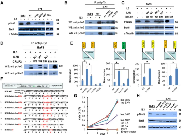
Biochemical analysis was consistent with the growth assays. Stat5
(Figure 2A), Jak1, Jak2, and Stat3 (Figure 2B) were phosphorylated
in the absence of cytokine in BaF3 cells transduced with the
IL7R
insPPCL(positive control that constitutively activates the
JAK-STAT pathway),
4IL7R
V253G, and IL7R
insEKVbut not in cells
expressing IL7R
WTor IL7R
insGEA. In IL7R
insEKVwe found higher
expression of Jak2, which correlated with the higher activation capacity
of this mutant in BaF3 cytokine withdrawal assays. Similarly,
coexpression of IL7R
WTwith CRLF2
insEIM, but not the expression
of CRLF2
insEIMby itself, caused constitutive phosphorylation of
Stat5 and Jak2 (Figure 2C,D) consistent with the known binding of
Jak2 to CRLF2
11.
To assess whether the functional and biochemical evidence of
receptor activation is associated with the oligomerization of IL7R
and CRLF2 mutated proteins in cell membranes, we used the
luciferase protein fragment complementation assay
10(supplemental
Figure 3A). HEK293 cells were transduced with homodimeric or
heterodimeric combinations of IL7R and CRLF2 fused to hGluc1 or
hGluc2 (supplemental Figure 3B). After transduction, luciferase
expression and receptor expression were analyzed in live cells
(supplemental Figure 4). Dimerization was calculated by dividing
luminescence by the mean fluorescence intensity of each receptor
thereby enabling normalization of the luminescence signal for
experimental variability resulting from transfection efficiency.
Figure 2. Noncysteine mutations in IL7R and CRLF2 cause constitutive activation of the JAK-STAT pathway and increase receptor dimerization. (A) Constitutive phosphorylation of Stat5 in BaF3 cells expressing IL7R mutants after 5 hours of cytokine deprivation. One mutant with insertion of cysteine (IL7RinsPPCL
) was used as a positive control. IL-31 indicates cells harvested after 5 hours of IL-3 deprivation followed by 20 minutes of IL-3 stimulation. (B) Identification of constitutive phosphorylation of Jak1, Jak2, and Stat3 in BaF3 cells expressing IL7R mutants after 5 hours of cytokine deprivation. IL-31 indicates cells harvested after 5 hours of IL-3 deprivation followed by 20 minutes of IL-3 stimulation. Cells were subjected to lysis and immunoprecipitation with anti p-Tyr antibody (sc-508; Santa Cruz Biotechnology). The presence of Jak1, Jak2, and Stat3 was visualized by western blotting with anti-Jak1/Jak2/Stat3 antibodies. (C) Constitutive phosphorylation of Stat5 in BaF3 cells expressing CRLF2 mutant after 5 hours of cytokine deprivation. IL-31 indicates cells harvested after 5 hours of IL-3 deprivation followed by 20 minutes of IL-3 stimulation. (D) Identification of constitutive phosphorylation of Jak2 and Stat5 in BaF3 cells expressing CRLF2 mutants after 5 hours of cytokine deprivation. IL-31 indicates cells harvested after 5 hours of IL-3 deprivation followed by 20 minutes of IL-3 stimulation. Cells were subjected to lysis and immunoprecipitation with anti p-Tyr antibody (sc-508; Santa Cruz Biotechnology). The presence of Jak2 and Stat5 was visualized by western blotting with anti-Jak2 or Stat5 antibodies. (E) Relative dimerization level of IL7R and CRLF2 from HEK293 cells transiently transfected with the WT or mutant receptor. Dimerization was calculated by dividing luminescence by the mean fluorescence intensity of each treatment, normalizing the luminescence signal for experimental variability resulting from transfection efficiency. * P, .01, 1-way ANOVA, and Student t test. (F) Alignment of WT, natural mutant (EKV), and 6 experimental mutants (numbered 1 to 6) in the TMD of IL7R. Numbers show the positions of nucleotides and corresponding amino acids. The inserted nucleotides and amino acids are shown in red. The deleted AL amino acids are shown in green. (G) Cytokine withdrawal assay of BaF3 cells transduced with IL7R experimental mutants. (H) Constitutive phosphorylation of Stat5 in BaF3 cells expressing IL7R experimental mutants after 5 hours of cytokine deprivation. IL-31 indicates cells harvested after 5 hours of IL-3 deprivation followed by 20 minutes of IL-3 stimulation. Del, deletion; Ins, insertion.
Consistent with the requirement of complementation for luciferase
expression, none of the single-receptor hGluc1 or hGluc2 constructs
that were individually transfected in HEK293 cells generated a signal
(supplemental Figure 3C). Yet when the IL7R
WThomodimer or
heterodimer was expressed, a signal was obtained reflecting a basal
level of receptor dimerization, as recently reported.
12We then
anal-yzed the influence of the somatic mutations of IL7R and CRLF2 on
receptor dimerization. As shown in Figure 2E, IL7R
ins EKV, IL7R
V253G,
and CRLF2
insEIMincreased receptors dimerization (P , .05)
whereas the IL7R
insGEAmutant that failed to provide
cytokine-independent survival of BaF3 cells, or to cause Stat5 phosphorylation,
also did not increase receptor dimerization (Figure 2E). Thinking that it
may be a “passenger” mutation, we looked for additional activating
mutations but found none (supplemental Table 3).
As CRLF2
insEIMalone increased receptor dimerization but did
not induce downstream constitutive signaling or transformed BaF3
cells, we concluded that there is no absolute correlation between
dimerization and activation.
We next proceeded to decipher the amino acids that are important
for the highly activating IL7R
insEKVmutation. We designed 6 variants
of the EKV mutation: AKV, EAV, EKA, EK, KV (including Del
ALlike the natural mutation), and ins V (Figure 2F). BaF3 lines expressing
each variant were created (supplemental Figure 5). The variants
IL7R
insEAVand IL7R
insEKAenabled cytokine-independent growth
of BaF3 cells, whereas IL7R
insAKV, IL7R
insEK, IL7R
insKV, and
IL7R
insVdid not transform BaF3 (Figure 2G). Biochemical analysis
was consistent with the growth assays. Stat5 was phosphorylated in
the absence of cytokine in BaF3 cells transduced with the IL7R
insEAVand IL7R
insEKAbut much less in cells expressing IL7R
insAKV,
IL7R
insEK, IL7R
insKV, and IL7R
insV(Figure 2H).
ConSurf
13calculations with IL7R showed that the TMD segment
manifests a unique evolutionary conservation pattern with i/i14
periodicity, equivalent to that of a perfect a-helix (supplemental
Figure 6). The conserved positions would reside in the same helix
face, providing an interface for IL7R homodimerization. All of the
natural and experimental mutations can be interpreted assuming that
activation requires IL7R dimerization with the cytosolic kinases in
predefined orientation with respect to each other. The conserved face
of the TMD helix in WT IL7R marks the orientation. Dimerization
along a different TMD helix face would not activate because the kinases
will not be facing each other. Molecular detailed interpretations of
the mutations’ effect are provided in supplemental Figures 7 and 8
and supplemental Table 4.
Taken together, these data indicate that the functional and
bio-chemical evidence for receptor activation by mutations lacking
cysteine correlates with ligand independent dimerization. There are
several examples of TMD mutations in receptors that cause
constitu-tive activation without introducing cysteine: V664E in the Neu
receptor tyrosine kinase
14-16and the dimerization inducing
muta-tions in MPL
17,18that was identified in rare familial thrombocytosis
patients.
19Our observations of noncysteine transmembrane
muta-tions in type I cytokine receptors in patients with ALL demonstrate
for the first time that such somatic mutations activate these receptors
through ligand-independent dimerization and are leukemogenic.
Acknowledgments
This work was supported by Israel Science Foundation Legacy and
iCORE programs, the Israel Cancer Research Foundation, Swiss
Bridge Foundation, USA Israel Binational Science Foundation,
Waxman Cancer Research Foundation, and William Lawrence and
Blanche Hughes Foundation. C.S. and N.T. are PhD candidates at
Tel Aviv University and this work is submitted in partial fulfillment
of the requirement for a PhD.
Authorship
Contribution: S.I. designed the study and wrote the paper; C.S.
performed research, analyzed data, and wrote the paper; N.T., N.G.,
Y.B., and N.B.-T. performed research and analyzed data; V.G., S.N.C.,
M.R.M., J.-C.T., and D.B. provided reagents and scientific expertise;
and O.R.B., G.C., A.E.K., A.B., and M.U.M. provided reagents
and data.
Conflict of interest disclosure: The authors declare no competing
financial interests.
Correspondence: Shai Izraeli, Sheba Medical Center, Tel Hashomer
Ramat Gan, 52621, Israel; e-mail: shai.izraeli@sheba.health.gov.il.
References
1. Tal N, Shochat C, Geron I, Bercovich D, Izraeli S. Interleukin 7 and thymic stromal lymphopoietin: from immunity to leukemia. Cell Mol Life Sci. 2014;71(3):365-378.
2. Wang X, Lupardus P, Laporte SL, Garcia KC. Structural biology of shared cytokine receptors. Annu Rev Immunol. 2009;27:29-60. 3. Yoda A, Yoda Y, Chiaretti S, et al. Functional
screening identifies CRLF2 in precursor B-cell acute lymphoblastic leukemia. Proc Natl Acad Sci USA. 2010;107(1):252-257.
4. Shochat C, Tal N, Bandapalli OR, et al. Gain-of-function mutations in interleukin-7 receptor-a (IL7R) in childhood acute lymphoblastic leukemias. J Exp Med. 2011; 208(5):901-908.
5. Zenatti PP, Ribeiro D, Li W, et al. Oncogenic IL7R gain-of-function mutations in childhood T-cell acute lymphoblastic leukemia. Nat Genet. 2011; 43(10):932-939.
6. Kubatzky KF, Liu W, Goldgraben K, Simmerling C, Smith SO, Constantinescu SN. Structural requirements of the extracellular to
transmembrane domain junction for erythropoietin receptor function. J Biol Chem. 2005;280(15): 14844-14854.
7. Lu X, Gross AW, Lodish HF. Active conformation of the erythropoietin receptor: random and cysteine-scanning mutagenesis of the extracellular juxtamembrane and transmembrane domains. J Biol Chem. 2006;281(11):7002-7011. 8. Hertzberg L, Vendramini E, Ganmore I, et al.
Down syndrome acute lymphoblastic leukemia, a highly heterogeneous disease in which aberrant expression of CRLF2 is associated with mutated JAK2: a report from the International BFM Study Group. Blood. 2010;115(5):1006-1017. 9. Bercovich D, Ganmore I, Scott LM, et al.
Mutations of JAK2 in acute lymphoblastic leukaemias associated with Down’s syndrome. Lancet. 2008;372(9648):1484-1492. 10. Remy I, Michnick SW. A highly sensitive
protein-protein interaction assay based on Gaussia luciferase. Nat Methods. 2006;3(12): 977-979.
11. Rochman Y, Kashyap M, Robinson GW, et al. Thymic stromal lymphopoietin-mediated STAT5 phosphorylation via kinases JAK1 and JAK2 reveals a key difference from IL-7-induced signaling. Proc Natl Acad Sci USA. 2010;107(45): 19455-19460.
12. Rose T, Pillet AH, Lavergne V, et al. Interleukin-7 compartmentalizes its receptor signaling complex to initiate CD4 T lymphocyte response. J Biol Chem. 2010;285(20):14898-14908. 13. Armon A, Graur D, Ben-Tal N. ConSurf: an
algorithmic tool for the identification of functional regions in proteins by surface mapping of phylogenetic information. J Mol Biol. 2001; 307(1):447-463.
14. Bargmann CI, Hung MC, Weinberg RA. Multiple independent activations of the neu oncogene by a point mutation altering the transmembrane domain of p185. Cell. 1986;45(5):649-657. 15. Beevers AJ, Nash A, Salazar-Cancino M,
Scott DJ, Notman R, Dixon AM. Effects of the oncogenic V(664)E mutation on membrane insertion, structure, and sequence-dependent
interactions of the Neu transmembrane domain in micelles and model membranes: an integrated biophysical and simulation study. Biochemistry. 2012;51(12):2558-2568.
16. Fleishman SJ, Schlessinger J, Ben-Tal N. A putative molecular-activation switch in the transmembrane domain of erbB2. Proc Natl Acad Sci USA. 2002;99(25):15937-15940.
17. Onishi M, Mui AL, Morikawa Y, et al. Identification of an oncogenic form of the thrombopoietin receptor MPL using retrovirus-mediated gene transfer. Blood. 1996;88(4): 1399-1406.
18. Defour JP, Itaya M, Gryshkova V, et al. Tryptophan at the transmembrane-cytosolic junction modulates thrombopoietin receptor
dimerization and activation. Proc Natl Acad Sci USA. 2013;110(7):2540-2545.
19. Ding J, Komatsu H, Iida S, et al. The Asn505 mutation of the c-MPL gene, which causes familial essential thrombocythemia, induces autonomous homodimerization of the c-Mpl protein due to strong amino acid polarity. Blood. 2009;114(15): 3325-3328.