Université de Montréal
Artificial Collagen for Cornea Repair
ParYasmina-Mia EL KHOURY
Département de pharmacologie et physiologie, Faculté de médecine
Mémoire présenté en vue de l’obtention du grade de Maîtrise ès scientifiques appliquées (M. Sc. A.) en génie biomédical
Mai 2020
Université de Montréal
Département de Pharmacologie et Physiologie, Faculté de Médecine
Ce mémoire intitulé
Artificial Collagen for Cornea Repair Présenté par
Yasmina-Mia EL KHOURY
A été évalué par un jury composé des personnes suivantes Alain Vinet Président-rapporteur May Griffith Directeur de recherche Marie-Claude Heuzey Membre du jury
1
Résumé
Les patients atteints de cécité cornéenne résultant d'une maladie ou d'une blessure dans de nombreux pays ne seront probablement pas transplantés avec des cornées de donneurs humains en raison d'une grave pénurie mondiale de tissus de donneurs. Cependant, même si des cornées de donneurs étaient disponibles, les patients présentant une inflammation ou une maladie grave ne seraient pas aidés car ils courent un risque élevé de rejet des cornées de donneurs car celles-ci contiennent des cellules allogéniques. Les implants cornéens sans cellules qui ne déclenchent pas de rejet ont été développés comme alternatives à la transplantation de donneurs humains par le laboratoire Griffith, et ont montré dans un premier essai clinique chez l'homme qu'ils régénèrent de manière stable le tissu et les nerfs cornéens. Ces implants comprenaient du collagène humain recombinant, la principale protéine structurelle trouvée dans la cornée humaine. Cependant, les collagènes de pleine longueur sont difficiles et coûteux à produire et ne peuvent pas être personnalisés. Une grande variété de peptides plus courts qui imitent le collagène et d'autres molécules de la matrice extracellulaire ont été développés et testés. Cela comprend les peptides hybrides combinant le collagène et la soie (VBsilk).
Le but de ma thèse est de confirmer les simulations de VBsilk d'un peptide hybride collagène-soie produit au Griffith Lab. Un autre objectif est de déterminer les conditions de production et de purification pour montrer que le peptide simulé peut être converti en un peptide réel.
En bref, l'ADN codant pour une séquence de VBsilk a été cloné dans ClearColi, une souche d'E. Coli à faible endotoxine. Les bactéries ont été cultivées dans des cultures à grand volume. Le VBsilk a été extrait et purifié par FPLC. SDS-PAGE a montré que des bandes de protéines de taille appropriée étaient obtenues. Par conséquent, il est possible de produire le peptide VBsilk.
3
Abstract
Patients with cornea blindness resulting from disease or injury in many countries are unlikely to be transplanted with human donor corneas due a worldwide severe shortage of donor tissues. However, even if donor corneas were available, patients with inflammation or severe disease would not be helped as they are at a high risk of rejecting donor corneas as these contain allogeneic cells. Cell-free corneal implants that do not trigger rejection were developed as alternatives to human donor transplantation by the Griffith lab, and shown in a first-in-human clinical trial to stably regenerate corneal tissue and nerves. These implants comprised recombinant human collagen, the main structural protein found in the human cornea. However, full-length collagens are difficult and expensive to produce, and cannot be customized. A wide variety of shorter peptides that mimic collagen and other extracellular matrix molecules have been developed and tested. This includes hybrid peptides combining collagen and silk (VBsilk).
The aim of my thesis is to is to confirm simulations of VBsilk, a hybrid collagen-silk peptide that was produced in the Griffith Lab. A further aim is to determine the conditions for the production and purification to show that simulated peptide can be converted into an actual peptide.
Briefly, the DNA coding for a VBsilk sequence was cloned into ClearColi, a strain of E. coli with low endotoxin. The bacteria were grown up in large volume cultures. The VBsilk was extracted and purified by FPLC. SDS-PAGE showed that appropriate-sized bands of protein were obtained. Hence, it is possible to produce VBsilk peptide.
5
Table of contents
Résumé ... 1 Abstract ... 3 Table of contents ... 5 List of Tables ... 7 List of Figures ... 9List of Acronyms and Abbreviations ... 11
Acknowledgements ... 17
Chapter 1 – Introduction ... 19
1.1 The human cornea ... 19
1.2 Corneal blindness and the need for implants ... 20
1.3 Collagen ... 22
1.4 Recombinant human collagen... 23
1.5 Peptide analogs of collagen and other extracellular matrix proteins for regenerative medicine ... 25
1.6 Designing peptides ... 28
1.7 Structural motifs in proteins ... 29
1.8 VBsilk design ... 28
1.9 Hypotheses and objectives ... 29
Chapter 2 – Materials and Methods ... 31
2.1 Protein modelling of VBsilk ... 31
2.2 Materials and equipment for VBsilk production ... 32
6
2.4 Growing up VBsilk producing bacteria ... 34
2.5 Preparation of the FPLC for peptide purification by Nickel Sulfate (NiSO4) Loading ... 37
2.6 FPLC Purification of VBsilk ... 38
2.7 SDS-PAGE ... 42
2.8 Western Blot... 43
Chapter 3 – Results... 47
3.1 VBsilk modelling ... 47
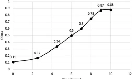
3.2 Growth Curve of VBsilk producing E. coli ... 57
3.3 FPLC Purification of VBsilk ... 57
3.4 SDS-PAGE ... 59
3.5 Western-Blot ... 62
Chapter 4 – Discussion ... 65
4.1 Protein modelling of VBsilk ... 65
4.2 VBsilk producing E. coli and induction of protein production ... 66
4.3 FPLC Purification of VBsilk ... 67
4.4 SDS-PAGE and Western Blot ... 69
7
List of Tables
Table 1. – Range and components of buffering systems tested in order to optimize yields of purified VBsilk protein. ... 39 Table 2. – SWISS-MODEL template search results for V-region of VBsilk... 47 Table 3. – List of enzymes that could cut VBsilk and the amino acid residue position affected. .. 54 Table 4. – Effect of different buffering conditions on VBsilk yield. ... 62
9
List of Figures
Figure 1. – Structure of the human cornea. ... 20
Figure 2. – Structure of collagen from amino acid to fibers. ... 23
Figure 3. – Plasmid map of pColdIII that was used to clone VBsilk.. ... 34
Figure 4. – Diagram showing the flow path within the AktaPure25 FPLC system. ... 38
Figure 5. – N-terminal region of VBsilk modelled using SWISS-MODEL based on SclB Protein from S. pyrogenes. ... 48
Figure 6. – Ramachandran Plot of VBsilk representing torsional angles of the peptide backbone. 49 Figure 7. – The first 317 amino acids of VBsilk graphed for likelihood for presence of a coiled coil using DeepCoil software. ... 50
Figure 8. – Cumulative gaussian curve of VBsilk CL domain (residue 96-317) stability profile (Collagen Stability Calculator V2). ... 51
Figure 9. – Jpred prediction of VBsilk C-terminal prediction. ... 52
Figure 10. – Growth curve of ClearColi transfected with pColdIII-VBsilk. ... 57
Figure 11. – FPLC chromatogram describing VBsilk loading on HisPrep FF 16/10 column. ... 58
Figure 12. – SDS-PAGE gel showing separation of fraction of VBsilk obtained after FPLC. ... 60
Figure 13. – Protein quantification of SDS-PAGE gels using Fij with ImageJ application. ... 61
Figure 14. – Western Blot of proteins obtained after FPLC purification using anti-His6 antibody. 63
11
List of Acronyms and Abbreviations
Å: angstrom
ALK: anterior lamellar keratoplasty B. mori: Bombyx mori
BSA: bovine serum albumin CLPs: collagen like peptides CMPs: collagen mimetic peptides CVs: column volume
DALK: deep lamellar keratoplasty dH2O: distilled water
DTT: Dithiothreitol E. coli: Escherichia coli
ECL: enhanced chemiluminescence ECM: extracellular matrix
EDC: 1-ethyl-3-(3-dimethylaminopropyl) carbodiimide EGF: Endothelial growth factor
ELPs: elastin like peptides
FPLC: Fast Protein Liquid Chromatography GAGs: glycosaminoglycans
GMEC: Global Minimal Energy Conformation HCl: Hydrochloric acid
12 HRP: horseradish peroxidase
IKVAV: isoleucine-lysine-valine-alanine IpTG: Isopropyl ß-D-1-thiogalactopyranoside KCl: Potassium Chloride
KPro: keratoprosthesis LB: lysogeny broth LPS: lipopolysaccharide M: molar
mL/min: millilitre per miniute mL: millilitres
mM: millimolar
MOPS: 3-(N-morpholino)propane sulfonic acid MPC: 2-methacryloyloxyethyl phosphorylcholine mS/cm: millisiemens per centimeter
ms: millisecond
Na-AC: sodium acetate NaCl: sodium chloride NaOH: sodium hydroxide NHS: N-hydroxysuccinimide NiSO4: nickel sulfate
OD: optical density
13 PEG: poly (ethylene glycol)
pI: isoelectric point
pKa: acid dissociation constant
PMSF: phenylmethanesulfonyl fluoride PVDF: polyvinylidene fluoride
QSQE: quaternary structure quality estimate RCF: relative centrifugal force
RGD: arginine-glycine-aspartic acid
RGDS: arginine-glycine-aspartic acid-serine RHCIII: recombinant human collagen type III RPM: revolutions per minute
S. pyrogenes: Streptococcus pyrogenes SclB/Scl2: collagen-like protein 2 TBS: Tris-buffered saline
TBS-T: Tris-buffered saline with Tween 20 TEE: translation enhancing element
Tris-Cl: Tris (hydroxymethyl) aminomethane (THAM) hydrochloride V: volt
YIGSR: tyrosine-isoleucine-glycine-serine-arginine μg/mL: microgram per millilitre
15
17
Acknowledgements
I would like to thank my colleagues from the Griffith Lab, especially Elle Edin and Fiona Simpson, with whom I have worked closely on several experiments. They helped me regain confidence in myself. Throughout the journey, I have learned to be independent and tougher.
I would like to thank my family and my close friends for encouraging me.
Principally, I would like to thank my supervisor, Dr. Griffith, for accepting me into her laboratory. She gave me the chance to work on this interesting research project.
19
Chapter 1 – Introduction
1.1 The human cornea
The cornea is the transparent outer layer of the eye. The human cornea has a diameter of approximately 11 mm. It is convex and aspherical, measuring about 500 µm in the center and gradually increasing to 550 µm in thickness at the periphery (Sridhar, 2018). The cornea serves as the main refractive surface of the eye, focusing light onto the retina for vision. Its ability to focus light is dependent upon its transparency.
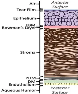
The cornea is composed of three cell-containing layers, an outermost epithelium, middle stroma, and an innermost endothelium (Figure 1). There are two acellular layers as well. The Bowman’s layer lies between the epithelium and the stroma, while the Descemet’s membrane lies between the stroma and endothelium. These acellular layers are composed mainly of collagen.
The corneal epithelium is a stratified epithelium that is non-keratinized. It comprises five to six layers of epithelial cells overlain by a tear film. The tear film keeps the epithelium moist and the epithelial cells healthy. The corneal epithelial cells have tight junctions that form a barrier between the eye and the external environment and protecting it against any pathogens in the environment.
The innermost layer of the cornea comprises a single-celled layer called the endothelium. The endothelial cells serve a pumping function by regulating the movement of molecules and ions between the aqueous humor and stroma, thereby maintaining the state of hydration, and in turn, the optical clarity of the cornea (Bonanno, 2003; Nishida, 1997).
In between the epithelium and the endothelium lies the stroma, which makes up 85% of the corneal thickness. The stroma consists of a hydrated extracellular matrix (ECM) surrounding an interconnected network of cells known as keratocytes. The main component of the stromal
20
ECM is collagen, which is arranged in regularly spaced parallel lamellae. Other major components are proteoglycans, which are glycosaminoglycans (GAGs) with protein cores. The highly organized collagen and proteoglycans are responsible for the structural integrity and transparency of the cornea (DelMonte et al., 2011). The proteoglycan-rich hydrated matrix and their associated GAGs strengthen the corneas swelling and compressive material properties (Meek et al., 2015). The main collagen components in the stroma are type I and type V fibrillar collagen.
Figure 1. Structure of the human cornea. EBM, Epithelial basement membrane ; PDM, Pre-Descemet’s membrane ; DM, Pre-Descemet’s membrane.
Image from: Blackburn, B.J., Jenkins, M.W., Rollins, A.M. & Dupps, W. A review of structural and biomechanical changes in the cornea in aging, disease, and photochemical crosslinking J. Front.
Bioeng. Biotechnol. doi.org/10.3389/fbioe.2019.00066
1.2 Corneal blindness and the need for implants
Any damage to the cornea caused by disease or injury that leads to permanent loss of transparency is likely to lead to vision impairment. There are many different causes of corneal
21
blindness, and it affects people of all ages, from the very young to the elderly. The causes can be unilateral (affects one eye) or bilateral (affects both eyes). Corneal blindness in high income (developed) countries is commonly due to aging and loss of endothelial cells and their pumping function. In low to middle income countries, infections or injuries (e.g. chemical injuries from the workplace) are more common. Globally, an estimated of 36 million people are blind, and 217 million have moderate to severe vision impairment (Bourne et al., 2017). The World Health Organization has named corneal vision impairment a priority eye disease (World Health Organization, 2018).
Transplantation with a human donor cornea from cadavers is the only widely accepted treatment worldwide for corneal blindness. There are an estimated 2000-3000 Canadians who are on waiting lists for up to two to three years in some provinces (Kramer, 2013). Globally, an estimated 12.7 million people are on waiting lists for corneal transplantation surgery, as there is only one donor cornea available for every seventy persons in need of transplantation (Gain et al., 2016). According to Gain et al. (2016), over 53% of the world’s population live in countries with no access to corneal transplantation.
In order to solve the issues of donor cornea shortage, graft failure and rejection, researchers have started the splitting of donor corneas into two pieces for use in two different surgical procedures in two different patients (Heindl et al., 2011). Corneas that are not of sufficiently high quality for transplantation have also been irradiated to sterilize and then cut into lenticules for use as partial thickness grafts and patches (Mathews et al., 2019). Expansion of human corneal cells into large sheets is another way to deal with the donor tissue shortage but at the present time, the cultured cells only address single layer issues. They still need a donor transplant if more than one cornea layer is damaged or diseased (Rama et al., 2010).
In addition to the severe human donor cornea shortage, there is another problem. Even if donor corneas were readily available, those patients with inflammation or severe diseases will not benefit from the donor corneas. This is because immune cells react against allogeneic cells in the donor corneas, and in corneas that are already inflamed, the immune system is already
22
primed to act against the foreign cells. This leads to a high chance of graft rejection or failure (49% of high risk patients are expected to have graft failure) (Yu et al., 2016).
Artificial corneas have also been developed for clinical use. Corneal prostheses known as keratoprostheses have been developed for use in patients who are at high risk for rejecting human donor corneas (Avadhanam et al., 2015). However, even the Boston Keratoprosthesis that is the most widely used device has many side effects (Aref et al., 2015; Avadhanam & Liu, 2015; Avadhanam et al., 2015; Nouri et al., 2001; Talajic et al., 2012). Their use also necessitates lifetime of antibiotics to prevent infection (Nouri et al., 2001). As such, they are only being used for corneas at the end-stages of damage or disease (Avadhanam & Liu, 2015).
Decellularized xenogeneic and human donor organs and tissues have been studied, both as scaffolds or as reconstituted implants re-seeded with stem cells (Islam et al., 2019). Decellularized corneas, and in particular xenogeneic porcine corneas, have now been tested in clinical trials as alternatives to human corneas (Shi et al., 2017).
Cell-free implants made from recombinant human collagen have also been developed tested in clinical trials by our laboratory and shown to stably regenerate the corneas of patients in clinical trials or regular and high-risk patients (Fagerholm 2009, 2010, 2014).
1.3 Collagen
Collagen is the most abundant protein in mammals. It is found in bones, hair, and skin and in the cornea. The main collagen in the human body and the cornea is type I collagen. Type I collagen is a structural collagen that forms fibrils. Each fibril is 300 nm long and is composed of three left-hand helices twisted in a right-left-handed manner. Each collagen fibril is composed of chains of repeating amino acids with the motifs glycine (Gly)-Xaa-Yaa, where Xaa and Yaa are most often proline (Pro) and hydroxyproline (Hyp) respectively (Brazel et al., 1987). The repeating sequence of amino acids with glycine as the third amino acid in the triplet, gives each collagen fibril its helical structure (Figure 2). The helix has a 9.6 Å rise per turn, 3.3 Å residues per turn and 2.9 Å rise per residue (Branden et al., 2012). The chains can geometrically twist together by forming the
23
triple-helix with a peptidyl-proline bond. The presence of hydroxyproline residues acts to stabilize the triple helical collagen molecules through stereoelectronic effects (Shoulders et al., 2009). Mutations that result from substitution of Gly in particular can results in the destabilizing of collagen fibers, leading to genetic diseases like Osteogenesis Imperfecta, where individuals have brittle bones (Shoulders et al., 2009).
In the body, collagen has a hierarchical structure. Triple helical collagen molecules are bundled together to form fibrils, and these in turn form collagen-fibers, which in the cornea are highly aligned (An et al., 2014) (Figure 2).
Figure 2. Structure of collagen from amino acid to fibers.
Image from: Nijhuis, W. & Eastwood, D. & Allgrove, J. & Hvid, I. & Weinans, H. & Bank, R. & Sakkers, R (2019). Current concepts in osteogenesis imperfecta: Bone structure, biomechanics and medical management. J. Children's Orthopaedics 13. 1-11. 10.1302/1863-2548.13.180190.
1.4 Recombinant human collagen
To prevent the possibility of animal to human zoonotic transmission of disease, laboratory produced recombinant DNA techniques have been used to produce various proteins, including
24
collagens. Recombinantly produced human collagens are essentially the same as native collagen as DNA that codes for the collagen is used to reproduce the entire collagen molecule (Olsen et al., 2003). Full-length human and recombinant-human collagens have been made in transgenic expression systems with different species such as yeast (Pichia pastoris), plants (tobacco), silkworm and human fibroblasts.
Hydrated gels or hydrogels made from recombinant human collagen have now been tested as cell-free implants in clinical trials to regenerate diseased and damaged corneas. Hydrogels made from recombinant human collagen that was crosslinked with water-soluble Ethyl-N′-(3-dimethylaminopropyl)carbodiimide hydrochloride (EDC) and its co-reactant, N-Hydroxysuccinimide (NHS) were tested in an early feasibility study on 10 human subjects (Fagerholm et al., 2010). In this study, 10 patients who were on waiting list for corneal transplantation, aged from 18 to 75, who had vision loss from advanced keratoconus (9 patients) and scarring from bacterial keratitis (1 patient). It was shown that all patients were able to regenerate their corneal tissues and nerves, and the regenerated corneas have been stable for over four years (Fagerholm et al., 2014). This shows that collagen-based implants are very effective at promoting regeneration of corneas in patients.
However, like native extracted full-length collagens, recombinant human collagen is a large macromolecule and not easy to manipulate chemically.In addition, because recombinant collagens reproduce full-length native collagens, it also contains the numerous hydroxyproline amino acids that are characteristic of the protein. To produce recombinant collagen with hydroxyproline amino acids, it is not only necessary to produce the collagen but also to produce prolyl 4-hydroxylase, the enzyme that catalyzes the formation of 4-hydroxyproline from proline amino acids (Vuorela et al., 1997). In addition, recombinant pepsin is needed to cleave the telopeptides from the full-length protein prior to use (Yang et al., 2004). This means expression of three different complex proteins to make recombinant collagen. Therefore, to produce recombinant human collagen, it is necessary to express and produce three different complex proteins.
25
1.5 Peptide analogs of collagen and other extracellular matrix proteins
for regenerative medicine
As discussed above, recombinant production of ECM proteins such as collagen is possible but can be complex. Many research groups including the Griffith laboratory has been developing short peptide equivalents to the much longer proteins of the ECM and many of these have been tested for different regenerative medicine applications (Rubert Perez et al., 2015). Among these are the collagen analogs.
Collagen-like peptides (CLPs) that are also known as collagen mimetic peptides (CMPs) have been developed as short peptide alternatives to native collagen. These range from a few amino acids in length to a few hundred amino acids. The shorter CLPs (40 amino acids or shorter) are usually synthesized on a peptide synthesizer using a solid support system. Longer CLPs, however, need to be produced either in segments that are then chemically linked together; or as a single long peptide chain using recombinant DNA technology (Edin et al., 2020).
Other ECM-derived peptides that have been used in place of much larger native proteins include cellular adhesion peptides such as arginine-glycine-aspartic acid-serine (RGDS) from fibronectin and isoleucine-lysine-valine-alanine (IKVAV) and containing tyrosine-isoleucine-glycine-serine-arginine (YIGSR) from laminin, which have been well-characterized. These have been used as integrin binding ligands in hydrogels. RGDS motifs have been used to improve keratinocytes and cell migration in skin (Bradshaw et al., 2014). The pentapeptide YIGSR promotes epithelial cell growth in cornea (Li et al., 2003), while IKVAV peptides and YIGSR stimulate neuraxial extension (Graf et al., 1987; Tashiro et al., 1989).
Another ECM macromolecule that has been widely tested for use as a biomaterial is silk. Silk is the fibrous secretion from the domestic silkworm Bombyx mori. Silk fibroin is the structural part of silk that is left after removal of the sticky sericin by degumming. Like collagen, it is made up of sequences of repeating amino acids sequence of (GAGAGS). However, silk shows better mechanical strength than collagen-based materials (Vepari et al., 2007). Silk fibroin also has controllable biodegradability (based on silk types and fabrication processes), can be transparent (with >95% transparency) while having high load bearing capacity (Vepari et al., 2007), and
26
tunable surface topography. In 1993, silk fibroin was recognized by the US Food and Drug Administration (FDA) as a biomaterial and has been used to make medical sutures (Melke et al., 2016). Silk can be easily and economically extracted using water-based mild processing and molded into various shapes, 3D constructs and micropatterns for use in regenerative medicine (Vepari et al., 2007), including corneal implants (Bray et al., 2011). However, unlike collagen which inherently has tripeptide arginine-glycine-aspartic acid (RGD) recognizable by the integrin receptors for cell adhesion, silk fibroin lacks specific cell adhesion motifs which limits its clinical application. One method to get around the inertness of silk is to hybridize it to collagen.
1.6 Modelling Protein structure
In silico methods are used to predict the 3D structures and behavior of proteins based on their primary amino acid sequence. Protein modelling relies mainly on two principles; homology-based predictions, and predictions based on physical interactions.
When predicting protein folding in silico, the Global Minimal Energy Conformation (GMEC) is the assumed actual protein conformation. Methods yielding GMEC follow some assumptions that need to be considered before running this kind of simulation. For large proteins, GMEC calculations are computationally heavy and might yield unrealistic predictions due to unaccounted for mechanistic properties. For designed proteins, a known structure often serves as the basis for the design. This allows for calculating GMEC for only shorter regions of the full strand. If computer-aided structure prediction is used for only parts of the protein, a good understanding of the folding mechanics is required. A change in a long sequence might destabilize the full motif, and even changes to flanking regions might inhibit the initial assembly of the structure. A large shortcoming of traditional computational prediction of folding is that in many proteins there are more than one individual strand involved in stabilizing a motif; many strategies utilize energy minima predictions for adjacent structures on a single strand but might fail when a structure can only be sustained in multimeric complexes. As the complexity of the assembly increases, the importance of combining design tools becomes more important. If assembly
27
dynamics for a given sequence or type of motif is known, these regions are often more easily and accurately designed by homology modeling compared to de-novo predictions (Samish, 2017).
There are a wide set of interconnected databases to turn to when performing homology-based predictions, the main ones being UniprotKB/SwissProt (going forward called by its original name; SwissProt), Uniprot/TrEMBL, RefSeq, NCBI Protein (includes RefSeq).
1.7 Structural motifs in proteins
The interactions within the amino acid chains of peptides result in a three-dimensional structure in the various segments of a protein. The most common secondary structures are alpha helices and beta-pleated sheets.
1.7.1 α-helixes
One of the most common and simplest protein secondary structures in naturally occurring proteins and peptides is the helix bundles. These are often found as coiled coils or as separate domains within larger, multi-domain proteins. Briefly, in an α-helix, the carbonyl (C=O) of one amino acid is hydrogen-bonded to the amine (Ludwiczak et al., 2019) of an amino acid that is located four places down the chain. This pattern of bonding pulls the polypeptide chain into a helical structure that resembles a curled ribbon. Each turn of the helix has 3.7 amino acids. The reactive organic or R groups of the amino acids stick outward from the α helix, where they are free to interact. α-helices are the only proteins that can exist as an individual strand with no internal strain (Berg et al., 2002). Examples of α-helix include short innate cationic peptides of the immune system like LL37, the cathelicidin produced by the human body.
Coiled coils
These are more complex, larger proteins. The sub-units of coiled coils are made up of alpha helixes with recognizable repeated motifs. Hydrophobic (H) and polar (P) residues are repeated in the heptad pattern HPPHPPP; where the individual positions are denoted abcdefg. Depending on
28
what position of the heptad starts the repeating pattern, the coiled-coil is designated an index; a pattern that starts with PPHPPPH would, for example, be a b-index coiled-coil. These motifs are common close to collagens in most organisms that produce them, and they act as assembly regions to initiate folding of collagens (Fletcher et al., 2012).
1.7.2 β-sheets
β-pleated sheets or β-sheets form when amino acid chains are stretched out and packed together to form sheet-like structures held together by hydrogen bonds. The hydrogen bonds form between carbonyl and amino groups of backbone, while the R groups extend above and below the plane of the sheet.
The peptide chains of a β-sheet may be parallel, pointing in the same direction (i.e., their N- termini are on one direction and their C-termini point the other way), or antiparallel, pointing in opposite directions (i.e., the N-terminus of one peptide chain is located next to the C-terminus of the other). They are often connected to each other by other structural elements such as loops, unstructured coils and helices. β-sheets are made up of regions rich in amino acids that are typically more hydrophobic, such as tyrosine, phenylalanine, tryptophan, threonine, valine, and isoleucine. Proline is usually only found in the unstructured regions between the secondary structures (Khakshoor et al., 2008; Nesloney et al., 1996).
1.8 VBsilk design
The overall three-dimensional structure or tertiary structure of a protein determines its final properties. In some cases, like collagen, multiple protein strands come together to form the quaternary structure of the final protein.
VBsilk was designed as a chimeric transgene protein. A series of “known” structures based on their amino acid sequence were combined to create a composite protein that in theory carries beneficial properties from several families of proteins that are regularly used for regenerative medicine applications. The focus in this design was to create a structure that had native folding
29
in each region were a derived sequence resided while also being appropriate to allow expression within a prokaryotic expression system such as Escherichia coli (E. coli).
VBsilk comprises a combination of sequences from a collagen (V-CL peptide), silk, elastin with RGD and YIGSR. The collagen region was selected due to collagens contribution to mechanical properties of ECM, and as collagens are recognizable to several cell surface receptors. Silk can confer high tensile strength. Elastin can both contribute elasticity that allows the material to retake its original form after repeated deformation, and while RGD and YIGSR are well-known fibronectin and laminin cell adhesion sites. During the design process, the focus was on feasibility of expression, and biological properties; advanced structural modelling was not performed. As discussed above, the combination of collagen and silk fibroin is considered to achieve improved mechanical and biological properties of hydrogels. Both collagen and silk are natural protein fibers that have been shown to be biocompatible and promote wound healing (Vepari et al., 2007; Fagerholm et al. 2010, 2014). VBsilk, which combines both collagen and silk amino acid sequences is also likely to be biocompatible. However, testing in vitro and in vivo is needed for confirmation.
1.9 Hypothesis and Objectives
This project focuses the translation of a novel collagen-silk-based hybrid protein, VBsilk, from an in silico design into a protein. The hypothesis I was testing is that through modelling and understanding folding, we can predict or explain behaviors in buffers and thereby optimize a purification strategy for VBsilk.
My first objective was to confirm simulations that a hybrid collagen-silk peptide that was produced in the Griffith Lab that we refer to as VBsilk, does contain collagenous helices in its predicted structure. My second objective was to determine the conditions for the production and purification of VBsilk, and to show that the design can be converted into actual peptides that can be purified and quantified.
31
Chapter 2 – Materials and Methods
2.1 Protein modelling of VBsilk
Protein BLAST searches were performed using BLASTp (NCBI) directly, or by proxy search in the MODEL online tool (Bienert et al., 2017; Guex et al., 2009; Waterhouse et al., 2018). SWISS-MODEL was the primary tool used for general homology modelling in cases where full length templates were available. The given quaternary structure quality estimate (QSQE) score reflected the expected accuracy of the interchain contacts. SWISS-MODEL outputs verification data in form of Ramachandran plots as a complement to the similarity data acquired. In the parts of the sequence where templates were missing, or where the known structure was too short for efficient homology modelling, a combination of manual review of primary sequence for known common elements (common turn, coil, or sheet forming units) and de novo, energy minima based folding was utilized. Specialized protein prediction tools were used in conjunction with homology folding for suspected collagenous or coiled coil regions, this was performed to determine stability and type of oligomerization of these regions. For analysis of coiled coils “DeepCoil”, a neural network-based tool for detecting coil-coiled domains in protein sequences (Ludwiczak et al., 2019; Zimmermann et al., 2018) was used. For analysis of collagenous regions Collagen Stability Calculator V 2.1 (https://compbio.cs.princeton.edu/csc/) was used (Persikov et al., 2005). The prediction of protein secondary structure was made with JPred v.4 Protein Secondary Structure Prediction Server (http://www.compbio.dundee.ac.uk/jpred/) that provided predicted Solvent Accessibility and Coiled-coil regions by Lupas method (Drozdetskiy et al., 2015).
The primary sequence of VBsilk was annotated, marking source sequences from which the protein was derived. The N-terminal region was an α-helix protein that was important for protein folding and its three-dimensional structure was predicted with SWISS-MODEL (https://swissmodel.expasy.org/repository). The study of coiled coils was used to predict the protein stability in the first 317 amino acids. The collagenous CL domain sequence was modelled
32
to analyse stability of its three identical sequence with the Collagen Stability Calculator. The C-terminal region after the collagenous CL domain was predicted with JPred. Likely enzyme cutting sites were predicted using ExPASy PeptideCutter (Gasteiger et al., 2005).
VBsilk was designed by Dr. Chyan-Jang Lee and modelling was performed by Dr. Elle Edin, post-doctoral fellow in the laboratory. What I did for this thesis was to confirm the remodelling by reproducing the simulations that were done.
2.2 Materials and equipment for VBsilk production
2.2.1 Materials
Synthetic DNA for VBsilk was prepared by GenScript (Leiden, Netherlands), expanded and stored as aliquots in a -80°C freezer until used. LB Broth Miller and ampicillin powders were purchased from BioShop Canada Inc. (Burlington, ON). Isopropyl ß-D-1-thiogalactopyranoside (IpTG) (Cat #BP1755-100), bovine serum albumin (Cat #AAJ6573118), thermo scientific gel code blue safe protein stain (Cat #PI24594), his-tagged protein standard (Cat #LC5606), secondary goat anti-mouse IgG horseradish peroxidase (HRP) antibody (Cat #PI32430), enhanced chemiluminescence (ECL) chemiluminescent substrate (Cat #WP20005) and pH buffer sachets (Cat #01-911-291) were bought from Thermo Fisher Scientific (St-Laurent, QC). Urea (Cat #800-140-DG) was purchased from Wisent Inc (St-Jean Baptiste, QC). The HisPrep FF 16/10 column (Cat #28-9365-52), was purchased from GE Life Sciences Canada (Mississauga, ON). SDS-PAGE and Western Blot kits, 2X Laemmli sample buffer (Cat #161-0737), 10X Tris/Glycine/SDS (Cat #1610772), mini Trans blot filter paper (Cat #1703932), immun-blot polyvinylidene fluoride (PVDF) membranes for protein blotting (Cat #1620177), 10% Mini-Protean precast gels (Cat #4561033) and roller for western blot (Cat# 1651279) were purchased from Bio-Rad (Mississauga, ON). HyClone HyPure Wifi Quality Water (Cat #SH30221.17) was bought from VWR (Mont-Royal, QC). Western blot box (Cat #B1200-7) was bought from Diamed Lab Supplies (Mississauga, ON). The color protein broad range (Cat #P7712S) was bought from New England Biolabs Ltd. (Ipswich, MA, USA). Recombinant protein A (Cat #AB52953) was purchased from AbCam (Toronto, ON). Anti-His-Tag antibody (Cat
33
#AM1010A) was purchased from FroggaBio (Toronto, ON). pH/Conductivity meter (Cat #RK-59200-04) was bought from Cole-Parmer Canada (Montreal, QC). Unless otherwise stated, all chemicals were purchased from Sigma-Aldrich Canada Ltd (Oakville, ON). Toxic chemicals as imidazole, phenylmethanesulfonyl fluoride (PMSF), hydrochloric acid (HCl), sodium hydroxide (NaOH) were handled under the fume hood.
2.2.2 Equipment
The FPLC AktaPure 25 system (GE Life Sciences, Waltham, MA, USA) with Unicorn version 7.2 software was used for protein purification. Other specialized equipment included a TecanSpark (Tecan, Männedorf, Switzerland) microplate reader was used with SparkControl method editor version 2.2 to read OD of bacteria. The ImageQuant LAS 4000 (GE Life Sciences, Waltham, MA, USA) was used for quantitative imaging for SDS-PAGE gels and Western blots.
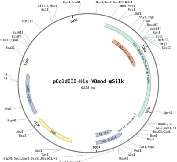
2.3 VBsilk producing plasmids
The pCold DNA family of vectors is a cold-shock recombinant protein expression system (Takara Bio Europe AB, Gothenberg, Sweden). A Cold Shock Protein A (cspA) promoter and related elements have been engineered into these vectors to increase target protein production at when these bacteria are given a cold shock (Qing et al. 2004). That is, they are switched from growing at 37 ℃ to 15 ℃. This cold shock suppresses the expression of bacterial cellular proteins and overall bacterial growth while allowing for the expression of target proteins at a high yield and with high purity as well as increased solubility as compared to conventional E. coli expression systems. The yield of target protein is expected to be 60% of all intracellular proteins produced.
34
Figure 3. Plasmid map of pColdIII that was used to clone VBsilk. The multiple cloning sites start at the NdeI site.
2.4 Growing up VBsilk producing bacteria
2.4.1 Solutions
For 4 L of LB Miller broth, 100 g of LB Miller was weighed on a precision balance and added to 4 L of dH2O in a 6 L sterilized glass flask. For 2.5 L, 62.5 g of LB Miller broth was weighed and
35
Aluminium foil was added to cover the flask and sealed with autoclaving tape to autoclave the solution at 121 °C for 30 mins.
1 M IpTG stock solution for 5 tubes of 15 mL was made was made by weighing on a precision balance 11.9 g of IpTG in 50 mL tube containing already distilled water (dH2O) and
vortexed to dissolve. The solution was moved into a 60 mL syringe and was filtered using a 0.22 μm syringe filter (Corning®) into 10 mL of 5 tubes of 15 mL each; The solution was stored in -20 °C.
2 M imidazole stock solution was prepared by adding 200 mL of dH2O in a 1L bottle. 27.23
g of imidazole was weighed on precision balance and combined to the 200 mL and topped 1 L of dH2O. Stock solution was filtered with 1 L filter bottle 0.2 μm then stored in 4 °C.
0.2 M PMSF stock solution was prepared by weighing 348.4 mg of PMSF on precision balance and added into a 50 mL tube where 10 mL of 100% ethanol was added then vortexed to dissolve. Solution was stored in -20 °C.
Ampicillin stock solution for 5 tubes of 15 mL was made by weighing on a precision balance 5 g of ampicillin in 50 mL distilled water (dH2O) and vortexed to dissolve. The solution was moved
into a 60 mL syringe and was filtered using a 0.22 μm syringe filter (Corning®) into 10 mL of 5 tubes of 15 mL each; The solution was stored in -20 °C.
2.4.2 Growing ClearColi©
The protocol used is a laboratory protocol that is described in Edin et al. (2020). ClearColi© are E. coli that have been genetically modified to expressed significantly reduced amounts of lipopolysaccharide (LPS) to not cause an endotoxic response in human cells. These cells were used to express VBsilk.
Flasks of 4 L or 2.5 L or 2 L LB Miller broth were sterilized and cooled to room temperature, 4 mL or 2.5 mL or 2 mL ampicillin was added since ampicillin has a final concentration of 100 μg/mL. With a 50 mL serological glass pipet, 40 mL of the LB miller and ampicillin prepared was pipetted in a 125 mL sterilized glass flask. The frozen aliquot tube of bacteria ClearColi pColdIII-VBsilk in -80 °C freezer was scratched with a 10 μm tip and placed in the mix then incubated in a shaking incubator with an aluminium foil at 250 rpm at 37 °C for, care was taken to not thaw the
36
bacteria pellet above -80 °C. 1 mL was measured with a serological glass pipet to load in a cuvette, the optical density (OD) was read with cuvette application on Tecan Spark, with the Spark control method editor version 2.2. The absorbance of bacterial growth was read at 600 nm since large particles, in the range of several microns, have high absorbance at 600 nm. When the smaller feeder culture reached OD 1, it was added to the 4 L LB Miller and ampicillin or 2.5 L or 2 L LB Miller and ampicillin then incubated at 250 rpm at 37 °C until OD600 of 1 was reached.
To induce VBsilk protein expression, 1 mL/L of IpTG stock was added to the bacterial cultures, yielding a final concentration of 1 M. A magnet stirrer was added to the flask and this was placed on a stirring plate and set to 300 rpm in a 4 °C cold room for 24 hours. After 24 hours, a 1 L phosphate lysis buffer prepared by combining 50 mM phosphate buffer, 690 mM NaCl, 13.5 mM KCl, pH 7.4, 20 mM imidazole from the 2 M stock, 5 M urea, 0.5% Triton X-100, 10% v/v glycerol and 1 mM PMSF in ethanol (added separately) then stored in 4 °C.
PMSF was added to inhibit serine peptidases that cleave peptide bonds and prevent denaturation. After ~24 hours, the culture was divided into 1 L centrifugal tubes and centrifuged at 4000 relative centrifugal force (rcf) for 10 mins at 4 °C. Supernatant was removed and a volume of phosphate lysis buffer that was five times greater than the amount of pellet collected was added to re-suspend the pellet. Re-suspended crude lysates were transferred in 50 mL tubes and frozen at -80 °C for 24 hours. The crude lysate was then thawed and sonicated at 60% amplitude; 1 s on, 200 ms off; 4 × 2 mins with 4 mins of cooling on ice between each cycle to further break down any bacterial cell membranes. Tubes were centrifuged at 15000 rcf for 30 mins at 4 °C and supernatant was collected in a sterile glass bottle.
All bacterial pellets were lysed as described above or stored frozen in a -20 °C freezer until used.
37
2.5 Preparation of the FPLC for peptide purification and Nickel Sulfate
(NiSO
4) Loading
20% ethanol was made in a 1 L cylinder by diluting 20 mL of 100% ethanol to 1 L dH2O then filtered
with 0.2 μm filter bottle of 1 L. The AktaPure 25 FPLC system (Figure 4) was cleaned according to the manufacturer’s directions as per the user’s manual.
Histidine increased affinity of nickel ions and made his-tag proteins to bind further and so column was loaded with NiSO4. The column was cleaned with 1 column volumes (CVs) of 20%
ethanol with a flow rate of 10 mL/min, with 5 CVs of distilled water with 5 mL/min of flow. 0.4CVs of NiSO4 was passed through the column at a flow rate of 1 mL/min.
Column cleaning was repeated after NiSO4 loading using 5 CVs of distilled water and 20%
ethanol were repeated. When the run was ended, the column cleaning was repeated with a further 2 CVs of 20% ethanol from tube A1 (shown in Figure 4) at 5 mL/min of flow and 10%
concentration of tube B1. Concentration of tube B1 was set at 0% and switched to tube A2 and run
with 2 CVs of 20% ethanol. The column was equilibrated with 20% ethanol to be stored for a longer period.
38
Figure 4. Diagram showing the flow path within the AktaPure 25 FPLC system.
Inlet valve: Two A and two B inlet ports. No integrated air sensor. InA/InB: Inlets delivering buffers or sample to system pumps. System pumps: Delivery of buffers or sample in purification runs. Pressure monitor: System pressure reading. Mixer: Mixing buffer composition homogeneously. Injection valve: Sample directing onto the column. Column valve: Column connection to instrument. UV: Measuring UV absorbance at a fixed wavelength of 280 nm. Cond.: Measuring conductivity of buffers and proteins. Fraction collector: Collection of multiple fraction tubes (GE Health Care Life Sciences, 2012b).
2.6 FPLC Purification of VBsilk
A range of buffers and different buffering conditions e.g. type of buffer (components of buffers), concentrations of buffers, detergents and other additives were tested. The buffers are important to prevent sudden pH changes that could irreversibly affect their folding, solubility, and future function. In general, the pH of the buffer solution should be within 1.0 pH unit of the pKa to provide appropriate buffering capacity, i.e. there is sufficient amount of the molecule in both its acidic and basic forms to neutralize the solution in case of a H+ or OH- influx. Common protein
39
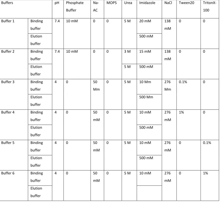
purification buffers include sodium or Tris phosphate. Tris-based buffers could not be used because they contain primary amines that could interfere with future amide crosslinking of the purified VBsilk to produce hydrogels. Hence, a sodium phosphate-based buffering system was selected. Table 1 shows the buffers that were tested.
Table 1. Range and components of buffering systems tested in order to optimize yields of purified VBsilk protein.
Buffers pH Phosphate
Buffer
Na-AC
MOPS Urea Imidazole NaCl Tween20 TritonX-100 Buffer 1 Binding buffer 7.4 10 mM 0 0 5 M 20 mM 138 mM 0 0 Elution buffer 500 mM Buffer 2 Binding buffer 7.4 10 mM 0 0 3 M 15 mM 138 mM 0 0 Elution buffer 5 M 500 mM Buffer 3 Binding buffer 4 0 50 Mm 0 5 M 10 Mm 276 Mm 0.1% 0 Elution buffer 500 Mm Buffer 4 Binding buffer 4 0 50 mM 0 5 M 10 mM 276 mM 1% 0 Elution buffer 500 mM Buffer 5 Binding buffer 4 0 50 mM 0 5 M 10 mM 276 mM 0 0.1% Elution buffer 500 mM Buffer 6 Binding buffer 4 0 50 mM 0 5 M 10 mM 276 mM 0 1% Elution buffer
40 Buffer 7 Elution buffer 7.9 0 0 50 mM 5 M 10 mM 276 mM 0.1% 0 Elution buffer 500 mM Buffer 8 Elution buffer 7.9 0 0 50 mM 5 M 10 mM 276 mM 1% 0 Elution buffer 500 mM
FPLC is used to separate macromolecules for purification of large amounts of protein mixtures. The present aim was to get rid of all undesired impurities. Briefly, 1 L of binding buffer and elution buffer were prepared for buffer 1 and 1 L of buffer 2 was made and filtered with 0.2 μm filter bottle of 1 L. For buffers 3 to 8, 3 L of binding buffers were prepared for sample dialysis (appendices A.1), 700 mL was filtered with 0.2 μm filter bottle of 1 L while 300 mL of elution buffer was made and was then filtered with 0.2 μm filter bottle of 250 mL.
VBsilk contains a his-tag for purification. Urea was added to provide denaturation conditions in which his-tag is completely exposed so it can bind to the nickel columns. Imidazole was used in binding buffer to protect the column resin and avoid excessive binding of non-target proteins. Imidazole is used in the elution buffer to be able to extract protein. System was cleaned according to FPLC system manufacturer handbook and many FPLC runs were made for optimization to obtain a purified VBsilk in a 4 °C fridge.
All buffers were used at the same temperature of 4 °C fridge. 1 L of buffer 1 was prepared respectively and filtered with 0.2 μm filter bottle of 1 L. As first run, 85 mL of the collected VBsilk supernatant was applied to the column containing NiSO4, pre-equilibrated with 5 CVs of binding
buffer 1. After washing the column with 5 CVs of loading buffer 1, the protein was linearly eluted in elution buffer 1 and 25 mL was collected in fraction collection tubes. The unpurified sample was collected in the flow through, in a clean collection flask, by running into an outlet valve. Column got cracked due to its clogging from previous runs made in the laboratory. A new column was charged with NiSO4 as method 2.5. Another run was made with a new method by
pre-equilibrating the column with 5 CVs of binding buffer 1, applying 75 mL of VBsilk supernatant into the column that was then washed with 5 CVs of binding buffer 1 and the protein was eluted with
41
4 different gradient elution steps of 3 CVs each. Fraction volume of collected tubes was set at 10 mL. With these parameters buffer 2 was tested using a sample volume of 200 mL. 10 mL of sample was used with the same previous buffers in another run to further purify the protein by changing the methods with 4 different gradient elution steps where first step was at 4 CVs and others of 3 CVs each. 20 mL of the resulted flow through was loaded and tested with buffer 2 in a new run using this new method. To differentiate sample purification, two same runs were made with this method before and after loading the column freshly with NiSO4: by charging the column with 80
mL and 100 mL of sample supernatant respectively. Since column was recently filled with NiSO4,
another attempt of undiluted 50 mL protein supernatant was applied to the column with buffer 1.
A fresh sample batch was produced and was performed in two different runs of buffer 1 with the new method, as sample volumes were 72 mL and 60 mL respectively. In run no. 14, the usual automated run was replaced by manual sample application (due to an issue with automation in the instrument). Manual loading and elution steps were reset to 3 CVs each. Manual sample application was used until run no. 17, using buffer 1 (shown in Table 1) with sample volumes at 80 mL, 45 mL, 70 mL and 7 mL each.
Different buffers with the addition of detergents to promote protein denaturation were tested. 3 L of binding buffers 3 to 8 were made and 2 L were used to dialyze sample against those buffers respectively. The remaining, 700 mL was filtered with 0.2 μm filter bottle of 1 L while 300 mL of elution buffer was made and was then filtered with 0.2 μm filter bottle of 250 mL.
As a final run, production of VBsilk was made with a different bacterium aliquot and was loaded in column using the last method with buffer 1. In all the runs the protein fractions containing peaks of purified protein in chromatogram were pooled and tested on gels.
42
2.7 SDS-Polyacrylamide Gel Electrophoresis
To check the size of the proteins produced by ClearColi to determine if VBsilk was being produced, Sodium dodecyl sulphate (SDS)-polyacrylamide gel electrophoresis (SDS-PAGE) was used to separate out proteins to check for VBsilk bands in the bacteria lysate
2.7.1 Solutions
Laemmli sample buffer (2X) used consisted of 4% SDS (w/v), 20% glycerol, 0.004% bromophenol blue (colour dye), 0.125 M Tris-Cl, pH 6.8 and 10% dithiothreitol (DTT).
1 M DTT was made by weighing 1.54 g of DTT in a precision balance. In a 15 mL tube, 4 mL of dH2O was added then the weighed DTT was poured in and topped to 10 mL with dH2O. The
stock solution was vortexed until dissolved. The solution was moved with a 10 mL syringe and filtered using a 0.22 μm syringe filter into a 10 mL tube then stored in -20 °C prior to use.
1 L 1X running buffer was prepared by diluting 1:10 10X Tris/Glycine/SDS in dH2O.
20 mg/mL bovine serum albumin (BSA) stock was made by weighing 20 mg of BSA in a 1.5 mL sterile tube with an analytical balance and added 1 mL of dH2O then vortexed.
0.5 μg/mL BSA was mixed in a 1.5 mL sterile tube by adding 39 μL of dH2O and 1 μL of 20
mg/mL BSA stock.
2.7.2 SDS-PAGE gel electrophoresis
A 1/10 dilution of the protein fractions with identifiable peaks on the FPLC chromatogram, as well as a sample from the flow through were prepared along with an undiluted flow through, and 0.5 µg BSA protein benchmark were separated out on SDS-PAGE gels. DTT was added to the samples to denature the proteins for faster separation. Glycerol was added to each sample to allow for easier loading. Sample dilutions and 0.5 µg BSA were prepared in a 1.5 µL sterilized Eppendorf tubes by adding 80 µL of dH2O, 10 µL of glycerol and 10 µL of sample. In a new sterile tube, 20 µL
of the dilution and undiluted flow through were added to 20 µL of 2X Laemmli sample buffer. Samples were boiled at 98 °C at 500 rpm for 10 mins with thermomixer.
43
Pre-cast “Mini-protean” 8% acrylamide gels were placed into the electrophoresis tank assembly. 1X running buffer was placed in the upper chamber and lower chambers. The combs were removed from the gels and samples were loaded into the wells. 20 µL samples was loaded in each well, apart from 3 μL ladder of color protein standard loaded in well 1. After connecting the tanks via electrodes to the power supply, the gels were run at 300 V for approximately 20 mins or until dye front of the samples was approximately 1 cm from the bottom of the gel. When run was done, the gel cassette assembly was opened, the precast gel got open with key according to arrows respectively and wells were cut with razor blade. Gel was put in western blot box, washed with 100 mL dH2O for 5 mins 3 times, with 20 mL blue stain and with 100 mL dH2O for 1h.
Densitometry was performed to quantify the proteins in the different lanes. This was done by placing the gel on the center of orange plate for colorimetry where digitalization and epi-illumination of the gel were captured by using azure c200 or Fuji image QuantLAS 4000 (GE Life Sciences). BSA weighed 66.5 kDa and VBsilk around 80 kDa. Bands were selected and analyzed for quantification on Fiji with ImageJ application: To outline the first lane, the rectangular selection tool was used. On the application status bar to analyze gels, the first lane was selected. The rectangular selection tool was moved to the next lane and to analyze gels on the status bar the next lane was selected. This step was repeated for each remaining lane. To generate the lane profile plots, analyze, gels then plot lanes were selected. The straight-line selection tool to draw base lines was used so that each peak of interest determined a closed area. Size was measured by clicking inside each peak with the wand tool and was copied on an Excel sheet. Protein concentrations were estimated by calculating the ratio of absorbance between the target fraction band and the BSA band; the BSA had a known loading amount, and thus enabled us to back calculate the amounts that had been in the fraction samples.
2.8 Western Blots
VBsilk produced in ClearColi© was tagged with a 6-His tag to allow the identification of target proteins in the subsequent bacterial lysate. The presence of the 6-His Tag allowed for the use of
44
an anti-His Tag antibody to localize and confirm using Western or immune-blotting that bands of protein of interest had been obtained in the SDS-PAGE separated protein samples.
2.8.1 Solutions
1 L of 1X standard transfer buffer or Towbin buffer was made by adding 25 mM Tris, 192 mM glycine, 20% (v/v) methanol (pH 8.3).
2 L of 1X tris-buffered saline (TBS) was composed of 20 mM Tris-HCl and 500 mM NaCl, with the pH meter the pH was adjusted to 7.5.
For 1 L tris-buffered saline with Tween20 (TBS-T), 1 L of 1X TBS was combined in a 1 L bottle with 0.05% Tween 20.
Blocking solution was made by weighing in a 50 mL tube 2.5 g BSA to 50 mL of 1X TBS-T. Solution was then vortexed until dissolved.
0.1 M NaOH was prepared by dissolving 400 μL of 12.5 M NaOH in a 50 mL tube of dH2O.
Primary antibody was 5 mL of blocking solution with 5 μL of His-Tag antibody. Secondary antibody was 5 mL of blocking solution with 10 μL HRP.
2.8.2 Western blotting of separated proteins
SDS-PAGE separated proteins were first transferred from the gels onto polyvinylidene fluoride (PVDF) membranes that bind proteins with high efficacy through hydrophobic interactions. The PVDF membrane was activated at room temperature with 20 mL of methanol in a western blot box for 5 mins. The submerged protein transfer was prepared on the gel holder cassette by preventing air bubbles on superposing respectively a black foam pad, 2 blotting filter papers, the SDS-PAGE gel, the activated PVDF membrane, 2 blotting filer papers and a second black foam pad. Each step was rolled on with the roller for western blot to prevent air bubbles. Gel holder cassette was locked and closed then put in the electrode assembly in the same way than another gel holder cassette that contained 2 black foam pads (cathode side was faced the cathode side of blotting module).
45
The electrode assembly was put in mini tank according to electrodes colors where transfer buffer was filled until top to the mark of 2 gels of the mini tank and a small stir bar in was added. In a medium plastic bucket, ice to cover bottom was put and the mini tank placed in. The bucket was settled on mini stir plate and the stirring was turned to 2 until magnet was spinning in the middle of the mini tank. Transfer buffer was filled to 4 gels of mini tank and closed electrode lid respectively to colors and connected in voltage power supply. Crushed ice was inserted in medium plastic bucket and mini tank and lid were covered. Power supply voltage was set to 100 V and run for 1 hour. When run was done, gel holder cassette containing the protein transfer was opened and membrane was taken out. If ladder was showing on membrane and gel was clear, transfer was correctly done. If not, it was run for further timing.
For immunodetection of the His-tagged VBsilk bands, PVDF membranes with transferred protein bands were washed with 10 mL of TBS-T to cover membrane for 5 mins on rotating table. With a 25 mL glass serological pipet 20 mL of Ponceau S was added for 5 mins on rotating table, then washed with 10 mL of dH2O for 5 mins on rotating table and bands were identified.
Membrane was washed with 5 mL of 0.1 M NaOH for 5 mins on rotating table. In order to avoid membrane-protein interaction, membrane was blocked with 5 mL of blocking solution to cover membrane for 1 hour on rotating table in room temperature. 5 mL of primary antibody His-Tag was added to detect the 6 x His VBsilk on a rotating table overnight in cold room. After 24h primary antibody was discarded, and the membrane washed 2 x 5 mins with 5 mL TBS-T. The membrane was then overlain with the secondary HRP antibody consisting of affinity-purified antibody that detected specified target, for one hour in a cold room, in an aluminium-covered western blot box due to the light sensitivity of the reagents. The PVDF membrane was then washed 2 x 5 mins with 5 mL TBS-T with a final wash of 5 mL TBS for 5 mins. An ECL high sensitivity chemiluminescent substrate detection kit was used for imaging the His-tagged VBsilk bands on a Fuji image QuantLAS 4000 densitometer. The densitometry was made by placing the membrane on top of a zip lock on a black tray for chemiluminescence by eliminating air bubbles. In a 50 mL tube with 1000 μL pipet, 2 mL of Reagent A was added of the ECL Chemiluminescent Substrate kit and 2 mL of Reagent B then mixed with pipet and spilled on surface of membrane to cover it eventually for 1 min (equal parts of reagents). Excess of substrate was absorbed with kimwipes.
46
The membrane was read on chemiluminescence with Fuji image QuantLAS 4000. Recombinant protein A weighed 39 kDa and bands were shown with a known size of 10 mg. Bands were selected and analyzed for quantification on Fiji with ImageJ application.
47
Chapter 3 – Results
3.1 VB silk modelling
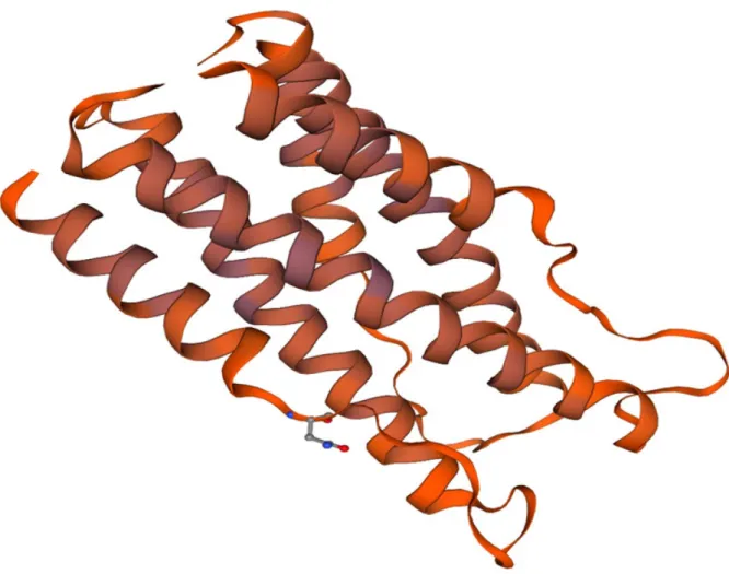
A high-quality template for the V-region of VBsilk was found using the SWISS-MODEL software, based on the Streptococcus pyrogenes (S. pyrogenes) protein Scl 2 (also referred to as “bacterial collagen”). Both the protein design source and the template were derived from the same sequence, but VBsilk had been “humanized” by substituting amino acids found in human collagen, so they were not identical (54.69% identity). The SWISS-MODEL template search is summarised in Table 2.
Table 2. SWISS-MODEL template search results for V-region of VBsilk. The source structure for the model was an x-ray crystallography model of SclB (/Scl2).
The resulting model for this region is a coiled coil homotrimer with an outer alpha helical bundle surrounding the coiled coil (Figure 5). The feasibility of this folding is validated by a Ramachandran plot (Figure 6). The Ramachandran plot represented torsional angles phi (Φ) and psi (Ψ) of the peptide backbone with residues that are likely to reside in α-helices or structures. The β-structures are few and likely interrupted as they reside in linker regions between α-helixes. The stability of the inner coiled coil is described by the second peak in (Figure 7).
Template Seq Identity
Oligo-state QSQE Method Resolution
Seq
Similarity Range Description
4nsm.1.A 54.69
homo-trimer 0.16 X-ray 1.60Å 0.44 44 - 106
Collagen-like protein SclB
48
Figure 5. N-terminal region of VBsilk modelled using SWISS-MODEL based on SclB Protein from Streptococcus pyrogenes. An inner tri-helical coiled coil is surrounded by three shorter coils,
49
Figure 6. Ramachandran Plot of VBsilk, representing torsional angles of the peptide backbone. The dark region in the top left indicates residues likely to reside in β-structures; the dark region intersecting the 0 on the Y-axis indicates residues likely to reside in α-helices. Residues residing in the upper right quadrant indicates turns. The residue found in the lower right quadrant is in a sterically un-favoured position.
50
Figure 7. The first 317 amino acids of VBsilk graphed for likelihood for presence of a coiled coil using as predicted using the “DeepCoil” software (Ludwiczak et al., 2019; Zimmermann et al.,
2018)
P-BLAST performed on the first third of the collagenous region of VBsilk yielded a close match to LPXTG cell wall anchor domain-containing protein (88% identity, 92% positives, Evalue: 2e-30) and
to collagen-like protein 2 (also known as SclB/Scl2) (88% identity, 92% positives, Evalue: 1e-29) both
of these sequences are S. pyrogenes proteins. The partial search was due to the VBsilk sequence containing three identical repeats of this region. The collagenous region downstream of the V-region was modelled for stability. Peak stability was found in the central V-regions of each of the three identical repeated collagen sequences (Figure 8).
51
Figure 8. Cumulative gaussian curve of VBsilk CL domain (residue 96-317) stability profile as determined using the Collagen Stability Calculator V2. (Persikov et al., 2005).
The C-terminal domain of VBsilk was difficult to model using traditional methods. P-BLAST searches could not find similar proteins due to its short repeated, non-naturally occurring motifs. Jpred prediction did not find any likely beta or alpha structures in this region but did yield a high confidence prediction for accessibility of peptide side chains (Figure 9). A high degree of buried residues were predicted across this span of the protein, only very short sequences were predicted as entirely exposed, the largest part of this part of the sequence has a partially buried nature, but with > 25 % of side chains being exposed, small regions (4 positions) formed peaks of extremely likely buried residues; GSGAGAGSASC, SGGGAGAGSGAGAG, SGGGAGAGSG, and VGVPGVGVPGGGA were the most likely to be buried.
53
Figure 9. Jpred prediction of VBsilk C-terminal prediction, sequence starting at residue 318 (Drozdetskiy et al., 2015). Lupas_21, Lupas_14 and Lupas_28 did not find any predicted α-helices or β-sheets. The JNETSOL25, 5 and 0, represents prediction of Solvent Accessibility. The levels are: Exposed, 25% or more Solvent Accessibility accessible, 5% or more Solvent Accessibility accessible and Buried (< 5% exposed). The JNetPRED is the consensus prediction. JNetCONF identified the confidence estimated for the prediction, high values mean high confidence prediction. The JNetHMM-HMM is the profile-based prediction and JNETPSSM-PSSM is the based prediction, no α-helices or β-sheets were found.
54
The ExPASy PeptideCutter was used to create a list of any likely enzyme targets since the aim is to produce biomaterials for use in the human cornea as implants. The results are summarized in Table 3 (Gasteiger et al., 2005). As seen, many potential degradation sites were found in the VBsilk sequence. Most of these enzymes are ones that are produced by E. coli, or other bacteria including skin dwelling bacteria. Examples include the Arg-C and Asp-N endopeptidases, and Staphylococcal peptidase I. Enzymes that are found in human tissue include thrombin, trypsin and pepsin.
Table 3. List of enzymes that could cut VBsilk and the amino acid residue positions affected.
Name of enzyme No. of
cleavages Positions of cleavage sites
Arg-C proteinase 21 18 60 68 70 129 141 153 207 219 231 285 297 309 330 357 403 430 476 503 549 576 Asp-N endopeptidase 23 8 41 43 45 76 80 95 127 149 173 205 227 251 283 305 322 358 395 431 468 504 541 577 Asp-N endopeptidase + N-terminal Glu 61 8 9 11 12 19 23 32 41 42 43 45 58 60 63 76 80 85 95 97 112 115 118 127 130 139 142 149 151 163 173 175 190 193 196 205 208 217 220 227 229 241 251 253 268 271 274 283 286 295 298 305 307 322 358 395 431 468 504 541 577 619 BNPS-Skatole 1 67 CNBr 5 1 50 108 186 264 Chymotrypsin-high specificity (C-term to [FYW], not before P)
7 49 56 67 326 399 472 545
Chymotrypsin-low specificity (C-term to [FYWML], not before P)
31 1 2 3 4 5 6 7 21 25 29 40 45 47 49 50 53 54 56 57 67 71 72 78 80 108 186 264 326 399 472 545 Clostripain 21 18 60 68 70 129 141 153 207 219 231 285 297 309 330 357 403 430 476 503 549 576 Formic acid 23 9 42 44 46 77 81 96 128 150 174 206 228 252 284 306 323 359 396 432 469 505 542 578 Glutamyl endopeptidase 38 10 12 13 20 24 33 43 59 61 64 86 98 113 116 119 131 140 143 152 164 176 191 194 197 209 218 221 230 242 254 269 272 275 287 296 299 308 620 Iodosobenzoic acid 1 67 LysC 29 14 16 34 35 52 69 73 84 95 117 126 135 138 149 162 173 195 204 213 216 227 240 251 273 282 291 294 305 615
55 LysN 29 13 15 33 34 51 68 72 83 94 116 125 134 137 148 161 172 194 203 212 215 226 239 250 272 281 290 293 304 614 NTCB (2-nitro-5-thiocyanobenzoic acid) 4 321 394 467 540 Pepsin (pH1.3) 16 21 24 25 28 29 40 44 45 52 53 56 57 79 92 170 248 Pepsin (pH>2) 24 21 24 25 28 29 40 44 45 48 52 53 55 56 57 66 67 79 92 170 248 326 399 472 545 Proteinase K 197 8 10 12 13 15 17 19 20 21 22 24 25 26 29 32 33 37 39 40 43 45 48 49 51 53 54 55 56 57 59 61 63 64 67 71 72 75 79 80 86 92 98 99 101 105 111 113 116 119 122 125 131 132 140 143 144 147 152 156 159 164 170 176 177 179 183 189 191 194 197 200 203 209 210 218 221 222 225 230 234 237 242 248 254 255 257 261 267 269 272 275 278 281 287 288 296 299 300 303 308 312 315 326 327 331 336 338 342 344 348 350 353 355 362 364 366 369 371 376 378 382 384 388 390 393 399 400 404 409 411 415 417 421 423 426 428 435 437 439 442 444 449 451 455 457 461 463 466 472 473 477 482 484 488 490 494 496 499 501 508 510 512 515 517 522 524 528 530 534 536 539 545 546 550 555 557 561 563 567 569 572 574 581 583 585 588 590 595 597 601 603 607 609 612 617 620 622 Staphylococcal peptidase I 37 10 12 20 24 33 43 59 61 64 86 98 113 116 119 131 140 143 152 164 176 191 194 197 209 218 221 230 242 254 269 272 275 287 296 299 308 620 Thermolysin 118 7 14 16 21 25 28 31 39 49 52 53 56 62 70 71 74 78 79 100 104 107 110 124 146 155 158 178 182 185 188 202 224 233 236 256 260 263 266 280 302 311 314 326 330 335 337 341 343 347 349 352 361 363 368 375 377 381 383 387 389 392 399 403 408 410 414 416 420 422 425 434 436 441 448 450 454 456 460 462 465 472 476 481 483 487 489 493 495 498 507 509 514 521 523 527 529 533 535 538 545 549 554 556 560 562 566 568 571 580 582 587 594 596 600 602 606 608 611 Thrombin 4 357 430 503 576 Trypsin 50 14 16 18 34 35 52 60 68 69 70 73 84 95 117 126 129 135 138 141 149 153 162 173 195 204 207 213 216 219 227 231 240 251 273 282 285 291 294 297 305 309 330 357 403 430 476 503 549 576 615
These chosen enzymes do not cut: Caspase1 Caspase10 Caspase2 Caspase3 Caspase4
56 Caspase5 Caspase6 Caspase7 Caspase8 Caspase9 Enterokinase Factor Xa GranzymeB Hydroxylamine Proline-endopeptidase Tobacco etch virus protease