Fundamental and practical aspects in the formulation of colloidal polyelectrolyte complexes of chitosan and siRNA
Running head: Nanosized complexes of chitosan and siRNA Tim Delas and Christophe Schatz
Laboratoire de Chimie des Polymères Organiques (LCPO), Univ. Bordeaux, CNRS, Bordeaux INP, UMR 5629, 33600 Pessac, France
Emails : [email protected], [email protected]
1. Introduction
The main approach to design non-viral vectors of siRNA relies on the electrostatic complexation of the negative charges of the siRNA backbone with positive charges carried by lipids or polymers.[1]
The positive charges arise from amine groups protonated at physiological pH or quaternized amine groups that are charged at any pH. When the complexation between siRNAs and cationic lipids or polymers is carried out under appropriate conditions of concentration, charge ratio and pH, the complex particles formed have colloidal sizes compatible with systemic administration. The electrostatic interaction ensures a high loading of siRNA without losses during the process which is advantageous in comparison to other encapsulation processes. The adjunction of specific chemical groups in the lipid or polymer structure endows the synthetic vector with biological functionalities of interest like cell targeting, cell internalization or endosomolytic activity. However, the use of synthetic cationic polymers like polyethyleneimine, polyamidoamine or poly(L-Lysine) faces some toxicity issues which considerably limits their applications for human therapy.[2] In this context the design of synthetic vectors from naturally occurring materials is highly desirable. Due to their biocompatibility, hydrophilicity and great variety of properties, polysaccharides are materials of interest for biological applications. Among them, chitosan is unique because it is the only native polysaccharide containing amine groups.[3] It is thus obvious that chitosan has been extensively studied for its complexing properties with nucleic acids.[4] Other polysaccharides can be cationized into chitosan-like polymers by chemical modification of functional groups on the polysaccharide chain but they have been less used than natural chitosan for the delivery of nucleic acids.[5]
This chapter focuses on the preparation of colloidal polyelectrolyte complexes of chitosan and siRNA with both fundamental and practical physico-chemical aspects. Only native chitosan is considered here, mainly for two reasons. Firstly, native chitosan has remarkable physicochemical properties that deserve to be used without having to modify its chemical structure, which in any case would alter its biological integrity. Second, a good understanding of the physical chemistry of the complexation of native chitosan with siRNA is a necessary prelude to study complexes obtained from chemically modified chitosans. In addition, excellent review papers have already reported on the state of the art of chitosan-based siRNA delivery systems including those based on modified chitosans.[6-9]
The first section of the chapter deals with the electrostatic complexation of polyelectrolytes which is the main approach considered here to form colloidal siRNA delivery systems. The goal is to give the reader the important facts about the morphology, kinetics and thermodynamics of polyelectrolyte complexes that can apply to any types of polyelectrolytes, synthetic or natural ones including chitosan and siRNA. A focus is also made on the specific conditions required for the formation of stable complex nanoparticles in aqueous media. In the second part, some important aspects related to chitosan and its solution properties are introduced. Chitosan does not behave as a regular polyelectrolyte, as evidenced by its low cationicity, its behaviour strongly dependent on the degree of acetylation and its propensity to form aggregates. The main trends observed in the complexation behaviour of chitosan with a model polyanion are also summarized in order to introduce the formation of nanosized complexes from chitosan. The third and main section deals with the electrostatic complexation of chitosan with siRNAs to form submicron-sized colloidal particles. The complexation thermodynamics and the colloidal aspects are described with emphasis on some important practical considerations.
2. General aspects of the polyelectrolyte complexation 2.1 Weak and strong polyelectrolyte complexes
Polyelectrolytes (PEs) are polymers containing ionic or ionizable groups. PEs can be classified as polyanions, polycations or polyampholytes depending on whether the chains carry negative, positive charges or both. They can also be classified as strong or weak whether they are charged at any pHs or in a limited pH range. PEs of opposite charges interact through electrostatic interaction to form complexes. The complexation is thermodynamically favored by the decrease of the Coulomb energy of PEs due to the charge pairing and the large entropy gain due to the release of counterions and water molecules initially bound to charges.[10-15] Secondary interactions like hydrogen bonding, hydrophobic effect or van der Waals forces may also take place. However, the electrostatic contribution generally dominates the interaction between the PEs. For example, it was shown by titration calorimetry that the electrostatic interaction represents approximately 90% of the total binding free energy of polycation-DNA complexes.[16] The polyelectrolyte complexation has a cooperative character, i.e. the stability of the complexes increases with the chain length of the PEs.
This can be explained by considering that the loss of translational entropy of PE chains is highest for the first binding between two oppositely charged units but then becomes less important for the binding of the neighboring units. Therefore, the complexation reaction proceeds according to a zipping mechanism.[17] When the total free energy change exceeds the kinetics energy of the polymer chains, a stable complex is formed. This condition is generally achieved when the PE chains contain a minimum of 5 to 8 units as found for various charged polymers including DNA. For comparison, hydrogen-bonded complexes require at least 40 monomer units, the bonding energy being much lower than for electrostatic complexes.[17] The cooperativity character of the complexation is important in that it allows the formation of stable complexes even under dilute conditions.
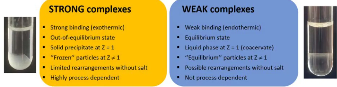
It is interesting to question the physical state of PE complexes i.e., whether the complexes are solid-like or liquid-like (Figure 1).[18,19] Although there is no theory yet to answer this fundamental question, the physical state can be simply assessed by observing the system at the charge stoichiometry where the interaction between PEs is maximized. Either a solid precipitate or a dense liquid phase should be seen to settle at the bottom of the tube. For very dilute systems, solid aggregates or micron-sized droplets should be observed by light microscopy. In a solid-like complex, the charges of PEs are in strong interaction with a complete release of counterions and water molecules. The complex behaves as a hydrophobic, dry material in which polymer chains have lost their mobility. In a liquid-like complex, also referred as complex coacervate, the interaction between the PEs is much weaker. The electrostatic bonds are only transient and the charges remain hydrated giving a high mobility to PE chains.[20,21] Note that the term ‘coacervate’ is often misused for any types of polyelectrolyte complexes. The reason why PEs can form either solid (strong) or liquid (weak) complexes is mainly due to the initial hydration of the PE chains. PEs with a large hydration shell tend to form weak complexes because the water molecules coordinated to the charges oppose the pairing of charges, resulting in a loose and still hydrated complex structure.[22] Other factors such as the ionic strength, the nature of counterions, the temperature or the dielectric constant of the medium can also influence the type of complex obtained. Thus, polyelectrolyte complexes form a continuum of morphologies, in relation with the total intensity of the interaction that can be varied by different means.[23]
Figure 1. Main characteristics of strong and weak polyelectrolyte complexes. Some variations are possible according to the chemical system considered.
Strong and weak complexes have distinctive thermodynamic signatures. For strong complexes, the electrostatic interaction leading to the formation of tight ion-pairs give rises to a typical negative enthalpy of complexation (exothermic) whereas for weak complexes, the hydration forces opposing the electrostatic interaction are often at the origin of a positive enthalpy of complexation (endothermic).[22] Considering, as a first approximation, that the entropy gain is similar for both types of complexes, the absolute value of the free energy and the associated complexation constant (K) are much higher for strong complexes than for weak complexes. As a result, in salt-free solutions, strong complexes tend to form frozen structures far from thermodynamic equilibrium while weak complexes are more likely to form equilibrium structures.[24,25] This has practical implications for the preparation of complexes. Indeed, the complexation reaction being an extremely fast process (<5 µs, i.e. nearly the diffusion-collision time of PE chains), experimental factors like the PE concentration, the mixing time, the charge ratio can greatly influence the final morphology of strong complexes while they exert little or no influence on weak complexes (coacervates).[26] In other words, the PE complexation proceeds under kinetic control for strong complexes and thermodynamic control for weak complexes.[25,24] Since complexes obtained from nucleic acids are often of the strong type as judged by their typical negative enthalpy of complexation,[27,16] their final structure is typically under kinetic control and thus dependent on the initial conditions.
2.2 Colloidal polyelectrolyte complexes
The complexation of polyelectrolytes is well suited to the design of colloidal particles as those used in gene delivery applications. The particle forming process relies on the charge neutralization of PE segments followed by their segregation through hydrophobic interaction into small aggregates stabilized by free unpaired charges (Figure 2).[26,28] PE systems forming either solid or liquid-like complexes can be used to form particles, the particle core being more hydrophobic and compact in the case of strong complexes. Two conditions are generally required to obtain stable particles of complex in aqueous solutions:[29] i) a relatively low PE concentration (< 1 wt. %) to avoid uncontrolled complexation and ii) an excess of one of the two PEs to have unpaired charges at particle surface. By assuming at first glance a complexation stoichiometry of 1:1 between cationic and anionic monomer units,[10] the charge excess of complex particles can be predicted from the mixing ratio, Z = [+]/[-] which corresponds to the molar ratio between cationic and anionic units of PEs introduced in the medium. Thus, positively charged complexes are formed at Z > 1 and negatively charged complexes at Z < 1. It is worth to notice that Z slightly differs from the nitrogen to phosphate ratio (N:P) used for nucleic acid complexation as the latter does not take into account the ionization rate of amine groups. The complex particles have typical sizes in the 50 to 500 nm diameter range
with levels of aggregation reaching up to several thousand chains.[25] The level of aggregation is essentially controlled by the concentration of PEs, higher concentrations systematically favoring the formation of bigger particles. Structural parameters of PE chains such as the chain length, the charge density and the chain flexibility also impacts on the particle size in such a way that all factors favoring the charge pairing between opposite PEs lead to complexed segments with a high density of neutral ions pairs, resulting in highly neutralized complexes and hence to small particle sizes.[29]
Furthermore, better particle stabilization is achieved when the polymer in excess is of higher molar mass than its default counterpart as this favors the formation of a larger outer shell (Figure 2). The addition of salt leads to subtle effects.[30] For relatively low salt concentrations, the screening effect of the salt makes the PE chains more flexible and the interaction between opposite PEs weaker.
Consequently, the charge complexation is favored by conformational adaptation of PE chains which leads to a lower level of aggregation. For higher salt concentrations, the screening of the charged, stabilizing shell of the excess component leads to aggregation of particles which can be irreversible.[25] In the specific case of weak complexes, the addition of a high salt concentration cause their dissolution rather than aggregation.[30]
Figure 2. Structure and mechanism of formation of colloidal complexes from a polycation in excess and a default polyanion. Primary complexes are rapidly formed by charge neutralization of PE segments of opposite charge. Their aggregation through hydrophobic interaction results into larger colloidal structures stabilized by the polycation in excess which is only partially complexed with the particle core. Further addition of polycation leads to the progressive shrinkage of the corona and the densification of the core.
The process of particle formation is easy to setup because it simply consists of mixing the two aqueous solutions of PEs under stirring. However, attention must be paid to the choice of mixing conditions in the case of strong complexes forming frozen structures like those based on nucleic acids. For these systems, the particle size is strongly dependent on the mixing time, i.e. the time required to homogenize components, with fast mixing conditions systematically leading to smaller particle sizes due to a better homogenization of PEs prior to complexation. This is similar to a precipitation process through nucleation and growth where fast mixing rates result in nucleation being favored over growth and hence smaller particle size. Therefore, all parameters influencing the mixing time like the PE concentration, the rate of addition, the stirring speed and even the size and shape of the reactor must be carefully considered.[31] In general, fast mixing conditions not only result in smaller but also more stable complex particles.[32] Another important parameter related to the formation of strong complexes is the order of addition. Indeed, it is advisable to always add the PE in default to the one in excess in order to avoid the system to reach, even transiently, the charge neutrality (Z=1) where irreversible aggregation may occur.[28,33] For PEs forming strong complexes,
the use of microfluidic tools is then particularly relevant for controlling the mixing time and neglecting the order of addition.[34] For PEs forming weak complexes, the mixing conditions should be less relevant since the complexes are formed at equilibrium.
In general, the formation of electrostatically stabilized particles of complex requires a significant excess of one of the two PEs since complexes tend to aggregate over time at Z close to 1. It is thus advisable to work at Z < 0.7 (excess of negative charges) or Z > 1.5 (excess of positive charges) to obtain stable suspensions of complexes. For Z values far away from the unity, the dispersions contain unreacted PE chains coexisting with complex particles. The amount of free PE is difficult to predict but it can be experimentally determined by gel electrophoresis or by titration of the supernatant after centrifugation of the dispersions.[35,36] The centrifugation is also a convenient technique to remove the excess of PE. It is noteworthy that complexes of nucleic acids are typically formed at N:P ratios above 10 and used without removal of excess polycation. In fact, the excess of free polycation can destabilize the cell membrane, thus favoring the cell internalization of particles.[37]
3. Chitosan 3.1 Origin of chitosan
Chitosan is obtained from the partial N-deacetylation of chitin under alkaline conditions. If chitin was first isolated from mushrooms (Braconnot, 1811), it is nowadays mainly produced from various marine seashell wastes and thus available in large amounts in the fishery industry.[38] Chitin and chitosan from fungal origin are also commercially available under the trade name Kitozyme.[39]
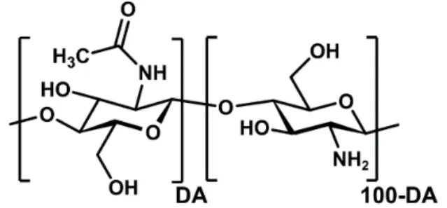
Chitin and chitosan have the same chemical structure as they are linear copolymers composed of D- glucosamine and N-acetyl-D-glucosamine linked by β-(1→4) glycosidic bonds (Figure 3). Their solubility properties allow to differentiate them: chitosan is soluble in slightly acid aqueous solutions like 1% acetic acid through the protonation of primary amines whereas chitin is insoluble in water, except for very low degrees of polymerization (DP < 6). Chitosan is therefore obtained when the degree of acetylation (DA) i.e., the molar fraction of acetylated units is sufficiently low so that enough glucosamine units can be protonated to solubilize of the polymer chain. This condition is typically achieved when the DA is lower than 60-70% depending on the molar mass and the origin of chitosan.
Figure 3. Chemical structure of chitosan. DA refers to the degree of acetylation
Controlling the DA and the molar mass is essential with regard to the physicochemical and biological properties of chitosan. The control of the DA is achieved either by heterogeneous deacetylation of chitin with concentrated sodium hydroxide solutions at high temperatures[40] or by homogeneous reacetylation of a highly deacetylated chitosan with acetic anhydride under mild conditions in hydro- alcoholic mixtures.[41] The deacetylation approach tends to favor an irregular structure with a block distribution of acetylated units due to the semi-crystalline nature of the initial chitin which decreases
the accessibility of the sodium hydroxide to reactive sites.[42] Conversely, the acetylation method gives a random distribution of the acetylated units along the chain. It is important to know the origin of the chitosan used because the pattern of the acetylated groups can strongly influence the solution properties of chitosan[42,43] and also the biological activity.[44,45] For example, a chitosan with DA=50% can be only partially soluble in acid solutions when heterogeneously deacetylated whereas it is soluble in neutral aqueous medium when homogeneously acetylated at the same DA. Most commercial chitosans have a DA around 20% and are obtained by heterogeneous deacetylation;
therefore, the presence of irregularities in the acetylation pattern cannot be excluded. The control of molar mass is achieved through hydrolysis of the polymer chain by chemical or enzymatic approaches[46-49] or by means of an ultrasound treatment.[50] The polymer degradation can be monitored by size exclusion chromatography or viscometry. These different approaches allow to prepare chitosan oligosaccharides with a good control of the degree of polymerization.[49,51]
3.2 Biological properties
Chitosan is known for its high biocompatibility, biodegradability, bioadhesivity (thanks to the presence of protonated glucosamine residues) as well as anti-tumour activity. This makes chitosan suitable for use in several applications in medicine. Biological properties of chitosan and chitosan oligosaccharides have been reviewed in several articles. [52-59] One of the mains advantages of chitosan is its low cationicity at physiological pH (see below) in comparison to polycations like polyethylene imine or polylysine. This unique property is associated to a low toxicity of chitosan both in oral and intravenous routes[60,61] and a limited activation of the complement system.[62] The low cationicity is also at the origin of a high buffering capacity that promotes the proton sponge effect in the endosomal compartments, which is relevant in the context of siRNA delivery.[63]
3.3 Solution properties
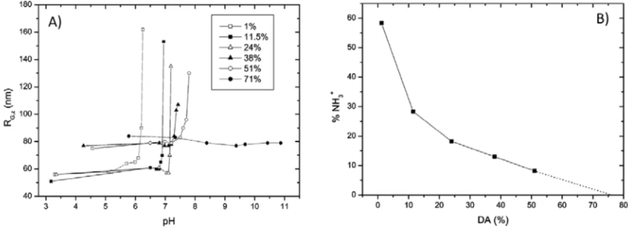
The solubility of chitosan is an important parameter to consider in the complexation with siRNAs because the polyelectrolyte chains need to be well solvated to perform the ion pairing in best conditions. It is generally stated that chitosan is a weak polybase with an intrinsic pK0 of 6.5 and is therefore soluble in slightly acidic solutions.[64] This unusually low pK0 value for a polyamine is at the origin of the low cationicity of chitosan. For comparison, PEI has pKa values between 8 and 10 depending on its molar mass and structure.[65] However, the solution behaviour of chitosan is more subtle as shown by the increase of the pK0 from 6.46 for DA 5% to 7.14 for DA 89% which illustrates an increase of the cationicity of amine groups in a more hydrophobic environment, as observed for simple aliphatic amines with the pKa increasing with the length of the alkyl chains.[66] As a consequence, the critical pH at which chitosan begins to precipitate increases from 6.2 for DA=1% to 7.5 for DA=50% (Figure 4.a). Meanwhile, the fraction of protonated glucosamine residues determined at critical pH decreases with DA thus evidencing that a lower degree of protonation is required to solubilize chitosans with higher DAs (Figure 4.b). [67] For 50% < DA < 70% the chitosan appears to be soluble at any pHs but the solutions then correspond more to a dispersion of highly solvated microgels instead of isolated polymer chains.[66-68] The chitosan microstructure has also an influence on solution properties as a random distribution of acetyl groups along the polymer chain improves the solubility compared to a block distribution.[42] Other parameters affecting the chitosan solubility are the molar mass, the polymer concentration, the nature of the acid used for protonation, the concentration and nature of the salt added in solution (if any).[68-71,67] Regarding the chain conformation, the chitosan behaves at acidic pHs as a semi-flexible polyelectrolyte with a
persistence length varying from about 5 nm to 10 nm when the DA increases.[72] Another important aspect of the solution behaviour of chitosan is its propensity to form aggregates in aqueous media.
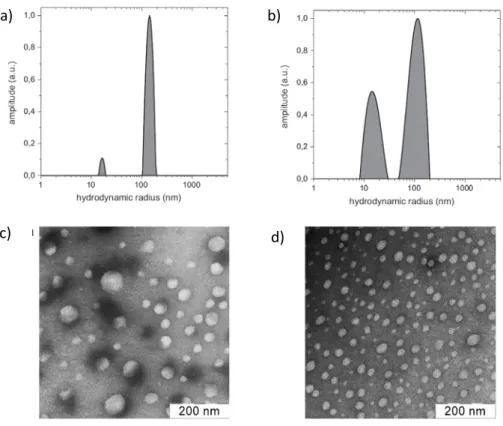
Even if they represent only a small fraction of the total material, the aggregates can strongly interfere with light scattering which is a common technique used to characterize polyelectrolyte complexes (Figure 5).[73-75] The aggregation is predominant for chitosan of high DA and/or molar mass but aggregates have been also found in solution of chitooligosaccharides or fully deacetylated chitosans.[76,77] Hydrogen bonds at low DAs and hydrophobic interaction at higher DAs are thought to be responsible of the aggregation of chitosan chains even though the exact mechanism of aggregation is not yet well understood.[77] The aggregates can be removed to a certain extent by membrane filtration with varying efficiency depending on the material and porosity of the filter.[78]
Figure 4. Solubility behaviour of chitosan varying in DA. A) variation of the gyration radius of chitosan as function of pH. B) Proportion of protonated glucosamine residues at the critical pH as a function of DA. Reprinted with permission from Schatz et al., Langmuir 2003, 19, 9896. Copyright 2003 American Chemical Society. [67]
Figure 5. Formation of aggregates in chitosan solution (Mw~120 kg/mol). a,b) Size distribution obtained by dynamic light scattering at 90° for 1.2.10-3 mM solutions of chitosan prepared in 0.3 M CH3COOH in the presence of 0.05 M CH3COONa. c,d) Transmission electron microscopy images of aggregates negatively stained with uranyl acetate. a) DA=0% , b,d) DA= 56%, c) DA=12%. The solutions are filtered with a 0.45 µm cellulose acetate membrane filter before analysis. Reprinted from Carbohydrate Polymers, Vol 87, Philippova et al., Aggregation of some water-soluble derivatives of chitin in aqueous solutions: Role of the degree of acetylation and effect of hydrogen bond breaker, 687-694, Copyright (2012), with permission from Elsevier.[77]
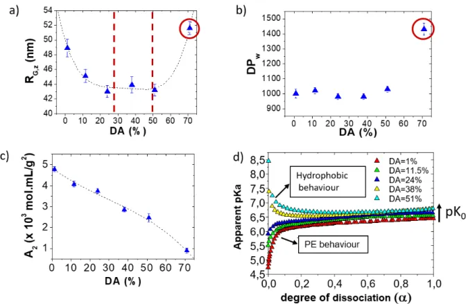
The solution properties of chitosan can be summarized through a general law of behaviour obtained through potentiometry, light scattering, interferometry and viscometry which highlights three distinct domains of DAs (Figure 6):[79,80,72] i) for DAs ≤ 28% which corresponds to the domain where the ionic condensation takes place according to Manning, the chitosan behaves as a hydrophilic polyelectrolyte whose charge density increases by decreasing the DA. In this domain, the conformation and properties of chitosan are almost fully determined by the intra- and intermolecular electrostatic interaction which can be screened by addition of salt. ii) for 28% < DA < 50%, hydrophilic and hydrophobic interactions are balanced, the physicochemical behaviour of chitosan is not strongly influenced by the DA; iii) For DAs > 50% the prominence of hydrophobic interactions results in a higher cationicity which is of interest for applications at neutral pH but the self-association of chains also increases in the same way.
a)
c)
b)
d)
Figure 6. Illustration of the solution behaviour of chitosan as function of the degree of acetylation (DA%). a-c) Static light scattering analysis of chitosan in acetate buffer at pH 4.5 providing the Z- average of the radius of gyration of chitosan (a), the weight average degree of polymerization (b) and the second virial coefficient (c). d) Apparent values of pKa and intrinsic pK0 as function of the dissociation degree of chitosan; the chitosan is fully deprotonated at α =1. Adapted with permission from Schatz et al., Biomacromolecules 2003, 4, 641-648 and Schatz et al., Langmuir 2003, 19, 9896- 9903. Copyright 2003 American Chemical Society. [80,67]
3.4 Complexation behaviour of chitosan
The electrostatic complexation of chitosan with polyanions of synthetic or natural origin has been the topic of numerous research works related to fundamental aspects and applications.[81,82] Chitosan can form either strong (solid) or weak (liquid) complexes depending on the nature of polyanion and the conditions of complexation. An illustrative example is the chitosan-hyaluronan system which is able to form both types of complexes depending on the pH and the charge ratio, thus highlighting the existence of a continuum of morphologies.[83] In the case of nucleic acids, chitosan form strong complexes with DNA, RNA and homopolynucleotides in standard conditions of complexation as shown by the formation of solid precipitates.[84] The negative enthalpy of complexation of the chitosan with DNA is also in agreement with complexes forming strong interaction.[85] Therefore, the formation of chitosan-nucleic acid complexes must be dependent on initial conditions such as the mixing time.
Chitosan-based colloidal complexes are obtained according to the complexation mechanism depicted in Figure 2. Some general trends related to the complex sizes can be deduced from a systematic study performed on the chitosan-dextran sulfate system used for vaccine delivery.[35,28,86,29,87]
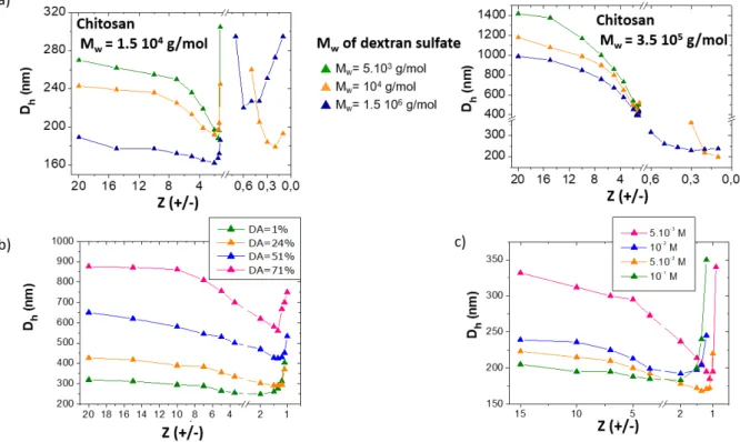
Dextran sulfate (DS) is a polyanion containing 2.2 sulfate functions per saccharidic residue. First, an increase of the molar mass of chitosan from 15 kDa to 350 kDa systematically leads to larger particle sizes when the chitosan is in excess ([+]/[-] > 1) (Figure 7.a). This behaviour illustrates the presence of
a rigid shell of chitosan around particles whose thickness directly depends on the chitosan chain size.[87,28] In comparison, the molar mass of DS has a minor influence on particle size, even when added in excess ([+]/[-] < 1), because it is a much less rigid polymer than chitosan. By increasing the DA, the particle size increases due to the mismatch in charge density between polycationic chitosan and polyanionic dextran sulfate ; more chitosan molecules are needed within complexes to achieve the ion pairing (Figure 7.b).[87] It is noteworthy that particles always become smaller and probably denser when the charge ratio approaches the unit, before the precipitation occurs, as a consequence of the densification of the core particle and the shrinking of the outer shell. Such a trend has been observed in many PE systems. When Z=1, the complex particles always precipitate as a consequence of the full charge neutralization. The increase of the ionic strength by addition of a salt favors the conformational adaptation of PE chains in complexes leading to smaller particles with a denser core (Figure 7.c) and the contraction of the chitosan shell. No flocculation of the complexes was observed at moderate salt concentrations ([NaCl] < 0.15 M).[28]
Figure 7. Role of various parameters on the hydrodynamic diameters (Dh) of PE complex particles obtained from chitosan and dextran sulfate at pH 4. a) Influence of the molecular weight (Mw) of PEs (DA chitosan = 15%). b) Influence of the degree of acetylation of chitosan (Mw chitosan = 1.5 105 g/mol, Mw dextran sulfate = 1.5 106 g/mol). c) Influence of the salt concentration (NaCl) (Mw chitosan
= 1.5 104 g/mol, DA= 15%, Mw dextran sulfate = 104 g/mol). Adapted with permission from Schatz et al., Biomacromolecules 2004, 5, 1882-1892 and Schatz et al., Langmuir 2004, 20, 7766-7778.
Copyright 2003 American Chemical Society.[28,87]
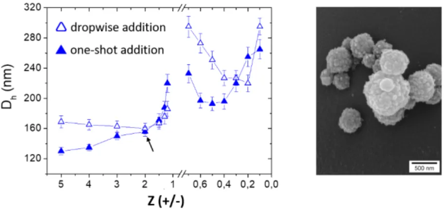
The chitosan-dextran sulfate system forms strong complexes as seen by the formation of a precipitate at charge stoichiometry. Then the mixing conditions deserved to be studied in details (mixing order and rate of addition of components). In general, the dropwise addition (or titration method) does not allow to form stable complexes on either side of Z=1 because of the quasi- irreversible aggregation occurring at the charge neutrality. Then, complexes with an excess of
positive charges must be prepared by addition of dextran sulfate to chitosan and vice versa for negatively charged complexes.[28] Conversely, the one-shot addition of components allows to reach any Z values without observing precipitation, except at Z=1 of course. The complex particles are also smaller compared to those obtained in a dropwise addition (Figure 8).[28] The PE composition in the complex particles can be accurately determined at any Z values through a rather simple depletion method where the suspensions of complexes were centrifuged and the free PEs in the supernatants assayed with specific dyes (Orange II for chitosan and Toluidine blue for dextran sulfate).[35]
Significant differences of composition in the complex particles were reported according to the polymer in excess, which could be attributed to various complexation mechanisms in relation with the reactivity and conformation of each polymer.[35]
Figure 8. (left) Influence of the rate of addition of PE components on the particle size (Dh : hydrodynamic diameter) for the chitosan (Mw=1.5 104 g/mol, DA= 15%) – dextran sulfate (Mw = 1.5 106 g/mol) system at pH 4. (right) SEM image of complex particles obtained at Z (+/-) =2 through a one-shot addition of dextran sulfate into chitosan. Adapted with permission from Schatz et al., Biomacromolecules 2004, 5, 1882-1892. Copyright 2004 American Chemical Society. [28]
A major issue of chitosan-based polyelectrolyte complexes is their poor stability under physiological conditions due to the presence of electrolytes and the pH value at which the degree of protonation of chitosan is low.[88-92] The effect of electrolytes on PE complexes depends on the type of complex : weak complexes tend to redissolve at a critical salt concentration whereas strong complexes precipitate.[30] Various strategies have been proposed to improve the stability of particles without modifying the chemical structure of chitosan. First, it has been shown that using rather highly acetylated chitosans (DA ~ 50%) considerably improves the long-term storage stability of positively charged complex particles in 150 mM NaCl solution or PBS buffer.[93,88,94] This can be explained by considering the two main modes of stabilization of particles according to the DA. In brief, for DAs <
50% the stabilization is predominantly electrostatic and thus the presence of salt may induce the aggregation of particles.[88] For DAs ~ 50%, the charge density is lower but the chain hydration remains high enough to allow steric forces to stabilize the particles. The strong increase of the cationicity of amine groups at DA=50% must also account for a better stability of the electrostatic linkages at physiological pH.[66] Using DAs higher than 50% is not advisable because of the strong tendency of the polymer chains to self-associate. Another method to achieve the stability in physiological conditions (PBS, 37°C) is obtained by introduction of multivalent ions like Zn2+ in the complex dispersion. These ions form coordination bonds with the hydroxyl and amine groups of chitosan and with some functional groups of the polyanion as well. This results in an ionic cross-
linking of the PEs chains in the complex which provides better stabilization.[95] Following this approach, zinc-stabilized complexes of chitosan/hyaluronan or chitosan/chondroitin sulfate have been developped for the HIV-1 inhibition.[96] Similarly, complexes of chitosan-ATP (adenosine triphosphate) or chitosan-TPP (tripolyphosphate) can be stabilized in physiological media with Fe3+
ions that bind strongly to amine groups of chitosan and phosphate groups of ATP.[97]
4. Chitosan-based siRNA delivery systems
When it comes to the delivery of siRNA, all parameters related to the chitosan structure and the conditions of complexation described in the previous sections need to be consider to optimize the formulation of siRNA complexes. There is some discrepancy among the data reported in literature which can be partly explained by various issues related to the origin of chitosan, the mixing conditions and other experimental factors. Anyway, the large body of data in the literature allows to establish a certain number of trends regarding the complexation thermodynamics, the colloidal characteristics of complexes, their stability in the biological environment, and their ability to disassemble and release siRNA in the cell to perform gene silencing.
4.1 Thermodynamics of complexation.
The strength of the interaction between chitosan and siRNA can be determined by using the isothermal titration calorimetry (ITC) technique which provides the enthalpy and entropy changes of the complexation (∆H and ∆S), the stoichiometry of complexation (n) and the binding constant (K) which is related to change in the Gibbs free energy change (∆G) through the equation: ∆G = ∆H-T∆S
= -RT ln K. ITC is therefore a performant technique to evaluate the thermodynamics of complexation under various conditions of pH, ionic strength, molar mass and DA of chitosan. The determination of the thermodynamic parameters allows to better anticipate the chemical stability of complexes in presence of serum proteins and their capacity to dissociate within the cell to release siRNA. However, the analysis of thermograms and their interpretation can be complicated by various heat effects related to buffer ionization, solvation, hydrophobic interaction, hydrogen bonds, most of which are difficult to quantify. The thermograms are usually fitted with a one binding site model but a two binding site model may be necessary to distinguish the contribution of the ionic complexation from the aggregation or condensation steps that may occur, particularly in the vicinity of the charge stoichiometry.[27,98] For chitosan oligosaccharides, aggregation phenomena have been also evidenced at early stage of the titration.[99]
The enthalpy change associated to the ion pairing between chitosan and siRNA is negative as expected for a polyion pair forming strong complexes.[99,100] However, it was shown for the chitosan-DNA system that a significant amount of the heat released arises also from the ionization of the buffer (BH+ B + H+) used as solvent. Indeed, the complexation of chitosan with a strong polyanion like DNA or siRNA leads to an increase of the pKa of chitosan because of its reduced electrostatic potential upon complexation, thus causing a proton transfer from the buffer to chitosan.[85] The entropy contribution T∆S is always higher than ∆H in absolute value which confirms that the release of counterions drives the electrostatic complexation. For a fully deacetylated chitosan, the binding constant K increases sharply when the degree of polymerization (DP) of the chitosan varies from 5 to 13, which highlights the cooperativity of the complexation.[99]
A similar behaviour has been observed for the complexation of chitosan with DNA, where the affinity between the two PEs strongly increases for a chitosan DP between 6 and 9.[101] The complex
stoichiometry varies from 5:1 (protonated glucosamine:phosphate) for DP 5 to nearly 1:1 for DP 13, which was confirmed by a chemical assay of the unbound phosphate groups of siRNA in the complex dispersion.[99] In fact, the existence of a critical DP was evidenced in the past years for several polyelectrolyte systems of natural or synthetic origin.[102-104] A patch of 5 to 10 consecutive charged units generates a high electrostatic potential which destabilizes the polyelectrolyte chain and thus strongly increases its reactivity for a polyion of opposite charge.[99] Above this threshold in DP, the binding constant continues to increase with the chitosan chain length, mainly for entropy reasons due to the increasing contribution of the hydrophobic interaction between complexed segments when the chitosan chain becomes longer. [100,85,99,17] Only for very high molecular weights (> 300 kDa), a decreased of K was reported and attributed to steric restrictions of chitosan chains that hamper their interaction with the polyanion.[98] When the ionization rate of chitosan is increased, either by lowering the pH or decreasing the DA, K increases too, which is expected for a complexation mechanism dominated by the electrostatics.[85] It was also shown that the siRNA sequence[8] and the nature of the counter ion associated with chitosan[105] can influence the binding affinity, which for the latter can be explained by considering the polyelectrolyte complexation as a kind of substitution reaction of counterions.[17] In general, there is a strong correlation between the binding constant and the encapsulation efficiency of siRNA in complexes.
Best efficiencies were found for complexes prepared at high N:P ratios (> 5) with chitosans of low DA and relatively high molar masses.[106]
4.2 Physicochemical properties of colloidal complexes.
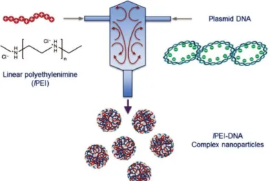
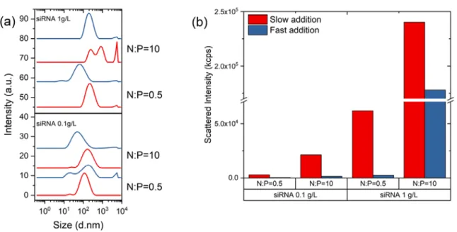
Mixing. The thermodynamic study is important for evaluating the intrinsic stability of complexes against various disassembly mechanisms occurring at the molecular level in presence of various proteins and other charged macromolecules. The study of the colloidal properties of complexes is as much important to determine the conditions of stability of particles in biological media. Before looking at the role of different parameters (molar mass, DA, N:P, pH and others) influencing the size and stability of particles, it is important to pay attention to the mixing conditions used to perform the complexation since chitosan and siRNA form out-of-equilibrium structures under the usual conditions of pH and salt concentration. As stated before, it is recommended to always add the chitosan solution to siRNA for N:P < 1 and vice versa for N:P > 1 even if the mixing step is fast. Such mixing order prevents the system to reach the neutrality where irreversible aggregation may occur. The rate of addition is equally important, as can be seen in Figure 9, where the hydrodynamic sizes of the complex particles are always smaller for a fast addition (one shot injection with a micropipette) than a slow addition (dropwise addition with a syringe).[99] Similarly, the light scattered intensity that is highly sensitive to the presence of aggregates (Iscattered∝ R6 in the Rayleigh scattering regime, with R the radius of particles) is systematically lower for a fast addition, regardless of the concentration of siRNA and N:P ratio.[99] This emphasizes that fast mixing conditions favor a good homogenization of components in the complexation medium, thus avoiding local over concentrations which may lead to aggregation phenomena. However, it is worth mentioning that the upscaling of the manufacturing process of such colloidal materials is not realistic by means of conventional mixing methods as those used here. Therefore, the implementation of micromixers or microfluidic devices to formulate polyelectrolyte complexes for gene delivery in a reproducible manner should be considered on an industrial scale (Figure 10).[107-109]
Figure 9. Light scattering analysis of chitosan (10 kDa, DA < 1%)/siRNA complexes prepared by a dropwise addition (red) or one-shot addition (blue) of the component in default to the excess one for two N:P ratios and two siRNA concentrations. a) Particle size distribution (PSD); b) Scattered intensities at 173° detection angle. Reprinted from Delas et al., Polymers 2019, 11, 1236 with permission from MDPI. [99]
Figure 10. Production of highly uniform PEI/DNA complex particles in large scale with high reproducibility using a Confined Impinging Jets (CIJ) device enabling fast and controlled mixing conditions. Reprinted with permission from Ref[107]. Copyright 2016, Wiley-VCH.
Molar mass and DA. Although the complexation reaction is essentially under kinetic control, the molar mass and DA of chitosan should not be neglected. Stable colloidal particles in the 50-200 nm size range nm can be obtained at N:P ≥ 5 with chitosans having low DAs (≤ 20%) and molar masses between 10 kDa and 150 kDa which corresponds, respectively to DP of 60 and 930 for a fully deacetylated chitosan (Figure 11).[100,110,111,106] Within this range, the chitosan chains are larger than the size of a siRNA duplex which can be assimilated to a 7 nm long rod.[112] According to the host-guest model proposed by Kabanov[113] and Tsuchida,[103] such a difference in size favors a
ladder-like complexation of the short siRNA in default along the long chitosan chains in excess. This results in the formation of neutral segments that can segregate and form the particle core, with the excess chitosan, only partially complexed, forming the outer shell (Figure 2). In fact, this is an ideal situation to prepare small and well-stabilized complexes.[106] Complex particles obtained from chitosans of lower molar masses (< 10 kDa) tend to be larger due to the poor stabilization of the short shell-forming chitosan chains.[99,114,110] For a molar mass corresponding to the critical DP, the complexes are not stable at all due to the lack of cohesion between the short complexed segments. [99] For chitosan of large molar mass (> 150 kDa), the particle size increases due to the formation of a prominent stabilizing shell.[114] Additionally the loss of conformational entropy of the long and rigid chitosan chains upon complexation prevents their compaction into a small and dense core. Besides, an increase of the chitosan molar mass also contributes to increase the positive charge of the particles (Figure 11).
Figure 11. Nanoparticle size and zeta-potential (ζ ) as a function of DDA (degree of deacetylation), the molar mass of chitosan, and N:P ratio at pH 5.5 for two concentrations of NaCl. Nanoparticles were prepared by manual mixing. Reprinted with permission from Alameh et al., Biomacromolecules 2018, 19, 112-131. Copyright 2018 American Chemical Society [106]
The role of the DA on the colloidal characteristics of complexes has been less studied. Chitosans used for siRNA delivery have typical DA values below 30 % to maximize the charge density. Within this range, the DA does not have a tremendous effect on the particle size. However, the DA strongly impacts on the surface charge as shown by the zeta potential values decreasing from +40 mV to +10 mV when the DA increases from 2% to 28% (Figure 11).[106]
Concentration and N:P. In addition to the molecular characteristics of chitosans, the formulation conditions must be considered as well. The importance of the role of the mixing method has already been discussed. Other factors to consider are the concentration of PEs and the N:P ratio, the pH and ionic strength of the complexation medium. The complexation is usually performed in diluted buffers adjusted to a pH comprised between 4 and 5 in order to maximize the ionization of chitosan. A salt can be added but the total ionic strength needs to be relatively low (< 0.1 M) to avoid extensive screening of the electrostatic interaction. The concentration of chitosan and siRNA has a strong influence on the particle size with larger particles being systematically formed when the concentration increases.[106] Ideally, the complexation should be performed in the dilute polymer regime, i.e. below the overlap concentration (c*) of polymer chains to favor as much as possible homogeneous complexation conditions. c* can be determined for conventional polymers by viscometry measurements using the equation, c*=1/[η] with [η] the intrinsic viscosity of the polymer solution. c* is around 2 g/L for a chitosan of DA=1% and Mw = 150 kDa.[80,72] For a thin rod macromolecule like siRNA, c* is given by c* = 1/L3 with L the contour length and c* expressed in units of number concentration.[115] For a siRNA of 23 bp (Mw ~ 15 kDa), c* is approximately equal to 70 g/L. Although such a high concentration of siRNA has never been reported, significant aggregation can already be seen at a concentration of 3.5 g/L where the complexes are quite big (~300 nm).[116]
Working in very dilute conditions (c << c*) is the best way to avoid local over concentration effects during the mixing step and thus obtain small complex particles. However, the counterpart can be a too low concentration of complexes for the targeted application. If needed, the particle dispersion can then be concentrated by freeze-drying followed by rehydration in a reduced volume of buffer.
The addition of various cryoprotective agents or the particle coating with hyaluronic acid can prevent aggregation phenomena during the freeze-drying.[117]
The N:P ratio is often varied in a relatively large range of values, from N:P=0.5 to N:P=50 or even higher. As previously mentioned, the N:P ratio is different from the charge ratio Z (+/-) as it does not consider the degree of ionization of chitosan which depends on the pH and DA. With a pKa of the phosphate groups close to 1.5, the charge density of siRNA is much less dependent on pH.[118] It is interesting to figure out how many chitosan chains coexist with siRNA for a defined N:P value.
Considering the degree of polymerization of chitosan (DPchitosan) and the number of base pairs in siRNA (bpsiRNA) the average number of chitosan molecules (n) per siRNA duplex can be estimated as n
= (2 * bpsiRNA/DPchitosan) * N:P. For a siRNA of 23 bp and a fully deacetylated chitosan of 25 kDa, n = 3 at N:P =10 and n = 15 at N:P = 50. Thus, depending on the chitosan molar mass, the excess of chitosan may appear less significant when considering the number of chains rather than the N:P ratio. The role of N:P on particle size is not clearly evidenced due to contradictory results in the literature. According to the complexation scheme in Figure 2 and size variations represented in the Figure 7, one should observe a compaction of the chitosan shell while approaching the stoichiometry.
Such a behaviour has been observed in some cases for N:P values between 5 and 50.[110,99,111,106] However, the extent of the particle compaction depends not only on the molar mass of chitosan but also on the intrinsic stability of the complexes which can vary for a system to another, especially in the vicinity of the charge neutrality. In general, complex particles tend to aggregate for N:P values below 5 and therefore working at higher N:P is recommended.[99] An excess of chitosan contributes to increase the positive charge of the complex as seen by zeta potential measurements and thus allows a better stabilization of the particles (Figure 11).[100,106] It is also known from studies on PEI that a large excess of polycation facilitates the transfection.[37]
The amount of free chitosan in the particle dispersion can be determined with analytical separation techniques like the asymmetrical flow field-flow- fractionation (A4F)[119] or by a using depletion
method where the particles are centrifuged and the unbound chitosan in the supernatant assayed by UV-vis spectroscopy at 260 nm[120] or 484 nm in presence of the Orange II dye.[35]
The morphology of complex particles is generally spherical (Figure 12) but elongated and less regular shapes have been also reported.[100,121,122] The size dispersity of the complexes can be evaluated by means of the so-called polydispersity index (PDI) determined by dynamic light scattering using the cumulant algorithm to analyze the correlation functions. PDI values obtained for chitosan-siRNA complexes are typically comprised between 0.1 and 0.3 which emphasizes a rather large dispersity.[110] In fact, polyelectrolyte complexes are not thermodynamic aggregates like micelles whose size is strictly given by the dimensions of the amphiphilic molecules. Rather, polyelectrolyte complexes form a continuum of structures ranging from loosely aggregated structures to fairly large aggregates (Figure 12).
Figure 12. Atomic force microscopy images of chitosan (M = 114 kDa, DA = 16%) /siRNA complex particles formed using 250 µg/mL chitosan, (A) N:P 71 and (B) N:P 6, and 1 mg/ml chitosan, (C) N:P 285 and (D) N:P 23. Reprinted from Molecular Therapy, Vol 14, Howard et al., RNA Interference in Vitro and in Vivo Using a Novel Chitosan/siRNA Nanoparticle System, Pages 476-484, Copyright (2006), with permission from Elsevier.[122]
Colloidal and thermodynamic stability. When it comes to the stability of complexes, a distinction must be made between the colloidal stability related solely to the particle behaviour and the thermodynamic stability related to the binding affinity of chitosan with siRNA. The two types of stability can be determined by studying the behaviour of complexes under various conditions of pH, ionic strength and concentrations of various polyanions. The colloidal stability of positively charged complexes against salt is reasonably good as seen from the limited aggregation observed in presence of 150 mM NaCl which corresponds to the ionic strength in physiological conditions (Figure 11).[106]
Conversely, the colloidal stability of similar complexes at physiological pH is very low due to the poor solubility of chitosan in general at pHs above 7 (Figure 4). The thermodynamic stability of the complex is also impacted at neutral pH as evidenced by gel retardation assays performed at pH 8
where the rapid dissociation of the complex and the concomitant release of siRNA molecules was clearly observed (Figure 13).[121] This highlights a dramatic weakening of the interaction between components which results from the decreasing charge density and the aggregation of chitosan molecules.[123] In fact, this is the main limitation of using chitosan for siRNA delivery. Conversely, at pH 6.5, the integrity of the complexes is effective for at least 20 hours at N:P ratios above 2 (Figure 13), which was further confirmed by the resistance of siRNA against nuclease degradation at same pH.[121] Competition assays with heparin or proteins (BSA, FBS) are also typically performed to evaluate the thermodynamic stability of complexes since large polyanionic molecules can compete with siRNA for binding to chitosan.[124] It is worth mentioning that these assays are relevant when performed at pH values where the complexes are in stable conditions. Competition assays performed in buffer like PBS at pH 7.2 can be biased by the precipitation of chitosan which leads either to the premature release of siRNA or on the contrary to its entrapment in the precipitate, especially at high N:P where there is a large excess of chitosan. That being said, best stability performances of complexes against heparin and serum are generally achieved with chitosans of high molar mass, low DA and with complexes prepared at high N:P, that is, all conditions favoring the strongest binding of siRNA to chitosan according to the thermodynamic study.[106]
Figure 13. Stability of complexes obtained from chitosan (Mn = 10 kDa, DA= 8%) and double-stranded oligodeoxynucleotides (dsODN) mimicking siRNA physicochemical properties assessed by gel electrophoresis at 0.5, 4 and 24 hours at pH 6.5 (A) and pH 8 (B) for different N:P ratios (0.5, 2, 10).[121]
Biological evaluation. Since the molar mass, DA and N:P ratios affect the size, charge and stability of particles, these parameters must indirectly impact on the transfection efficiency and gene silencing.
Some contradictory results have been reported in the literature, possibly due to variation in the conditions of complexation and transfection protocols.[9] In a recent comprehensive study, all relevant parameters linked to the formulation of chitosan-siRNA complexes, as discussed above, have been investigated in a systematic manner.[106] Authors showed enhanced nanoparticle uptake and gene silencing by increasing the surface charge of complexes, which is primarily obtained at low DA (Figure 11), as previously shown.[111] This can be understood by considering that highly positively charged particles have better interaction with cell membranes and higher endosomal buffer capacity. The molar mass and N:P ratio have a positive but marginal role on knockdown efficiency. A minimal molar of chitosan of 10kDa is required for particle stability in the presence of serum, particle internalization and knockdown. This is in contrast to plasmid DNA complexation where a balance between the molar mass and the DA of chitosan must be found in order to complex
and condense DNA into small particles while allowing their intracellular unpacking at targeted sites.
In the case of siRNA, the critical condition for obtaining small and stable complex particles is to use a chitosan with a chain length longer than that of siRNA, which is equivalent to choosing a chitosan molar mass greater than or equal to 10 kDa. Then, the strength of the binding with siRNA can be tuned via the protonation rate of glucosamine units, either by varying the DA or the pH.
5. Conclusion
Despite its relatively low cationicity, chitosan is primarily studied and used for its capacity to electrostatically interact with oppositely charged substrates like siRNA molecules. The intensity of the interaction primarily depends on the charged density related to the DA but the molar mass of chitosan is also important due to the cooperative nature of the complexation. Small and well- stabilized particles of complex can be formed in a relatively large range of sizes and surface charges.
However, one must pay attention to the mixing conditions to ensure a good homogenization of components in the medium since complexes of chitosan and siRNA do not form equilibrium structures, but rather frozen aggregates. The complexation is a simple and safe procedure to achieve high encapsulation of siRNA in nanoparticles. The downside is the limited particle stability in physiological conditions that relates to the insolubility of chitosan itself at neutral pH. Various chemical or physical approaches have been proposed to circumvent the poor stability of complexes at physiological conditions. These include covalent modifications of chitosan like quaternization, PEGylation, glycosylation and also non-covalent reactions as the so-called ionic gelation with tripolyphosphate anion, which is actually a complexation reaction. However, this adds complexity to the system and possible cytotoxic effects. Recent studies have shown that native chitosan with a low degree of acetylation and a reasonably low molar mass can efficiently deliver siRNA in vitro and in vivo with minimal toxicity.
References
1. Yadava P, Roura D, Hughes JA (2007) Evaluation of two cationic delivery systems for siRNA.
Oligonucleotides 17 (2):213-222. doi:10.1089/oli.2006.0062
2. Monnery BD, Wright M, Cavill R, Hoogenboom R, Shaunak S, Steinke JHG, Thanou M (2017) Cytotoxicity of polycations: Relationship of molecular weight and the hydrolytic theory of the mechanism of toxicity. Int J Pharm 521 (1-2):249-258. doi:10.1016/j.ijpharm.2017.02.048
3. Ravi Kumar MNV (2000) A review of chitin and chitosan applications. Reactive and Functional Polymers 46 (1):1-27. doi:https://doi.org/10.1016/S1381-5148(00)00038-9
4. Buschmann MD, Merzouki A, Lavertu M, Thibault M, Jean M, Darras V (2013) Chitosans for delivery of nucleic acids. Advanced Drug Delivery Reviews 65 (9):1234-1270.
doi:https://doi.org/10.1016/j.addr.2013.07.005
5. Sizovs A, McLendon PM, Srinivasachari S, Reineke TM (2010) Carbohydrate polymers for nonviral nucleic acid delivery. Topics in current chemistry 296:131-190. doi:10.1007/128_2010_68
6. Serrano-Sevilla I, Artiga Á, Mitchell SG, De Matteis L, de la Fuente JM (2019) Natural Polysaccharides for siRNA Delivery: Nanocarriers Based on Chitosan, Hyaluronic Acid, and Their Derivatives. Molecules (Basel, Switzerland) 24 (14):2570. doi:10.3390/molecules24142570
7. Singh B, Choi YJ, Park IK, Akaike T, Cho CS (2014) Chemical modification of chitosan with pH- sensitive molecules and specific ligands for efficient DNA transfection and siRNA silencing. Journal of nanoscience and nanotechnology 14 (1):564-576. doi:10.1166/jnn.2014.9079
8. Vauthier C, Zandanel C, Ramon AL (2013) Chitosan-based nanoparticles for in vivo delivery of interfering agents including siRNA. Current Opinion in Colloid & Interface Science 18 (5):406-418.
doi:https://doi.org/10.1016/j.cocis.2013.06.005
9. Ragelle H, Vandermeulen G, Préat V (2013) Chitosan-based siRNA delivery systems. Journal of controlled release : official journal of the Controlled Release Society 172 (1):207-218.
doi:10.1016/j.jconrel.2013.08.005
10. Ou Z, Muthukumar M (2006) Entropy and enthalpy of polyelectrolyte complexation: Langevin dynamics simulations. The Journal of Chemical Physics 124 (15):154902. doi:10.1063/1.2178803 11. Dautzenberg H, Hartmann J, Grunewald S, Brand F (1996) Stoichiometry and structure of polyelectrolyte complex particles in diluted solutions. Ber Bunsenges Phys Chem Chem Phys 100 (6):1024-1032
12. Thünemann AF, Müller M, Dautzenberg H, Joanny JF, Löwen H (2004) Polyelectrolyte Complexes.
Advances in Polymer Science, vol 166.
13. Philipp B, Dautzenberg H, Linow KJ, Kötz J, Dawydoff W (1989) Polyelectrolyte complexes - recent developments and open problems. Progress in Polymer Science 14 (1):91-172. doi:10.1016/0079- 6700(89)90018-X
14. Fundamentals of Polyelectrolyte Complexes in Solution and the Bulk. In: Multilayer Thin Films. pp 47-86. doi:10.1002/3527600574.ch2
15. Tsuchida E (1994) Formation of Polyelectrolyte Complexes and their Structures. Journal of Macromolecular Science, Part A 31 (1):1-15. doi:10.1080/10601329409349713
16. Ehtezazi T, Rungsardthong U, Stolnik S (2003) Thermodynamic Analysis of Polycation−DNA Interaction Applying Titration Microcalorimetry. Langmuir 19 (22):9387-9394. doi:10.1021/la0268799 17. Tsuchida E, Abe K (1982) Interactions between macromolecules in solution and intermacromolecular complexes. In: Tsuchida E, Abe K (eds) Interactions Between Macromolecules in Solution and Intermacromolecular Complexes. Springer Berlin Heidelberg, Berlin, Heidelberg, pp 1- 119. doi:10.1007/BFb0017549
18. Comert F, Malanowski AJ, Azarikia F, Dubin PL (2016) Coacervation and precipitation in polysaccharide-protein systems. Soft Matter 12 (18):4154-4161. doi:10.1039/c6sm00044d
19. Tirrell M (2018) Polyelectrolyte Complexes: Fluid or Solid? ACS Central Science 4 (5):532-533.
doi:10.1021/acscentsci.8b00284
20. Bohidar H (2008) Coacervates : A novel state of soft matter - An overview. J Surface Sci Technol 24:105-124
21. Priftis D, Tirrell M (2012) Phase behaviour and complex coacervation of aqueous polypeptide solutions. Soft Matter 8 (36):9396-9405. doi:10.1039/C2SM25604E
22. Alonso T, Irigoyen J, Iturri JJ, larena IL, Moya SE (2013) Study of the multilayer assembly and complex formation of poly(diallyldimethylammonium chloride) (PDADMAC) and poly(acrylic acid) (PAA) as a function of pH. Soft Matter 9 (6):1920-1928. doi:10.1039/C2SM26884A
23. Wang Q, Schlenoff JB (2014) The Polyelectrolyte Complex/Coacervate Continuum.
Macromolecules 47 (9):3108-3116. doi:10.1021/ma500500q
24. Liu X, Chapel JP, Schatz C (2017) Structure, thermodynamic and kinetic signatures of a synthetic polyelectrolyte coacervating system. Adv Colloid Interface Sci 239:178-186.
doi:10.1016/j.cis.2016.10.004
25. Dautzenberg H (1997) Polyelectrolyte Complex Formation in Highly Aggregating Systems. 1. Effect of Salt: Polyelectrolyte Complex Formation in the Presence of NaCl. Macromolecules 30 (25):7810- 7815. doi:10.1021/ma970803f
26. dautzenberg H (2001) Polyelectrolyte complex formation in highly aggregating systems:
Methodical aspects and general tendencies. In: Radeva T (ed) Physical Chemistry of Polyelectrolytes, vol 99. Marcel Dekker Inc., New York, USA, pp 743-792
27. Utsuno K, Uludağ H (2010) Thermodynamics of polyethylenimine-DNA binding and DNA condensation. Biophysical journal 99 (1):201-207. doi:10.1016/j.bpj.2010.04.016
28. Schatz C, Domard A, Viton C, Pichot C, Delair T (2004) Versatile and efficient formation of colloids of biopolymer-based polyelectrolyte complexes. Biomacromolecules 5 (5):1882-1892.
doi:10.1021/bm049786+
29. Delair T (2011) Colloidal polyelectrolyte complexes of chitosan and dextran sulfate towards versatile nanocarriers of bioactive molecules. European Journal of Pharmaceutics and Biopharmaceutics 78 (1):10-18. doi:10.1016/j.ejpb.2010.12.001
30. Dautzenberg H, Kriz J (2003) Response of Polyelectrolyte Complexes to Subsequent Addition of Salts with Different Cations. Langmuir 19 (13):5204-5211. doi:10.1021/la0209482
31. Buchhammer H-M, Mende M, Oelmann M (2003) Formation of mono-sized polyelectrolyte complex dispersions: Effects of polymer structure, concentration and mixing conditions. Colloids and Surfaces A: Physicochemical and Engineering Aspects 218:151-159. doi:10.1016/S0927- 7757(02)00582-4
32. Dragan ES, Mihai M, Schwarz S (2006) Polyelectrolyte complex dispersions with a high colloidal stability controlled by the polyion structure and titrant addition rate. Colloids and Surfaces A:
Physicochemical and Engineering Aspects 290 (1):213-221.
doi:https://doi.org/10.1016/j.colsurfa.2006.05.022
33. Müller M, Keßler B, Fröhlich J, Poeschla S, Torger B (2011) Polyelectrolyte Complex Nanoparticles of Poly (ethyleneimine) and Poly (acrylic acid): Preparation and Applications. Polymers 3:762-778.
doi:10.3390/polym3020762
34. Debus H, Beck-Broichsitter M, Kissel T (2012) Optimized preparation of pDNA/poly(ethylene imine) polyplexes using a microfluidic system. Lab on a chip 12:2498-2506. doi:10.1039/c2lc40176b 35. Drogoz A, David L, Rochas C, Domard A, Delair T (2007) Polyelectrolyte complexes from polysaccharides: Formation and stoichiometry monitoring. Langmuir 23 (22):10950-10958.
doi:10.1021/la7008545
36. Karibyants N, Dautzenberg H, Cölfen H (1997) Characterization of PSS/PDADMAC-co-AA polyelectrolyte complexes and their stoichiometry using analytical ultracentrifugation.
Macromolecules 30 (25):7803-7809. doi:10.1021/ma970802n
37. Boeckle S, von Gersdorff K, van der Piepen S, Culmsee C, Wagner E, Ogris M (2004) Purification of polyethylenimine polyplexes highlights the role of free polycations in gene transfer. The Journal of Gene Medicine 6 (10):1102-1111. doi:10.1002/jgm.598
38. Alabaraoye E, Achilonu M, Hester R (2018) Biopolymer (Chitin) from Various Marine Seashell Wastes: Isolation and Characterization. Journal of Polymers and the Environment 26 (6):2207-2218.
doi:10.1007/s10924-017-1118-y
39. Gautier S (2009) Ultra-pure chitosan: Insight on new non-animal sources for use in advanced drug delivery & cell therapy. Drug Delivery Technology 9:20-22
40. Lamarque G, Cretenet M, Viton C, Domard A (2005) New Route of Deacetylation of α- and β- Chitins by Means of Freeze−Pump Out−Thaw Cycles. Biomacromolecules 6 (3):1380-1388.
doi:10.1021/bm049322b
41. Vachoud L, Zydowicz N, Domard A (1997) Formation and characterisation of a physical chitin gel.
Carbohydrate Research 302 (3):169-177. doi:https://doi.org/10.1016/S0008-6215(97)00126-2
42. Aiba S-i (1991) Studies on chitosan: 3. Evidence for the presence of random and block copolymer structures in partially N-acetylated chitosans. International Journal of Biological Macromolecules 13 (1):40-44. doi:https://doi.org/10.1016/0141-8130(91)90008-I
43. Ottey MH, Vårum KM, Smidsrød O (1996) Compositional heterogeneity of heterogeneously deacetylated chitosans. Carbohydrate Polymers 29 (1):17-24. doi:https://doi.org/10.1016/0144- 8617(95)00154-9
44. Aiba S-i (1993) Studies on chitosan: 6. Relationship between N-acetyl group distribution pattern and chitinase digestibility of partially N-acetylated chitosans. International Journal of Biological Macromolecules 15 (4):241-245. doi:https://doi.org/10.1016/0141-8130(93)90044-M
45. Cord-Landwehr S, Richter C, Wattjes J, Sreekumar S, Singh R, Basa S, El Gueddari NE, Moerschbacher BM (2020) Patterns matter part 2: Chitosan oligomers with defined patterns of
acetylation. Reactive and Functional Polymers 151:104577.
doi:https://doi.org/10.1016/j.reactfunctpolym.2020.104577
46. Vårum KM, Ottøy MH, Smidsrød O (2001) Acid hydrolysis of chitosans. Carbohydrate Polymers 46 (1):89-98. doi:https://doi.org/10.1016/S0144-8617(00)00288-5
47. Ilyina AV, Tikhonov VE, Albulov AI, Varlamov VP (2000) Enzymic preparation of acid-free-water- soluble chitosan. Process Biochemistry 35 (6):563-568. doi:https://doi.org/10.1016/S0032- 9592(99)00104-1
48. Chapelle C, David G, Caillol S, Negrell C, Durand G, Desroches le Foll M, Trombotto S (2019) Water-Soluble 2,5-Anhydro-d-mannofuranose Chain End Chitosan Oligomers of a Very Low Molecular Weight: Synthesis and Characterization. Biomacromolecules 20 (12):4353-4360.
doi:10.1021/acs.biomac.9b01003
49. Tømmeraas K, Vårum KM, Christensen BE, Smidsrød O (2001) Preparation and characterisation of oligosaccharides produced by nitrous acid depolymerisation of chitosans. Carbohydrate Research 333 (2):137-144. doi:https://doi.org/10.1016/S0008-6215(01)00130-6
50. Popa-Nita S, Lucas J-M, Ladavière C, David L, Domard A (2009) Mechanisms Involved During the Ultrasonically Induced Depolymerization of Chitosan: Characterization and Control.
Biomacromolecules 10 (5):1203-1211. doi:10.1021/bm8014472
51. Trombotto S, Ladavière C, Delolme F, Domard A (2008) Chemical Preparation and Structural Characterization of a Homogeneous Series of Chitin/Chitosan Oligomers. Biomacromolecules 9 (7):1731-1738. doi:10.1021/bm800157x
52. Xia W, Liu P, Zhang J, Chen J (2011) Biological activities of chitosan and chitooligosaccharides.
Food Hydrocolloids 25 (2):170-179. doi:https://doi.org/10.1016/j.foodhyd.2010.03.003
53. Adhikari HS, Yadav PN (2018) Anticancer Activity of Chitosan, Chitosan Derivatives, and Their Mechanism of Action. International Journal of Biomaterials 2018:2952085.
doi:10.1155/2018/2952085
54. Fathi M, Majidi S, Zangabad PS, Barar J, Erfan-Niya H, Omidi Y (2018) Chitosan-based multifunctional nanomedicines and theranostics for targeted therapy of cancer. Medicinal Research Reviews 38 (6):2110-2136. doi:10.1002/med.21506
55. Briones Nieva CA, Villegas M, Cid AG, Romero AI, Bermúdez JM (2019) Chitosan applications on pharmaceutical sciences: A review. Drug Delivery Letters 9 (3):167-181.
doi:10.2174/2210303109666190404143906
56. Shariatinia Z (2019) Pharmaceutical applications of chitosan. Advances in Colloid and Interface Science 263:131-194. doi:10.1016/j.cis.2018.11.008
57. Ojeda-Hernández DD, Canales-Aguirre AA, Matias-Guiu J, Gomez-Pinedo U, Mateos-Díaz JC (2020) Potential of Chitosan and Its Derivatives for Biomedical Applications in the Central Nervous System.
Frontiers in Bioengineering and Biotechnology 8. doi:10.3389/fbioe.2020.00389
58. Liaqat F, Eltem R (2018) Chitooligosaccharides and their biological activities: A comprehensive review. Carbohydrate Polymers 184:243-259. doi:https://doi.org/10.1016/j.carbpol.2017.12.067 59. Raftery R, Brien FJ, Cryan S-A (2013) Chitosan for Gene Delivery and Orthopedic Tissue Engineering Applications. Molecules 18 (5). doi:10.3390/molecules18055611
60. Tapola NS, Lyyra ML, Kolehmainen RM, Sarkkinen ES, Schauss AG (2008) Safety Aspects and Cholesterol-Lowering Efficacy of Chitosan Tablets. Journal of the American College of Nutrition 27 (1):22-30. doi:10.1080/07315724.2008.10719671
61. Baldrick P (2010) The safety of chitosan as a pharmaceutical excipient. Regulatory Toxicology and Pharmacology 56 (3):290-299. doi:https://doi.org/10.1016/j.yrtph.2009.09.015
62. Marchand C, Bachand J, Périnêt J, Baraghis E, Lamarre M, Rivard GE, De Crescenzo G, Hoemann CD (2010) C3, C5, and factor B bind to chitosan without complement activation. Journal of Biomedical Materials Research Part A 93A (4):1429-1441. doi:10.1002/jbm.a.32638
63. Richard I, Thibault M, De Crescenzo G, Buschmann MD, Lavertu M (2013) Ionization behavior of chitosan and chitosan-DNA polyplexes indicate that chitosan has a similar capability to induce a proton-sponge effect as PEI. Biomacromolecules 14 (6):1732-1740. doi:10.1021/bm4000713
64. Domard A (1987) pH and c.d. measurements on a fully deacetylated chitosan: application to CuII—polymer interactions. International Journal of Biological Macromolecules 9 (2):98-104.
doi:https://doi.org/10.1016/0141-8130(87)90033-X
65. von Harpe A, Petersen H, Li Y, Kissel T (2000) Characterization of commercially available and synthesized polyethylenimines for gene delivery. Journal of Controlled Release 69 (2):309-322.
doi:https://doi.org/10.1016/S0168-3659(00)00317-5
66. Sorlier P, Denuzière A, Viton C, Domard A (2001) Relation between the Degree of Acetylation and the Electrostatic Properties of Chitin and Chitosan. Biomacromolecules 2 (3):765-772.
doi:10.1021/bm015531+
67. Schatz C, Pichot C, Delair T, Viton C, Domard A (2003) Static Light Scattering Studies on Chitosan Solutions: From Macromolecular Chains to Colloidal Dispersions. Langmuir 19 (23):9896-9903.
doi:10.1021/la034410n
68. Vårum KM, Ottøy MH, Smidsrød O (1994) Water-solubility of partially N-acetylated chitosans as a function of pH: effect of chemical composition and depolymerisation. Carbohydrate Polymers 25 (2):65-70. doi:https://doi.org/10.1016/0144-8617(94)90140-6
69. Rinaudo M, Pavlov G, Desbrières J (1999) Influence of acetic acid concentration on the solubilization of chitosan. Polymer 40 (25):7029-7032. doi:https://doi.org/10.1016/S0032- 3861(99)00056-7
70. Rinaudc M, Pavlov G, Desbrières J (1999) Solubilization of Chitosan in Strong Acid Medium.
International Journal of Polymer Analysis and Characterization 5 (3):267-276.
doi:10.1080/10236669908009742
71. Kubota N, Eguchi Y (1997) Facile Preparation of Water-Soluble N-Acetylated Chitosan and Molecular Weight Dependence of Its Water-Solubility. Polymer Journal 29 (2):123-127.
doi:10.1295/polymj.29.123
72. Lamarque G, Lucas J-M, Viton C, Domard A (2005) Physicochemical Behavior of Homogeneous Series of Acetylated Chitosans in Aqueous Solution: Role of Various Structural Parameters.
Biomacromolecules 6 (1):131-142. doi:10.1021/bm0496357
73. Anthonsen MW, Vårum KM, Hermansson AM, Smidsrød O, Brant DA (1994) Aggregates in acidic solutions of chitosans detected by static laser light scattering. Carbohydrate Polymers 25 (1):13-23.
doi:https://doi.org/10.1016/0144-8617(94)90157-0