HAL Id: tel-01253854
https://tel.archives-ouvertes.fr/tel-01253854
Submitted on 11 Jan 2016
HAL is a multi-disciplinary open access archive for the deposit and dissemination of sci-entific research documents, whether they are pub-lished or not. The documents may come from teaching and research institutions in France or abroad, or from public or private research centers.
L’archive ouverte pluridisciplinaire HAL, est destinée au dépôt et à la diffusion de documents scientifiques de niveau recherche, publiés ou non, émanant des établissements d’enseignement et de recherche français ou étrangers, des laboratoires publics ou privés.
biomimetic surfaces which can control the host
response : Study of the biological mechanisms from the
origin of the observed activity
Helena Felgueiras
To cite this version:
Helena Felgueiras. Synthesis and grafting of bioactive polymers to create biomimetic surfaces which can control the host response : Study of the biological mechanisms from the origin of the observed activity. Materials. Université Paris-Nord - Paris XIII, 2014. English. �NNT : 2014PA132023�. �tel-01253854�
Institut Galilée, Université Paris XIII
PhD THESIS Presented by Helena FELGUEIRAS
To obtain the degree of
DOCTEUR DE L’UNIVERSITÉ PARIS XIII Discipline: Science de l’Ingénieur
Spécialité: Génie Biologique et Médical
Synthesis and Grafting of Bioactive Polymers
to Create Biomimetic Surfaces Capable of
Controlling the Host Response
Study of the Biological Mechanisms from the Origin of the Observed Activity
Defended on October 2nd of 2014, before the examination committee:
Reviewers Mrs. Margaret Evans, Professor at CSIRO, Sydney, Australia
Mrs. Brigitte Grosgogeat, Professor (PUPH) at Université Claude Bernard Lyon 1
Examiners Mrs. Véronique Migonney, Professor at Université Paris 13 and Thesis Director
Mrs. Nadine Varin-Blanc, Professor at Université Paris 13, INSERM
i This research project was conducted at the Laboratory of Biomaterials and Specialty Polymers (LBPS-CSPBAT UMR 7244, CNRS), Institut Galilée, from Université Paris 13.
First of all, I would like to express my sincere gratitude to my supervisor Professor Véronique Migonney, director in functions of the LBPS/CSPBAT laboratory, for taking the risk and accepting me as her PhD student. Her help, support, patience and friendship were very encouraging during the development of this research. It was a privilege to work with and be guided by her. I will always cherish these three years that I spent in the LBPS team.
I thank Professor Margaret Evans (CSIRO, Australia) for the insightful observations, work suggestions and availability to discuss this research along the years but most importantly for agreeing to be one of the juries of this thesis. I am truly honored by her presence.
To Professor Brigitte Grosgogeat (Université Claude Bernard Lyon 1) I extend my gratitude for her prompt acceptance to become a jury of this thesis and for the attention and time given to this work.
I thank Professor Nadine Varin-Blank (Université Paris 13) and Doctor Muriel Vayssade (Université de Technologie de Compiègne) for their valuable insights and constant availability. I am very grateful for their participation as members of the jury. I would like to give a special word of recognition and gratitude to Professor Joachim Kohn from the New Jersey Center for Biomaterials (NJCBM), Rutgers University (USA), for allowing my participation in two Summer International Programs at his lab. His kindness and warm welcome were very reassuring during the entire time in the USA.
I am very grateful to Professor Sanjeeva Murthy from the NJCBM group for his guidance during the Summer International Programs. His pertinent suggestions, availability and continuous discussions helped me greatly during this investigation.
ii
I would like to acknowledge Dr. Gerard Hélary, Dr. Géraldine Rohman, Dr. Danielle Geldwerth, Dr. Sylvie Changotade and Dr. Didier Lutomski, all from the LBPS team, for their encouragement, prompt advices, patience and above all friendship. I would like to extend this thank you note to all the former and current students and researchers at the LBPS, namely Ines Ben Aissa, Stephane Huot, Jean Sebastien Baumann, Céline Daudre, Amélie Auge, Gabriella Bostan, Miriam Machado e Joana Gomes for their constant support and great work atmosphere.
To Professor David G. Castner (University of Washington, USA) and Professor Robert Latour (Clemson University, USA) I extend my gratitude for the pertinent questions and advices during discussion meetings.
To Sven Sommerfeld, PhD student at the NJCBM team, I express my gratitude for the patience, help and enriching suggestions on the QCM-D work. I am thankful as well to the exchange students with whom I shared the six months I spent in the NJCBM group and to the various NJCBM researches that kindly offered their time and help in many occasions. Their support and enthusiasm resulted in wonderful memories. To all of them I wish the best of luck.
I also thank the CERAVER society for their collaboration and material production and to the ANR TECSAN, program ACTISURF, and the École Doctorale Galilée for the financial support provided along my studies.
Finally, I thank my family and friends for their support and constant motivation, especially in the most difficult times, and for being always unconditionally present in my life.
iii
List of Figures vii
List of Tables xi
List of Abbreviations/ Nomenclature xiii
I. GENERAL INTRODUCTION 1
General Introduction 2
II. LITERATURE REVIEW 5
1. Bone Tissue 6
1.1 Composition 6
1.2 Structure and Organization 7
1.2.1 Cortical Bone 9
1.2.2 Periosteum and Endosteum 9
1.2.3 Trabecular bone 10 1.3 Cell Types 10 1.3.1 Osteoclasts 10 1.3.2 Bone-lining Cells 11 1.3.3 Osteocytes 12 1.3.4 Osteoblasts 12 1.4 Bone Dynamics 14
1.4.1 Formation and Growth 14
1.4.2 Modeling 15
1.4.3 Remodeling 15
1.5 Bone formation at the interface: Biological Mechanisms 18
1.5.1 Osteoinduction 18
1.5.2 Osteoconduction 18
1.5.3 Osteointegration 19
2. Interface Bone – Biomaterial 21
2.1 Protein Adsorption 21
2.1.1 Function and Structure 22 2.1.2 Interactions with the Surface 25 2.1.3 Kinetics of Protein Adsorption 26 2.1.4 Conformational Changes and Stability 27 2.1.5 Reversibility and Irreversibility 27 2.1.6 Competitive behavior 28 2.2 Dynamics of Cell Adhesion 30
2.2.1 Focal Adhesions 31 2.2.2 Adhesion Molecules 32 2.2.3 Non-Adhesion Molecules 38 2.2.4 Cell Signaling 39 3. Biomaterials 42 3.1 Recommended properties 43 3.2 Biocompatibility 44 3.3 Classes of Biomaterials 45 3.3.1 Composites 45 3.3.2 Ceramics 46 3.3.3 Polymers 46 3.3.4 Metals 47
3.4 Titanium Metal and Alloys 48 3.4.1 Titanium Alloy, Ti6Al4V 49
iv
4.1 Role of Surface Properties on the Cellular Behavior 52
4.1.1 Topography 52
4.1.2 Wettability and Surface Energy 55 4.1.3 Chemical Composition 57 4.2 Biochemical Modification of Ti Surfaces 59 4.2.1 ECM Molecules Immobilization 60 4.2.2 Peptides Immobilization 61 4.3 Grafting of Bioactive Polymers 63 4.3.1 Grafting of Poly(sodium styrene sulfonate) 64
III. MATERIALS & METHODS 67
– 1st Part. Substrates – 68 1. Materials 68 1.1 Disks 68 1.1.1 Substrates Preparation 68 1.1.2 Surface Modification 68 1.2 Quartz Crystals 69 1.2.1 Substrates preparation 69 1.2.2 Surface Modification 70 1.2.3 Data Treatment 71 2. Surface Characterization 72 2.1 Morphology 72 2.2 Topography 72 2.3 Chemical Composition 73
2.4 Toluidine Blue Colorimetric Method 74 2.5 Surface Energy and Wettability 76 2.6 Fourier-transformed infrared spectroscopy 77
2.7 Crystalline Structure 78
– 2nd Part. Biological Testing – 79
1. Surfaces Conditioning and Sterilization 79
1.1 Disks 79
1.2 Quartz Crystals 79
2. Interface Protein – Surface 80
2.1 Absence of proteins 80
2.2 Fetal Bovine Serum Proteins Adsorption 81 2.3 Single Protein Adsorption 81
2.3.1 Disks 81 2.3.2 Quartz Crystals 81 2.4 Proteins’ Conformation 82 2.5 Protein Competition 82 2.5.1 Sequentially 82 2.5.2 Mixture 83
3. Osteoblasts-like cells Culture 83
3.1 Cell Line 83
3.2 Cellular Expansion 83
3.3 Cell – Material Interactions 83
3.3.1 Viability 84
3.3.2 Morphology 85
3.3.3 Attachment and Proliferation 86 3.3.4 Attachment Strength 87
3.3.5 Differentiation 88
v
IV. RESULTS AND DISCUSSION 93
– 1st Part. Surface Characterization – 94
1. Disks 94
1.1 Surface Morphology 94
1.2 Topography 95
1.3 Chemical Composition 98
1.4 Toluidine Blue Colorimetric Method 105 1.5 Surface Energy and Wettability 106 1.6 Fourier-Transformed Infrared Spectroscopy 108
1.7 Crystalline Structure 109
2. Crystals 110
2.1 Morphology 110
2.2 Chemical Composition 111
2.3 Toluidine Blue Clorimetric Method 112
2.4 Wettability 112
2.5 Crystalline Structure 113
– 2nd Part. Biological Testing – 115
1. FBS Supplemented Medium VS Serum Free Medium 115
1.1 Viability – Alamar Blue Analysis 116 1.2 Viability – Calcein and Propidium Iodide Staining 117
1.3 Cell Attachment 120
1.4 Attachment Strength 121
Main Conclusions 123
2. Single Protein Adsorption and Cell Behavior 123
- General Cell Behavior - 124
2.1 Cells Viability 124
2.2 Spreading and Morphology 125
2.3 Attachment Strength 129
2.4 Proliferation 131
2.5 Differentiation and Mineralization 132
- Interface Cell-Biomaterial - 135
2.6 Disks 135
2.6.1 Spreading and Morphology 136
2.6.2 Attachment 145
2.6.3 Attachment Strength 146
2.7 Sensors 147
2.7.1 Cell Attachment 147
2.7.2 Protein Adsorption 149 2.7.3 Antibody Interference with Integrin-Mediated Adhesion 150 2.7.4 Effect of Poly(NaSS) Coating on the Fn Conformation 153 2.8 Cell Behavior in Double Depleted Medium 154 2.8.1 Spreading and Morphology 155
2.8.2 Attachment 158
2.8.3 Attachment Strength 159
Main Conclusions 161
3. Protein Competition 162
3.1 Competitive Protein Adsorption 162 3.1.1 Sequential Adsorption 162 3.1.2 Adsorption from Mixtures 166
Main Conclusions 168
4. Adsorption From Protein Mixtures and Cell Behavior 169
vi
4.4 Proliferation 173
4.5 Differentiation and Mineralization 174
Main Conclusions 176
V. CONCLUSIONS 177
APPENDICES 181
vii
Figure 1. Bone tissue macroscopic organization [7]. ... 8
Figure 2. Bone tissue microscopic organization [3]. ... 8
Figure 3. Bone cells [1]. ... 10
Figure 4. Mechanism of osteoclastic bone resorption [13] ... 11
Figure 5. Different stages of osteoblasts formation and differentiation, and respective molecular signals [18]. ... 13
Figure 6. Endochondral ossification process [22]. ... 14
Figure 7. Bone remodeling cycle [26]. ... 16
Figure 8. (A) Schematic representation of an osteointegrated hip implant [36] and (B) real life radiography of a similar outcome [37]. ... 19
Figure 9. Biomaterial interaction with the living system and correspondent intermediary biochemical and biophysical phenomena [39]... 21
Figure 10. Four levels of protein structure [48] ... 23
Figure 11. Schematic representation of proteins’ domains and respective interactions with substrates of different nature, hydrophilic, hydrophobic, positively charged and negatively charged [51]. .... 24
Figure 12. Schematic representation of the Vroman effect, with protein B that arrives first to the surface being displaced by protein A which creates more stable bonds with the available binding sites of the surface [42]. ... 29
Figure 13. Schematic representation of the long-term cell adhesion to biomaterials process and respective proteins and molecular events associated [63]. ... 30
Figure 14. Molecules involved in the formation of focal adhesions [69]... 31
Figure 15. Sbunit structure of an integrin cell-surface matrix receptor, with a large extracellular domain with specificity to fibronectin RGD motifs and a short cytoplasmic domain with specificity to talin proteins [69]. ... 33
Figure 16. Representation of the fibronectin protein molecular organization and respective binding sites [81] ... 34
Figure 17. Vitronectin domains, N-terminal, Central and C- terminal [86]. ... 36
Figure 18. Collagen type I hierarchical organization [69] and molecular structure of the triple helix of α-chains [89]. ... 37
Figure 19. Cell-to-cell adhesion mediated by cadherins, with α- and β-catenin intracellular partners [69]. ... 38
Figure 20. List of implants with human anatomy significance [103]. ... 43
Figure 21. Implant material requirements in orthopedic applications [105]. ... 44
Figure 22. Global biomaterials market in 2002 [112]. ... 47
Figure 23. Chemical structure of the sodium styrene sulfonate monomer. ... 68
Figure 24. (A) Schematic representation of the chemical grafting process. Chemical oxidation of Ti6Al4V substrates: sulfuric acid/dH2O action (B) and addition of hydrogen peroxide(C). ... 69
Figure 25. Gold and Ti6Al4V sensors. ... 70
Figure 26. Poly(DTEc) chemical structure. ... 71
Figure 27. Polystyrene chemical structure. ... 71
Figure 28. Schematic representation of Ra, Rz, Rp and Rq, four roughness (2D) parameters [187]. . 73
Figure 29. Representation of the (A) XPS [190] and (B) EDS principles [191]. ... 74
Figure 30. Toluidine Blue molecule. ... 74
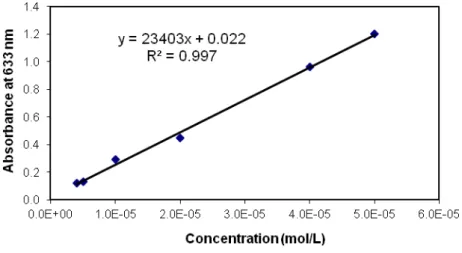
Figure 31. Toluidine blue calibration curve, with equals to 23403 L.mol-1.cm-1. ... 75
Figure 32. Schematic representation of a contact angle measurement [194]. ... 76
Figure 33. SEM micrographies of the (A) ungrafted and (B) grafted Ti6Al4V surfaces. ... 95
Figure 34. 2D profile delineation of (A) ungrafted and (B) grafted Ti6Al4V surfaces. ... 96
Figure 35. 3D model area of (A) ungrafted and (B) grafted Ti6Al4V surfaces. ... 97
Figure 36. XPS survey spectra of (A) ungrafted and (B) grafted Ti6Al4V surfaces and respective elemental identification. ... 101
Figure 37. High resolution XPS spectra of carbon on ungrafted materials and respective peak identification (arrows). ... 102
viii
Figure 40. ATR-FTIR spectrum from Ti6Al4V surfaces, ungrafted and grafted with poly(NaSS). ... 108
Figure 41. XRD spectra of (A) ungrafted and (B) grafted Ti6Al4V substrates. ... 110
Figure 42. SEM micrography of a Ti6Al4V QCM-D crystal. ... 111
Figure 43. XRD spectra of a Ti6Al4V QCM-D sensor. ... 114
Figure 44. MC3T3-E1 cells viability on ungrafted and grafted Ti6Al4V dics cultured for 30, 240, 1440 and 4320 min in the presence of FBS proteins (CM) and in its absence (NCM). Significant differences between CM and NCM cultures are indicated by # while between ungrafted and grafted surfaces in equal conditions (CM or NCM) are indicated by * (#/* p<0.05; ##/** p<0.001; ###/*** p<0.0001). ... 116
Figure 45. MC3T3-E1 cells morphology and spreading on ungrafted and grafted Ti6Al4V surfaces cultured from 30 to 4320 min in CM and NCM conditions (20x resolution). ... 118
Figure 46. MC3T3-E1 cells spreading on grafted Ti6Al4V after 4320 min (3 days) in CM conditions. The symbolizes the orientation preferably followed by the cells and the highlights particular cells with cytoplasm protrusions already extending in that direction. ... 119
Figure 47. MC3T3-E1 cells attachment on ungrafted and grafted Ti6Al4V surfaces cultured in CM and NCM conditions, from 5 min to 3 days. Significant differences between CM and NCM cultures are indicated by # (### p<0.0001). ... 121
Figure 48. Percentage of remaining attached cells after the application of a shear stress of 10 dyn/cm2 for 15 min. Significant differences between: CM and NCM cultures are indicated by #; ungrafted and grafted in the same culture conditions (CM or NCM) by *; ungrafted surfaces in different conditions (CM vs NCM) by +; and between grafted surfaces in different conditions (CM vs NCM) by ^ (#/*/+/^ p<0.05, ##/**/++/^^ p<0.001 and ###/***/+++/^^^ p<0.0001). ... 122
Figure 49. Percentage of viable cells attached to the ungrafted and grafted Ti6Al4V surfaces after 4 h of culture. ... 125
Figure 50. MC3T3-E1 cells morphology on ungrafted and grafted Ti6Al4V surfaces after 30 min of culture (50x magnification)... 126
Figure 51. MC3T3-E1 cells morphology after 4h of culture on ungrafted and grafted Ti6Al4V substrates, pre-adsorbed with FBS, BSA, Fn and Col I. Focal adhesion points were stained with anti-vinculin, the actin fibers with phalloidin and the nucleus with DAPI (50x magnification). .... 128
Figure 52. MC3T3-E1 cells area after 4 h of culture. Significant differences between ungrafted and grafted surfaces pre-adsorbed with the same proteins are indicated by * (*p<0.05 and ***p<0.0001). ... 129
Figure 53. Percentage of cells that remained attached to the substrates after the application of a shear stress of 10 dyn/cm2 for 15 min. MC3T3-E1 were cultured for 30 min. Significant differences between ungrafted and grafted surfaces pre-adsorbed with the same proteins are indicated by * (*p<0.05, **p<0.001 and ***p<0.0001). ... 130
Figure 54. Evolution of the MC3T3-E1 osteoblastic cells number on ungrafted and grafted surfaces, pre-adsorbed with FBS, BSA, Fn and Col I, from 4 h to 14 days of culture. Significant differences between ungrafted and grafted surfaces pre-adsorbed with the same proteins are indicated by * (*p<0.05 and **p<0.001). ... 132
Figure 55. ALP concentration after 14 days of culture. Significant differences between ungrafted and grafted surfaces pre-adsorbed with the same proteins are indicated by * (***p<0.0001). ... 132
Figure 56. (A) Calcium and (B) phosphate production after 28 days of culture. Significant differences between ungrafted and grafted surfaces pre-adsorbed with the same proteins are indicated by * (*p<0.05, **p<0.001 and ***p<0.0001). ... 134
Figure 57. MC3T3-E1 cells morphology on ungrafted and grated surfaces pre-coated with Fn, after 4 h of culture. The N-Terminal and C-Terminal HB domains and the RGD peptide were blocked using monoclonal antibodies (50x magnification). ... 138
Figure 58. MC3T3-E1 cells area on surfaces pre-adsorbed with unblocked and blocked Fn (N-HB, RGD and C-HB), after 4 h of culture. Significant differences between ungrafted and grafted surfaces pre-adsorbed with the same proteins are indicated by * (*p<0.05 and ***p<0.0001). .. 140
Figure 59. MC3T3-E1 cells morphology on ungrafted and grafted Ti6Al4V surfaces pre-coated with the RGD peptide, after 4 h of culture (50X magnification). ... 140
ix
(50X magnification)... 143 Figure 61. MC3T3-E1 cells attachment on ungrafted and grafted substrates pre-adsorbed with Fn, Col
I and Vn, after 4 h of culture. Antibodies against binding domains of Fn (N-HB, RGD and C-HB) and integrins (α5β1, α2β1, αvβ1, respectively Fn, Col I and Vn) were used. ... 145
Figure 62. Percentage of cells that remained attached to the substrates after the application of a shear stress of 10 dyn/cm2 for 15 min. MC3T3-E1 were cultured for 30 min. Significant differences between ungrafted and grafted surfaces pre-adsorbed with the same proteins are indicated by * (*p<0.05, **p<0.001 and ***p<0.0001). ... 146 Figure 63. (A) MC3T3-E1 cells attachment (2h, 37°C) onto Ti6Al4V, Ti6Al4V physisorbed poly(NaSS), gold, and poly(DTEc) sensors pre-adsorbed with FBS, BSA, Fn and Col I, under static conditions. (B) Pattern of frequency shift during cell attachment tests in static conditions. Though the image represents the cell attachment on gold sensors pre-adsorbed with Fn, all sensors behaved similarly with the 3 proteins (CM = complete medium or MEM- supplemented with 10%FBS). 148 Figure 64. Adsorption of BSA, Fn and Col I onto Ti6Al4V, Ti6Al4V physisorbed poly(NaSS), gold, and
poly(DTEc) at 37°C and 25 µL/min flow, until saturation. ... 149 Figure 65. Percentage of cell attachment inhibition on Ti6Al4V and poly(NaSS) physisorbed sensors,
pre-adsorbed with (A) Fn and (B) Col I, by the presence of anti-integrins (2 h at 37ºC and 0 µL/min). (C) Morphological characteristics of cells cultured for 2 h on Fn pre-adsorbed substrates, in the absence (control) and presence of anti-integrins. Significant differences between surfaces are indicated by * (* p<0.05, ** p<0.001 and *** p<0.0001). ... 152 Figure 66. (A) Schematic structure of a Fn fragment, with identification of binding domains of interest
[adapted from 41]. (B) Percentage of active sites, namely RGD peptide and heparin domains N-terminal (N-HB) and C-N-terminal (C-HB), exhibited by Fn pre-adsorbed on Ti6Al4V sensors with and without poly(NaSS) (25 µL/min). Significant differences between surfaces are indicated by * (** p<0.001 and *** p<0.0001). ... 153 Figure 67. MC3T3-E1 cells morphology on ungrafted and grafted surfaces cultured in (A) FBS and
SFM and (B) DD, DD + Fn and DD + Vn (50X magnification). ... 157 Figure 68. MC3T3-E1 cells attachment on ungrafted and grafted substrates cultured in FBS, SFM, DD,
DD + Fn and DD + Vn medium conditions and Fn and Vn pre-adsorbed surfaces. ... 158 Figure 69. Percentage of cells that remained attached to the substrates after the application of a shear stress of 10 dyn/cm2 for 15 min. MC3T3-E1 were cultured for 30 min in FBS, SFM, DD, DD + Fn
and DD + Vn conditions. Significant differences between ungrafted and grafted surfaces pre-adsorbed with the same proteins are indicated by * (*p<0.05, **p<0.001 and ***p<0.0001). ... 159 Figure 70. (Left) Percentage of a second protein adsorption onto Ti6Al4V, poly(NaSS), gold,
poly(DTEc) and PS after pre-adsorption of a first until saturation (sequential tests, at 37°C and 25 µL/min). The percentage values were determined by relating the sequential adsorption rates of the second protein with its own individual values. (Right) Representation of the changes in frequency (overtone 9) registered on all surfaces in the sequential tests ((A) BSA + Fn and Fn + BSA; (B) Fn + Col I and Col I + Fn; (C) BSA + Col I and Col I + BSA). ... 164 Figure 71. BSA (1st column) and Fn (2nd column) protein solutions stained in yellow by the FITC
conjugate. Liquids recovered from the QCM-D apparatus waste 20 min after Col I injection. ... 165 Figure 72. Mass density (left) and frequency changes (right) on Ti6Al4V, poly(NaSS), gold, poly(DTEc)
and PS resultant from the adsorption of two proteins in a mixture: (A) BSA & Fn; (B) Fn & Col I; (C) Col I & BSA, at 37°C and 25 µL/min. Significant differences between surfaces pre-coated with the same protein are indicated by * (* p<0.05, ** p<0.001 and *** p<0.0001). ... 168 Figure 73. Percentage of viable cells attached to the ungrafted and grafted Ti6Al4V surfaces after 4 h
of culture. ... 169 Figure 74. MC3T3-E1 cells morphology on ungrafted and grafted Ti6Al4V surfaces pre-adsorbed with
protein mixtures after 4 h of culture. ... 170 Figure 75. Percentage of cells that remained attached to the substrates after the application of a shear stress of 10 dyn/cm2 for 15 min. MC3T3-E1 cells were cultured for 30 min. Significant differences between ungrafted and grafted surfaces pre-adsorbed with the same protein combinations are indicated by * (*p<0.05 and ***p<0.0001). ... 172
x
differences between ungrafted and grafted surfaces are indicated by * (*p<0.05). ... 174 Figure 77. ALP concentration after 14 days of culture. Significant differences between ungrafted and
grafted surfaces pre-adsorbed with the same protein mixtures are indicated by * (*p<0.05 and ***p<0.0001). ... 174 Figure 78. (A) Calcium and (B) phosphate production at 28 days of culture. Significant differences
between ungrafted and grafted surfaces pre-adsorbed with the same protein mixtures are
xi
Table 1. Mechanical properties of CP Ti, Ti6Al4V and cortical bone [39,116-118]. ... 49
Table 2. Ti6Al4V chemical composition [114,118]. ... 50
Table 3. P-Nitrophenol (p-NP) range of concentration in AMP. ... 89
Table 4. BSA range of concentrations in TBS-Triton. ... 89
Table 5. Calcium range of concentrations in TCA. ... 90
Table 6. Phosphate range of concentrations in TCA. ... 91
Table 7. Profile delineation (2D) of the ungrafted and grafted Ti6Al4V disks roughness. ... 96
Table 8. Surface area (3D) evaluation of the ungrafted and grafted Ti6Al4V disks roughness. ... 97
Table 9. XPS elemental composition of ungrafted and grafted Ti6Al4V surfaces. ... 98
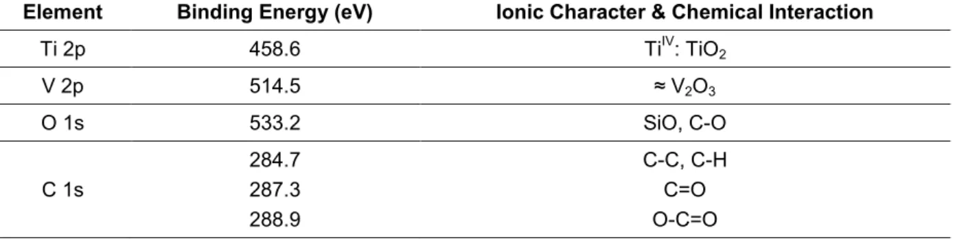
Table 10. Binding energies and composition of the high energy spectra of each chemical element, Ti2p, Al2p, V2p, O1s and C1s, present on Ti6Al4V surfaces [adapted from 202]. ... 100
Table 11. Binding energies and composition of the high energy spectra of each chemical element, Ti2p, Al2p, V2p, O1s and C1s, found on the ungrafted Ti6Al4V surfaces. ... 102
Table 12. Binding energies and composition of the high energy spectra of each chemical element, Ti2p, V2p, O1s and C1s, found on the grafted Ti6Al4V surfaces. ... 103
Table 13. EDS elemental composition of ungrafted and grafted Ti6Al4V surfaces. ... 103
Table 14. Liquid contact angles of ungrafted and grafted Ti6Al4V surfaces. ... 106
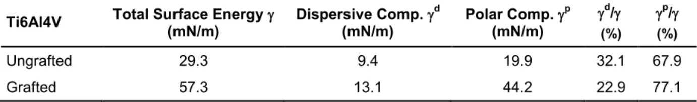
Table 15. Surface energy and quantification of dispersive and polar components of ungrafted and grafted Ti6Al4V surfaces. ... 107
Table 16. Adsorption bands characteristics of poly(NaSS) [adapted from 170]. ... 109
Table 17. XPS elemental composition of Ti6Al4V and poly(NaSS) coated sensors. ... 111
Table 18. Water contact angles of Ti6Al4V, poly(NaSS) physisorbed, gold, poly(DTEc) and PS QCM-D sensors. ... 113
Table 19. Percentage of inhibition in cell spreading and focal adhesions by cells cultured on blocked Fn (N-HB, RGD and C-HB binding regions) relatively to unblocked Fn. ... 139
Table 20. Percentage of inhibition in cell spreading and focal adhesions by cells cultured with anti-integrins (α5β1, α2β1 and αvβ1) relatively to cells cultured in unblocked conditions. ... 144
xiii
Poly(NaSS) – Poly(sodium styrene sulfonate)
Ti6Al4V – Titanium alloy with aluminum and vanadium ECM – Extracellular matrix
Col I – Collagen type I OCN – Osteocalcin HAP - Hydroxyapatite
RANKL – Receptor activator of NK-кB ligand M-CSF – Macrophage colony-stimulating factor OPN – Osteopontin
BSP – Bone sialoprotein
BMPs – Bone morphogenetic proteins FGF – Fibroblast growth factor ALP – Alkaline phosphatase BMU – Basic multicellular unit
RGD – Arginine-glycine-aspartate motif/peptide Fn – Fibronectin
Vn – Vitronectin
cDNA – complementary DNA
PAI-1 – Plasminogen activator inhibitor-1 BSA – Bovine serum albumin
FAK – Focal adhesion kinase PKC – Protein kinase C
MAPK – Mitogen-activated protein kinase
ISO – International Organization for Standardization ASTM – American Society for Testing Materials PMMA – Poly(methyl methacrylate)
CP Ti – Commercially pure titanium Ra – Average roughness
RUNX2 – Runt-related transcription factor 2 mRNA – Messenger RNA
PGE2 – Prostaglandin E2
TGF-β1 – Transforming growth factor-β1 TNF- – Tumor necrosis factor-
VCAN-1 – Vascular adhesion molecule-1 OMD – Osteomodulin
SULF1 – Sulfatase-1
QCM-D – Quartz crystal microbalance
Poly(DTEc) – Poly(desamino tyrosyl-tyrosine ethyl ester carbonate) PS - Polystyrene
xiv
Rq – Quadratic mean roughness
XPS – X-ray Photoelectron Spectroscopy EDS – Energy Dispersive x-ray Spectroscopy TB – Toluidine blue
FTIR – Fourier transformed InfraRed ATR – Attenuated total reflection XRD – X-ray Diffraction
PBS – phosphate buffered saline solution SDS – Sodium dodecyl sulfate
DMEM – Dulbecco’s modified eagle medium FBS – Fetal bovine serum
HB – Heparin binding domain
ATCC – American type culture collection SFM – Serum free medium
DD – Double depleted medium TCPS – Tissue culture polystyrene
TBS-Triton X100 – Tris buffered saline-triton X100 VEGF-A – Vascular endothelial growth factor TCA – Trichloroacetic acid
2
G
ENERALI
NTRODUCTIONThe continuing aging of the population has brought with it an ever-increasing need for materials specifically suited for biomedical applications. The fact that this need is increasing is no surprise if we compare the life expectancy of a hip implant, for instance, with the ever-increasing life expectancy of a patient. With normal implant longevity of 12 to 15 years, it is expected a patient that receives a hip implant at the age of 65 to endure at least one more surgical intervention, before the end of his life. Nowadays, this is one of the biggest concerns of biomedical researchers.
In the orthopedic domain, the search for specialized materials that respond both to the mechanical and chemical solicitations of the human body but assure a proper host response have lead to outstanding and revolutionary breakthroughs over the years. It is now clear that titanium and in particular the titanium alloy Ti6Al4V are the most favorable choices for bone substitution. They possess excellent corrosion resistance due to their ability to interact with water and air to form a spontaneous oxide layer, which also protects against ion release; great mechanical properties which approximate the material to the human bone (i.e. young modulus); and are compatible with the living tissue. Despite being continuously used in the medical field, titanium may present some challenges to a long-term implantation. Possible aseptic loosening due to inadequate tissue response (i.e. fibrous tissue formation and/or infection) may occur. Therefore, it is imperative to find solutions that, on one side, maintain the mechanical and chemical resistance and properties of the materials and, on the other side, improve the biological response. This is the “heart” of the present research work.
Based on previous investigations conducted by the Laboratory of Biomaterials and Specialty Polymers (LBPS-CSPBAT) team, we now know that the long-term response of polymeric and metallic implants can be improved by grafting bioactive polymers bearing anionic groups onto their surfaces. The interactions between the bioactive materials and the living tissue have been followed in vivo and in vitro and their potential to modulate the attachment, adhesion, spreading and differentiation of various cell types has been demonstrated. The mechanism by which the cells respond to the bioactive model surfaces has been investigated, as well. The results
3 point to a selective protein adsorption (i.e. fibronectin and vitronectin) with favorable affinity and conformations that allow to control the host response. By following this line of investigation, the LBPS developed a strategy that consists in the grafting of bioactive macromolecular chains from the poly(sodium styrene sulfonate) or poly(NaSS) polymer. It has been seen that this polymer aside from enhancing the cells activity also decreases bacterial adhesion. Data from in vivo testing has confirmed the stability of this bioactive polymer in physiological environments and the needless of protection against enzymatic degradation, overcoming limitations of pre-existing strategies of incorporation or release of bone-promoting proteins (BMPs, collagen…) and antibiotic drugs (gentamycin). Preliminary results on titanium have shown that this polymer induces the creation of new active sites along the surface for protein adsorption from the extracellular matrix aside from enhancing the osteoblastic differentiation, both alkaline phosphatase activity and mineralization. Nevertheless, much needs to be understood about the role of the surface chemistry on the cells development and its interaction with important proteins from the blood plasma and the extracellular matrix.
In this investigation, the authors:
(1) studied the impact of poly(NaSS) grafted onto Ti6Al4V substrates on the general osteoblastic cell behavior (from attachment to mineralization);
(2) followed the cells development on grafted Ti6Al4V materials pre-adsorbed with three important and well characterized proteins, albumin, fibronectin and collagen type I, both adsorbed individually and from a mixture, and we inferred about the effect poly(NaSS) exerts on cells when exposed by itself (without a protein interface);
(3) demonstrated the influence of two adhesive proteins, fibronectin and vitronectin, on the cells early adhesion on regular and double depleted media;
(4) put in evidence the importance of integrin-mediated cell attachment on fibronectin, vitronectin and collagen type I pre-adsorbed surfaces
(5) characterized the proteins conformation by assessing the amount of their active binding sites and the variations in cell morphology;
(6) and finally, determined the competitive character of albumin, fibronectin and collagen type I.
4
This thesis was subdivided in five chapters:
I) General Introduction: in this section a brief introduction to the main subjects of investigation was made and the objectives of the research were established. II) Literature Review: this chapter includes a complete analysis of all topics related to the main subject of investigation. It followed a hierarchical orientation starting from the simplest and general aspects and continuously evolving to more complex and detailed information.
III) Materials and Methods: here a description of the techniques and materials employed in the project was provided. This included characterization sections, only related to the materials, and biological sections, composed of all necessary tests to understand the initial cellular behavior on treated Ti6Al4V materials.
IV) Results and Discussion: this section deals with the results from the entire experiments, with each one being exposed in the company of insightful observations and fundamented discussions.
V) Conclusions and Future Perspectives: as the title suggests, the main conclusions retrieved from the experimental data were provided and topics for future work were suggested.
6
1.
B
ONET
ISSUEThe bone tissue is a specialized form of connective tissue composed of mineral and organic phases that provide a great rigidity and hardness to the bone. Despite its inert appearance and high resistance, bone tissue is in fact a very dynamic structure. For the entire organism life, bone is in constant remodeling so its mechanical properties and metabolic functions remain viable.
Different cell types are involved in the bone tissue formation, namely osteoclasts, bone lining cells, osteocytes and osteoblasts. These cells are embedded within the mineralized extracellular matrix (ECM), formed primarily by fibrils of collagen type I (Col I), glycoproteins and proteoglycans, which allow the bone to maintain its functions of support and protection of the organism. Besides those, bone tissue is also involved in the skeletal muscles and tendon connections needed for the body movement; works as a lever system amplifying the strength generated during muscle contraction; and possesses a storage for calcium, phosphate and other ions, so important for several metabolic functions. More than 90% of the calcium in the human body is present in bones to guaranty the stiffness of the skeleton, supply the organism with the mineral, and preserve vital functions through bone resorption [1-3]. Recent reports have shown the remodeling process of bone to be an important regulator of glucose levels in blood, since the osteocalcin (OCN) hormone liberated by bone favors the synthesis and secretion of insulin [1].
1.1 Composition
Biochemically, the bone tissue is divided in two phases, organic ( 20% w/w)
and inorganic ( 60% w/w). It is also composed of water ( 10% w/w) and several
other organic molecules such as glycosaminoglycans, glycoproteins, lipids, peptides, enzymes and different ions.
Col I (86%) is the predominant constituent of the organic phase of bone. It provides elastic and viscoelastic capacities to the bone, enabling its resistance to tension stress. Small amounts of collagen types III, V and X can also be found in this phase, aside from various glycoproteins, one of which, the OCN, believed to play an important role during mineralization [3].
The collagen molecules are organized in parallel fibers, forming interfibrillar cross-links at the extremities of bone that provide stability to the matrix (three
7 dimensional structures). Their orientation goes towards the primary lines of stress by which the bone structure is regularly submitted. In certain stages of the bone matrix formation, the small amounts of other collagen types can be involved in the collagen fibrils size regulation (diameter). There are several gap regions or “holes” in between those collagen formations that work as initial deposits for hydroxyapatite (HAP) crystals, during mineralization. This way, the organic matrix will work as a molecular and structure basis for the initial deposition of the inorganic components, defining its efficiency [4].
The inorganic phase of bone is highly resistant to compression stress due to its main composition in calcium and phosphate ions. These are usually organized as HAP crystals.
The HAP present in the human bone is a ceramic of following composition
Ca10(PO4)6(OH)2, with a hexagonal crystalline structure. In its natural form, HAP
crystals display different impurities, typically as a way to overcome possible deficiencies in calcium and phosphate. Potassium, magnesium, strontium and/or sodium are usually found in the place of calcium ions; carbonate can be found substituting phosphate ions; whereas chloride and fluoride can be found replacing hydroxyl ions. These elements are very soluble and, as consequence, the bone is able to resolubilise and release its ions into the extracellular fluids as needed.
The incorporation of other elements in the crystalline structure of the HAP slows down during the organism life, becoming more and more crystalline. This phenomena explains the slower remodeling process of bone tissue correlated with aging [3,5,6].
1.2 Structure and Organization
The bone tissue can be classified according to its porosity and structural organization into cortical, also known as compact bone, and trabecular bone, also known as cancellous or spongy bone (Figure 1) [3,5].
8
Figure 1. Bone tissue macroscopic organization [7].
The dense outer cortical bone encloses an irregular medullary space containing the cancellous bone, which is composed of branching networks of interconnecting bony trabecular elements. Both cortical and trabecular bone are composed of osteons (Figure 2).
Figure 2. Bone tissue microscopic organization [3].
The adult human skeleton is composed of 80% cortical bone and 20%
trabecular bone. However, different bones possess different ratios of cortical and trabecular bone. For instance, the vertebra has a ratio of 25/75 cortical to trabecular bone, while the femoral head has a 50/50 ratio. Despite the very distinct structural and functional tasks, their cellular elements are equal [3,5,7,8].
9 1.2.1 Cortical Bone
The cortical bone is a dense and solid hierarchical organization of osteons, also known as Haversian systems, that surrounds the marrow space. The Haversian systems are cylindrical in shape. They are composed of 6 to 8 concentric lamellae sheets of approximately 400 mm long and 200 mm wide that follow a parallel orientation according to the longitudinal axis of bone, creating a branching network within the cortical bone. In between the various lamellae there are gaps named lacunas where the osteoblasts differentiated into osteocytes are located. There are
approximately 2.1x10⁷ cortical osteons in a healthy human adult and its porosity
ranges between 5 and 10%. Cortical bone facilitates bone’s main functions, support and protection, as well as system of levers of movement and chemical elements storage. It forms the cortex, or outer shell, of most bones and possesses a complex vascular system of blood vessels and nerves, which provide high sensitivity and regenerative capacities to the bone [3,5,7,8].
The cortical bone possesses an outer periosteal surface or periosteum and an inner endosteal surface or endosteum.
1.2.2 Periosteum and Endosteum
The periosteum is a fibrous connective tissue, highly vascularized, that covers the outer surface of all bones, with the exception of joints where bone is covered by joint cartilage. It attaches to the bone by means of strong collagenous fibers also named Shapey’s fibers and provides an attachment site for muscles, ligaments and tendons. It is divided in an outer fibrous layer (containing fibroblasts) and an inner osteogenic layer (containing osteoprogenitor cells). In adults, it is responsible for bone remodeling (new bone formed after injuries), while in children is critical for bone formation and development.
The endosteum is a soft, thin, membranous layer of non-mineralized connective tissue that surrounds the inner cavity of cortical bone, trabecular bones and blood vessels (Volkman’s canals) present in bone. It contacts directly with the bone marrow space and contains blood vessels, osteoblasts and osteoclasts. As the periosteum, the endosteum is also involved in the bone remodeling process by immobilizing osteoclasts during bone growth, preventing the formation of unnecessarily thick bones [2,3,9].
10
1.2.3 Trabecular bone
The trabecular or cancellous bone has a higher surface area to mass ratio than the cortical bone. Consequently, exhibits more flexible, light, softer and weaker characteristics. It is composed of osteons, also known as packets (semilunar shape), plates, and rods averaging from 50 to 400 mm in thickness that are organized as honeycomb-like networks. Bone marrow (major producer of blood cells) can be found in between these spaces. Due to its organization and greater surface area, trabecular
bone is preferable for metabolic functions. There are approximately 1.4x10⁷
trabecular osteons in a healthy human adult. The trabecular bone is commonly found in the end of long bones, joints or within vertebrae, and contributes with 20% to the total skeletal mass [2,3,5].
1.3 Cell Types
From a histological point of view, the human bone is composed of several distinct cell types (Figure 3). Considering their impact in bone formation, special attention is provided to the osteoclasts, bone-lining cells, osteocytes and osteoblasts [1].
Figure 3. Bone cells [1].
1.3.1 Osteoclasts
Osteoclasts are large multinucleated cells with diameters ranging from 20 to 100 µm, only responsible for bone resorption (Figure 4). They possess an acidophilic cytoplasm including various vesicles and vacuoles. Osteoclasts derive from the fusion of mononuclear monocyte-macrophage precursor cells, a hematopoietic type of cells. Its formation occurs in association with the stromal cells in the bone marrow that secrete essential cytokines, the RANKL (receptor activator of NF-кB ligand) and the M-CSF (macrophage CSF), which trigger the differentiation of the hematopoietic
11 cells into osteoclasts. While RANKL is crucial for the osteoclast formation, the M-CSF is responsible for their survival and for cytoskeleton rearrangement required for bone resorption [1,3,10].
The newly formed osteoclasts must be activated to set the bone-resorbing cells activity. During that process, they become highly polarized cells since bone
resorption depends on the osteoclasts ability to secret hydrogen ions (H+) and the
cathepsin K enzyme. The H+ ions are used to dissolve the mineralized matrix of
bone, whereas the cathepsin K digests the organic phase of bone (mainly Col I). The osteoclasts bind to bone matrix via the integrin receptors, in particular the
v3 integrin that interacts with osteopontin (OPN) and bone sialoprotein (BSP). Upon
contact with the bone matrix, the osteoclasts actin cytoskeleton is rearranged so the acidified resorption (secretion of hydrochloric acid and acidic proteases aside from
the H+ and the cathepsin K that favors the hydroxyapatite crystals dissolution) is
sealed to the surrounding bone tissue. After their task is complete, the osteoclasts migrate into the adjacent marrow space, where they undergo apoptosis (they can live up to seven weeks), and the calcium and phosphate ions are liberated to the extracellular fluids [1, 10-13].
Figure 4. Mechanism of osteoclastic bone resorption [13]
The equilibrium between the osteoclasts and osteoblasts activity defines the velocity in which bone regeneration occurs.
1.3.2 Bone-lining Cells
Bone-lining cells are flat and highly interconnect with each other, possess an attenuated cytoplasm and a reduced metabolic activity (small number of organelles).
12
They derive from inactive osteoblasts, which are no longer involved in the remodeling process, and, as the name suggest, form a cellular layer on the surface of bone, protecting it and controlling ions exchange aside from assuring the nutritional support of osteocytes embedded in the underlying bone matrix. In addition, these cells are of importance in the bone remodeling process, since they possess various hormones and growth factors essential to its initiation [1,3,14].
1.3.3 Osteocytes
Osteocytes are star-shaped cells, derived from osteoblasts that become completely surrounded by osteoid. They are usually smaller than their precursors (loss of organelles) and occupy spaces named lacuna, which define the cells shape. They are able to extend their cytoplasm along the canaliculi (small channels) and communicate with neighboring osteocytes and bone-lining cells by means of gap junctions. Osteocytes can also communicate with distant cells through signaling molecules such as nitric oxides and glutamate transporters. These cells are the most abundant in the bone tissue (represent 95% of the total cells in a mature bone) with a life expectancy of approximately 25 years [1-3,15].
Osteocytes are the cells responsible for maintaining the bone matrix, since they can both synthesize new matrix and participate in its degradation. This function is also responsible for their longevity. In case of death, the resorption of bone matrix is assured by the osteoclasts while the remodeling is dependent on the osteoblasts activity. The osteocytes are also involved in the response to mechanical stimulation of bone, translating the stress signals from bending or stretching into biological activity, which can change gene expression and the cells apoptotic mechanism [15,16].
1.3.4 Osteoblasts
Osteoblasts are bone-forming cells responsible for the secretion of the bone matrix and the major cellular component of bone. They exhibit a cuboidal or polygonal shape (15-30 µm), possess a large nucleus and a cytoplasm rich in organelles with strong activity (granular endoplasmic reticulum, rounded mitochondria and well-developed Golgi apparatus). These cells are commonly found in the inner or outer surfaces of forming bone sites, typically as covering monolayers [1-3].
13 Osteoblasts arise from osteoprogenitor cells, self-renewing pluoripotent stem cells as mesenchymal stem cells, located in the bone marrow that induced by different local and systemic factors (bone morphogenetic proteins (BMPs), fibroblast growth factor (FGF) and others) differentiate into non-functional pre-osteoblastic cells and then into mature osteoblasts. Depending on the stimulant factor, stem cells can differentiate into several other types of cells (Figure 5) [17,18].
Figure 5. Different stages of osteoblasts formation and differentiation, and respective molecular
signals [18].
Active osteoblasts are very versatile and the responsible for the mineralization of the bone matrix. They secret Col I, the main component, exhibit an intense alkaline phosphatase (ALP) activity that intensifies the concentration of calcium and phosphate ions aside from the hydroxyapatite formations, and are capable of synthesizing various specialized proteins such as OCN, BSP, OPN and BMPs that work as biomarkers of osteoblastic function and development. The osteoblasts are involved in the bone remodeling process, supervising the resorption and promoting the osteoclasts contact with the mineralized bone surface. In fact, they synthesize the cytokine RANKL to induce their differentiation into osteoclasts [3,17,19,20].
In the end of the bone remodeling process, most of the osteoblastic cells undergo apoptosis. Still, there is part that take new functions, differentiating into osteocytes or bone-lining cells.
14
1.4 Bone Dynamics
1.4.1 Formation and Growth
Bone formation (or ossification) is initiated during the first weeks after conception and its development is traditionally classified as endochondral (cartilage intervenes as bone precursor) or intramembranous (without cartilage intervention).
The endochondral ossification involves the formation of bone by means of hyaline cartilage models that are, with time, progressively substitute by bony tissue (Figure 6). After the third month of gestation, the cartilage models become highly vascularized and the osteoblasts start the formation of a collar of compact bone around the cartilage, leading it to disintegrate. At this point, osteoblasts can penetrate into the cartilage model and initiate its replacement with trabecular bone. The ossification process continues from the center to the extremities of the bone, until only cartilage remains at articular joints and growth regions. Most bones are formed this way [1-3,21].
Figure 6. Endochondral ossification process [22].
The intramembranous ossification consists in the substitution of connective tissue membranes with bony tissue, in other words, bone is formed by differentiation of mesenchymal stem cells into osteoblasts. Here, bone formation starts at the eight week of gestation. Mesenchymal stem cells migrate and aggregate at specific areas, and start differentiating into osteoprogenitor cells. The region becomes highly vascularized and the aggregate enlarges. The cytoplasm of the osteoprogenitor cells changes, resulting in a more evident and complex structure, which induces the differentiation of the cells into osteoblasts. These, on its turn, synthesize collagen and other components of the bone matrix, expanding it. With time, the matrix calcifies and organizes as described in section 1.2. Most of the flat bones from skull and face are generated by intramembranous ossification.
15 As formation, bone growth starts during gestation but only ends in the early adulthood. Two stages of bone growth can be identified, the longitudinal and the radial. The longitudinal growth is mainly responsible for increasing bone length, while the radial is in charge of the bone cross-sectional area enlargement.
Important hormones are involved in the rapid formation and growth of bone. In females the estrogen is the predominant, whereas in males testosterone plays a major role. Several other growth hormones are implicated as well. Environmental factors such as physical activity and good nutrition can also be of influence during bone formation and growth [3,21,22].
1.4.2 Modeling
Bone modeling governs the enlargement, shape and size of individual bones during growth. This process allows bones to shape in response to inner or outer physiologic influences or mechanical forces, leading to the gradual adjustment of the skeleton. Here, bone formation, instigated by osteoblasts, and resorption, activated by osteoclasts, may occur in combination. Despite the antagonism of the two mechanisms, during bone modeling, they can work together to guarantee the appropriate shape and size of each individual bone (by adding or removing bone), allowing them to fit in harmony in one big and cohesive structure. While in childhood bone modeling is very frequent, in adults the remodeling process overlaps the modeling. Still, under certain circumstances such as renal osteodystrophy or hypoparathyroidism, the opposite can happen and the modeling stage may increase [3,21].
1.4.3 Remodeling
Bone remodeling is the process by which bone is renewed to maintain strength and mineral homeostasis. It involves continuous removal of old bone and its replacement by newly synthesized protein based matrix that later undergoes mineralization to form new bone. Bone remodeling occurs during the entire human life and, as in the modeling process, osteoblasts and osteoclasts work in close collaboration adding and removing bone, respectively. The balance of bone resorption and bone deposition is controlled/regulated by the activity of these two cell types.
16
During childhood, immature bone is replaced by bio-mechanically and metabolically competent bone through this process, whereas in adulthood bone remodeling is responsible for the replacement of aged, damage and/or mechanically unfit bone, preventing accumulation of microdamaged bone (from normal activity). In one year of an adult’s life between 5 and 10% of total bone is renewed. Remodeling responds also to functional demands of mechanical loading by adding bone where needed and removing it where it is not necessary. Aside from maintaining the structural integrity of the skeleton and adapting its architecture to the environment demands, the remodeling is also required to supervise its metabolic functions by means of calcium and phosphorus storage. This constant resorption and formation of new bone makes bone a very dynamic tissue with a highly plasticity level.
The remodeling cycle in adults lasts approximately four months and takes place in the “basic multicellular unit” or BMU – small packets of cells located in the cortical or trabecular surface. It comprises five stages, activation, resorption, reversal, formation and mineralization (Figure 7), and involves three types of bone cells, the osteoblasts, osteoclasts and the osteocytes [3,21,23-25].
Figure 7. Bone remodeling cycle [26].
1. Activation or Quiescence: the resting bone surface is activated by the retraction of bone lining cells and digestion of the endosteal membrane by collagenase action. This phase lasts two to three days and is instigated by different mechanical and environmental stimuli. Precursors of osteoclasts are recruited and activated, differentiating into multinucleated osteoclasts (mature), which will then attach to the mineralized bone and initiate its resorption.
17 2. Resorption: the osteoclasts attached to the bone initiate the erosion of the surface, leading to the digestion of the mineral and osteoid matrix. This task is completed by the macrophages and allows the release of various important growth factors contained within the matrix, for instance the BMPs fundamental for the bone forming cells activation. This phase lasts between 2 to 4 weeks. Since the life expectancy of osteoclasts is limited to approximately 12 days, the progression of bone remodeling requires the continual addition of these cells.
3. Reversal: this is the intermediary phase between resorption and bone formation and the responsible for the transmission of bone inducing signals to various cells found in bone resorption cavities, including monocytes, osteocytes and pre-osteobasts. These signals are not yet known, however there are some who assume BMPs, bone matrix-derived factors or FGFs as prospective candidates. During this phase, the osteoclasts disappear and macrophage-like cells are seen at the bone surface area. These cells aside from inhibiting the osteoclasts resorption action can also stimulate the osteoblasts activity.
4. Formation: bone formation results from a complex cascade of phenomena. At first, osteoclasts are inactivated and detach from the bone surface, being afterwards replaced by osteoprogenitor cells or pre-osteoblasts. These cells are attracted to the surface by means of various cell signals during reversal phase, which simultaneously stimulate their proliferation. The BMPs induce cell differentiation onto osteoblasts that start the synthesis of the osteoid matrix, filling the pre-existent resorption cavities. This matrix is mainly composed of Col I and non-collagenous proteins (i.e. growth factors). Eventually, the remaining osteoblasts in function stop their activity and give rise to lining cells that completely cover the newly formed bone.
5. Mineralization: this phase is characterized by the secretion of ALP, an enzyme associated with the early ECM maturation, which increases the calcium and phosphate ions concentration and allows their deposition as hydroxyapatite crystals over the osteoid matrix. This stage begins 30 days after deposition of the osteoid and takes almost 90 days in the trabecular bone and 130 days in the cortical bone. At the end, the new bone returns to a resting state, until new remodeling is initiated [23-25].
18
1.5 Bone formation at the interface: Biological Mechanisms 1.5.1 Osteoinduction
As the name suggests, osteoinduction is the process by which osteogenesis is induced. Primitive osteoprogenitor cells, undifferentiated and pluripotent, such as mesenchymal stem cells differentiate into osteoblasts and new bone is generated. This phenomenon requires appropriated stimuli. High molecular weight glycoproteins as BMPs (abundant in the cortical bone) are the most common osteoinductive cell mediator. Osteoinduction is a basic biological mechanism that occurs frequently in fracture healing and implant incorporation. In bone grafts, the recreation of this process is of particular interest for a faster integration as it triggers the formation of new osteoblasts and, this way, new bone tissue at the site. Despite the osteoblasts presence before implantation/injury, their intervention during bone formation tends to be minor, being the newly osteoinduced osteoblasts the main executors [27-29].
The most common osteoinductive biomaterials are the natural HAPs and the bioceramics.
1.5.2 Osteoconduction
In layman terms, osteoconduction can be defined as the process by which bone cells are directed towards a material’s surface and are allowed to grow on it. This is a three-dimensional phenomena characterized by the ability of growing bone in apposition or above pre-existing bone.
Osteoconduction occurs frequently in implantable materials. The biomaterial works as a passive physical support/structure, waiting for the cells to act without stimulating the formation of bone tissue. Bone is allowed to growth freely on the surface or down in pores, channels or pipes through cellular and vascular local invasion. However, and despite continuous debate, many believe osteoconduction cannot occur by itself and to be dependent on the osteoinduction process. The differentiation of osteoprogenitor cells into osteoblasts is indeed required for bone formation. Still, in practice, an injury is sufficient to recruit undifferentiated and pluripotent bone cells to the site. This raises the debate on whether or not a biomaterial acts as an osteoinductor [29,30].
Titanium and its alloys, in particular the Ti6Al4V, as well as stainless steel are favored as osteconductors. To the contrary, bone conduction cannot take place at copper and silver substrates [29].
19 1.5.3 Osteointegration
The term osteointegration was introduced for the first time by Branemark (1969) which described this phenomenon as the “direct structural and functional
connection between the living bone and the surface of a load-carrying implant”, after
observing a total and permanent integration of titanium screws on a rabbit femur [31]. Later, Donald’s Illustrated Medical Dictionary defined osteointegration from a histological perspective as “the anchorage of an implant by the formation of bony
tissue around the implant without the growth of fibrous tissue at the bone-implant interface” [32]. The Williams Dictionary of Biomaterials offered another description of
osteointegration as “the concept of a clinically asymptomatic attachment of a
biomaterial to bone, under conditions of functional load” [33]. Over the years, this
terminology has changed and adapt. Despite their continuous application nowadays, limitations to those definitions have been detected. Neither took into consideration that, in particular situations, osteointegration happens despite the absence of a direct or chemical bond between the implant and bone, and just a physical contact is observed. For that reason, generally an implant is accepted as osteointegrable when its anchorage to the bone is such that no relative movements are detected at the interface and the bond generated endures under normal load conditions (Figure 8) [29,31,34,35].
(A) (B)
Figure 8. (A) Schematic representation of an osteointegrated hip implant [36] and (B) real life
radiography of a similar outcome [37].
Osteointegration is not an isolated phenomenon and is dependent on both osteoconduction and osteoinduction. For instance, toxic materials that do not allow osteoconduction will not osteointegrate. Osteointegration implies that bone anchorage is maintained over time, through the consolidation of bone at the implant
20
site in normal and healthy conditions and without intermediate fibrous tissue or fibrocartilage formation [29,31,34,35].
These are the foundations for a successful osteointegration:
1. Viable and healthy bone, highly vascularized for a good ossification process (must not cause necrosis or inflammation);
2. Small space, without fibrous tissue, between bone and implant to keep the stability of the implant without micromovements (it should be less than 150 µm);
3. Implant biointegrable and compatible with the living tissue with mechanical stability and resistance similar to the natural bone, as well as appropriate design/conception, surface chemical composition, topography and morphology [38].
After implantation, the injured area should be allowed to heal without load for a considerable time, which can go up to several months depending on its location and dimension [34,35].
21
2.
I
NTERFACEB
ONE–
B
IOMATERIALThe interaction of an implant with the living tissue is a very sensitive phenomenon, which in a short period of time triggers a cascade of events (determine the biocompatibility of the material) that culminate with the cells attaching to the surface of the material (Figure 9).
An interface that is formed between two different phases, solid (biomaterial), and liquid (the surrounding biological environment, i.e. blood), usually exhibits high energy that can only be stabilized by the adsorption of substances from the medium, such as proteins. Water molecules bond to the biomaterial, originating an ionic layer that allow the proteins to adsorb, modifying their structure and functions according to the materials’ outermost layer properties, coat the substrate and establish a reliable connection with cells. This might appear a simple mechanism but many are the implications to the cells response these events may represent. In consequence, a complete analysis of each one of those phenomena is required [39-42].
Figure 9. Biomaterial interaction with the living system and correspondent intermediary biochemical
and biophysical phenomena [39].
2.1 Protein Adsorption
It is commonly state the first event taking place at the interface bone-biomaterial is protein or macromolecule adsorption from the biological fluids. Evidently this phenomenon is not the first, since water adsorption, ion bonding and adsorption of low molecular weight solutes, such as amino acids, occurs prior to it.
22
Still, protein adsorption remains the most important. Due to its rich, competitive and complex nature, these biological structures have the ability to disrupt the entire system at the interface changing the host response. The tendency of proteins to accumulate in between phases has led scientists to argue that the comprehension of the proteins behavior at interfaces is in fact a big step towards the comprehension of their normal and natural behavior [43-45].
Proteins are large complex amphipathic molecules composed of amino acids that contain combinations of ionic, polar and apolar regions exposed to the environment by a three dimensional arrangement. These regions grant the protein a “surface active” character allowing it to interact with various materials, both in vitro and in vivo. The biomaterials surface properties induce changes in the proteins behavior during adsorption, particularly in their conformation, by means of intermolecular forces. Van der Waals forces, Lewis acid-base forces and hydrophobic interactions are some of the many intermolecular events that affect the intrinsic structural stability of the proteins. In consequence, those alterations are reflected by the biological responses that follow, i.e. cell attachment. As proteins are the primary elements to be recognized by cells, their reaction towards the implantable material is believe to be a consequence of the interfacial protein layer [44,45].
2.1.1 Function and Structure
The treatment and understanding of protein adsorption requires familiarity with the concepts of protein function and structure, at least its basis.
Proteins are high molecular weight macromolecules resultant from the co-polymerization of up to 20 different amino acids (8 have apolar side chains, 7 have polar and 5 have charged polar). As general rule, a chain of amino acids is only named protein if superior to 40 units; otherwise it is identified as a peptide. In the
human body there are near 105 different proteins, each one with a specific role.
Proteins are involved in nearly all biological processes, managing the transport and storage of vital substances, providing mechanical support and protection, acting as catalysts of biochemical reactions, and others. Thus, in order to accommodate their functions, proteins fold into one or more spatial conformations driven by different non-covalent interactions. Their specificity is therefore determined by the acquired structure and chemical composition [44-46].
23 There are four levels of protein structure, the primary, the secondary, the tertiary and the quaternary (Figure 10). The primary structure refers to the amino acid organization as a linear sequence. The unique and specific amino acid organization is particular to each protein and it is hold together by way of covalent bonds (peptide bonds). Hydrogen interactions are then created between amino acids from different positions along the protein chain, causing it to bend and fold resulting in various secondary structures (i.e. α-helix or β-pleated sheet). A three dimensional conformation is here created but only reaches full potential by means of intramolecular associations [42,44,47]. Hydrophobic/hydrophilic interactions, ionic interactions, hydrogen bonding, salt bridges and covalent disulfide bonds are the most common associations – tertiary structures. On its turn, the quaternary structure refers to the regular association of two or more polypeptide chains, at different structure levels, to form a complex. A multi-subunit protein is generated that tends to be stabilized mainly by weak interactions between residues exposed by polypeptides within the complex [46-48].
Figure 10. Four levels of protein structure [48]
2.1.1.1 Structure and Orientation of the Adsorbed Protein Layer
The structure of proteins adsorbed on a biomedical substrate is hard to identify and even harder to predict. The majority of proteins adsorb as monolayers
generating close-packed formations of mass density between 1 to 5 mg/m2.
However, this range cannot be assumed as absolute. It all depends on the protein molecular orientation, conformation state and, as expected, the properties of the
![Figure 5. Different stages of osteoblasts formation and differentiation, and respective molecular signals [18]](https://thumb-eu.123doks.com/thumbv2/123doknet/14676305.742474/29.892.173.764.331.623/figure-different-osteoblasts-formation-differentiation-respective-molecular-signals.webp)
![Figure 9. Biomaterial interaction with the living system and correspondent intermediary biochemical and biophysical phenomena [39]](https://thumb-eu.123doks.com/thumbv2/123doknet/14676305.742474/37.892.252.685.608.902/figure-biomaterial-interaction-correspondent-intermediary-biochemical-biophysical-phenomena.webp)
![Figure 13. Schematic representation of the long-term cell adhesion to biomaterials process and respective proteins and molecular events associated [63]](https://thumb-eu.123doks.com/thumbv2/123doknet/14676305.742474/46.892.203.650.788.1073/schematic-representation-adhesion-biomaterials-respective-proteins-molecular-associated.webp)
![Figure 16. Representation of the fibronectin protein molecular organization and respective binding sites [81]](https://thumb-eu.123doks.com/thumbv2/123doknet/14676305.742474/50.892.179.674.633.960/figure-representation-fibronectin-protein-molecular-organization-respective-binding.webp)
![Table 1. Mechanical properties of CP Ti, Ti6Al4V and cortical bone [39,116-118].](https://thumb-eu.123doks.com/thumbv2/123doknet/14676305.742474/65.892.117.821.297.509/table-mechanical-properties-cp-ti-ti-cortical-bone.webp)