RESEARCH OUTPUTS / RÉSULTATS DE RECHERCHE
Author(s) - Auteur(s) :
Publication date - Date de publication :
Permanent link - Permalien :
Rights / License - Licence de droit d’auteur :
Institutional Repository - Research Portal
Dépôt Institutionnel - Portail de la Recherche
researchportal.unamur.be
University of Namur
Ir-CPI, a coagulation contact phase inhibitor from the tick Ixodes ricinus, inhibits
thrombus formation without impairing hemostasis
Decrem, Yves; Rath, Géraldine; Blasioli, Virginie; Cauchie, Philippe; Robert, Séverine;
Beaufays, Jérôme; Frère, Jean Marie; Feron, Olivier; Dogné, Jean Michel; Dessy, Chantal;
Vanhamme, Luc; Godfroid, Edmond
Published in:
Journal of Experimental Medicine
DOI:
10.1084/jem.20091007
Publication date:
2009
Document Version
Publisher's PDF, also known as Version of record
Link to publication
Citation for pulished version (HARVARD):
Decrem, Y, Rath, G, Blasioli, V, Cauchie, P, Robert, S, Beaufays, J, Frère, JM, Feron, O, Dogné, JM, Dessy, C,
Vanhamme, L & Godfroid, E 2009, 'Ir-CPI, a coagulation contact phase inhibitor from the tick Ixodes ricinus,
inhibits thrombus formation without impairing hemostasis', Journal of Experimental Medicine, vol. 206, no. 11,
pp. 2381-2395. https://doi.org/10.1084/jem.20091007
General rights
Copyright and moral rights for the publications made accessible in the public portal are retained by the authors and/or other copyright owners and it is a condition of accessing publications that users recognise and abide by the legal requirements associated with these rights. • Users may download and print one copy of any publication from the public portal for the purpose of private study or research. • You may not further distribute the material or use it for any profit-making activity or commercial gain
• You may freely distribute the URL identifying the publication in the public portal ?
Take down policy
If you believe that this document breaches copyright please contact us providing details, and we will remove access to the work immediately and investigate your claim.
The Rockefeller University Press $30.00
The classical cascade of coagulation describes
two converging enzymatic cascades that are
triggered either by exposure of blood to a
damaged vessel wall (the extrinsic pathway) or
by blood-borne components of the vascular
system (the intrinsic pathway). In fact, the
ex-trinsic coagulation pathway starts by the
bind-ing of FVII (plasma factor VII) to tissue factor
(TF) produced by subendothelial cells. The
re-sulting complex activates FX (factor X), which
leads to thrombin generation and, ultimately,
initiates the process of fibrin formation (Davie
and Ratnoff, 1964; MacFarlane, 1964). In
contrast, the intrinsic pathway is initiated by
contact phase proteins, including the zymogens
factor XII, factor XI, and prekallikrein, as well
as the cofactor high molecular weight
kinino-gen (HK; Yarovaya et al., 2002). FXII
under-goes autoactivation when bound to polyanionic
surfaces, generating activated factor XII by a
conformational change (Silverberg et al., 1980;
Tankersley and Finlayson, 1984). FXIIa then
converts prekallikrein into kallikrein by
cleav-age of a single peptide bond. Once small amounts
of kallikrein are formed, they catalyze the
con-version of surface-bound FXII into FXIIa,
CORRESPONDENCEEdmond Godfroid: [email protected]
Abbreviations used: ANOVA, analysis of variance; aPTT, activated partial thromboplastin time; cDNA, complementary DNA; GST, glutathione S-transferase; HK, high molecular weight kininogen; Ir-CPI,
Ixo-des ricinus contact phase
inhibi-tor; IVC, inferior vena cava; PL, phospholipid; PPP, platelet-poor plasma; PT, prothrombin time; RU, resonance unit; TF, tissue factor; t-PA, tissue plas-minogen activator.
Ir-CPI, a coagulation contact phase inhibitor
from the tick Ixodes ricinus, inhibits thrombus
formation without impairing hemostasis
Yves Decrem,
1Géraldine Rath,
2Virginie Blasioli,
1Philippe Cauchie,
3Séverine Robert,
4Jérôme Beaufays,
1Jean-Marie Frère,
5Olivier Feron,
2Jean-Michel Dogné,
4Chantal Dessy,
2Luc Vanhamme,
1,6and
Edmond Godfroid
11Service de Biologie Moléculaire des Ectoparasites, Institut de Biologie et Médecine Moléculaires, Université Libre de Bruxelles, Gosselies B-6041, Belgium
2Unit of Pharmacology and Therapeutics (FATH 5349), Université Catholique de Louvain, Brussels B-1200, Belgium 3Experimental Medecine Laboratory, Université de Bruxelles, Montigny-Le-Tilleul B-6110, Belgium
4Department of Pharmacy, Facultés Universitaires Notre Dame de la Paix Namur, B-5000, Belgium 5Centre d’Ingénierie des Protéines, Institut de Chimie B6a, Université de Liège, B-4000 Liège, Belgium
6Service de Parasitologie Moléculaire, Institut de Biologie et Médecine Moléculaires, Université Libre de Bruxelles, Gosselies B-6041, Belgium
Blood coagulation starts immediately after damage to the vascular endothelium. This
system is essential for minimizing blood loss from an injured blood vessel but also
contrib-utes to vascular thrombosis. Although it has long been thought that the intrinsic
coagula-tion pathway is not important for clotting in vivo, recent data obtained with genetically
altered mice indicate that contact phase proteins seem to be essential for thrombus
forma-tion. We show that recombinant Ixodes ricinus contact phase inhibitor (Ir-CPI), a
Kunitz-type protein expressed by the salivary glands of the tick Ixodes ricinus, specifically interacts
with activated human contact phase factors (FXIIa, FXIa, and kallikrein) and prolongs the
activated partial thromboplastin time (aPTT) in vitro. The effects of Ir-CPI were also
exam-ined in vivo using both venous and arterial thrombosis models. Intravenous administration
of Ir-CPI in rats and mice caused a dose-dependent reduction in venous thrombus
forma-tion and revealed a defect in the formaforma-tion of arterial occlusive thrombi. Moreover, mice
injected with Ir-CPI are protected against collagen- and epinephrine-induced
thromboem-bolism. Remarkably, the effective antithrombotic dose of Ir-CPI did not promote bleeding or
impair blood coagulation parameters. To conclude, our results show that a contact phase
inhibitor is an effective and safe antithrombotic agent in vivo.
© 2009 Decrem et al. This article is distributed under the terms of an Attribu-tion–Noncommercial–Share Alike–No Mirror Sites license for the first six months after the publication date (see http://www.jem.org/misc/terms.shtml). After six months it is available under a Creative Commons License (Attribution–Noncom-mercial–Share Alike 3.0 Unported license, as described at http://creativecommons .org/licenses/by-nc-sa/3.0/).
Many blood-sucking ectoparasites synthesize substances
to thwart the defense mechanisms of the hosts on which they
feed (Ribeiro and Francischetti, 2003). Ticks, in particular,
produce salivary substances capable of modulating the host
immune responses and maintain blood in a sufficiently fluid
state to effectively acquire and digest their blood meal (Brossard
and Wikel, 2004). As indicated earlier, hemostasis involves a
network of factors organized in different pathways that can
be activated independently. Ticks are, therefore, confronted
with a redundant system and must simultaneously block
sev-eral different steps to obtain effective antihemostasis. This has
been achieved during the long tick–host coevolution, as ticks
produce a variety of compounds with antihemostatic activity
(Ribeiro and Francischetti, 2003; Brossard and Wikel, 2004;
Steen et al., 2006).
In this study, we characterize a novel serine protease
inhibitor from the tick Ixodes ricinus. This protein, Ixodes ricinus
contact phase inhibitor (Ir-CPI), has one Kunitz domain and
is capable of effectively inhibiting the intrinsic coagulation
pathway and, to a much lesser extent, fibrinolysis in vitro.
We also showed that Ir-CPI can inhibit the reciprocal
activa-tion of FXII, prekallikrein, and FXI in human plasma by
spe-cifically binding to these three factors when they are activated.
Lastly, tests in animal models showed that Ir-CPI has
anti-thrombotic properties in vivo and inhibits the formation of
both venous and arterial thrombi without disturbing the
clot-ting balance. To our knowledge, this is the first time that a
selective inhibitor of the coagulation contact phase has been
shown to protect against the formation of both venous and
arterial thrombi.
RESULTS
Sequence analysis of Seq7
Expression and purification of recombinant Seq7. To identify
complementary DNAs (cDNAs) coding for proteins
specifi-cally expressed in the salivary glands of female I. ricinus ticks,
a subtractive cDNA library was set up using messenger RNAs
extracted from salivary glands of unfed and 5-d-fed female
I. ricinus ticks (Leboulle et al., 2002). One clone, formerly called
seq7 (GenBank accession no. AJ269641), was selected for
fur-ther characterization as it coded for a predicted protein (Seq7)
with similarities to the second Kunitz domain of human TF
pathway inhibitor. The predicted Seq7 amino acid sequence
comprised the typical consensus Kunitz motif
F-x(3)-G-C-x(6)-[FY]-x(5)-C. Moreover, SignalP and TargetP programs
predicted a signal peptide cleavage site at position 23 and the
absence of a hydrophobic transmembrane region, suggesting
that the protein was secreted.
To produce a recombinant form of Seq7, the open
read-ing frame deleted from the sequence predicted to code for a
signal peptide was cloned in the expression vector pGEX-6P-1
in frame with the coding sequence of glutathione
S-transfer-ase (GST) and expressed in bacteria. Affinity purification
fol-lowed by cleavage with PreScission protease and further fast
protein liquid chromatography yielded pure recombinant
protein (unpublished data).
leading to strong positive feedback on the system (Cochrane
et al., 1973; Dunn et al., 1982). During this process, FXII is
activated by a succession of proteolysis steps leading to the
production of a series of different active enzymes: FXIIa
and FXIIf (Kaplan and Austen, 1971; Revak et al., 1977;
Silverberg et al., 1980; Dunn and Kaplan, 1982). In vitro,
the contact phase system initiates the intrinsic coagulation
pathway by cleavage of FXI into activated factor XI by FXIIa
(Bouma and Griffin, 1977; Kurachi and Davie, 1977). FXIIf
may also activate FVII, the proenzyme initiating the
extrin-sic coagulation pathway (Kisiel et al., 1977; Radcliffe et al.,
1977). Propagation of clotting involves several
positive-feedback mechanisms. For example, thrombin can activate
FXI, which activates additional factor IX, therefore
amplify-ing the coagulation process (von dem Borne et al., 1995). The
surface-associated FVIIa–TF complex can activate FIX in
addition to FX. This activation loop provides an
impor-tant contribution to the blood clotting process (Josso and
Prou-Wartelle, 1965; Osterud and Rapaport, 1977; Bauer
et al., 1990). It later became clear that activation of the
con-tact–phase system is critically involved in proteolytic
mecha-nisms distinct from coagulation, namely the kallikrein–kinin,
complement, and fibrinolytic systems (Mandle and Kaplan,
1979; Ghebrehiwet et al., 1983; Kaplan et al., 2002; Moreau
et al., 2005).
The search for new anticoagulants is a major challenge in
medicine. In practice, this involves identifying drugs capable
of preventing thrombus formation without increasing the
risk of hemorrhage. For the past 40 yr, anticoagulant
treat-ment has been dominated by two classes of agents: heparins
and antivitamins K. Heparins accelerate the inhibitory action
of antithrombin on some activated coagulation factors
(spe-cifically FXa and thrombin) by indirect inhibition of these
factors and are only active when administered parenterally.
The latter prevent the final synthesis of four coagulation
fac-tors (prothrombin, FVII, FIX, and FX). These compounds
require careful laboratory testing to guarantee sufficient
anti-thrombotic effectiveness while avoiding the risk of
hemor-rhage. This disadvantage, together with the relatively narrow
therapeutic margin of these drugs, has considerably stimulated
research into other types of agents. The choice of
anticoagu-lant therapy, either for venous thromboembolism or arterial
thrombosis (myocardial infarction or stroke), is based on how
well a drug inhibits thrombosis and its hemorrhagic side
effects. The recent discovery that FXI and FXII deficiency
protects against thrombosis without causing spontaneous
bleed-ing in mice makes FXII a unique and ideal target for drug
design (Gailani and Renné, 2007; Renné and Gailani, 2007).
Indeed, FXII-deficient mice were found to be protected
against arterial thrombosis, collagen- and epinephrine-induced
thromboembolism (Renné et al., 2005), and ischemic brain
stroke (Kleinschnitz et al., 2006). In all these models, the
pro-tection was abolished by the infusion of human FXII into
FXII-null mice. Moreover, as in the case of their human
coun-terparts, the FXII-null mice do not suffer from impaired
hemostasis (Pauer et al., 2004).
to these results, Seq7 was renamed Ir-CPI (Ixodes ricinus
con-tact phase inhibitor).
Ir-CPI inhibits thrombin generation
As thrombin is the key player in clot formation, the effects of
Ir-CPI were investigated on its activity during the
coagula-tion process. The intrinsic coagulacoagula-tion pathway was induced
using a mixture of ellagic acid and phospholipid (PLs) as
trig-gers. The addition of recombinant Ir-CPI to the assay caused
a dependent prolongation of the lag time and a
dose-dependent decrease in the peak concentration of active
throm-bin (Cmax) compared with the control curve (i.e., without
inhibitor; Fig. 2 A). At a final Ir-CPI concentration of
9.1 µM, the lag time was prolonged 3.6-fold compared with
the control curve. Regarding Cmax, the effect was maximal
at 0.7 µM and did not increase at higher concentrations
(2.2 µM, 6.6 µM, and 9.1 µM). At a concentration of 0.7 µM,
Recombinant Ir-CPI prolongs activated partial
throm-boplastin time (aPTT) and fibrinolysis time. As Seq7
dis-played homology to a factor involved in hemostasis, the
activity of the recombinant protein was first assessed using
classical global hemostasis tests in vitro. The recombinant
protein had no effect on PFA closure time or platelet
aggre-gation with whole blood (unpublished data). This suggested
that Seq7 did not interfere with primary hemostasis. The
anti-coagulant activity of Seq7 was then assessed using four
rou-tine tests of plasma clotting time. Prothrombin, thrombin,
and RVV times remained unchanged (unpublished data),
suggesting that Seq7 did not interfere with extrinsic or
com-mon factors of the coagulation cascade. However, recombinant
Seq7 prolonged aPTT (7.7 times at 2 µM) in a dose-dependent
manner (Fig. 1 A), showing interference with the intrinsic
pathway. Finally, the activity of Seq7 was investigated on
fi-brinolysis. The results showed that the fibrinolysis time was
slightly increased by 1.18-fold in the presence of Seq7 at
2 µM (Fig. 1 B). All these experiments were also performed
with GST used as a negative control. No interference was
observed with any of the tests (unpublished data). According
Figure 1. In vitro anticoagulant activity of Ir-CPI. Increasing
amounts of Ir-CPI were incubated with human plasma for 2 min. Coagu-lation aPTT (A) and clot lysis times (B) were determined as described in Materials and methods. Each point represents the mean ± SEM of three independent determinations.
Figure 2. Effect of Ir-CPI on the thrombin activity profile during the coagulation process induced either by intrinsic or extrinsic coagulation pathway. (A) Human plasma was incubated with various
concentrations of Ir-CPI (0, 0.001, 0.003, 0.009, 0.027, 0.081, 0.243, 0.729, 2.187, 6.561, and 9.077 µM), and the mixture was activated with ellagic acid and PL to initiate the intrinsic coagulation pathway. (B) Human plasma was incubated with various concentrations of Ir-CPI (0, 0.729, 2.187, 6.561, and 9.077 µM), and the mixture was activated with 5 pM TF and PL to initiate the extrinsic coagulation pathway. The amidolytic activ-ity of thrombin was determined by adding its specific fluorogenic sub-strate and recording the increase in fluorescent signal. Results are presented as the mean of three independent experiments.
factors and their specific chromogenic substrates. In each of
these assays, we compared the activation of a nonactivated
factor (zymogen) by an activated factor in the presence or
ab-sence of Ir-CPI. The results showed that Ir-CPI inhibited the
activation of prekallikrein into kallikrein by FXIIa, activation
of FXI into FXIa by FXIIa, and activation of FXII into FXIIa
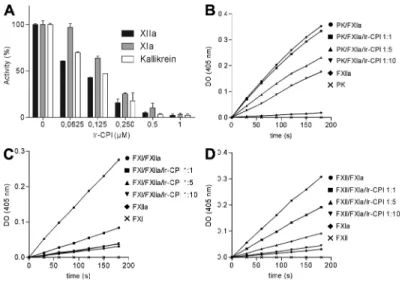
by FXIa (Fig. 3, B–D). In contrast, Ir-CPI failed to inhibit the
activation of FXII into FXIIa by kallikrein (unpublished data).
Collectively, the results of these experiments show that
Ir-CPI impacts the activation of some factors (e.g., FXIa and
FXIIa) participating in the contact phase of coagulation.
Ir-CPI binds to FXIa, FXIIa, kallikrein, and plasmin
We then asked whether the inhibitory activity of Ir-CPI
in-volved a direct interaction with one or more blood coagulation
factors by evaluating the capacity of Ir-CPI to bind to
coagula-tion or fibrinolysis factors or cofactors by surface plasmon
reso-nance. The assays demonstrated a specific interaction between
Ir-CPI and four factors: FXIIa, FXIa, plasmin, and kallikrein
(Fig. 4). No interaction was observed with any of the other
factors or cofactors tested (prekallikrein, HK, FXII, FXI, FIX,
FIXa, FX, FXa, prothrombin, thrombin, FVIIa, t-PA, and
plas-minogen; unpublished data). Except for kallikrein, these data
were confirmed by using dot blot assays (Fig. S1). Moreover,
the kinetics of interaction between Ir-CPI and the four targeted
factors (FXIIa, FXIa, plasmin, and kallikrein) were measured
after immobilization of Ir-CPI. In assays designed to determine
the binding kinetics, the quantity of immobilized Ir-CPI was
deliberately kept at a low level (200 resonance units [RU])
to avoid problems caused by limitation of the reaction rate by
a mass-transport effect. The initial binding rate was shown to
the Cmax was reduced by 37% and the lag time was
pro-longed 2.7-fold.
In contrast, when the coagulation cascade was triggered by
the extrinsic pathway (5 pM TF and 4 µM PL), a slight
dose-dependent decrease in Cmax and a dose-dose-dependent
prolonga-tion of the lag time were observed (Fig. 2 B). At a final Ir-CPI
concentration of 9.1 µM, the Cmax was reduced by 30% and
the lag time was prolonged 1.6-fold. Similar results were
ob-tained when a lower concentration of TF (1 pM) and PL (4 µM)
were used as triggers. Overall, these results show that Ir-CPI
is an inhibitor of thrombin generation.
Ir-CPI inhibits the activation of contact system factors
To determine the targets of Ir-CPI, its effect on seven
proco-agulant active serine proteases (namely kallikrein, FXIIa, FXIa,
FIXa, FXa, FIIa, and FVIIa) and two fibrinolytic serine
pro-teases (tissue plasminogen activator [t-PA] and plasmin) was
measured in amidolytic tests using specific substrates for each
of these serine proteases. These assays failed to reveal any
ef-fect of the Ir-CPI protein on the amidolytic activity of these
factors (unpublished data).
The capacity of Ir-CPI protein to inhibit the activation of
these factors was then analyzed on human plasma contact
phase factors by incubating human plasma with Ir-CPI and
then adding a contact phase activator. The activation of
con-tact factors (FXII, FXI, and kallikrein) was evaluated using a
specific substrate for each factor. The results showed that
Ir-CPI inhibited the generation of the active form of these
three factors in a dose-dependent manner (Fig. 3 A).
The effect of Ir-CPI on contact phase factors was further
dissected and examined in reconstituted systems using purified
Figure 3. Inhibitory effect of Ir-CPI on generation of FXIIa, FXIa, and kallikrein in human plasma and on reconstituted systems. (A) Diluted
human plasma was incubated with various concentrations of Ir-CPI (0.0625, 0.125, 0.25, 0.5, and 1 µM), and the mixture was activated with aPTT reagent to initiate the contact system. The amidolytic activities of generated FXIIa, FXIa, and kallikrein were determined by adding their specific chromogenic sub-strates and recording the increase in absorbance at 405 nm. (B–D) The effect of Ir-CPI was examined in reconstituted systems using purified factors and their specific chromogenic substrates. The activation of a nonactivated factor by an activated factor, in the presence or absence of Ir-CPI, was analyzed in each experiment. (B) Activation of prekallikrein into kallikrein by FXIIa. (C) Activation of FXI into FXIa by FXIIa. (D) Activation of FXII into FXIIa by FXIa Results are presented as the mean ± SEM of three independent determinations.
mation of AB, and k
a2and k
d2the forward and backward rate
constants for the AB
↔(AB) conformation change. The values
of these rate constants are shown in Table I. These results
in-dicate that Ir-CPI specifically binds to FXIIa, FXIa, kallikrein,
and plasmin but not to their zymogenic forms (Fig. 4). It
should, however, be noted that the values in Table I are only
indicative of the orders of magnitude. Indeed, large variations
of the constants are observed when the association curves
(Fig. 5, 0–84 s) are analyzed individually. Similarly, when the
dissociation curves are fitted to a sum of two exponentials
(y = y
f+ Ae
-kt+ Be
-kt, where t is the time and k and k are
complex functions of k
a1, k
a2, k
d1, and k
d2), the best fits are
obtained with nonzero values of y
f. This indicates that, with
two exponentials, dissociation is not complete when time
increases, which suggests a third, irreversible or very slowly
reversible, step after the AB ↔(AB) conformation change. This
irreversible component represents 20–40% for kallikrein,
53–60% for fXIa, 10–24% for fXIIa, and 19–27% for plasmin.
be independent of flow variations by linear regression
measure-ments at the start of kinetics with injections of analytes at
in-creasing flows from 30 to 70 µl/min. This confirmed that there
was no limitation of the reaction (unpublished data). Interaction
kinetics were determined for each analyte at six different
con-centrations (from 5 to 300 nM). The data were processed with
the BIA evaluation software and the best fits obtained with the
following two-state binding model, which implies the binding
of the plasma protein to immobilized Ir-CPI in a 1/1 ratio
fol-lowed by a reversible conformation change:
A B AB AB k k k k d a d a +
↔ ↔
1 1 2 2 ( ),
where A is the plasma protein, B is the immobilized
Ir-CPI, and (AB) is the complex form after conformational
change in the initial complex AB. In this model, k
a1and k
d1are the association and dissociation rate constants for the
for-Figure 4. Sensorgrams for interactions between targeted factors (kallikrein, FXIa, FXIIa, and plasmin) and immobilized Ir-CPI measured by surface plasmon resonance. Ir-CPI was immobilized on the surface of a CM5 sensor chip at a level of 200 RU. Factors (from 5 to 300 nM) were injected
at a flow rate of 70 µl/min in HBS buffer, and association was monitored for 84 s. After return to buffer flow, dissociation was monitored for 150 s. The sensor chip surface was regenerated by a pulse injection of 25 mM NaOH after each experiment. For each interaction, one representative result from three independent determinations is shown.
Table I. Kinetic constants for Ir-CPI interactions with kallikrein, FXIa, FXIIa, and plasmin
Ir-CPI–targeted factors ka1 (1/Ms) kd1 (1/s) ka2 (1/s) kd2 (1/s) K (1/M) Kallikrein 3.49 ± 0.54 × 10⁴ 5.32 ± 0.92 × 10² 8.34 ± 0.07 × 10³ 1.42 ± 0.11 × 10³ 4.51 ± 0.19 × 10⁶
FXIa 2.31 ± 0.59 × 10⁶ 1.02 ± 0.51 × 101 2.98 ± 0.29 × 10² 2.32 ± 0.28 × 10³ 3.83 ± 2.37 × 10⁸
FXIIa 2.19 ± 0.06 × 10⁵ 9.46 ± 0.04 × 10² 6.55 ± 0.66 × 101 1.05 ± 0.04 × 101 1.68 ± 1.10 × 10⁸
Plasmin 9.00 ± 0.39 × 10⁵ 5.35 ± 1.33 × 10³ 5.82 ± 0.99 × 10³ 6.14 ± 0.96 × 10³ 3.30 ± 0.57 × 10⁸
were 40.8 ± 9.9% of the maximum value 20 h after i.v.
ad-ministration of the recombinant protein (unpublished data).
The anticoagulant effect of Ir-CPI was also investigated with
plasma from rats and mice. The results showed that Ir-CPI
prolongs both the rat and the mouse aPTT by 2.18- and
2.77-fold at 2 µM, respectively.
The antithrombotic in vivo action of Ir-CPI was first
evaluated in a mouse model of lethal pulmonary
thrombo-embolism, induced by the infusion of a mixture of collagen
and epinephrine. All vehicle mice (15 out of 15) died within
5 min, with a dramatic reduction in circulating platelet counts
within 2 min of the challenge (Fig. 5, A and B). In contrast,
the two groups of Ir-CPI–treated mice showed a significant
increase of survivors (3 out of 15 [20%] at 1 mg/kg Ir-CPI
and 4 out 15 [26.7%] at 10 mg/kg Ir-CPI), although their
peripheral platelet counts were reduced to a similar degree as
those of mice injected with PBS. This suggests that the
pro-tection conferred by Ir-CPI administration is not caused by a
platelet adhesion/activation defect but by a defect in
throm-bin generation or some other FXIIa/FXIa-related activity.
Consistent with this premise, the analysis of the effect of
Ir-CPI on primary hemostasis by using plasma and total blood
from Ir-CPI–treated mice demonstrated no effect of Ir-CPI
on platelet aggregation (unpublished data). In addition, the
examination of histological sections of lung tissue showed a
reduction in the number of occluded vessels in Ir-CPI–treated
mice (survivors and nonsurvivors), whereas the majority of
vessels were obstructed in vehicle (Fig. 5 C).
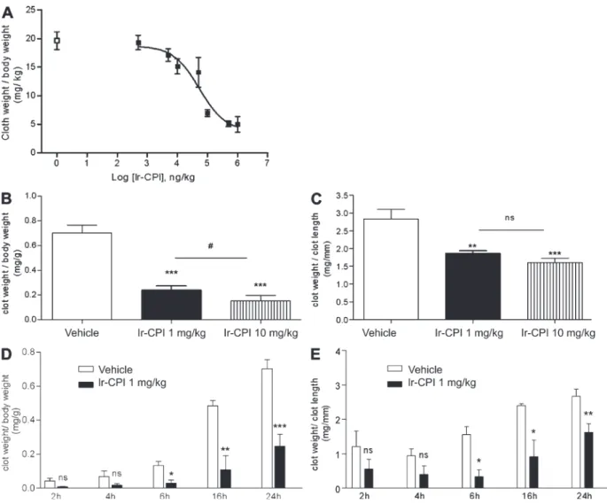
The effect of Ir-CPI on venous thrombus formation was
then assessed using two thrombosis animal models. In the first
model in rats, venous thrombosis was induced by stasis after
vessel ligation and activation of thrombosis by severe
endo-thelial damage and vessel occlusion with ferric chloride (see
Materials and methods). The control group showed 100%
thrombus formation, with a mean thrombus weight of 19.6 ±
1.6 mg/kg (n = 6). In contrast, prior i.v. administration of
Ir-CPI induced a dose-dependent progressive decrease in
thrombus formation, with a calculated effective
concentra-tion (EC50) at 49.2 µg/kg and with a maximum effect starting
at 100 µg/kg (Fig. 6 A).
The efficacy of Ir-CPI in inhibiting thrombus formation
was also measured on a murine model of venous thrombosis
in which complete stasis is induced by ligation of the inferior
vena cava (IVC). 5 min before surgery, mice were given an
i.v. injection of either Ir-CPI (1 or 10 mg/kg) or the vehicle
(PBS) in the caudal vein. Mice were sacrificed 24 h after
thrombosis induction and the thrombosed IVC fragments
were harvested and weighed. The clot weight/body weight
ratio was significantly reduced in the presence of Ir-CPI,
from 0.70 ± 0.11 in the control group to 0.24 ± 0.06 in the
group of mice treated preventively with Ir-CPI at a dose of
1 mg/kg (Fig. 6 B). Moreover, the clot weight/clot length
ratio was 2.83 ± 0.54 for a thrombus formed in the control
group, whereas it was only 1.86 ± 0.13 in mice treated with a
dose of 1 mg/kg of Ir-CPI (Fig. 6 C). At a dose of 10 mg/kg
of Ir-CPI, the clot weight/body weight and the clot weight/
Ir-CPI interferes with thrombus formation in animal models
of venous thrombosis
Before testing the antithrombotic action of Ir-CPI, we
evalu-ated the half-life of Ir-CPI in vivo. A semiquantitative estimate
of Ir-CPI pharmacokinetics was obtained using
125I-Ir-CPI.
The results showed that plasma
125I-Ir-CPI concentrations
Figure 5. Effect of Ir-CPI on pulmonary embolism model. (A)
Mor-tality associated with i.v. of collagen and epinephrine after administration of PBS (vehicle) or Ir-CPI. All PBS-treated mice died within 5 min. Animals alive 30 min after the challenge were considered survivors. Data represent the mean ± SEM of the results obtained with 15 animals per group from two independent experiments. *, P ≤ 0.05. (B) Platelet counts in mice 2 min after infusion of collagen/epinephrine. The control group was not injected with collagen/epinephrine. Data represents the mean ± SEM of results obtained with five animals per group from two independent ex-periments. Values on the number of circulating platelets in the three groups of injected mice are significantly alike. (C) Number of thrombi in the lungs of mice 2 min after infusion of collagen/epinephrine. Thrombi per visual field were counted at 20×. Data represent the mean for 20 fields per mice (n = 3 per group) from two independent experiments.
of the blood vessels. Thrombus generation in 1-mg/kg Ir-CPI–
treated and untreated mice was initiated by i.v. injection of
10 mg/kg of rose bengal followed by 20 s of high light
inten-sity exposition. In control mice, thrombus formation could
be observed as soon as 30 min after light excitation and, 2 h
later, blood flow was reduced or stopped in the major part of
the chamber. However, in Ir-CPI–treated mice, no changes
in the arterioles’ aspect or flow could be recorded after 2 h
(Fig. 7 A) or after 24 h (not depicted). Fluorescent
micros-copy confirmed these results (Videos 1–4) and provided
additional flow velocity data with help of Cap Image, a
com-puter-assisted image analysis program. Because flow velocity
is not the same in each arteriole, results were expressed as
a percentage of their own flow recorded before thrombus
induction. Fig. 7 B shows an 86.2 ± 4.7% flow reduction in
the control mice, whereas, in Ir-CPI-treated mice, the flow
velocity remained unchanged.
DISCUSSION
Most of the antihemostatic mechanisms used by hematophagous
parasites have evolved as adaptations to evade the vertebrate
blood coagulation system (Mans et al., 2002). The importance of
this selection pressure is illustrated by the wide range of strategies
developed by these ectoparasites, enabling them to thwart
primary hemostasis, clotting, and fibrinolysis during feeding
(Ribeiro and Francischetti, 2003). Ticks are obligate
hematoph-agous ectoparasites and, when they feed, their mouthparts cause
extensive damage to the tissues surrounding the bite site as they
break the vessels locally and establish a nutrition cavity rich in
cells and host blood factors. These factors must, therefore, be
inhibited if ticks are to accomplish their blood meal.
In vitro experiments showed that Ir-CPI considerably
prolongs the aPTT (7.7-fold at 2.0 µM), which measures the
intrinsic pathway of coagulation, without modifying the PT,
which measures the extrinsic pathway, the thrombin time,
which measures the conversion of fibrinogen to fibrin and its
subsequent polymerization, or the RVV time, which
mea-sures the activation of FX into FXa and the subsequent
coagulation steps. Coagulation contact phase factors may,
therefore, be the targets of Ir-CPI. Surface plasmon
reso-nance analysis and dot blot assays showed that Ir-CPI
specifi-cally binds to factors of the contact phase (namely, FXII, FXI,
and kallikrein) but not to factors belonging to the common
and TF pathways. In addition, Ir-CPI binds with a very high
clot length ratios reached 0.15 ± 0.11 and 1.60 ± 0.33,
respec-tively. We also performed time-course experiments of clot
formation in untreated and Ir-CPI treated mice. In Ir-CPI
1 mg/kg-treated mice, the clot weight/body weight ratio and
the clot weight/clot length ratio were significantly lower than
in untreated mice after 6, 16, and 24 h stasis, whereas no
dif-ference was observed after 2 and 4 h. (Fig. 6, D and E).
To examine the effect of Ir-CPI on blood coagulation
parameters after i.v. administration, the effects of Ir-CPI were
then tested on ex vivo clotting assays. For the rats, Table II
shows that aPTT values were similar in comparison with
con-trols for Ir-CPI EC50 and 100-µg/kg doses, whereas aPTT
values were statistically higher in comparison with controls for
Ir-CPI doses >1 mg/kg, showing a 1.4-fold increase in the
latter case. In contrast, prothrombin time (PT) was not
af-fected by 1 mg/kg Ir-CPI. Moreover, this high dose of Ir-CPI
had no effect on the fibrinolysis time (Table II). Finally, the
bleeding effect of Ir-CPI was evaluated using a tail-transection
model. No statistically significant blood loss was observed
5 min after administration of 1 mg/kg Ir-CPI (Table II).
The effects of Ir-CPI in mice on ex vivo clotting assays
were also tested. These showed that Ir-CPI at a dose of 1 mg/kg
did not modify aPTT, PT, and fibrinolysis times. However,
aPTT was increased in the presence of Ir-CPI at a dose of
10 mg/kg (21.7 ± 2.4 in the control group vs. 39.8 ± 5.2 in
the group of Ir-CPI–treated mice), whereas PT and
fibrino-lysis times remain unchanged at this dose. Finally, the bleeding
effect of Ir-CPI was also evaluated by using a tail-transection
model. In this case, the bleeding time was not affected after
administration of 1 mg/kg Ir-CPI. Although we observed a
slight increased of the bleeding time at a dose of 10 mg/kg,
this value was not significant as compared with those obtained
with untreated mice (Table III). Taken as a whole, these
re-sults show that Ir-CPI has a significant antithrombotic effect
in vivo without increasing bleeding or impairing blood
coag-ulation parameters.
Ir-CPI interferes with thrombus formation in mouse arterial
thrombosis model.
To further investigate the antithrombotic in vivo effect of
Ir-CPI on arterial thrombosis, we performed intravital
video-microscopy, using a skinfold chamber model in which a
transparent window is surgically placed into the dorsal skin of
NMRI mice to allow direct visualization and light excitation
Table II. Hemostasis and coagulation in Ir-CPI–treated rats
Inhibitors aPTT PT Fibrinolysis Bleeding effect (O.D. 450 nm)
s s min
PBS 20.0 ± 3.8 21.4 ± 1.5 53 ± 27 0.026 ± 0.006
Ir-CPI 0.1 mg/kg 20.7 ± 3.9 21.3 ± 1.1 66 ± 29 0.067 ± 0.060
Ir-CPI 1 mg/kg 28.8 ± 3.4a 21.6 ± 1.6 51 ± 28 0.021 ± 0.008
LMWH 0.5 mg/kg 36.2 ± 3.8a 26.9 ± 1.4a 57 ± 19 0.174 ± 0.091a
Values are mean ± SEM (n = 5 for each test). LMWH, low molecular weight heparin.
block the amidolytic activity of target proteases by binding
the amino acid P1 of the inhibitors to the catalytic site of the
enzyme (Laskowski and Kato, 1980). On the contrary, our
results suggest that Ir-CPI does not directly interact with the
site responsible for the amidolytic activity of FXIIa, FXIa,
and kallikrein but probably acts on these factors by binding to
an exosite, thereby preventing enzyme activity by steric
hin-drance. This action mechanism seems to be similar to that of
two other anticoagulant factors: the Ixolaris protein, which is
isolated from the tick I. scapularis (Francischetti et al., 2002),
and bothrojaracin, which is isolated from the venom of the
Brazilian snake jararaca (Zingali et al., 1993).
By binding to FXIIa, Ir-CPI prevents both activation of
FXI into FXIa and activation of prekallikrein into kallikrein.
affinity (Table I) to the activated form of the target enzymes
and does not bind to the zymogenic form. This indicates that
Ir-CPI does not block the activation of the target factor but,
rather its action on the next factor in the clotting cascade.
This was confirmed by showing that recombinant Ir-CPI
protein inhibits the reciprocal activation of FXI, FXII, and
prekallikrein in human plasma in a dose-dependent manner.
Furthermore, Ir-CPI inhibits activation of these three factors
in reconstituted systems (activation of a zymogen factor by an
activated factor, activation of prekallikrein into kallikrein by
FXIIa, and activation of FXI into FXIa by FXIIa) without
affecting the amidolytic activity of these proteases. This was
unexpected, as inhibitors belonging to the Kunitz-type serine
protease inhibitor family (e.g., BPTI and TFPI) generally
Figure 6. Effect of Ir-CPI on venous thrombosis in rats and mice. (A) Ir-CPI at the indicated doses was administered i.v. 5 min before induction of
thrombosis by 10% FeCl3 and complete stasis. The control group received PBS () instead of Ir-CPI (). Each point represents the mean ± SEM for five or
six animals. (B and C) Ir-CPI at the indicated doses was administered i.v. before IVC ligature. The control group received PBS instead of Ir-CPI. The throm-bosed IVC fragments were harvested and weighed 24 h later. Results were expressed by dividing the thrombus weight with mouse weight (mg/g; B) or thrombus length (mg/mm; C). (D and E) Time-course experiments of clot formation. Results were expressed by dividing the thrombus weight with mouse weight (mg/g; D) or thrombus length (mg/mm; E). The experiment shown in A is representative of two independent experiments. The data shown in B–E are presented as the mean ± SEM of at least four independent experiments. *, P ≤ 0.05; **, P ≤ 0.01; ***, P ≤ 0.001 (relative to vehicle); #, P ≤ 0.05 (between 1 and 10 mg/kg Ir-CPI dose).
amount of thrombin generated or the kinetics of the process.
Moreover, in this test, a large and physiologically irrelevant
excess of activator is used, leading to a massive activation
of the coagulation cascade. Consequently, effective inhibitor
concentrations must usually be relatively high. The results
obtained with Ir-CPI confirm that this protein is a potent
in-hibitor of thrombin generation.
Contact phase defects are manifested by a prolongation of
aPTT in vitro. However, deficiencies affecting these factors
do not cause excessive bleeding (Colman, 1984). As an
excep-tion to this rule, FXI deficiency leads to mild bleeding after
trauma or injury. This is in agreement with the suggestion that
the main role of FXI is not the initiation of coagulation but
the insurance of a positive-feedback mechanism ensuring the
secondary production of thrombin, which is essential for
effi-cient hemostasis (Gailani and Broze, 1991). A thrombophilia
study confirmed this hypothesis. It showed that an increased
level of FXI is a risk factor for thrombosis. It also suggested
that FXI had a twofold role: secondary generation of
throm-bin and down-regulation of fibrinolysis via TAFI (Meijers
et al., 2000). The ability of thrombin to activate FXI may also
explain why FXII is not required for normal blood clotting in
Likewise, by binding to FXIa, Ir-CPI prevents both
activa-tion of FXII into FXIIa and activaactiva-tion of FIX into FIXa.
Moreover, Ir-CPI also has an affinity for kallikrein (Table I)
so that it has an inhibitory effect on the kallikrein–kinin
sys-tem as well as an immediate inhibitory effect on activation of
FXII into FXIIa. Thus, the specificity of Ir-CPI for the
acti-vated factors initiating the contact phase alone is remarkable
in that it only targets the contact phase during activation.
To better evaluate the potential anticoagulant effect of
Ir-CPI in human plasma, we used the recently described
throm-bin generation method as a pharmacological tool (Robert
et al., 2009). Thrombin generation, which is more sensitive
to the action of anticoagulant (Prasa et al., 1997a,b), has long
been known to be a valid physiological function test for
in-vestigating thrombin activity during the coagulation process
(MacFarlane and Biggs, 1953; Pitney and Dacie, 1953). This
test has major advantages in comparison with the classical PT
or aPTT clotting assays. Only the initiation phase is
investi-gated in the latter assay and its endpoint is plasma clotting,
which occurs when the burst of thrombin generation has not
yet taken place (Mann et al., 2003). Measurement of PT or
aPTT therefore provides no information about the total
Table III. Hemostasis and coagulation in Ir-CPI treated mice
Inhibitors aPTT PT Fibrinolysis Bleeding effect (O.D. 450 nm)
s s min
PBS 21.7 ± 2.4 25.4 ± 1.6 26 ± 9.7 8.6 ± 3.3
Ir-CPI 1 mg/kg 25.8 ± 5.8 25.1 ± 0.1 25 ± 11.3 7.6 ± 4.7
Ir-CPI 10 mg/kg 39.8 ± 5.2a 27.5 ± 2.6 21 ± 4.6 14.5 ± 4.8
LMWH 0.5 mg/kg 47.5 ± 6.3a 28.9 ± 3.3 23 ± 5.0 >30.0 ± 0.0a
Values are mean ± SEM (n = 5 for each test). LMWH, low molecular weight heparin.
aP < 0.05, as compared to values observed in the control (PBS) by one-way ANOVA and Student’s t test–Newman-Keuls test.
Figure 7. Intravital microscopy images from mouse dorsal skinfold window chambers illustrating the effect of Ir-CPI on high light inten-sity–induced thrombosis. (A) Representative images of microvessels of 1-mg/kg Ir-CPI–treated (bottom) and untreated (top) mice. The vessels after
in-jection of 10 mg/kg of rose bengal but before high light intensity stimulation (time, 0) are shown on the left. The same vessels 2 h after high light intensity excitation (time, 2 h) are shown on the right. Representative images of five experiments are shown. Bars, 100 µm. (B) Bar graph showing changes in blood flow velocity in untreated and Ir-CPI–treated mice (n = 5). Results are expressed as a percentage of initial flow measured in each vessel.
Ir-CPI acts mainly on clot propagation. This observation is
consistent with the in vitro interactions of Ir-CPI with FXIIa,
FXIa, and kallikrein, with these contact factors being mainly
implicated in the clot propagation. The change in thrombus
weight and density is probably related to the inhibitory effect
of Ir-CPI on thrombin generation. Furthermore, the
inhibi-tory effect of Ir-CPI on thrombus formation in the mouse
model shows the efficacy of Ir-CPI over a 24-h period,
con-firming the results obtained in the rat showing the presence
of 41% of Ir-CPI 24 h after its i.v. injection. From the
point of view of clotting physiology, the ex vivo values of
aPTT, PT, and fibrinolysis times were unchanged at an Ir-CPI
concentration of 0.1 mg/kg. At higher doses (1 mg/kg and
10 mg/kg), only the value of aPTT was significantly increased
as it was observed with FXII-KO mice (Renné et al., 2005).
Importantly, it should also be pointed out that the bleeding
time of animals treated by Ir-CPI was not significantly changed
even at the highest doses.
In the arterial model, thrombosis was induced by i.v.
in-jection of rose bengal followed by high light intensity
exposi-tion. In Ir-CPI–treated mice, no changes in the arterioles’
aspect or flow could be recorded after 2 h (Fig. 7 A) or after
24 h (not depicted). In addition, we observed a flow
reduc-tion in the control mice, whereas in Ir-CPI-treated mice, the
flow velocity remained unchanged. Therefore, in these in
vivo models used to study platelet recruitment and thrombus
formation at sites of arteriole injury in Ir-CPI–treated mice,
we observed a profound defect in the formation and
stabiliza-tion of platelet-rich thrombi.
Although contact phase factors have long been
consid-ered to not be required for in vivo coagulation, recent studies
show that mice genetically deprived of one of these factors
are protected against the formation of both venous and
arte-rial thrombi (Renné et al., 2005). Results obtained with the
Ir-CPI protein confirm that a specific coagulation contact
phase inhibitor is effective in animal thromboembolic
mod-els. Therefore, our study confirms the important role of
con-tact phase factors in the coagulation propagation phase that
causes the secondary generation of active thrombin.
How-ever, results obtained with the Ir-CPI protein do not exclude
the possibility that the effects observed in vivo may result
from the reciprocal inhibition of contact phase factors. These
results could be the result of FXIa inhibiting the activation of
FIX in the amplification phase, depending on the former’s
activation by thrombin.
The selection of anticoagulant therapy, whether for
venous thromboembolism or arterial thrombosis (myocardial
infarction or stroke), is based on how well the drug inhibits
thrombosis and on the extent of the bleeding side effects
(Colman, 2006). The discovery that FXII deficiency protects
against thrombosis without causing spontaneous bleeding
makes FXII a unique and attractive target for drug design
(Gailani and Renné, 2007; Renné and Gailani, 2007). The
data of the current study demonstrate that an exogenous
mol-ecule targeting the contact phase factors (FXII and FXI) can
protect against the formation of a thrombus without altering
vivo as shown in patients deficient for FXII. Similarly, Renné
et al. (2005) showed that the coagulation balance of FXII KO
mice is not disturbed in any way and is similar to that observed
in wild-type mice. However, FXII KO mice are protected
from thrombus formation, an essential element in venous and
arterial thrombosis. In FXI-deficient mice, fibrin formation in
ischemic vessels in the brain is reduced in comparison with
wild-type mice, suggesting that the thrombosis-inducing
ef-fects of FXII are mediated through FXI and the contact phase
(Renné et al., 2005, 2006; Colman, 2006; Kleinschnitz et al.,
2006). Moreover, a recent study indicates that, in a plasma
environment, thrombin or TF do not activate FXI, even in
the presence of platelets (Pedicord et al., 2007). These results,
together with the identification of an antithrombotic
pheno-type in FXII KO mice, suggest that FXI activation by the
contact system may be physiologically relevant. Although
dif-ferences may exist between mouse and human systems, FXII
or FXI probably have similar roles during thrombosis in mice
and in humans. These proteases may, therefore, provide
at-tractive targets for the prevention or treatment of
thrombo-embolic diseases with a minimal risk of therapy-associated
bleeding (Gailani and Renné, 2007; Renné and Gailani,
2007). Kleinschnitz et al. (2006) used a synthetic inhibitor of
FXIIa (Silverberg and Kaplan, 1982), d-Pro-Phe-Arg-CH
2Cl
(PCK), to highlight FXIIa as a new drug target. However, the
selectivity of this compound should be challenged. Indeed, it
also inhibits in vitro the amidolytic activity of plasma
kalli-krein, plasmin, factor Xa, thrombin, TF/FVIIa, and
uroki-nase. Moreover, the peptidic structure, as well as the alkylating
behavior of the chloromethyl function, makes PCK unsuited
for oral clinical use as anticoagulant agent (Kettner and Shaw,
1978; Robert et al., 2008).
We therefore evaluated the inhibitory effect of Ir-CPI on
both venous and arterial thrombus formation in animal
mod-els closer to physiological venous and arterial thrombus
for-mation. In venous models, thrombosis was induced by the
infusion of a mixture of collagen and epinephrine into the
jugular vein of the mouse, by ferric chloride and complete
stasis of the posterior vena cava in the rat, and by complete
stasis of the vena cava for 24 h in the mouse. Consistent with
the results obtained with FXII-deficient mice (Renné et al.,
2005), Ir-CPI seems to protect against collagen/epinephrine
thromboembolism (20 and 26.7% of survivors at a dose of
1 and 10 mg/kg of Ir-CPI, respectively) without any effect on
primary hemostasis, confirming that Ir-CPI does not inhibit
the platelet adhesion/activation and aggregation processes. In
the two other venous models, it was also shown that Ir-CPI
reduced thrombus formation in a dose-dependant manner
with an EC50 calculated in the rat of 50 µg/kg and a
maxi-mum effect starting from 0.1 mg/kg (mean reduction of 71.0 ±
5.1% in the weight of the clot). Moreover, the clot weight/
body weight ratio and the clot weight/clot length ratio were
significantly lower in Ir-CPI–treated mice at a dose of 1 mg/kg
than in untreated mice after 6, 16, and 24 h of stasis, whereas
no difference was observed after 2 and 4 h, showing that
Ir-CPI also impacts clot density. These data suggest that
dition of an agonist (collagen; Chrono-Log Corporation). Platelet aggregation was measured as the maximal per 10-min change in impedance.
Anticoagulant activity
The anticoagulant activities of Ir-CPI were determined by four coagulation tests using a Start8 coagulometer (Diagnostica Stago). Blood samples were collected in 3.8% trisodium citrate from healthy human donors, rats, and mice. Platelet-poor plasma (PPP) was obtained by further centrifugation at 4,000 g for 10 min.
aPTT. 25 µl of plasma and 25 µl Ir-CPI were preincubated for 2 min at
37°C. Mixtures were activated for 4 min with 25 µl actin FS (Siemens). Clotting was initiated by adding 50 µl of 25 mM CaCl2.
PT. 25 µl of plasma and 25 µl Ir-CPI were preincubated for 2 min at 37°C.
Mixtures were activated for 4 min with 25 µl innovin, 1/10 from human plasma or 1/100 from animal plasma (Siemens). The clotting reaction was started by adding 50 µl of 25 mM CaCl2.
Russel viper venom time. 25 µl of plasma, 50 µl Hepes buffer (25 mM
Hepes, 2% Glycine, and 145 mM NaCl, pH 7.35), and 25 µl Ir-CPI were preincubated for 2 min at 37°C. Clotting was initiated by the addition of 25 µl LA 1 (Siemens).
Thrombin time 25 µl of plasma, 50 µl Hepes buffer, and 25 µl Ir-CPI were
preincubated for 2 min at 37°C. Clotting was initiated by the addition of 25 µl thrombin (Diagnostica Stago).
Determination of clot lysis times
Clot lysis times on PPP were determined as previously described (Boudjeltia et al., 2002).
Thrombin activity profiles
Materials. PPP reagent (5 pM TF and 4 µM PL in the final mixture), PPP
LOW reagent (1 pM TF and 4 µM PL in the final mixture), and thrombin calibrator were purchased from Synapse BV. For each experiment, a fresh mixture of fluorogenic substrate/calcium chloride buffer solution was pre-pared as follows: 2,275 µl of buffer (20 mM Hepes, pH 7.35) containing 60 mg/ml of bovine serum albumin (Sigma-Aldrich) and 240 µl of 1 M calcium chloride were mixed with 60 µl of 100 mM DMSO solution of fluorogenic thrombin substrate (Z-Gly-Gly-Arg-AMC; Bachem). Actin FS was diluted 25-fold with distilled water.
Preparation of human plasma. Blood from healthy male volunteers, who
were free from medication for at least 2 wk, was taken by venipuncture and collected into 0.105 M sodium citrate (9:1 vol/vol). PPP was obtained by centrifugation at room temperature for 10 min at 2,500 g and was used im-mediately after centrifugation.
Calibrated automated thrombin activity measurement. Thrombin
activity measurement was performed using the previously reported CAT procedure (Hemker et al., 2003; Robert et al., 2009). In brief, 80 µl PPP, 10 µl PBS or Ir-CPI, and 20 µl PPP reagent, PPP LOW reagent, or diluted Actin FS were mixed in a 96-well microtiter plate (Immulon 2HB; Thermo Fisher Scientific) and incubated for 5 min at 37°C. The coagulation process was triggered by the addition of 20 µl of substrate/calcium chloride buffer at 37°C. A calibration condition was also realized. In this latter case, the same protocol as that described using PBS was followed, but the activator was re-placed by 20 µl of thrombin calibrator. The reaction of fluorogenic thrombin substrate hydrolysis was monitored on a microplate fluorometer (Fluoroskan Ascent FL; Thermo Fisher Scientific) with a 390/460-nm (excitation/emis-sion) filter set. Fluorescence was measured every 20 s for 60 min. The com-mercially available Thrombinoscope software (Synapse BV) automatically processed the acquired data to give thrombin activity profile curves and mea-surement parameters (lag time and Cmax). 10 Ir-CPI concentrations ranging from 0.001 to 9.077 µM were tested in each experiment in triplicate.
coagulation and bleeding times. Effectively, Ir-CPI has a
dose-dependent protective action against thrombin-induced
thromboembolism in four animal models without disturbing
the clotting balance. At the effective doses, no effect was
ob-served on aPTT and PT values, fibrinolysis time, or,
remark-ably, on bleeding time. To our knowledge, this is the first
time that an inhibitor of the coagulation contact phase has
been shown to protect against the formation of venous and
arterial thrombi. Ir-CPI may, therefore, provide an
impor-tant therapeutic tool by protecting patients at risk from
dis-eases such as pulmonary embolism, cerebral ischemia, deep
vein, and arterial thrombosis.
MATERIALS AND METHODS Animals
Animal care and experimental procedures were performed in accordance with the Helsinki Declaration (Publication 85–23, revised 1985), local insti-tutional guidelines (laboratory license LA 1500474), and the Belgian law of 14th August 1986, as well as the royal decree of the 14th of November 1993 on the protection of laboratory animals. Animal protocols were ap-proved by the local ethic commission of the Université Libre de Bruxelles. Studies were performed using male Sprague-Dawley OFA rats weighing 250–300 g (Harlan) and 8-wk-old NMRI female mice weighing 20–25 g (Elevage Janvier).
Expression and purification of recombinant Ir-CPI in Escherichia coli
The coding region of Ir-CPI cDNA was cloned in frame with GST in the pGEX-6P-1 vector and transformed into E. coli strain BL21. Production of the recombinant protein was induced by the addition of 1 mM IPTG at 37°C for 2 h. After centrifugation of bacterial lysates prepared using a French press, the resulting supernatant, which contained the GST-Ir-CPI fusion protein, was incubated with Glutathione Sepharose High Performance (GE Healthcare). Ir-CPI was released by cleaving with PreScission protease ac-cording to the manufacturer’s specifications and then purified to homogene-ity by gel filtration chromatography using a HiLoad Superdex 75 column (GE Healthcare). Purified proteins were then tested for endotoxin contami-nation using the Limulus Amebocyte Lysate QCL-1000 kit (Cambrex). En-dotoxin levels were <0.4 enzyme units/mg of protein in all preparations used. Samples containing endotoxin amounts superior to that threshold were loaded on Detoxi-Removal endotoxin gel columns according to the manu-facturer’s instructions (Thermo Fisher Scientific) to ensure the removal of endotoxins. This was followed by dialysis against buffers appropriate for the subsequent experiments. Protein concentrations in the endotoxin-purified batches were determined using a Micro BCA kit (Thermo Fisher Scientific) according to the manufacturer’s instructions.
Primary hemostasis
Human blood samples were collected from healthy donors in 0.102-M triso-dium citrate tubes (9:1 vol/vol). Global platelet function was measured on a PFA-100 machine (Siemens) with a collagen/epinephrine or collagen/ADP cartridge. The sample (1/10 protein in HBSS and 9/10 citrated whole blood) was aspirated through a capillary under steady high shear rates within 45 min of sample collection. Platelet plug formation was induced by the presence of the platelet agonists and the high shear rates, and this gradually occluded the aperture. The closure time was considered to be the time required to obtain full occlusion of the aperture. This time is called the closure time (CT) and is measured in seconds. The test is therefore a combined measure of platelet adhesion and aggregation. Platelet aggregation was performed using the im-pedance channel of a Whole Blood Lumi-Aggregometer (Chrono-Log Corporation; Ingerman-Wojenski and Silver, 1984). With this method, the electrical impedance between two fine electrodes immersed in the sample in-creases with platelet coating and aggregation on these electrodes after the
ad-The kinetics of interactions between Ir-CPI and the four interacting factors were performed after a new immobilization of Ir-CPI. The quantity of Ir-CPI immobilized for measurements of kinetics was deliberately maintained at a low level (to 200 RU) to avoid the problems of limitation of the reaction by the process of mass transport. Independence with respect to differences in flow of the initial rate of connection, measured by linear regression at the start of the ki-netics after injections of analytes with increasing flows (30–70 µl/min), con-firmed that the reactions were not limited by such a process. Interaction kinetics were determined for each analyte, with six different concentrations (from 5 nM to 300 nM). Binding data were analyzed using BIA evaluation software to de-termine the kinetic constants. The specificity of the reaction was evaluated using the GST protein (1,000 RU immobilized on the surface of the sensor chip) as negative control.
Determination of radioactivity of 125I–Ir-CPI in rat blood
125I-labeled Ir-CPI was prepared by iodination with [125I] sodium iodide in 20 mCi/mg of protein, using IODO-BEADS Iodination Reagent (Thermo Fisher Scientific), according to the manufacturer’s instructions. Free iodide was removed by extensive gel filtration on Sephadex G10.
The in vivo distribution of 125I–Ir-CPI in rat blood was evaluated after i.v. administration. Samples containing 10 × 106 cpm were resuspended in 200 µl of PBS and administered to rats. Blood was collected after 3, 20, 40, or 60 h by cardiac puncture in 3.8% trisodium citrate. Plasma was obtained by centrifuga-tion, and aliquots of 500 µl were placed in glass test tubes. Radioactivity was de-termined in a gamma counter.
Ex vivo effect of Ir-CPI on aPTT, PT, and fibrinolysis
Ir-CPI was administered i.v. to rats and mice, and blood was collected after 5 min by cardiac puncture in 3.8% trisodium citrate. PPP was obtained by cen-trifugation at 4,000 g for 10 min. The aPTT, PT, and fibrinolysis times were measured using theses procedures.
Collagen/epinephrine-induced pulmonary thromboembolism
20–25-g C57BL/6J mice were anesthetized by i.p. injection of a mixture of 80 mg/kg ketamine and xylazine (5 mg/kg). 1–10 mg/kg Ir-CPI or vehicle was injected in the iliac vein 5 min before challenge. A mixture of 0.8 mg/kg colla-gen and 60 µg/kg epinephrine was then injected into the jugular vein. Platelet counts were determined by flow cytometry.
Histopathologic analyses
C57BL/6J mice were killed and lungs were fixed at 4°C for 24 h in buffered 4% formaldehyde. Tissues were dehydrated and embedded in paraffin, cut into 5-µm sections, and stained with Mayer’s hematoxylin and eosin (Sigma-Aldrich).
Complete stasis combined with vessel injury induced venous thrombosis model in the rat
Thrombus formation was induced by a combination of complete stasis and vessel injury by ferric chloride according to the modification of the method described by Peternel et al. (2005). Rats were anesthetized i.p. with 70 mg/kg pentobarbital sodium. The abdomen was opened by making an incision along the linea alba toward the sternum, followed by exposition of the pos-terior vena cava. Surgical threads, 1 cm apart, were placed loosely around the vena cava beneath the renal veins and above the bifurcation of the iliac veins to form a snare. Complete stasis was induced in the posterior vena cava by tightening the downstream snare firmly around the posterior vena cava. Si-multaneously, a piece of filter paper (0.3 × 0.8 cm) saturated with 10% wt/vol ferric chloride solution was applied to the external surface of the posterior vena cava caudally of the ligature for 10 min, 10 min after the removal of the filter paper, the upstream snare was firmly tightened around the posterior vena cava, and the rat was then euthanized. The ligated venous segment was excised and the thrombus was removed and immediately weighed after blot-ting off excess blood. Results were expressed in milligrams of thrombus per kilogram of rat body weight. Ir-CPI (0.5–1,000 µg/kg, corresponding to 2 nM to 4 µM) or vehicle were injected in the left femoral vena 5 min be-fore the induction of the thrombus formation.
Assay of the inhibitory effect of Ir-CPI on coagulation factors
The inhibitory activity of Ir-CPI was examined on seven procoagulant serine proteases (plasma kallikrein, FXIIa, FXIa, FIXa, FXa, thrombin, and FVIIa) and two fibrinolytic serine proteases (t-PA and plasmin). Each serine protease was preincubated with Ir-CPI in a 1:5 molar ratio for 5 min at 37°C, followed by the addition of the appropriate chromogenic substrate (final concentration 0.5 mM). Final concentrations in a total volume of 200 µl in 96-microwell plates were the following: 3 nM kallikrein/S-2302, 62.5 nM FXIIa/S-2302, 31.25 nM FXIa/S2366, 500 nM FIXa/Spectrozyme FIXa, 10 nM FXa/S-2222, 35 nM thrombin/Spectrozyme TH, 100 nM FT-FVIIa/ Spectrozyme FVIIa, 35 nM t-PA/Spectrozyme t-PA, and 30 nM plasmin/ Spectrozyme PL. The kinetics of substrate hydrolysis were measured over 20 min. Chromogenic substrates S-2302, S-2366, and S-2222 were supplied by Chromogenix, and Spectrozyme FIXa, TH, FVIIa, t-PA, PL were ob-tained from American Diagnostica Inc.
Assay of the effects of Ir-CPI on contact system activation in plasma
The effects of Ir-CPI on the activation of the contact system in human plasma were assessed from the generation of activated contact factors (FXIa, FXIIa, and kallikrein). Human plasma was treated with acid to inactivate plasma serine protease inhibitors (De La Cadena et al., 1987) and then diluted 1:10 in buffer. 50 µl of diluted plasma was incubated with 20 µl of various concentrations of Ir-CPI for 5 min and then activated with 5 µl aPTT reagent (actin FS). After 10 min, a chromogenic substrate mixture at a final concentration of 0.5 mM and one or two inhibitors, 100 nM of Maize trypsin inhibitor or 50 µM kallis-top, were added, and the amidolytic activity of the generated enzyme was de-termined at 405 nm for 3, 10, and 30 min for kallikrein, factor XIIa, and factor XIa, respectively. Sets of added chromogenic substrate and inhibitors were the following: S-2366, kallistop, and CTI for FXIa assay; S-2302 and kallistop for FXIIa assay; and S-2302 and CTI for kallikrein assay.
Effect of Ir-CPI in a reconstituted system
A reconstitution assay of the kallikrein–kininogen–kinin system was per-formed using purified coagulation factors (FXIIa and prekallikrein). 12.5 nM FXIIa was preincubated with Ir-CPI in Hepes buffer for 2 min at 37°C. 12.5 nM prekallikrein was added to the mixture, and then prekallikrein activation was started. After 10 min, chromogenic substrate S-2302 and CTI were added, and the increase in absorbance at 405 nm was recorded over 3 min. Reconstitution assays of the intrinsic coagulation pathway were performed using purified coagulation factors FXI/FXIa and FXII/FXIIa. The effect of Ir-CPI on the activation of FXI by FXIIa was tested by incubating 15 nM FXI, 60 nM FXIIa, and Ir-CPI for 10 min at 37°C. After incubation, sub-strate S-2366 was added and the increase in absorbance was measured. The effect of Ir-CPI on the activation of FXII by FXIa was tested by incubating 15 nM FXIa, 60 nM FXII, and Ir-CPI for 10 min at 37°C. After incubation, substrate S-2302 was added and the increase in absorbance was measured.
Binding analysis using surface plasmon resonance
The interaction between Ir-CPI and coagulation (HK, plasma prekallikrein, plasma kallikrein, FXII, FXIIa, FXI, FXIa, FIX, FIXa, FX, FXa, prothrom-bin, thromprothrom-bin, and FVIIa) or fibrinolytic (t-PA, plasminogen, and plasmin) factors was monitored using a BIAcore X instrument (GE Healthcare). 15 µM Ir-CPI was immobilized on the surface of a CM5 sensor chip in 10 mM acetate buffer, pH 5.0, by the amine coupling procedure according to the manufacturer’s instructions. 1,500 RU of immobilized Ir-CPI were used for the assay. To subtract the nonspecific component from the apparent binding response, a blank flow cell was prepared using the same immobilizing proce-dure without Ir-CPI. Binding analyses were performed using HBS buffer (10 mM Hepes, 150 mM NaCl 3 mM, and EDTA, pH 7.4, with 0.005% surfactant P20) as running buffer at 25°C. 100 µl of each 100-nM analyte was injected on the sensor chip at a flow rate of 70 µl/min. Association was monitored during an 84-s injection of analyte. Dissociation was monitored for 3 min after return to the running buffer. Regeneration of the sensor chip surface was achieved with a 15-µl pulse injection of 25 mM NaOH.