THESE
Pour obtenir le grade de Docteur
opéré par l’Université de Rouen Spécialité :Microbiologie
Pseudomonas adaptation to stress factors: role of membrane lipids
and Pseudomonas fluorescens response to NO
2Présentée et soutenue publiquement par
Tatiana Kondakova
Thèse dirigée par le Pr Nicole ORANGE et le Dr Frédéric Dionnet, co-endadrée par les Dr Cécile Duclairoir Poc et David Préterre
aux Laboratoire de Microbiologie Signaux-Microenvironnement, EA 4312 de l’Université de Rouen, et Centre d'Étude et de Recherche Technologique en Aérothermique et
Moteurs
Thèse soutenue publiquement le 20 novembre 2015 devant le jury composé de
Madame Angela CORCELLI
Professeur / Coordonnateur du Centre Interdépartemental de la Recherche de la Paix / Département des Sciences Médicales Fondamentales, Université de Bari, Bari, Italie
Rapporteur
Madame Janine FRÖHLICH Docteur / Chef du groupe de recherche / Département de chimie Polyphasique
/ Institut Max Planck Chimie, Mayence, Allemagne Rapporteur
Monsieur Hermann HEIPIEPER
Docteur / Chef du Groupe des Processus Microbiens / Département Biotechnologie de l'Environnement / Centre Helmholtz pour la recherche environnementale – UFZ, Leipzig, Allemagne
Examinateur
Madame Nicole ORANGE Professeur / Enseignant-chercheur / Laboratoire de Microbiologie Signaux et
Microenvironnement / Université de Rouen, Evreux, France Examinateur
Monsieur Frédéric DIONNET Docteur / Directeur du Centre d'Étude et de Recherche Technologique en
Aérothermique et Moteurs, St Etienne du Rouvray, France Examinateur
Madame Cécile DUCLAIROIR POC
Docteur / Enseignant-chercheur / Maître de conférences/ Laboratoire de Microbiologie Signaux et Microenvironnement / Université de Rouen, Evreux, France
THESE
Pseudomonas adaptation to stress factors: role of membrane lipids and
Pseudomonas fluorescens response to NO
2The high distribution of Pseudomonas fluorescens group is linked to its ability to adapt to stress factors. This work goaled the response of an airborne P. fluorescens MFAF76a, and its clinical standard P. fluorescens MFN1032 to environmental changes in order to refine the specific adaptation of airborne bacteria. First the HPTLC-MALDI TOF MSI tool defined glycerophospholipid (GP) composition of both strains. In stationary growth phase, an unknown GP, in short UGP, was found and seemed to be involved in temperature adaptation for the clinical strain. After exposure to 0.1, 5 and 45 ppm concentrations, the bacterial response to NO2 was defined through motility, biofilm formation, antibiotic resistance and expression of several chosen target genes. While no change in parameters was seen in bacteria exposed to 0.1 and 5 ppm of NO2, several alterations were occurred with a bacterial exposure to 45 ppm. NO2 seemed to bias the UGP production, reduced P. fluorescens swim and decreased swarm only for MFN1032 strain. Biofilm formed by NO2-treated MFAF76a showed increased maximum thickness, with no change in c-di-GMP intracellular level. Expression of the hmp-homologue gene involved in NO detoxification was upregulated in response to NO2, suggesting a possible common pathway between NO and NO2 detoxification. Finally, NO2 was found to increase bacterial resistance to ciprofloxacin and chloramphenicol. Thus the resistance nodulation cell division (RND) MexEF-OprN efflux pump encoding genes were highly upregulated in both strains. Together these findings implement the first model of bacterial response to NO2 toxicity and the role(s) of GP in bacterial adaptation to environmental changes.
Keywords: Pseudomonas fluorescens, adaptation, glycerophospholipid, fatty acids, pollution, nitrogen oxides
Adaptation de Pseudomonas aux facteurs de stress : rôle des lipides
membranaires et réponse de Pseudomonas fluorescens au NO
2La large distribution des Pseudomonas fluorescens est liée à leur grande adaptabilité aux facteurs de stress tels que les variations environnementales. Ce travail avait pour objet la réponse spécifique au milieu aéroporté de P. fluorescens, comme l’aéroportée MFAF76a et la clinique MFN1032, comme standard. La technique récente HPTLC-MALDI-TOF MSI a permis de caractériser les divers glycérophospholipides (GP) des deux souches. En phase stationnaire de croissance, un GP inconnu (UGP - unknown GP) a été isolé et semble intervenir dans l’adaptation à la température de la souche clinique MFN1032. Quant au stress NO2 gaz, les deux souches ont été exposées aux concentrations: 0.1, 5 et 45 ppm. Leurs phénotypes ont été confrontés à l’expression de quelques gènes ciblés. Pour les valeurs standard 0,5 and 5 ppm en NO2, aucun paramètre n’est modifié. Par contre, une réponse bactérienne est constatée suite à l’exposition à 45 ppm de NO2. Cette exposition semble impacter la production d’UGP. De plus hmp et mexEF-oprN, codant respectivement pour la flavohémoglobine et pour la pompe à efflux RND, se trouvent surexprimés, corroborant l’évolution de la résistance bactérienne aux antibiotiques. Contrairement au NO, aucune altération de la biomasse de biofilm n’est observée pour le NO2, qui favorise, cependant, l’augmentation de son épaisseur chez MFAF76a, mais aussi l’inhibition du swarming et la diminution du swimming, avec un taux de c-di-GMP stable. Ce faisceau de résultats offre, pour la première fois, la réponse bactérienne et le rôle des GP lors de stress comme NO2 ou à la température, autre modification environnementale.
Mots clefs: Pseudomonas fluorescens, adaptation, glycérophospholipide, acides gras, pollution, oxydes d’azote,
La science ouvre à l’esprit humain une voie infinie, et le lance, par une série d’étapes sans nombre, sur l’Asymptote de la Vérité.
Remerciements
This work was realized in Laboratory of Microbiology Signals and Microenvironment EA4312, of University of Rouen. I am grateful to the Haute Normandie region for the financial support for this research work.
I would like to thank Pr. Angela CORCELLI from University of Bari Aldo Moro, and Dr.
Janine FRÖHLICH from Max Planck Institute for Chemistry of Mainz for agreeing to participate in my PhD defense and examine my work. It is my honor to be examined by you and I would like hereby to express my sincere gratitude. Special thanks to Dr. Hermann HEIPIEPER from Helmholtz Centre for Environmental Research of Leipzig for agreeing to participate in my PhD defense but also for your help in this work, your advices and support. Thank you to accepting me working in your lab, and for your help in FAME analysis and results interpretation. This allow me not only to acquire invaluable professional and personal experience, but also to obtain the European PhD grade. I am very grateful for your help in the review writing. I am glad you are present for my PhD defense today.
Je remercie également mes directeurs de thèse, le Pr. Nicole ORANGE et le Dr. Frédéric DIONNET, pour m’avoir fait confiance et acceptée dans leur équipe. Je vous suis profondément reconnaissante de m’avoir accordé du temps et des conseils tout au long de ma thèse. Merci pour toutes vos corrections pertinentes des oraux, posters et articles. Je tiens à vous exprimer ma plus profonde reconnaissance.
Je tiens également à remercier mes encadrants, le Dr. Cécile DUCLAIROIR POC et David PRETERRE pour avoir aiguillé mes travaux pendant ces trois années. Merci Cécile pour ta convivialité et ton soutien même dans les moments difficiles ! Merci de m’avoir fait confiance. J’ai particulièrement apprécié tes conseils, nos discussions, ton écoute ainsi que ta disponibilité à chaque moment dans ton bureau. J’ai beaucoup appris à tes cotés !
Un grand merci à toi Nadine MERLET-MACHOUR ! L’interprétation des spectres MALDI sans toi aurait été moins drôle ! Merci pour tes conseils et le partage de tes connaissances. Ce fut une expérience très enrichissante.
Je remercie également le Pr. Marc FEUILLOLEY, directeur du LMSM, pour m’avoir accueillie dans son laboratoire, pour ses conseils lors de ces trois années, son aide rédactionnelle et ses encouragements. Je n’oublierai jamais les petites pauses café et les moments de rire.
Un grand merci à tous les membres du laboratoire LMSM, Audrey, Chloé, Alex, Jérémy,
Lucille, Gwen, Irina, Lilly, Manuela, Noémie, Camille, Victorien, Andréa, Baoloc, Kelly,
Awa, Florence, Thomas… Je ne cite sûrement pas tout le monde, la liste est longue. Merci
pour votre soutien, votre gentillesse et votre bonne humeur ! Je vous adresse mes chaleureuses pensées et je vous souhaite à tous beaucoup de chance pour la suite.
Merci beaucoup Olivier et Magalie, vous étiez toujours à l’écoute, disponible et arrangeante !
Merci à toi Magalie pour les expositions au NO2, pour tes conseils et ton sourire. Merci à toi
Olivier pour ta bonne humeur, ton rire va beaucoup me manquer !
Merci à vous mes amis, Dorian et Célia, pour votre bonne humeur, votre soutien, amitié et tout et tout ! Merci Corinne, pour tes conseils, le partage de tes connaissances, nos soirées. Merci à toi Mathias, pour les bons moments à Maastricht et ta gentillesse. Merci à vous Charlène et
Nicolas, pour votre bonne humeur et amitié. Merci à toi Thibaut pour ta gentillesse, ton aide et soutien, nos soirées ensemble, et tout et tout ! Merci particulièrement à toi Rachel. Merci pour ta présence, ton amitié, ta gentillesse. Tu m’a beaucoup apporté lors de ces trois années ensemble. Tu es et resteras une personne exceptionnelle pour moi. En tous cas, il me faudrait plusieurs pages pour exprimer le bonheur d’être à vos côtés mes amis ! Vous restez pour toujours dans mon cœur.
J’adresse une chaleureuse pensée à tous mes amis. Je te remercie, François pour ton soutien tout au long de la thèse, ta présence, ta compréhension et ton aide précieux. Pendant ces trois années, tu étais mon amour, mon collègue et mon ami. Sans toi tout cela n’aurait pas la même valeur. Я от всего сердца благодарю Вас, мои родители. Без Вас всего этого никогда бы не случилось. Благодарю Вас за ваше терпение, безграничную любовь, поддержку и заботу! Целой диссертации не хватит, чтобы выразить мою признательность и любовь к Вам! Спасибо тебе Наташа за любовь, понимание и поддержку, тебе Леша за дружеское отношение, за ревю, и пивные посиделки, Вам Алла Федоровна и Леонид Устинович за хорошее настроение и помощь во всем. Спасибо вам, мои маленькие солнышки, Ярослава и Даша. Один взгляд на вас вызывает у меня улыбку. Наконец, спасибо огромное Вам, Владимир Константинович, Катя и Марина. Вы научили меня самому главное в нашей профессии: любить микробиологию.
Table of content
List of abbreviations... 11
List of figures and tables ... 13
Figures ... 13
Tables ... 14
I. Introduction ... 15
Chapter 1 Pseudomonas genus ... 17
1. Properties of Pseudomonas genus ... 19
2. Taxonomic diversity ... 20
3. Pseudomonas fluorescens strains ... 23
Highlights ... 55
Chapter 2 Stress factors and Pseudomonas fluorescens lipid adaptation ... 57
1. Stressors and bacterial survival ... 59
2. Impact of physical factors on membrane properties ... 59
3. Impact of chemical factors on membrane properties ... 62
4. Impact of biological factors on membrane properties ... 65
Highlights ... 68
Chapter 3 Nitrogen oxides, their chemistry, biological roles and atmospheric pollution ... 71
1. Nitrogen oxides (NOx) ... 73
2. NO formation in living organisms ... 78
3. NO targets in cells ... 86
4. Bacterial strategies of response to RNS ... 93
5. NO-mediated regulation in bacteria ... 102
6. Abiotic RNSand air pollution ... 108
Highlights ... 113
II. Goals ... 115
III. Materials and methods ... 117
Materials ... 119 1. Chemicals ... 119 2. Bacterial strains ... 119 Methods ... 119 1. Methods of microbiology ... 119 2. Physico-chemical methods ... 121 3. Methods of biochemistry ... 123
4. Methods of molecular biology ... 125
5. Methods of bioinformatics ... 130
Chapter 4 Study of lipidome of airborne P. fluorescens ... 137
Chapter 5 Effect of stress factors on membrane of P. fluorescens ... 149
Chapter 6 Pseudomonas fluorescens response to NO2 toxicity ... 183
V. General discussion ... 205
VI. Conclusions and perspectives ... 211
VII.References ... 215
VIII. Annexes ... 241
Supplementary data ... 243
Chapter 4 Study of lipidome of airborne P. fluorescens ... 243
Chapter 5 Effect of stress factors on membrane of P. fluorescens ... 244
Chapter 6 Pseudomonas fluorescens response to NO2 toxicity ... 246
Scientific contributions ... 249
1. Publications ... 249
2. Oral communications... 265
List of abbreviations
[Fe-S]: Iron-Sulfur cluster
µM: Micromole
2-acyl-GPE:
2-acyl-GlyceroPhosphoEthanolamine
8-oxoG: 8-oxoguanine
AA: Amino Acids
Aas: Acyl-ACP synthase
Abs: Absorbance
ACP: Acyl Carrier Protein
ADEM: French environment and energy management agency
AHL: N-Acyl Homoserine Lactone
AK: Adenylate Kinase
Ala-PG: Alanyl-PhosphatidylGlycerol
ANR: ANaerobic Regulator of arginine deiminase and nitrate reductase
ATP: Adenosine TriPhosphate
BER: Base Excision Repair
BF3: Boron trifluoride
bNOS: bacterial Nitric Oxide Synthase
bp: base pair
BPG: BisPhosphatidylGlycerol
C20:4ω6: Arachidonic acid
C20:5ω3: Eicosapentaenoic acid
C22:5ω3: Docosapentaenoic acid
CAM: Contact Angle Measurements
cAMP: cyclic Adenosine MonoPhosphate
C-di-GMP: bis-(3’-5’)-Cyclic dimeric guanosine MonoPhosphate
cDNA: complementary DNA
CDP: Cytidine DiPhosphate
Cfa: Cyclopropane fatty acid synthase
CFP: Cyan Fluorescent Protein
CFU: Colony-Forming Unit
CiLy: Citrate Lyase
cis-UFA: cis-Unsaturated Fatty Acid
CITEPA: Interprofessional technical center for studies on air pollution
CL: CardioLipin
CLP: Cyclic LipoPeptides
CLSM: Confocal Laser Scanning Microscopy
CO2: Carbon dioxide
CO3-: Carbonate radical anion
Ct: Cycle threshold
Cti: cis–trans-isomerase
cXMP: Xanthosine-(3’,5’)-cyclic MonoPhosphate
cyclo-FA: Cyclopropane Fatty Acid
DEPC: DiEthylPyroCarbonate
DGC: DiGuanylate Cyclase
DHB: 2,5-DiHydroxyBenzoic acid (MALDI matrix)
DMB: Davis Medium Broth
DNA: DeoxyriboNucleic Acid
DNR: Dissimilatory Nitrate respiration Regulator
DSB: Double-Strand Break in DNA
E: PCR Efficiency
eDNA: extracellular DNA
EDTA: EthyleneDiamineTetraacetic Acid
EGP: Exponential Growth Phase
eNOS: endothelial Nitric Oxide Synthase
EPS: ExoPolySaccharides
ESI: ElectroSpray Ionization
ETC: Electron Transport Chain
FA: Fatty Acid
FAD: Flavin Adenine Dinucleotide
FAME: Fatty Acid Methyl Esters
Fap: Familial Amyloidotic Polyneuropathy
FAS: Fatty Acid Synthesis
FAST: Fragment Analysis and Structural TOF
Fe2+: Ferrous iron
Fe3+: Ferric iron
FeB: non-heme iron
FlavoHb: FlavoHemoglobin
Fur: Ferric uptake protein
GC: Gas Chromatography
GCyc: Guanylate Cyclase
GlyC: GlycoCardiolipin
GP (or PL): GlyceroPhospholipid
GSH: Reduced Glutathione
GSNO: S-nitrosoGlutathione
GSNOR: S-nitrosoGlutathione Reductase
GTR: Generalised Time-Reversible
HG: Head Group
HNO: Nitroxyl
H-NOX: Heme-Nitric OXide/oxygen binding domain
HONNOH: Hyponitrous acid
HONO: Nitrous acid
HOONO: Peroxynitrous acid
HPLC: High-Performance Liquid Chromatography
HPTLC-MALDI TOF MSI: High Performance
Thin-Layer Chromatography - Matrix-Assisted Laser Desorption Ionization - Time Of Flight Mass Spectrometry Imaging
HR: Homologous Recombination
IM: Inner Membrane
INERIS: French National Institute for Industrial Environment and Risks
iNOS: inducible, macrophage Nitric Oxide Synthase
ISC: Iron-Sulfur Cluster system
K+: Potassium ion
KDO: 3-deoxy-D-manno-Octulosonic acid
L(O)NO2: Lipid epoxide
LB: Luria-Bertani medium
LO: Lipid alkoxyl radical
LOO-: Lipid peroxyl radical
LOONO-: Lipid peroxynitrite
LPA: LysoPhosphatidic Acid
LPE: LysoPhosphatidylEthanolamine
LPS: LipoPolySaccharide
MALDI: Matrix-Assisted Laser Desorption Ionization
MCP: Methyl-accepting Chemotaxis Protein
MDO: Membrane-Derived Oligosaccharides
MIC: Minimum Inhibitory Concentration
ML: Maximum Likelihood
MLSA: Multi-Locus Sequence Analysis
MMR: MisMatch Repair
MRM: Multiple Reaction Monitoring
MS/MS: Tandem Mass spectrometry
MS: Mass Spectrometry
MSI: Mass Spectrometry Imaging
N2O: Nitrous oxide
N2O3: Dinitrogen trioxide
N2O4: Dinitrogen tetroxide (dimer of
NO2)
N2OR: Nitrous Oxide Reductase
NAD(P)H: Nicotinamide Adenine Dinucleotide
NAD+: Oxidized form of Nicotinamide
Adenine Dinucleotide (NAD)
NADH: Reduced form of Nicotinamide Adenine Dinucleotide (NAD)
NADP+: Nicotinamide Adenine
Dinucleotide Phosphate
NAP: Periplasmic Nitrate Reductase
NAR: Respiratory membrane-bound NitrAte Reductase
Nas: Nitrate Assimilation Enzymes
NCBI: National Center for Biotechnology Information
NDPK: Nucleoside DiPhosphate Kinase
NER: Nucleotide Excision Repair
NH3: Ammonia
NH4+: Ammonium
NHEJ: Non-Homologous End-Joining
Ni:NOR: Nitrite: Nitric Oxide Reductase
NIR: NitrIte Reductase
nM: Nanomolar
NMR: Nuclear Magnetic Resonance Spectroscopy
nNOS: Neuronal Nitric Oxide Synthase
NO: Nitrogen monoxide
NO2-: Nitrite
NO2: Nitrogen dioxide
NO3-: Nitrate
NOR: Nitric Oxide Reductase
NOS: Nitric Oxide Synthase
NOx: Nitrogen Oxides
NR: Nitrate Reductase
O2NOCO2-: Nitrocarbonate ion
OD: Optical Density
OM: Outer Membrane
ONOO-: Peroxynitrite
ONOOCO2-: Nitrosoperoxycarbonate anion
OP: Oxidative Phosphorylation
PA: Phosphatidic Acid
PBAP: Primary Biological Aerosol Particles PC: PhosphatidylCholine Pcs: PhosphatidylCholine Synthase PEPC: PhosphoEnolPyruvate Carboxylase PG: PhosphatidylGlycerol PGP: PhosphatidylGlycerol Phosphate pGpG: 5′-PhosphoGuanylyl-(3'-5')-Guanosine PI(3,4,5)P3: PhosphatidylInositol 3,4,5-trisPhosphate PI(4,5)P2: PhosphatidylInositol 4,5-biPhosphate PI: PhosphatidylInositol
PIP: PhosphatidylInositol Phosphate
pKa: Acidity constant
PLD: PhosphoLipase D
PM: Particulate Matter
Pmt: Phospholipid N-MeThylation
poly-UFA: polyUnsaturated Fatty Acid
PPDK: Pyruvate Phosphate DiKinase
ppGpp: Guanosine
3’,5’-bisPyroPhosphate
ppm: Part Per Million
PS: PhosphatidylSerine
PSD: Post Source Decay
qRT-PCR: Quantitative RetroTranscription Polymerase Chain Reaction
Rf : Retention Factor
RNA: RiboNucleic Acid
RND: Resistance-Nodulation-cell Division
RNS: Reactive Nitrogen Species
ROS: Reactive Oxygen Species
rpm: Rotation Per Minute
RR: Response Regulator
rRNA: Ribosomal RNA
RS-NO: S-Nitrosothiols
RS-NO2: S-Nitrothiol
RT: Reverse Transcription
S-DGD-5-PA: Sulfated Diglycosyl
DiphytanylGlycerol Diether
SDS: Sodium Dodecyl Sulfate
SFA: Saturated Fatty Acid
SGP: Stationary Growth Phase
SM: SphingoMyelin
SSB: Single-Strand Break in DNA
S-TGD-1: Sulfo-TriGlycosyl-Diether
T3SS: Type 3 Secretion System
T6SS: Type 6 Secretion System
TAE: TetraAcetic Acid
TCA: TriCarboxylic Acid
TLC: Thin-Layer Chromatography
Tm: Transition temperature
TOF: Time Of Flight
trans-UFA: trans-Unsaturated Fatty Acid
UFA: Unsaturated Fatty Acid
UGP: Unknown GlyceroPhospholipid
V: Viscosinamide
vol: Volume
WHO: World Health Organization
List of figures and tables
Figures
Figure 1. Functional range and environmental niches of the Pseudomonas genus, highlighting the broad
distribution of the P. fluorescens species ... 20
Figure 2. Phylogenetic relationships of Pseudomonas genus bacteria among 16S ribosomal RNA genes ... 22
Figure 3. Microscopic and macroscopic aspect of P. fluorescens ... 23
Figure 4. Swimming, swarming, and twitching motilities of P. aeruginosa PAO1 ... 25
Figure 5. Pathway of biofilm development in P. aeruginosa ... 26
Figure 6. Scanning electron micrographs of P. fluorescens biofilms ... 27
Figure 7. Structure and physiological functions of c-di-GMP ... 28
Figure 8. A proposed model for regulation of P. fluorescens biofilm formation ... 29
Figure 9. Pseudomonas cell envelope ... 31
Figure 10. Adaptation of glycerophospholipid structure and composition in Pseudomonas ... 49
Figure 11. Modifications of fatty acids structure in Pseudomonas spp. ... 52
Figure 12. Schematic model of regulation of the Cti enzymatic mechanism by membrane fluidity... 53
Figure 13. Enzyme stabilization at high osmolarity ... 61
Figure 14. Scheme of the toxicity of hydrocarbons in the cell membranes ... 63
Figure 15. Interactions of cell envelope with metals ... 64
Figure 16. Glycerophospholipid rearrangement that can occur on binding cationic antimicrobial agents... 66
Figure 17. Lewis structure of (A) NO and (B) NO2 ... 73
Figure 18. Redox relationships of NOx with other RNS species ... 75
Figure 19. Scheme of biosynthesis of nitric oxide from L-arginine in mammalian cells ... 79
Figure 20. Schematic model of NOx synthesis and assimilation in plants ... 80
Figure 21. Bacterial nitrogen cycle ... 82
Figure 22. Denitrification pathway in Pseudomonas spp. species and enzymes, catalyzed the NOx reduction .... 82
Figure 23. Schematic representation of the denitrification genes locus in Pseudomonas aeruginosa PAO1 ... 83
Figure 24. Schematic model of the regulatory network controlling the denitrification genes in P. aeruginosa ... 86
Figure 25. Bacterial targets of RNS ... 87
Figure 26. Schematic representation of NO2 reaction with polyunsaturated lipids ... 88
Figure 27. Reaction of NO and lipid peroxyl radicals ... 88
Figure 29. Free radical pathways to tyrosine nitration ... 91
Figure 30. DNA damage pathways resulting from RNS exposure... 93
Figure 31. Permeability of reactive species across lipid membrane ... 94
Figure 32. Model of NalA- and NasT/S-dependent expression of nitrate assimilation operon in P. aeruginosa PAO1 ... 96
Figure 33. Schematic illustration of the MexEF-OprN RND efflux pump ... 98
Figure 34. Reprogramming of P. fluorescens metabolism in order to adapt to nitrosative stress ... 99
Figure 35. The proposal model of the KatA-mediated NO sensing ... 101
Figure 36. Model for the transcriptional regulation of nbdA through NO... 104
Figure 37. GcbA-mediated modulation of the BdlA-dependent biofilm dispersion ... 105
Figure 38. H-NOX-dependent control of biofilm formation through the regulation of c-di-GMP level ... 106
Figure 39. NOx sources in atmosphere ... 108
Figure 40. Evolution of NOx emission in metropolitan France from 1990 to 2013 ... 110
Tables
Table 1. Chemical structures of membrane fatty acids ... 50Table 2. Summary table of pathways, involved in bacterial lipid adaptation to various stress factors ... 67
Table 3. Threshold values for NOx ... 111
Table 4. NO2 air quality standards ... 112
Table 5. Composition of growth media ... 120
Table 6. Primer sequences used for quantitative RT-PCR assay ... 127
Chapter 1
Pseudomonas genus
1. Properties of Pseudomonas genus
Bacteria of genus Pseudomonas belong to the phylum Proteobacteria, class γ-Proteobacteria, order Pseudomonadales. Pseudomonas are unicellular Gram-negative straight or lightly curved rods, with rounded extremity and average size of 2x0.5 µm (Palleroni, 1984). These bacteria are motile with one or several polar flagella, able to growth in minimum medium and metabolize several hydrocarbon substrata as energy and carbon source. These aerobic bacteria have a strictly respiratory type of metabolism with oxygen as the terminal electron acceptor. However, nitrate can be used by several Pseudomonas strains as an alternate electron acceptor, allowing an anaerobic growth. Pseudomonas possess oxidase and catalase activity (Brenner et
al., 2005). The features of these organisms are the degradation of a large number of organic
compounds (Barathi & Vasudevan, 2001; Foght & Westlake, 1988; Leahy & Colwell, 1990), the interactions with plants (Mercado-Blanco & Bakker, 2007; Nomura et al., 2005; Tao et al., 2003) and the associations in the rhizosphere, which are advantageous for agriculture (Lugtenberg et al., 2001; Rainey, 1999). In addition, Pseudomonas include one of the most dangerous opportunistic pathogens among all bacteria P. aeruginosa (Driscoll et al., 2007; Srour et al., 2014). Pseudomonas comprise taxa of metabolically versatile organisms capable of living under diverse environmental conditions. Consequently, the members of this group are able to growth in diverse environments (Ringen & Drake, 1952) as illustrated in Figure 1, including water (Mena & Gerba, 2009), soil (Couillerot et al., 2009; Kiely et al., 2006), air (Duclairoir Poc et al., 2014; Morris et al., 2007), food, and hospital environment (Shooter et
al., 1969, 1971). This worldwide distribution seems to be due to a high physiological and
genetic adaptability (Spiers et al., 2000), being the presence of numerous genomic islands in the genus, a key adaptability traits of individual strains to such ubiquitous environments (Peix
et al., 2009). Thus, the genomes of several species like P. aeruginosa, P. fluorescens, P. syringae, P. putida or P. stutzeri include between 3.7 and 7.1x10⁶ base pairs (bps) (Spiers et
Figure 1. Functional range and environmental niches of the Pseudomonas genus, highlighting the broad distribution of the P. fluorescens species
Adapted from Scales et al., 2014.
2. Taxonomic diversity
The name Pseudomonas originates from the work of Migula, 1894 describing this genus as “Cells with polar organs of motility strictly”. Defined in this way, the genus Pseudomonas was very heterogeneous and suffers several taxonomic reclassifications. In 1966 Stainer established the Pseudomonas classification based on the capacity of strains to metabolize the different energy sources. This fundamental work described 267 Pseudomonas strains with their biochemical, physiological and phenotypic properties (Stanier et al., 1966). With the emergence of molecular biology, the Pseudomonas genus was reorganized. The study of genomic sequence
homology by DNA-DNA (Palleroni et al., 1972; Palleroni & Doudoroff, 1971) or DNA-RNA (Palleroni et al., 1973) hybridizations, as well as the phylogenetic analysis based on 16S ribosomal RNA (rRNA) sequence (Anzai et al., 2000) have led to the division of Pseudomonas genus on five distinct 'rRNA homology' groups. This division explains an important heterogeneity of the Pseudomonas genus (Vos et al., 1985, 1989; Vos & Ley, 1983). The species belonging to the 'group I' conserved the genus name and termed the Pseudomonas sensu
stricto; the other species (‘group II-V’) Pseudomonas sensu lato have been reclassified and
generically renamed according to their phylogenetic relationships, include nowadays the related genus, including Ralstonia, Comamonas, Agrobacterium, Brevundimonas, or Burkholderia (Anzai et al., 2000). The genus Pseudomonas sensu stricto was divided on two major lineages (Figure 2) represented by typical species, namely Pseudomonas aeruginosa and Pseudomonas
fluorescens (Bodilis et al., 2012; Mulet et al., 2010). The P. aeruginosa lineage is the more
homogenous and consists of the P. aeruginosa species, but also of more distant species, like P.
oleovorans or P. stutzeri. Basing on study of the 16S rRNA gene and targeting one or two
hypervariable regions, Bodilis and coworkers divided P. fluorescens lineage on two r-clusters (“r”-clusters, the clusters based on “r”RNA gene data): the “putida” and the “fluorescens” (Bodilis et al., 2012). The putida r-cluster corresponds to the P. putida group; the fluorescens r-cluster contains the P. fluorescens, P. syringae and P. lutea groups, as shown in Figure 2. The fluorescens r-cluster is the largest in terms of species number (about 60% of the
Pseudomonas species described), and the most heterogeneous Pseudomonas group in terms of
Figure 2. Phylogenetic relationships of Pseudomonas genus bacteria among 16S ribosomal RNA genes
3. Pseudomonas fluorescens strains
3.1. General information
The name Pseudomonas fluorescens was chosen because of the production of complex fluorescent peptidic siderophores called pyoverdines (Figure 3), which are very efficient iron scavengers in bacteria (Cornelis & Matthijs, 2002). This species is described as psychrotrophic germ with optimum growth temperature between 25°C and 30°C, able to growth at 4°C, but unable to growth at 41°C, as thermophilic bacteria. Several strains of P. fluorescens are sequenced (58 sequenced genomes, 5 genomes annotated without gap, in April 2015, according to NCBI, http://www.ncbi.nlm.nih.gov/). A whole variety of phenotypes and genotypes allows the classification of P. fluorescens as heterogeneous bacteria, with several biovars and sub-biovars. The percentage of DNA/DNA hybridization between strains of the same biovar is frequently less than 50%. Thus, the genotype of this species is difficult to define. The taxonomy of Pseudomonas is currently being renewed (Palleroni, 2010), as evidenced by the reclassification of CHAO or Pf-5 strains to a novel species named P. protegens (Ramette et al., 2011). Here, we defined P. fluorescens as bacteria, belonging to the fluorescens r-cluster and previously published at the name P. fluorescens.
Figure 3. Microscopic and macroscopic aspect of P. fluorescens
A. Scanning electron micrograph of P. fluorescens (www.scienceclarified.com). B. Macroscopic aspect of P.
fluorescens SBW25, pyoverdine production (Spiers et al., 2013).
3.2. Distribution and adaptive capacity of Pseudomonas
fluorescens
The members of P. fluorescens group are highly distributed in all major environments, including water, soil and air as illustrated in Figure 1 (Bodilis et al., 2004; Duclairoir Poc et
in soil, are abundant on the surfaces of plant roots and leave. This applies also the role of P.
fluorescens as rhizosphere biocontrol agents (Walsh et al., 2001). Biocontrol strains have
noticeably been observed at the root surface (i.e. the rhizoplane), often forming microcolonies or discontinued biofilms in the grooves between epidermal cells (Couillerot et al., 2009). Some of the plant-colonizing strains, such as SBW25, positively affect plant health and nutrition (Silby et al., 2009).
The ability to growth at refrigerated temperatures opens to P. fluorescens the possibility to colonize the ‘cold environments’. Thus, P. fluorescens was isolated from Antarctic glacier ice melt (Attard et al., 2012) or from clouds and rain water (Ahern et al., 2007), where these bacteria can catalyze freezing of supercooled water at a temperature as warm as -2°C (Attard et
al., 2012; Joly et al., 2013). The low temperature adaptation makes P. fluorescens
contamination a particular problem for the food-processing (Dogan & Boor, 2003; Jonghe et
al., 2011). Indeed, at low temperature P. fluorescens is able to product degradation enzymes,
including proteases, lipases and gelatinases, modifying organoleptic food quality (Russell, 2002). In addition, P. fluorescens can cause blood infusion-related infections and outbreaks, contaminating the blood products or the equipment associated with intravenous infusions during storage (Gibaud et al., 1984; Khabbaz et al., 1984; Scales et al., 2014).
Being psychrotrophic bacteria, several P. fluorescens strains still grow at increasing temperature (at least 37°C) (Picot et al., 2004). Bodilis and coworkers proposed to split P.
fluorescens into two groups: (1) “true psychrotrophs” able to multiply at temperatures from 4°C
to at least 30°C (but no grow at 37°C); and (2) “thermo-tolerant psychrotrophs” able to grow at temperatures from 4°C to at least 37°C (but no grow at 42°C). Group (1) contains only strains from soil and water. Group (2) is composed of clinical isolates and also certain strains with soil origins (Bodilis et al., 2004). This thermotolerance associated with virulence factors expression and biofilm formation (Figure 1) allows P. fluorescens to tame eukaryotic hosts. While far less virulent than P. aeruginosa, P. fluorescens can cause acute infections (opportunistic) in humans and was reported in clinical samples from the mouth, stomach and lungs of patients with compromised immune status (Chapalain et al., 2008; Scales et al., 2014), as shown in Figure 1. Species of P. fluorescens are associated to agents of pulmonary diseases (Bahrani-Mougeot
et al., 2007; Choi et al., 2011; Dickson et al., 2014), urinary infections (Osawa et al., 2002),
eye infections (Durbán et al., 1996), and bloodstream infections (Benito et al., 2012). Several strains are able to link to eukaryotic cells, such as intestinal cells Caco2/TC7, to modify their permeability, inducting cytotoxic and pro-inflammatory responses (Madi et al., 2010a, b). Indeed, P. fluorescens possesses a number of functional traits, which provide to this bacterium
secondary metabolites (Gross & Loper, 2009; Mavrodi et al., 2006; Ramette et al., 2003; Rossignol et al., 2008; Weller et al., 2007), synthesis of siderophores (Cornelis & Matthijs, 2002), type III secretion system (Marchi et al., 2013; Mavrodi et al., 2011; Rezzonico et al., 2005; Sperandio et al., 2012), ability to form biofilms (Barahona et al., 2010; Baum et al., 2009), and plasticity of some strains to adapt to grow at higher temperature.
3.3. Biofilm formation as P. fluorescens adaptation mode
Bacteria respond to fluctuating environmental signals to coordinate adaptive changes in metabolic pathways and physiological outputs. Integration of environmental factors affords bacteria the ability to make important decisions regarding how to respond to the constantly changing environments. One of the possibilities is the change of the lifestyle. Bacteria can live in a free movement (planktonic) existence or commit to a surface attached lifestyle (biofilm). There are three major types of bacterial motility (Figure 4), depending on medium viscosity, namely, swimming in aqueous environments, twitching on solid surfaces, and swarming on semisolid, viscous media, such as those containing 0.4 to 0.7% (wt/vol) agar (Overhage et al., 2008). Figure 4. Swimming, swarming, and twitching motilities of P. aeruginosa PAO1 A. Macroscopic motility assays. Swimming motility on a semi-liquid agar plate (Left); Swarming motility on a semi-solid agar plate (Middle); Macroscopic assay of twitching motility (Right). B. Microscopic aspect of Pseudomonas motility. Modified according M. Harunur and A. Kornberg, 2000. Swimming and swarming motility needs the synthesis of flagella. The swimming is brought about by individual cells independently perceiving chemical signals that trigger adaptive chemotactic responses. The swarming motility is characterized by a multicellular movement of bacteria that migrate above solid substrates in groups of tightly bound cells (Calvio et al., 2005). Swarming motility requires both the functional flagella and the production of surface-wetting agent biosurfactant (Caiazza et al., 2007; Kearns, 2010). Production of the biosurfactants serves
as an essential aid to swarming motility by acting as a wetting agent to overcome the surface tension of water and facilitates movement across the most surface (Caiazza et al., 2005). The nature of Pseudomonas biosurfactants was described as strain-dependent (Duclairoir-Poc, 2011). Unlike P. aeruginosa, which products biosurfactant rhamnolipid, P. fluorescens secretes cyclic lipopeptides (CLP), including viscosine and viscosinamide (Duclairoir-Poc, 2011; Nielsen et al., 1999). Twitching motility is a flagella-independent form of bacterial translocation over moist surfaces. It occurs by the extension, tethering, and then retraction of polar type IV pili (Mattick, 2002).
When coming in contact with a surface, whether in natural, industrial, or clinical settings, bacteria must evaluate whether the surface and the environment are favorable for attachment and biofilm formation (Wei & Ma, 2013).
Figure 5. Pathway of biofilm development in P. aeruginosa A. Scheme of biofilm development. 1. Initial attachment; 2. Irreversible attachment; 3. Microcolony formation; 4. Biofilm maturation; 5. Biofilm dispersion. B. Confocal microscopy of biofilm development in P. aeruginosa. Extra-cellular polysaccharide matrix (red fluorescence) enmeshes bacterial cells (green
fluorescence) during biofilm development.
Adapted from following sources: Ma et al., 2009; Wei and Ma, 2013.
The biofilm is defined as a structured community of bacterial cells enclosed in a self-produced polymeric matrix and adherent to inert or living surfaces (Costerton et al., 1999; O’Toole & Ha, 2015). The formation of biofilms occurs as a series of regulated steps displayed in Figure 5 (Ma et al., 2009; O’Toole et al., 2000). Bacteria swim using flagellar-mediated motility toward a surface and initiate a reversible attachment (Figure 5.1) (O’Toole & Kolter, 1998). A subpopulation of transiently attached bacteria becomes irreversibly attached to the surface to form a monolayer, followed by the formation of small microcolonies (Figure 5.2) (Hinsa et al., 2003). The microcolonies grow into a mature biofilm with a developed architecture (Figure 5.3
and 4). The mature biofilm is typically characterized by macrocolonies, located in extracellular matrix and separated by fluid-filled channels as shown in Figure 6. These channels transport
et al., 2003). The biofilm cycle is completed by the biofilm dispersion, when bacterial cells are
shed from the biofilm into the medium (Figure 5.5), to found best environmental conditions (Chua et al., 2014; O’Toole & Kolter, 1998).
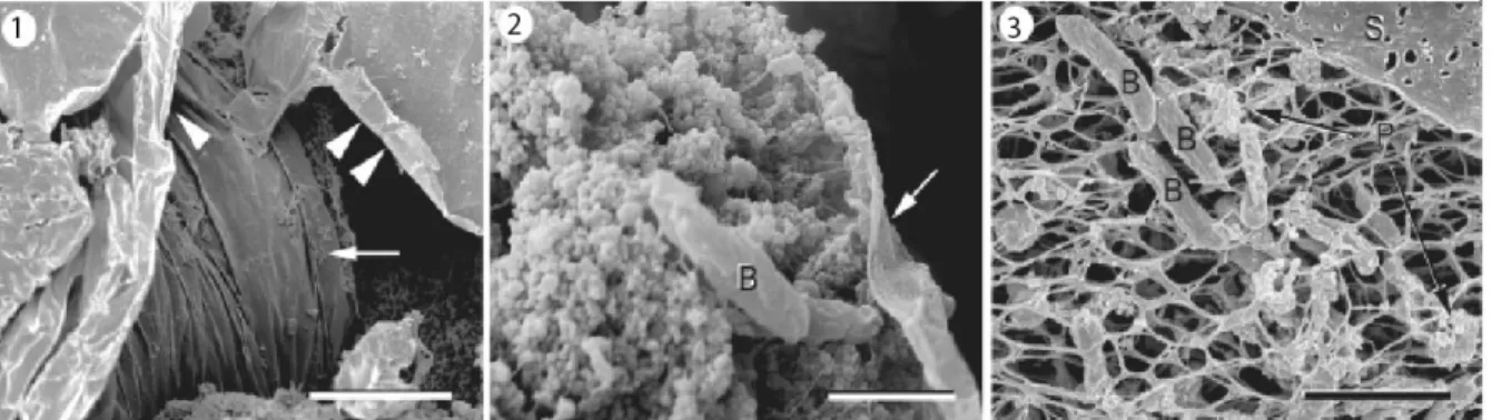
Figure 6. Scanning electron micrographs of P. fluorescens biofilms
The monocultures of 14-day biofilms form P. fluorescens EvS4-B1 were shown. 1. Flat sheets of material (arrowheads), with some of the sheets wrapped around other structures (arrow). Bar = 1 µM. 2. The inside core of the “wrapped” structures, consisting of bacteria (B) embedded in an extracellular matrix and thin sheet of material (arrow). Bar = 1 µM. 3. A sheet of material (S), consisting of extracellular matrix, covering and attaching to the fiber network (potentially fluid-filled channels) and including associated bacteria (B) and particulate matter (P). Bar = 2µM.
Adapted from Baum et al., 2009; Scales et al., 2014.
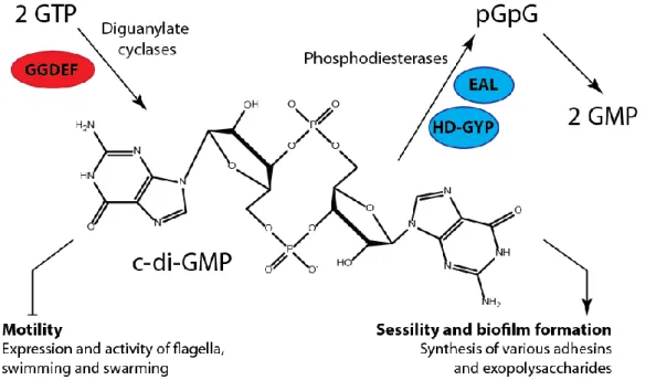
The biofilm formation involves an array of cellular factors and highly regulated mechanisms, specific for each bacterial species (Monds et al., 2007). Several extracellular components such as exopolysaccharides (EPS), proteins (e.g. adhesins, amyloid familial amyloidotic polyneuropathy (Fap) fibrils), and extracellular DNA (eDNA) are involved in adhesion and biofilm formation by the pseudomonads (Dueholm et al., 2013). However, a unifying theme across bacterial species is the synthesis of the cellular signaling molecule bis-(3’-5’)-cyclic dimeric guanosine monophosphate (c-di-GMP), which regulates both biofilm formation and motility (D’Argenio & Miller, 2004; Boyd et al., 2012; Schirmer & Jenal, 2009). The di-GMP is a soluble molecule, which functions as a second messenger in bacteria. In general, c-di-GMP stimulates the biosynthesis of adhesins and EPS matrix in biofilms and inhibits various forms of motility. It controls switching between the motile planktonic and sessile biofilm-associated ‘lifestyles’ in bacteria (Hengge, 2009). This molecule is synthesized by diguanylate cyclases (DGCs) (Paul et al., 2004), and is broken down into 5′-phosphoguanylyl-(3'-5')-guanosine (pGpG) by specific phosphodiesterases (PDEs) as presented in Figure 7. The active DGC is a dimer of two subunits with GGDEF domains. The PDE harbors EAL or HD-GYR domains (Kulesekara et al., 2006; Stelitano et al., 2013). While the accumulation of c-di-GMP in bacterial cells promotes the biofilm formation, favoring EPS production and suppressing motility, reduction of c-di-GMP level favors planktonic growth and/or dispersion of established biofilm (Christen et al., 2005; Newell et al., 2011a).
Figure 7. Structure and physiological functions of c-di-GMP
In bacteria, bis-(3'-5')-cyclic dimeric guanosine monophosphate (c-di-GMP) is controlled by diguanylate cyclases (DGC) with GGDEF domains (red) and specific phosphodiesterases that carry EAL or HD-GYP domains (blue). The high c-di-GMP level reduces motility and stimulates various biofilm-associated functions, such as the formation of fimbriae and other adhesins and various matrix exopolysaccharides. pGpG, 5'-phosphoguanylyl-(3'-5')- guanosine. pGpG is subsequently split into two GMP molecules.
Adapted from Hengge, 2009.
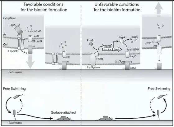
Biofilm research has utilized P. fluorescens as a model, because of its high adaptation capacity and typical growth on surface in nature (Newell et al., 2011b). O’Toole and coworkers identified a mechanism of regulation of biofilm formation by phosphate in P. fluorescens
Figure 8. This mechanism is based, in majority, on two proteins: a large adhesive protein,
LapA, required for the attachment, and a periplasmic cysteine protease LapG that cleaves LapA from the cell surface, as shown in litterature (Boyd et al., 2014; O’Toole & Ha, 2015). Under high phosphate level, the c-di-GMP accumulates in the cell. LapG activity is regulated by the inner membrane GMP effector protein LapD (O’Toole & Ha, 2015). LapD binds c-di-GMP and sequesters LapG at the inner membrane, promoting the maintenance of LapA on the cell surface. Under these conditions, LapA remains at the cell surface, forms irreversible attachments to the substratum, promoting biofilm formation (Newell et al., 2011a).
Figure 8. A proposed model for regulation of P. fluorescens biofilm formation
At high concentrations of phosphate, c-di-GMP accumulates in the cell. LapD binds c-di-GMP, sequesters LapG at the inner membrane, promoting the maintenance of LapA on the cell surface. In these conditions, cells attach to the surface and form the biofilm. Conversely, low phosphate concentrations are sensed by PhoR/Pst system complex, the PhoR kinase phosphorylates PhoB, which activates the transcription of rapA. The RapA degrades c-di-GMP to pGpG. This leads to dissociation of c-c-di-GMP from LapD, and the cleavage of the LapA in the periplasm, promoting the detachment of attached to substratum bacteria, and/or inhibition of future surface attachment of planktonic cells.
According Newell et al., 2011a.
Systematic analysis of P. fluorescens genomes revealed, that these species harbors over 20 genes, coding for putative DGCs, with 4 confirmed to be involved in the regulation of surface attachment and/or biofilm formation (Newell et al., 2011b). These genes include wspR gene. WspR is the response regulator (RR) of the Wsp chemosensory system, which is involved in production of c-di-GMP, as its effector. The Wsp system includes the DGC WspR, a predicted membrane-bound methyl-accepting chemotaxis protein (MCP)-like receptor (WspA), two CheW-like scaffolding proteins (WspB and WspD), histidine sensor kinase (WspE), and two methyltransferases WspC and WspF (Hickman et al., 2005). In addition to WspR, three other DGCs were found to promote biofilm formation in P. fluorescens. GcbA mainly controls swimming motility, GcbB preferentially affects localization of the LapA adhesin, and GcbC affects both LapA and motility (Newell et al., 2011b).
Although the clear establishing of positive correlation between biofilm formation and c-di-GMP level, the regulation of c-di-c-di-GMP, and as result, the switch between planktonic and biofilm growth, is increasingly complex (Petrova et al., 2014). Several genes (flgZ, sadB, fgtA,
flhH or fap) and regulation pathways (AdnA, GacA/GacS, AlgU or FuBA) were described as
Martínez-Granero et al., 2012; Mastropaolo et al., 2012; Nian et al., 2007; Robleto et al., 2003). These systems do not precise here, and themselves can be a subject of another work.
Biofilm is not only a simple bacterial lifestyle, but also a good adaptation mode to changing environmental conditions. The highly permeable water channels interspersed throughout the biofilm allow bacterial nutrient availability and metabolic cooperativity (Davey & O’Toole, 2000); the extracellular matrix protects bacteria against environmental stressors (O’Toole & Ha, 2015), including pollution (Baumgarten et al., 2012; Baum et al., 2009) or temperature changes (White-Ziegler et al., 2008), allowing bacterial survival in inconvenient environments. Biofilm formation is one of the strategies to colonize eukaryotic host. As a result, infections caused by bacterial biofilms are persistent and very difficult to eradicate (Drenkard, 2003; Mah
et al., 2003).
3.4. Envelope role in adaptation of Pseudomonas fluorescens
3.4.1. Introduction
To be faced with an unpredictable, dilute and often hostile environment, bacteria have evolved a sophisticated and complex cell envelope that protects them playing a role of a first selective barrier between the cell and its environment (Russell et al., 1995; Silhavy et al., 2010). The barrier functions of bacterial envelope are known to be depend critically on the physical state of lipid bilayer, including its structure and fluidity (Mansilla et al., 2004). Microbial envelopes are responsible for a plethora of physiological processes, such as regulating the substances traffic in and out of the cells (Uratani & Aiyama, 1986). It stabilizes protein structures for a correct functioning of membrane embedded enzymes and provides a matrix for many biological reactions (Bernal et al., 2007; Pogozheva et al., 2014). The cell wall plays also a role of the first sensor in activating a stress response (Los & Murata, 2004) and participates in bacteria/host interactions (van der Meer-Janssen et al., 2010; Vromman & Subtil, 2014; Wenk, 2006). Therefore, the precise regulation of membrane structure and fluidity, known as homeoviscous adaptation, in the face of a constantly changing environment is an important challenge for all bacteria (Baysse & O’Gara, 2007).
3.4.2. Properties of P. fluorescens membrane
Pseudomonas envelope, as well as envelope of all Gram-negative bacteria, is composed of two
membranous structures (Figure 9) (Bos et al., 2007; Glauert & Thornley, 1969). The inner one (IM) is composed of a glycerophospholipids (GPs) bilayer (Chatterjee & Chaudhuri, 2012). The outer membrane (OM) is an asymmetrical bilayer, consisting of GPs and lipopolysaccharides (LPS) in the inner and outer leaflet respectively (Freulet-Marrière et al.,
2000). The GPs bilayer of OM immerges proteins, including porins, and receptors (Bos et al., 2007). The outer leaflet is composed primarily of LPS projecting outside and the inner leaflet containing GPs.
Figure 9. Pseudomonas cell envelope
Inner membrane (IM) is a typical glycerophospholipid (GP) bilayer, playing a role of permeability barrier of the cell. The outer membrane (OM) is composed of an inner monolayer of GP and an outer monolayer of the Lipid A component of lipopolysaccharide (LPS). Lipid A is a fatty acid substituted phosphorylated disaccharide, connected via the unusual sugar 3-deoxy-D-manno-octulosonic acid (KDO) to a polysaccharide to build up the inner core, outer core and the O-antigen repeat. The prismatic space contains the amino acid-sugar cross-linked peptidoglycan layer, which gives a structural rigidity to the cell envelope. Many proteins and membrane-derived oligosaccharides (MDO) can also be found in periplasm.
Adapted from W. Dowhan and M. Bogdanov, 2002.
The LPS consists of three different sectors: (i) lipid-A (a fatty acid substituted phosphorylated disaccharide), (ii) the core polysaccharide comprising the inner and the outer cores, and (iii) the O-specific polysaccharide chains, a heterogeneous polymer of branched oligosaccharide units, as shown in Figure 9 (Chatterjee & Chaudhuri, 2012; Freulet-Marrière et al., 2000). The lipid portion of LPS serves as the lipid anchor and is commonly composed of fatty acids (FAs), sugars, and phosphate groups. The P. fluorescens LPS have been described as able to associate to the major outer membrane porin OprF (Freulet-Marrière et al., 2000) and essential for the outer membrane functions, particularly during host – pathogen interactions (Picot et al., 2003). The P. fluorescens LPS is an infamous molecule because they are responsible for the endotoxin shock associated with the septicemia (Raetz & Whitfield, 2002), apoptosis and a concomitant and limited necrosis (Picot et al., 2003).
Peptidoglycan layer is made up of repeating units of the disaccharide N-acetyl glucosamine and N-acetyl muramic acid, which are cross-linked by pentapeptide side chains (Heilmann, 1972;
Mirelman & Nuchamowitz, 1979; Vollmer et al., 2008). Because of its rigidity, it determines the cell shape. Agents, such as enzymes or antibiotics, that damage the peptidoglycan cause cell lysis owing to the turgor pressure of the cytoplasm (Silhavy et al., 2010).
P. fluorescens possesses a relatively simple GP composition with phosphatidylethanolamine
(PE, 60-80%), phosphatidylglycerol (PG; 10-25%), phosphatidylcholine (PC; not defined) and cardiolipin (CL; 8-25%) being the four major GPs found in the cell envelope (Cullen et al., 1971; Gill, 1975). Their relative ratio varies according experimental conditions and bacterial strains. However, these data are based on the experiments, performed in the 1970s using the thin-layer chromatography (TLC) (Cullen et al., 1971; Gill, 1975). The FAs composition of P.
fluorescens has been well described (Fouchard et al., 2005). Like all members of Pseudomonas
genus, P. fluorescens contains 16-18 carbons in FA chains with or without unsaturation (only one unsaturation is found). In the absence of well systematized review about Pseudomonas GP synthesis and homeostasis we decided to summarize all known today information about this topic in the review format.
3.4.3. Glycerophospholipid synthesis and functions in Pseudomonas
Tatiana Kondakova; François D'Heygère; Marc J. Feuilloley; Nicole Orange; Hermann J. Heipieper;Cécile Duclairoir Poc
3.4.4. P. fluorescens lipidome adaptation
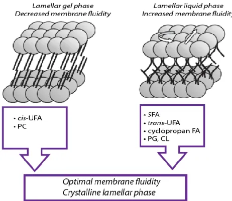
Environmental changes directly affect structural membrane characteristics (Baysse & O’Gara, 2007). As demonstrated in Figure 10, GPs’ bilayer forms different phases (i.e. a gel phase or a liquid-crystalline phase) (Hazel & Williams, 1990). The normal cell function requires membrane GPs bilayers that are largely fluid, in crystalline lamellar phase (planar bilayer) (Mansilla et al., 2004; Russell et al., 1995).
Figure 10. Adaptation of glycerophospholipid structure and composition in Pseudomonas
In the lamellar liquid-crystalline state, the lipid molecules are melted and disordered. Upon transition to the gel state, the glycerophospholipids (GPs) become ordered and the frequency of rotation and lateral movement is reduced. Bacteria can regulate their membrane fluidity adjusting the GPs head groups and fatty acids (FAs). In order to increase membrane fluidity, the increasing level of cis-unsaturated fatty acids (cis-UFAs) and phosphatidylcholine (PC) is required. In contrast, to decrease membrane fluidity, the levels of saturated (SFAs), trans-unsaturated (trans-UFAs), or cyclopropane FA are increased. Concerning the GP head groups, the increasing of phosphatidylglycerol (PG) and/or cardiolipin (CL) levels negatively regulates membrane fluidity.
Adapted from Baysse and O’Gara, 2007.
Environmental changes can cause the suboptimal membrane fluidity resulting in the subnormal membrane functions. The temperature at the midpoint of this transition is called the transition temperature (Tm), and the change of phase accompanying an increase in temperature is called
the lipid phase transition, or most properly, the order-disorder transition. The Tm is a function
of the membrane GP composition and mainly depends on the FA composition (Mansilla et al., 2004). The (overly simplified) rule of thumb is that GPs that contain unsaturated fatty acids (UFAs) have much lower Tm than those lipids made of saturated fatty acids (SFAs). The effect
is due to different packing of the two types of GP acyl chains as demonstrated in Table 1. SFA acyl chains can pack tightly, but the steric hindrance imparted by the rigid kink of the cis double bond results in much poorer chain packing of UFAs, even below the phase transition
temperature. The Tm depends also on the glycerophospholipid head group: Tm (CL) > Tm (PE)
> Tm (PG) (Rühl et al., 2012).
Table 1. Chemical structures of membrane fatty acids
Fatty acid Structure Effect on membrane fluidity
C16:0 Decreases membrane
fluidity
cis-C16:1 Increases membrane
fluidity trans-C16:1 Increases resistance to solvents and temperature cyclopropane-C17:0 Increases resistance to osmotic stress and
solvents
The ratio of GP head groups plays a pivotal role in maintain of optimal membrane properties and results in the balance between zwitterionic or neutral GPs (such as PE) and anionic GP (i.e. PG or CL) (Parsons & Rock, 2013). These changes affect the physico-chemical properties of membranes due to the differences in melting temperature between the GP head groups. The increase of the PG level comparing to PE in the membrane, allows the decrease of membrane permeability for lipophilic and polar molecules, and, at the same time, makes membrane more stable. This is because the PG and the CL have a higher transition temperature than PE and are more ordered and less densely packed GPs (Fang et al., 2000; Murzyn et al., 2005). The PC level plays also an important role in membrane properties. The increase of the PC level, comparing to PE, coincides positively with membrane fluidity. Basing on the size of the GP head group relative to the FA chains, the PC increases the membrane fluidity. Giving that the chemical structure of the choline HG is larger, than that of the PE, the PC has a relatively similar area of head group compared to FA chains. In contrast, the PE decreases the membrane fluidity, because the HG of PE is smaller, than the FA chains, resulting in a larger area of FA chains relative to the HG (Fajardo et al., 2011).
From these considerations, it seems to be clear, that bacteria and the most (if not all) poikilothermic organisms must regulate their membrane fluidity in response to environmental modifications. For this, the members of genus Pseudomonas have various pathways, several examples of which will be discussed below.
Control of fatty acid de novo synthesis
The control at the level of FAS is crucial for membrane homeostasis, because the biophysical properties of bacterial membrane are determined in large part by the composition of the FAs that are produced by de novo synthesis (Zhang & Rock, 2008). In this way, the long-chain acyl-ACP (see part 3.4.3. Glycerophospholipid synthesis and functions in Pseudomonas) plays a role of a key regulator of the FAS. In Pseudomonas spp., the three most significant enzymes are regulated by acyl-ACP: ACC, FabH and FabI. The ACC activity is inhibited by the long-chain acyl-ACP (Davis & Cronan, 2001) and lower ACC activity decreases the quantity of malonate groups essential for the initiation and the elongation steps of FAS. The acyl-ACP-mediated inhibition of catalytic activity of FabH prevents the initiation of new acyl-chains and, as consequence, limits the production of FAs (Heath & Rock, 1996a). The FabI catalytic activity, involved in the FA elongation, is also inhibited by acyl-ACP that could also reduce the total rate of bacterial FAs (Heath & Rock, 1996b). However, it should be noted, that P.
aeruginosa possesses the FabY and FabV enzymes (see part 3.4.3. Glycerophospholipid synthesis and functions in Pseudomonas), and the regulation of de novo FA synthesis in this bacterium could be more complex than in other Pseudomonas spp. species. The intracellular acyl-ACP level is controlled by PslB enzyme, which, in turn, is regulated by guanosine 3’,5’-bispyrophosphate (ppGpp) (Wahl et al., 2011). The ppGpp is a global regulator of gene expression and RNA synthesis in bacteria (Poole, 2012; Zhang & Rock, 2008), responsible also to peptidoglycan production and antibiotic resistance (Nguyen et al., 2011). It should be nevertheless noted, that only growing Pseudomonas cells can modify their GP and FA composition by de novo synthesis (Diefenbach & Keweloh, 1994).
Regulation of membrane biophysical properties
As previously demonstrated, the mainly studied membrane regulation pathway in Pseudomonas spp. strains is based on the FAs modifications (Heipieper & Fischer, 2010; Pepi et al., 2008; Zhang & Rock, 2008). Several physico-chemical factors (i.e. contamination by organic solvents or increasing temperature) can increase the membrane fluidity. To maintain the optimal membrane functions, this increase is regulated by the production of SFAs. The GPs containing 16:0 SFAs display a transition temperature that is higher than those of the 16:1 cis-UFAs (Heipieper et al., 2003; Zhang & Rock, 2008). The double bond of a cis-UFA causes an unmovable 30° bend in the acyl chain as presented in Table 1 and Figure 11 (Roach et al.,
2004), leading to the increase of the membrane fluidity. The SFA/UFA ratio is commonly quantified by the degree of FA saturation (Heipieper et al., 1996). The pathways, including FabAB and Des enzymes for synthesis of UFA, are discussed in the part 3.4.3. Glycerophospholipid synthesis and functions in Pseudomonas and will not be included below.
Figure 11. Modifications of fatty acids structure in Pseudomonas spp.
Bacteria can introduce a double bond into a saturated fatty acid (SFA) increasing membrane fluidity, via catalytic properties of Des enzymes. The rigid cis- double bond, introduces a pronounced kink of 30° in the acyl chain. GPs that contain cis-unsaturated fatty acids (cis-UFAs) occupy a greater molecular volume (blue shading) and do not pack as densely in the bilayer as GPs that contain SFAs. The cyclopropane FAs (a methyl group introduction across the double bond) formed by Cfa catalysis, have similar biophysical properties to the cis-UFAs, but render the membrane more stable to environmental insults. Pseudomonas cells can replace a cis-double bond with a trans-double bond via a mechanism, catalyzed by Cti isomerase. The trans-UFAs have properties that resemble SFAs, but give rise to the membranes that have higher transition temperatures. Phosphatidylethanolamine modifications are shown as an example.
Adapted from Zhang and Rock, 2008.
The conversion of cis- to trans-UFAs is an another adaptive mechanism documented for several
Pseudomonas spp. species, enabling bacteria to decrease their membrane fluidity, (Heipieper et al., 2003; Heipieper & Fischer, 2010). The advantage of the conversion is due to steric
differences between cis- and trans-UFAs (Figure 11, Table 1). Contrary to the cis-UFA, the steric structure of the trans-configuration lacks the kink in the FA chain and is able to insert into the membrane as for the SFAs (Roach et al., 2004). The conversion from the cis- to the
trans-unsaturated double bond does not have the same quantitative effect on membrane fluidity
as the conversion to SFAs, but still substantially influences the rigidity of the membrane, as illustrated in Figure 12 (Heipieper et al., 2003).
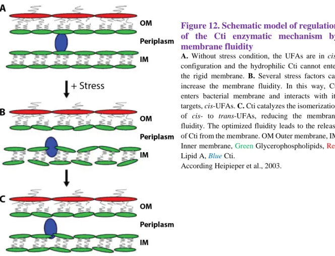
Figure 12. Schematic model of regulation of the Cti enzymatic mechanism by membrane fluidity
A. Without stress condition, the UFAs are in
cis-configuration and the hydrophilic Cti cannot enter the rigid membrane. B. Several stress factors can increase the membrane fluidity. In this way, Cti enters bacterial membrane and interacts with its targets, cis-UFAs. C. Cti catalyzes the isomerization of cis- to trans-UFAs, reducing the membrane fluidity. The optimized fluidity leads to the release of Cti from the membrane. OM Outer membrane, IM Inner membrane, Green Glycerophospholipids, Red
Lipid A, Blue Cti.
According Heipieper et al., 2003.
Besides FabA and FabZ enzymes discussed previously (see part 3.4.3. Glycerophospholipid synthesis and functions in Pseudomonas), the cis–trans-isomerase (Cti) was described in several Pseudomonas species (Holtwick et al., 1999; Pedrotta & Witholt, 1999), including P.
fluorescens (Heipieper et al., 2003). It is a constitutively expressed periplasmic enzyme that, to
exert its action, necessitates neither adenosine triphosphate (ATP) nor other cofactors, and consistently, is independent of the de novo FAS.
In several conditions, Pseudomonas spp. are able to form the cyclopropane FA (a methyl group across the double bond to form a cyclopropane ring, as presented in Table 1 and Figure 11). The required methylation reaction is carried out by cyclopropane FA synthase (Cfa), which uses S-adenosylmethionine as the methyl donor to create the cyclopropane group. Studies of acyl chain dynamics by nuclear magnetic resonance spectroscopy (NMR) indicate that the cyclopropane rings restrict the overall mobility and the disorder of the acyl chain between the
cis segment and the polar HG, compared to the cis- double bonds (Chang & Cronan, 1999;
Dufourc et al., 1983). The cyclopropane FAs have similar biophysical properties to the UFAs, but render the membrane more stable to environmental insults, such as acid stress (Zhang & Rock, 2008). Recent investigations, used molecular dynamics simulation showed, that the cyclopropane FAs may fulfill a dual function: stabilizing membranes against adverse conditions while simultaneously promoting their fluidity. Marked differences in the effect of cis- and