HAL Id: tel-02489811
https://tel.archives-ouvertes.fr/tel-02489811
Submitted on 24 Feb 2020
HAL is a multi-disciplinary open access
archive for the deposit and dissemination of sci-entific research documents, whether they are pub-lished or not. The documents may come from teaching and research institutions in France or abroad, or from public or private research centers.
L’archive ouverte pluridisciplinaire HAL, est destinée au dépôt et à la diffusion de documents scientifiques de niveau recherche, publiés ou non, émanant des établissements d’enseignement et de recherche français ou étrangers, des laboratoires publics ou privés.
Herve Rabille
To cite this version:
Herve Rabille. Biophysical and cellular mechanisms of tip-growth in the brown alga Ectocarpus sp.. Cellular Biology. Sorbonne Université, 2018. English. �NNT : 2018SORUS597�. �tel-02489811�
Sorbonne Université
Ecole doctorale 515 Complexité du Vivant
UMR 8227 (SU – CNRS) Laboratoire de Biologie Intégrative des Modèles Marins / Equipe de recherche « Morphogenesis of Macroalgae »
Biophysical and cellular mechanisms of tip-growth in the
brown alga Ectocarpus sp.
Par Hervé Rabillé
Thèse de doctorat de Biologie
Dirigée par Dr. Bénédicte Charrier
Présentée et soutenue publiquement le 3 décembre 2018
Devant un jury composé de :
M. Arezki Boudaoud, Professeur de l’ENS de Lyon, France : Rapporteur
Mme. Siobhan Braybrook, Project Investigator at the University California Los Angeles, USA : Rapporteuse
M. Benedikt Kost, Professeur de l’Université Friedrich Alexander Universität, Erlangen, Allemagne : Examinateur
M. Bernard Kloareg, Professeur de Sorbonne Université de Paris, France : Examinateur Mme Bénédicte Charrier, Directrice de Recherche CNRS : Directrice de thèse
Preamble
Contribution
This thesis project is by no means the only product of my own work, but is instead the fruit of the collective work of many people. The contribution of each is detailed below. At first, the project has involved the effort of all the members of the Morphogenesis of MacroAlgae research team. Dr Bénédicte Charrier, researcher in plant biology, performed a substantial part of the cytology experiments and data analysis. Dr Bernard Billoud, lecturer in bioinformatics, contributed to the analysis of some quantitative data and carried out all the computational work related to the mechanical modelling of Ectocarpus tip-growth. Élodie
Rolland, research technician in tissue culture, performed most of the algae cultivation tasks,
especially the preparation of the parthenosporophytes of Ectocarpus grown on glass coverslips, which were essential for most of the experiments.
The staining of the actin cytoskeleton using the phalloidin-based probe has been performed in collaboration with Pr. Christos Katsaros and his PhD student Maria
Koutalianou, both from the National and Kapodistrian University of Athens (Greece).
Actin and tubulin immunolocalisation experiments have been carried out with Dr. Adeel
Nasir (Friedrich Alexander Universität, Erlangen-Nurnberg, Germany), who supplied us with
an alternative protocol for cytoskeleton staining during a short visit.
All the observations of cell ultrastructure by Transmission Electronic Microscopy (TEM) have been conducted by Dr. Sophie Le Panse, from the “MerImage” microscopy plateform at the Roscoff Marine Biology Station.
The AFM data that are briefly presented and discussed in this report have been acquired by Benoit Tesson (Scripps Institution of Oceanography, University of California, San Diego, USA).
The identification of the causal mutation in the mutant étoile (etl) and the bioinformatic analysis (with the help of B. Billoud) of the candidate ETOILE gene which is briefly discussed in this report, is mostly the work of Zofia Nehr (former PhD student in the team). I performed the final completion of her substantial work with the help of the L2 student
Quentin Rochas, whom I supervised.
Finally, the results presented on the effect of drugs depolymerizing the cytoskeleton on the growth and morphogenesis of the apical cell were from time-lapse pictures of Ectocarpus filaments grown by Carole Duchêne, a former L3 internship in our team.
Articles
The Part 1 (Introduction section) of the report contains a large chapter reviewing the biomechanical models of tip-growth across the tree of life. It will be submitted as a review for an annual series or a book (Rabillé & Charrier, in preparation). The Part 1 also comprises an Opinion paper discussing the extent to which the cell wall composition and its intrinsic mechanics impact growth (Charrier, Rabillé & Billoud, in press in Trends in Plant Science).
The Part 2 deals with the biomechanics of tip-growth and of the cell wall. It contains two Original Research papers. The first article (Part 2.1) presents a visco-elastic model accounting for the tip growth in Ectocarpus, highlighting the role of the cell wall thickness
(Rabillé et al., in revision in PLoS Biology). The second one (Part 2.2) reports the role of alginates in the mechanics of the cell wall along the filament of Ectocarpus (Rabillé et al., in preparation).
Acknowledgements
First I must thank the two organisms that have funded my PhD: The Presidency of the
former Université Pierre and Marie Curie (now fused with Sorbonne Université) and the Brittany Region. I am also indebted to the Phycomorph Network (EC funded COST Action
FA1406) for having funding my Short-Term Scientific Mission from the 12th to the 29th of October, 2016, in the laboratory of Pr. Christos Katsaros (see below).
I thank warmly Dr Bénédicte Charrier, head of the Morphogenesis of MacroAlgae (MMA) team and my PhD supervisor, for having given me the opportunity to do work on such an exciting research project, at the very forefront of the current knowledge in cellular and developmental biology. Thank also you a lot for all the time you have dedicated to supervising my thesis, for all the (often very long) theoretical discussions about the mechanisms of cell growth, and especially on wall expansion mechanisms, the very hot topic that repeatedly came at the center of our thoughts in the course of these three years. Also, thank you for founding the very last weeks of this thesis project.
In the same vein, I have to thank you a lot, Dr Bernard Billoud, for your dedication into the project, and for the HUGE work for the numerical modelling of Ectocarpus tip-growth. Thank you also for, again, all the theoretical discussions mentioned above, and for you many informed, enlightened, and often stated loud and clear, points of view on the numerous hot technical and theoretical issues this research project has given rise to. More generally, I would like to tell all my gratitude to both B. Charrier and B. Billoud, for the overwhelming work that was the redaction and formatting of the two original papers which drafts are included in this report, in which I have only played a small part.
I also would like to thank Dr Catherine Boyen for having funded one extra month and a half of my thesis, that have allowed me to have time to write my report (extra time that I have largely abused).
I would also like to thanks all the member of my thesis committee, for their GREAT patience and indulgence (my apologies, again, for the almost 2 hours’ lateness at the second meeting, and my complete lack of professionalism), for their interest in the project and their kind pieces of advice. So thanks to Pr Bruno De Reviers, Dr Hayat Bouteau, Dr Thierry
Comtet. Thank also to Dr Arezki Boudaoud and Dr. B. Billoud for attending the first
meeting in February 2017.
I thank also all the members of my thesis comitee, that have kindly accepted to attend and evaluate my future thesis defence, that is to take place on the 3rd of December 2018. So thank you Dr Siobhan Braybrook, Dr Arezki Boudaoud, Dr Bernard Kloareg and Dr Benedikt
Kost. Many thanks especially for two former for tackling the task of evaluating this huge
report.
Here and now, I would like to thanks all the other people that have got involved in the thesis project, and without whom nothing, or so few, would have been possible. First I would like to thanks Élodie Rolland, research technician in the team, for preparing loads of
Ectocarpus cultures necessary for virtually all the experiments. Also thanks a lot to Dr
Sophie Le Panse for the massive TEM work. Thank a lot to Pr Christos Katsaros and to
(future Dr) Maria Koutalianou, for your dedication and help with the actin fluorescent staining. Also thank you very much, Dr Adeel Nasir, for your charitable help with actin staining, and providing us with some very good complementary results regarding this delicate. I have also to thanks to the two internship students, whose works have contributed to this project: first Carole Duchêne, L3 internship, for all her time-lapses of Ectocarpus growth and
development in presence of various drug, that I used to measure the impact of LatB on the morphogenesis of the apical cell. Secondly, thank to Quentin Rochas, L2 internship, for all his molecular biology work (much better than mine) to complete the positional cloning of
étoile. Regarding this task (that is not addressed in this report), I have also to thank Dr Zofia
Nehr for her help, especially regarding the use of the GeneMapper software. Finally, thanks
to Dr Thomas Torode, Dr Cécile Hervé and Pr. Paul Knox for providing the anti-alginate monoclonal antibodies (BAMs) and, at last, thanks to Murielle Jam, for having prepared and furnished the stock solution of the G-specific alginate-lyase (AlyA1, designated as AlyG in this report), that was central in our work on the links between the alginates and wall mechanics.
Now I must thank all my mates (students and young doctors), three years, for the soirées, restaurant sessions, coffee breaks, and fun and moral support in general, that have enlightened these three harsh years (especially the last one). Thanks first to Dr Maria Matard-Mann,
Laure Mignerot, Dr Adèle James (A.K.A “Miss l’Oréal”), Anaïs Naretto, and Eugénie Grigorian, that all experience (or have experienced recently) the ups and downs of the thesis.
Thanks also to Elodie Rolland, Dr Jonathan Dorival (ouai, c’est pas faux !!!!), Dr Yacine
Badis, Dung Nguyen Thi Ngoc, Dr Zofia Nehr, and Émilie Guilloud. Thank you, all of
you, for your kindness, your good mood, and more simply for having being there!
Special thanks to Maria, for your kindness, cheerfulness, and your constant support and numerous pieces of advice. Please never change the way you are! And thank you also for the anti-stress essential oils! Special thanks to Yacine, too, just for being that crazy, for the political discussion and for having taught me the rudiments of boxing. And special thanks to, you Eugénie.
Also thanks to my two office neighbours, Ludovic Delage and Gaëlle Correc, for your general kindness, happiness, and jokes. Thank you for having endured my antisocial, and sometimes grumpy, temperament.
To finish, I wish to thanks my few friends and my family members, that have been shield against my loneliness, even though we have stayed in contact through social networks (Roscoff is so damn far from the rest of France!). So thank you very much to Emmanuel
Daoud-Hoareau, Mathieu Pierre and Gwendoline Birot, my master best friends, members
of the RDP team (you know what I mean)! Thank you, Mathieu David and Conrad
Hillairet, my only friends from the “Classe Préparatoire”. We are far from one another, and
have somewhat lost touch each other, but I never forgot you. Thank you to you, mummy and
daddy, and to Armelle and Gwenaëlle, my two sisters to which I am so close. Without all of
Table of content
1. Introduction ... 15
1.1. “The mechanics of tip-growth: an overview over the Tree of Life”... 17
1.1.1. Introduction ... 17
1.1.2. General concepts of biomechanics of cell morphogenesis and tip-growth ... 19
1.1.3. The cytoskeleton as the main mechanical factor of the growth patterning... 25
1.1.4. The cell wall as the main mechanical factor of growth patterning ... 34
1.1.5. Turgor and associated hydrodynamic flows as the main mechanical factor of the growth patterning ... 58
1.1.6. Conclusion and perspectives ... 62
1.2. Brown algae: an ideal and stimulating groups for discovering alternative morphogenetic mechanisms ... 67
1.2.1. General overview of brown algae ... 67
1.2.2. Morphological diversity and morphogenetic pathways in brown algae ... 69
1.2.3. Physical constraints on the development and morphogenesis of brown algae ... 72
1.2.4. Cellular peculiarities ... 73
1.3. About the transposition of canonical mechanical models of cell wall expansion to brown algae 79 1.3.1. Abstract ... 79
1.3.2. Cell wall expansion: does the known matter really matter? ... 79
1.3.3. Uncoupling cell wall growth from the intrinsic properties of the wall ... 80
1.3.1. Cell wall growth: demystifying polysaccharide chemistry ... 83
1.3.2. Concluding remarks and future prospects ... 91
1.3.3. Glossary ... 92
1.4. Ectocarpus as a model system to study cellular morphogenesis in brown algae ... 93
1.4.1. A model species for the brown algae ... 93
1.4.2. Ectocarpus (partheno)sporophytes are ideal for the study of cell morphogenesis ... 94
1.4.3. Cellular morphogenesis and tip-growth in prostrate filaments ... 94
1.4.4. étoile: a tip-growth mutant of Ectocarpus ... 97
1.5. Thesis objectives ... 98
2. Biomechanics of the apical cells and biomechanical strategy of the apical cell tip-growth ... 99
2.1. A mechanical model of Ectocarpus tip-growth ... 99
2.1.1. Abstract ... 99
2.1.2. Author summary ... 100
2.1.3. Introduction ... 100
2.1.4. Results ... 102
2.1.1. Discussion ... 113
2.1.2. Materials and Methods ... 121
2.2. The mechanical role of alginates in Ectocarpus cell walls ... 127
2.2.1. Introduction ... 127
2.2.2. Results ... 129
2.2.3. Discussion ... 146
2.2.4. Materials and Methods ... 150
3. Molecular underpinning of apical cell tip-growth: the role of the (actin) cytoskeleton 155 3.1. Background ... 155
3.2. Organization of the microtubules (MTs) in the apical cell ... 156
3.3. Role of the actin cytoskeleton in tip-growth of Ectocarpus ... 156
3.3.1. Organization of the actin in the apical cell ... 156
3.3.2. Impact of depolymerization of F-actin on apical cell organisation and growth ... 162
4. General discussion and perspectives ... 173
4.1.1. Cell wall thickness gradient as a mechanical patterning factor in tip-growth ... 173
4.1.2. How to generate a stable thickness gradient? ... 176
4.2. The importance of wall mechanics in tip-growth of Ectocarpus apical cells ... 178
4.2.1. Mechanical features of the cell wall in the apical cell ... 178
4.2.2. In muro molecular determinism of wall mechanical properties ... 182
4.2.3. Cytoplasmic determinism of wall mechanical properties: direct mechanical role of the actin cytoskeleton? ... 184
4.2.4. Conclusion: differential role of the cytoskeleton and of the wall chemistry and mechanics in the control of growth ... 186
5. Material and Methods ... 191
List of Figures
Part 1 – Introduction
Figure 1.1 Basic characteristic of a tip-growing cell p. 21
Figure 1.2 Mechanisms of amoeboid locomotion p. 27
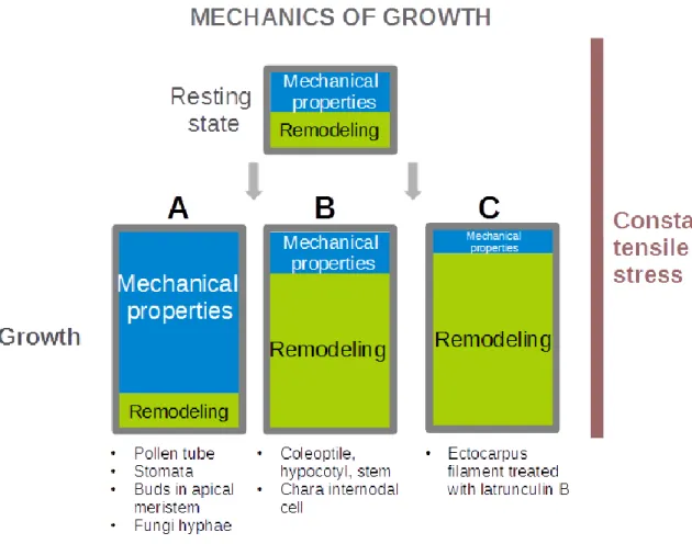
Figure 1.3 Classification of the different mechanical “strategies” observed or envisioned for tip-growing cells
p. 64
Figure 1.4 Phylogenetic positions of brown algae (Phaeophyceae) p. 68
Figure 1.5 Overview of the diversity of thallus construction modes
in brown algae
p. 70
Figure 1.6 Cell wall mechanical properties involved in cell wall
expansion
p. 81
Figure 1.7 Comparison of the cell wall chemical composition and
structure in land plants and brown algae
p. 86
Figure 1.8 Ectocarpus general morphology and early pattern of
growth
p. 96
Part 2 – Biophysics of tip-growth in Ectocarpus
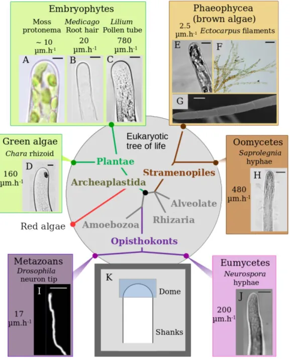
Figure 2.1 Diversity of tip-growth in the Eukaryotic tree p. 103
Figure 2.2 Position and direction of cell wall expansion during
growth
p. 105
Figure 2.3 Viscoplastic model of tip growth p. 107
Figure 2.4 Turgor and curvature of the apical cells. p. 108
Figure 2.5 Cell-wall thickness of the apical cell p. 111
Figure 2.6 Schemes summarizing the biophysical properties of two
tip growing cells: Ectocarpus filament apical cell and tobacco pollen tube
p. 112
Figure 2.7 Contribution of the cell wall biophysical parameters in
Ectocarpus and pollen tube tip growth
Figure 2.8 Impact of yield threshold (σy) and extensibility (Φ)
variations on Ectocarpus tip growth
p. 118
Figure 2.9 Impact of the cell wall thickness gradient and pattern of cell wall biosynthesis
p. 120
Figure 2.10 Filament organisation and cell morphologies observed
by scanning electronic microscopy
p. 133
Figure 2.11 Mannuronan-rich alginate blocks labelled with BAM6
antibody
p. 134
Figure 2.12 Mannuronan-Guluronan alginate blocks labelled with
BAM7 antibody
p. 135
Figure 2.13 Guluronan-rich alginate blocks labelled with BAM10
antibody
p. 136
Figure 2.14 Summary of alginate mapping along the filament of
Ectocarpus
p. 137
Figure 2.15 Cell wall thickness and structure along the filament p. 139
Figure 2.16 Wall stress along the filament p. 140
Figure 2.17 Alginate location in response to a hypotonic shock p. 142
Figure 2.18 Alginate location in response to a hypertonic shock p. 145
Figure 2.19 Stiffness in the dome by dilatation/retractation p. 146
Part 3 – Molecular basis of tip-growth: the role of actin cytoskeleton
Figure 3.1 Organization of microtubules in the apical cells of theWT and étoile, revealed by immunofluorescence microscopy
p. 159
Figure 3.2 Distribution and organization of the actin cytoskeleton
in the apical cells of WT and étoile, revealed by (immuno)fluorescence microscopy
p. 160
Figure 3.3 Summary of the general organization of the actin and
microtubule cytoskeleton in the Ectocarpus prostrate filaments of the apical cell
p. 163
Figure 3.4 LatB effectively depolymerizes the AFs in the apical
cells of Ectocarpus prostrate filaments
Figure 3.5 AF depolymerization of LatB abolish tip-growth but not surface expansion in apical cells, and zonal organization of the apical cells according to the dependence of the shape upon the presence of AFs
p. 166
Figure 3.6 AF depolymerization by LatB reduces cell wall
deformability after ~19h of treatment
p. 170
Figure 3.7 AFs depolymerization by LatB does not inhibit wall
deposition but alter its structure
p. 171
Figure 3.8 AFs depolymerization by LatB enhances cellulose
deposition in the wall of the apical cell
p.172
Part 4 – General discussion
Figure 4.1 Global overview of the results on the integrated
mechanism of tip-growth in the apical cell of
Ectocarpus sporophytic vegetative filaments
List of Tables
Part 1 – Introduction
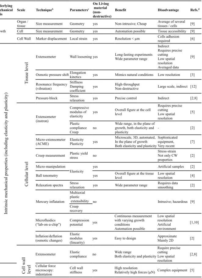
Table 1.1 Techniques employed for the study of cell wall
mechanics during expansion.
p. 84
Table 1.2 Cell wall components of land plants and brown algal
cell walls
p. 88
Part 2 – Biophysics of tip-growth in Ectocarpus
Table 2.1 Elastic Modulus E of the four virtual cell wall layers
L1-4 inferred from the force curves obtained by atomic force microscopy.
1. Introduction
In order to carry out this pioneering study about the biomechanical mechanism of tip-growth in Ectocarpus, a considerable analysis of the literature has been necessary to put our results in the context of research and theories about cell growth and morphogenesis. In this introductory session, the huge literature about the biomechanics of tip-growth, especially in walled-cell organisms (land plants, all fungal or fungal-like organisms, algae, and bacteria to a lesser extent) is critically reviewed in a first part (1.1, a review paper in preparation). In this chapter the main mechanical models are presented, with their advantages and pitfalls, and the diversity of models is discussed in the context of the origin and the evolution of tip-growth.
In a second part (1.2), the general characteristic of brown algae in term of phylogeny, ecology, morphogenesis and cellular characteristics (cytoskeleton and cell wall) are reviewed, in order to expose the particularly exciting challenges and opportunities that this special group offers to study alternative cellular and tissular morphogenetic mechanisms.
In a third part (1.3), the problem of wall expansion and its molecular control during walled cell growth and morphogenesis is tackled, in the form of an Opinion Paper that is in press in the journal Trends in Plant Science. In this part, the traditional methods and concepts pertaining to the mechanism of wall expansion, developed mainly for terrestrial plants, are questioned. A detailed comparison of cell wall structure and chemical composition between land plants and brown algae is presented, to show that the mechanisms at play during cell growth must be radically different between the two groups. The latter must be studied anew, without a priori hypothesis drawn from the land plant literature, in order to discover novel mechanisms of cell wall expansion, and their link with cell and tissue morphogenesis.
In a fourth part (1.4), the brown algal model species to study cell morphogenesis and tip-growth, Ectocarpus sp., is described, with the current knowledge about its development.
1.1. “The mechanics of tip-growth: an overview over the Tree of
Life”
In preparation (this part is to be published as a book chapter).
1.1.1.
Introduction
From the sub-cellular to the organism levels, growth and morphogenesis are fundamental mechanical processes, and the developing organisms have to comply with the rules of the physical world to acquire their final form and size (Boudaoud, 2010; Mirabet et al., 2011; Ali
et al., 2014). One of the most fundamental issue in the field of evolutionary developmental
biology (evo-devo) is to decipher how living things adapted to make use of the inescapable physical laws to achieve functional morphologies essential to their fitness, and how those mechanisms have emerged and evolved afterwards. Evolution works mainly at the genomic level, while growth and morphogenesis result from the physical transformation of living structures that imply in part (but not only) their mechanical deformations (Niklas, 2000; Ali et
al., 2014). Thus, an outstanding issue is to understand to what extent the evolution of
“macroscopic” biomechanical processes at play during morphogenesis have been constrained by i) the genes and the molecular machinery behind the metabolic networks, that build the cell structural components, and that control the cell spatial organization and dynamics, and ii) by the “physical world”, including the cytomechanical properties (resulting from the composition and structure of cellular components) and those of the external abiotic environment (Hamant and Traas, 2010; Mirabet et al., 2011; Ali et al., 2014). Different trade-off between these factors could have resulted in the range of morphogenetic strategies observed in today’s living organisms.
The study of morphogenesis of isolated cell types not embedded into a multicellular tissue is a good approach for this aim, because these cells are easily accessible for experimental manipulations and microscopic observations, have a limited number of interacting physical components and thereby represent simplified systems for modelling (Harold, 1990; Niklas, 2000; Geitmann, 2006a; Geitmann and Ortega, 2009). In this respect, tip-growth represents an ideal case, because it is extremely polarised, yet simple and robust (see an overview of the general characteristics of tip-growing cells in Fig 1.1). Tip-growing cells are generally “invading cells” exploring external environments and thus are easily isolated and cultivated in laboratory for in-vivo studies. Tip (or apical) growth is one of the most common polarised cell elongation form in the living world (Heath, 1990), and is encountered in a large range of taxonomic groups, both in prokaryotes (actinomycetes, Prosser, 1990) and in eukaryotes (land plants, metazoans, eumycetes, oomycetes, the three major groups of macroalgae and several minor algal clades, Heath, 1990). Its wide phylogenetic occurrence is a testament to the large adaptive advantage it provides to the organisms, such as the exploration and colonization of vast surfaces or the invasion of hard solid media like soils or living tissues. Extremely elongated filaments insure critical functions as diverse as colonization, anchorage, water and nutriment uptake or delivery of particular cargos or chemical signals between distant spots in the organism, to cite just a few (Money et al., 2004; Harris, 2011; Sanati Nezhad and Geitmann, 2013; Bezanilla et al., 2015). It represents a unique chance for evo-devo studies of basic cellular morphogenetic phenomena spanning many branches of the tree of life, and for digging into its deepest evolutionary roots. In this context, the most important question is
whether such a distinctive polarized growth form could have been generated by different biomechanical morphogenetic strategies. This would allow deciphering the degree to which the “physical world” carries weight on the mechanisms of tip growth. In addition, exploring in parallel the molecular aspect of tip-growth functioning would indicate to what extent the variations of biomechanical mechanisms are correlated to variations in the set of available molecular regulators and pathways in different groups.
Until now, it is not clear whether tip-growth has emerged repeatedly in the course of evolution, or if it only appeared once and has thereafter been conserved in the various diverging lineages. From a molecular point of view, the invasive growth processes in Eukaryotes (at least in land plants, fungi and metazoans) are thought to be controlled by a common, evolutionary conserved molecular “toolkit” (Vaškovičová et al., 2013). This molecular toolkit involves the actin cytoskeleton, cellular trafficking, the exocyst, some molecular pathways including Rho-GTPases and lipid signalling. The evolutionary distance between land plants, metazoans and eumycetes suggests that the molecular toolkit was already present in the Last Eukaryotes Common Ancestor (LECA), so any eukaryotic taxa may have had the opportunity to inherit it. However, even if these molecular players are homologous, it is still possible that the regulatory network they built emerged from convergent evolution, rather than having a unique origin (Vaškovičová et al., 2013). In the future, the involvement and degree of conservation of such core toolkit remains to be investigated in more details, including other, underexplored phylogenetic groups. The scarce data existing about underexplored groups, like brown algae (Coelho et al., 2002; Fowler et al., 2004; Hable and Kropf, 2005; Bogaert et al., 2013; Muzzy and Hable, 2013; Hable, 2014) and oomycetes (Jackson and Heath, 1989; Garrill et al., 1993) suggest that at least some of these molecular factors are again involved in tip growth in these distant clades, belonging to the Stramenopiles “kingdom”, further supporting the hypothesis of a conserved molecular toolbox. Although some molecular players are also found in polarly growing prokaryotes (Zhang et al., 2010), those involved in tip-growing Actinomycetes seem specific to them (Flärdh, 2003, 2010; Flärdh et al., 2012), suggesting independent evolutionary roots for tip-growth between the major domains of the tree of life. However, more research will be needed in the future before concluding about the degree of conservation and divergence of the molecular factors involved in tip-growth regulation, and about their effect on tip growth biomechanics.
At a physical level, cellular growth and morphogenesis result from the combined action of “protruding” forces generated by the protoplast to expand the cell surface at localized areas, and of “resisting” forces, that tend to oppose the firsts. The latter are those generated in reaction by either the cellular envelope (cell membrane and extracellular matrix, Mirabet et
al., 2011) and by the external medium in which the cell is growing (Money, 1999; Sanati
Nezhad and Geitmann, 2013, Fig 1.1). Thus, it would be sensible that only few, or maybe a unique, biomechanical strategy, could account for such a robust, conserved cellular morphogenesis as tip-growth. Nonetheless, for more than one century of research, a surprising plethora of alternative biomechanical models of tip-growth have been imagined and put forward by different authors. However, most of the putative mechanisms are, for now, only theoretical, and some models lack clear experimental support. Should such diversity turn out to be real, it would be interesting to test whether it is more correlated to the phylogenetic position or to the abiotic environment of the organism, or to the particular physical conditions encountered by apically-growing cell types. At least, because of the fundamental difference in cell size and structure between eukaryotes and prokaryotes, tip-growth mechanisms are likely to be completely different between the two domains (Prosser, 1990). In the eukaryotes, an interesting modelling paper by Campàs and colleagues (2012) pointed toward a disparity in the physiological and biophysical strategies adopted by land plants (Archaeplastida) and
hyphal eumycetes (Opistokontes) on the one hand, and by fungi-like oomycetes (Stramenopiles) on the other hand. In prokaryotes, some Actinomycetes also form hyphae very similar to eumycetes and oomycetes; all the three groups form complex mycelial networks able to invade host tissues or soils (Prosser, 1990; Flärdh, 2010; Cameron et al., 2015). Yet, these three groups do not share any close phylogenetic ancestor and evolved completely independently. This simple noticing suggests that tip-growth has emerged multiple times by convergent evolution, always leading to the same final, reproducible morphology.
Unfortunately, the current literature on tip-growth lacks of a broad view on the emergence and evolution of tip-growth across the tree of life. A large majority of studies have indeed focused only on some favoured taxonomic groups, i.e. the angiosperms, eumycetes and metazoans, each only represented by a small set of model species. The few papers offering an evo-devo comparison of tip-growth mechanisms generally remained focused on these few groups (Vaškovičová et al., 2013; Honkanen and Dolan, 2016; Honkanen et al., 2016; Rensing, 2016) while other taxonomic groups are still largely neglected. Deciphering if, and how, the physical constraints and the genomic baggage of an organism have influenced the biomechanical strategies to produce tip-growing cells will require more work in the future, and need to encompass understudied taxa, and to cross-correlate the biomechanical processes and their regulators into single, integrated models.
The aim of the present review is to browse the current knowledge about the diversity of biomechanical strategies of tip-growth drawn from both experimental evidences (cell mechanics, ultrastructure and chemistry) and theoretical models. A first chapter will briefly present the basic characteristics of tip-growing cells, in term of growth kinetics and mechanics (Part 1.1.2). The various biomechanical models will then be presented and classified according to the main cellular component or physical parameters involved. Those mechanical players, namely the cytoskeleton (Part 1.1.3), the cell wall (Part 1.1.4) and the turgor pressure (Part 1.1.5) are thus successively described as the main “mechanical patterning factor” of tip-growth. The models are critically evaluated to uncover their strength and limits. For the sake of conciseness, the various experimental approaches and details about the implementation of mathematical and computational models are left apart. We rather focus on the concepts, theories and ideas that have been supported or validated by experimental data. When possible, the reader is redirected toward the relevant papers for more information.
1.1.2.
General concepts of biomechanics of cell morphogenesis and
tip-growth
1.1.2.1. Diversity of tip-growing cell shapes
All tip-growing cell type share the same basic architecture: an elongated, generally stable tubular region terminated by a differentiated apical region, where the expansion of the cell envelope ─ a cell membrane completed with the internal cortical cytoplasm and the outer extracellular materials ─ is restricted (Heath, 1990; Martin et al., 2001; Fig 1.1). The restriction of the surface expansion to the apex implies that the tubular regions become increasingly older as the distance from the tip increases. These non-growing regions are
defined as being “distal” to the apex that is generally designated as the proximal pole. This terminology can be sometimes confusing as the growing tip is generally the farthest extremity of a cellular projection that emerged from a basal cell body, like the pollen tube emerging from the pollen grain or the root hair from a root epidermal cell (Gilroy and Jones, 2000; Rounds and Bezanilla, 2013), and thus should rather be designated as the distal pole. By commodity, we will keep the traditional terminology used by authors working on tip-growing cells, that is, the apex being the proximal pole.
Tip-growing walled cells are generally considered as perfectly axisymmetric shells. From the extreme tip of the cell (the apical pole per se), meridians can be drawn toward the distal directions, more or less parallel to the longitudinal axis of the cell. This direction is called the meridional direction. The orthogonal direction to the meridional one is called the hoop, transversal or circumferential direction. Because of the axial symmetry of the cell, most molecular, physiological and physical parameters occurring at the cell surface during tip-growth (like cell wall deposition, ion flux, membrane and in muro enzymatic activities) can be comprehensively quantified only as a function of the meridional position. However, some geometrical, structural and kinetics parameters (at least surface curvature and strain rates) can be anisotropic, i.e. can be different between the meridional and circumferential directions. Thus, those properties must be, at any point of the cell surface, quantified in both meridional and circumferential directions (Fig. 1.1).
The tubular region below the apical growth site is traditionally designated as the “shanks” or the “shaft”, and has generally the basic form of an isodiametric elongated cylinder. In some cases low residual growth can still occur in subapical regions, generally contributing to a slight and gradual increase in diameter, for example in Medicago truncatula root hair (Shaw
et al., 2000) or in Saprolegnia ferax hyphae (Jackson and Heath, 1990). Beyond this basic and
highly conserved shape, a large diversity of diameters is found between taxa, ranging from the narrow hyphae of Actinomycetes (less than 1 µm in diameter; Prosser, 1990; Goriely and Tabor, 2003a), to the wide giant cells (several hundreds of µm) of the sporangiophore of
Phycomyces (Castle, 1958) and the giant siphonous cell of the alga Vaucheria
(Xanthophyceae; Mine and Okuda, 2003; Mine et al., 2008).
In contrast to the tubular shanks, the growing apices show a large diversity of shapes. In the non-walled axon of metazoan neurons, it grows as a cone, a highly complex motile device projecting numerous filipodia in all directions (Heidemann, 1990; Franze and Guck, 2010). This is in striking contrast to walled cell organisms, which the vast majority of tip-growing cells belongs to, where the growing apical region is much simpler and generally takes the form of a demi-spheroid or a prolate demi-ellipsoid dome (Fig 1.1). However, the dome shape usually appears significantly divergent from a truly ellipsoid shape, as in the M. truncatula root hair, (Shaw et al., 2000; Dumais et al., 2004) and the Phycomyces sporangiophore (Castle, 1958). Thus, even among walled cell organisms, a large diversity of dome shapes exists between distantly related taxa, and this has been pinpointed as the sign of fundamentally different biomechanical strategies between distantly related groups (Campàs and Mahadevan, 2009; Campàs et al., 2012). To wholly quantified the shape and the wall strain of tip-growing cells, both the circumferential and meridional curvatures must be quantified as a function of the meridional distance from the apical pole (see for examples Chen, 1973; Hejnowicz et al., 1977; Dumais et al., 2004).
Figure 1.1 - General organisation of a tip-growing cell
The schema represents a walled cell tip-growing cell (such as found in land plants, fungi or algae) with a strong turgor pressure, but the same principles apply to animal cells, that have fairly only a cell membrane. In this situation, turgor pressure is still present but with much lower values (maximum some hundreds of Pa). The cell is organised into an apical region and tubular shanks. Growth activity, i.e. deposition and expansion of the cellular envelope, is restricted to the apical tip, especially for walled cells, where cell wall does are stabilized at the base of the dome, and no more expansive growth occurs in the shank. The turgor results from a difference of osmotic pressure between the internal and the external medium (πi and πe, respectively). The cytoskeleton is particularly
abundant, with often long microtubules (MTs) and actin filaments (AFs) more or less parallel to the longitudinal axis in the shanks, and a complex network of AFs in the apical region. Expansive growth at the apex (apical dome for walled cell organism) results from a local unbalance between the outward directed “protruding” mechanical forces that tend to expand the cell envelope surface and volume, and “resisting forces” that opposes the firsts. The “protruding” forces are generated by the cytoskeleton or the turgor pressure. The resisting forces are primarily exerted by the cell envelope, namely the cortical cytoskeleton and / or the cell wall when present. The external medium in which the cell is growing can also exert significant mechanical impedance on the growing apex. In walled cells, the resistance of the external wall to the turgor generates a high tensile stress in the wall, that is generally thought to provide most of the work of wall expansion. However, in cases where the cell wall has a constant thickness across the cell surface, the tensile stress is lower in the apical dome because of local curvature, making it a priori unfavourable to restrict growth in this area.
1.1.2.2. Diversity of cell envelopes in tip-growing cells
Tip-growth implies that the expansion of the cell envelope is essentially restricted to the apical region, while the sub-apical region adopts a stable tubular shape. In plant and fungal organisms, the envelope includes a cell wall that is the key element to be deformed to actuate tip-growth (Hamant and Traas, 2010; Mirabet et al., 2011; Chebli and Geitmann, 2017). The presence of such extracellular compartment is paralled by the presence of a high internal hydrostatic pressure, called turgor. The internal hydrostatic pressure in cell results from the difference of osmotic pressure between the cytoplasm (πi) and the external medium (πo), the former being more concentrated in osmotically active solutes than the second. Higher internal osmolarity lower the water potential of the cytoplasm compared to that of the external environment (Fig 1.1). The water thence permanently tends to flow inside the protoplast, developing a hydrostatic pressure. At any given point on the inner face of the cell envelope, it generates a constant outward directed force orthogonal to the tangent to the cell surface at this point. The cytoplasm is traditionally conceptualised as an aqueous fluid and, because of the water incompressibility, the turgor is thought to be a scalar, not a vectorial (i.e. oriented) force, meaning that it is constant throughout the cytoplasm (Winship et al., 2010), and thus exert the same protrusive force at any point of the cell surface. In principle, turgor alone cannot, then, generate a cell shape different than a purely spherical cell (Hamant and Traas, 2010; note, however, that this is not entirely true, see for example Boudaoud, 2003; Bigan, 2015).
However, for the sake of mechanical equilibrium, the outward-directed protrusive force of the turgor is contained by the cell wall, in which a huge tensile stress that builds up in the outer cell envelope, until a mechanical equilibrium is reached. As the cell wall has a certain thickness, not negligible compared to the cell radius, the turgor-generated tensile force is, at any point of the cell surface, distributed on a cross-section of wall. The tensile force per unit cross-sectional area of wall is called the tensile stress and is equivalent to a pressure (Pa; Fig
1.1). The wall tensile stress that build in response to turgor is even larger than the turgor itself,
because of the thinness of the cell wall compared to the cell radius (Castle, 1937) and provides the mechanical energy necessary to stretch the wall (Geitmann and Ortega, 2009; Guerriero et al., 2014). At any point of the cell surface, the tensile stress will depend on mainly 4 factors: 1) the turgor pressure (MPa), 2) the local cell surface shape, represented by the curvature measured in a least two, perpendicular directions (m-1) and 3) the local cell wall thickness (m) that effectively bears the tensile stress (Dumais et al., 2006). The tensile stress is positively correlated with turgor but negatively regulated by the others two parameters, and all could be locally modified to control the local expansion of cell surface at sub-cellular scales, underlying heterotropic growth like tip-growth (Green, 1965, 1969; Geitmann and Ortega, 2009). In the cylindrical part of a tip-growing cells, the circumferential stress is twice as large as the meridional stress. In the apical dome, if the wall thickness is considered constant, the tensile stress progressively decreases toward the apical pole in both the circumferential direction because of the increasing curvature, and both components gradually converge to the same value at the extreme pole (Hejnowicz et al., 1977; Dumais et al., 2004). This, indeed, represents the main paradox of tip-growth in walled cell organisms: surface expansion is restricted to the apical dome, where the tensile stress that make the work of expansion is the lowest in the whole cell! Most of the experimental and theoretical models dedicated to the biomechanic of tip-growth have indeed aimed to solve this conundrum (Harold, 2002; Geitmann, 2006b; Kroeger and Geitmann, 2012b).
In non-walled cells, the outer layer of the cell envelope is the cell membrane, that is a fluid layer of phospholipids and proteins (even though the existence of lipid rafts and the
molecules in the plane of the membrane; Janmey, 1995; Adler et al., 2010; Levental and Veatch, 2016). In this case, the cell “surface”, even if deposited in specific areas, can flow laterally, preferentially toward the site of active expansion. Such processes most likely occur in the animal migrating cells (Lauffenburger and Horwitz, 1996), growing axons (Franze and Guck, 2010), and the amoebas (Taylor and Condeelis, 1979; Grębecki, 1994). In these cell types, the “cell envelope” includes the cortical actomyosin that is connected to the extracellular medium (see Part 1.3 below). Yet, the same mechanical principles apply to the cell membrane and cortical cytoskeleton in those kinds of cell. The cell membrane is under more or less tension because of the protrusive forces exerted by the weak hydrostatic pressure or the cytoskeleton, and those forces regulates the shape, motility, migration, and finally morphogenesis, of the cell (Houk et al., 2012; Diz-Muñoz et al., 2013; Lieber et al., 2013; Kim et al., 2015).
1.1.2.3. Biomechanical theories of growth and morphogenesis control in walled cell organisms
A vast majority of tip-growing cell types occurs in walled organisms, like plants, fungi, and algae (Heath, 1990). As stated above, it is the cell wall that must be stretched at the growing tip to expand forward the cell, and this mechanical work is thought to be done by the tensile stress that is built in this compartment in reaction to the turgor pressure inside the protoplast (see above). In this context, several biophysical theories have been developed to explain growth and morphogenesis in walled cell (Cleland, 1971, page 19; Cosgrove, 1986; Geitmann and Dumais, 2009; Mirabet et al., 2011). For a proper understanding of the mechanical models that will be developed in the rest of the review, these two theories are briefly exposed here.
1.1.2.3.1. The canonical theory of Lockhart
The canonical biophysical theory of plant cell growth established by Lockhart (Lockhart, 1965) and further extended by Ortega and other authors (Ortega, 1985; Passioura et al., 1992; Geitmann and Ortega, 2009), stated that the turgor prevents the water from entering the cell. As a consequence, the cell growth can occur only when the turgor is decreased (Winship et al., 2010, 2011). Turgor cannot be regulated directly, though; rather, it is decreased by stress relaxation mechanisms into the cell wall, pointing toward this compartment as the major mechanical patterning agent involved in walled-cell organisms (Cleland, 1971; Cosgrove, 2016; see Part IV). In this theory, the cell wall expansion is considered as the flowing of a viscous material put under tensile stress generated by the differences in internal and external hydrostatic pressures (i.e, turgor). The cell wall will, however, only yield if the tensile stress is above a limit tensile stress value called the yield-threshold. As the wall expansion is irreversible, the deformation is considered plastic in nature, and traditionally represented as a purely plastic flows. The rate of viscous extension is proportional to the difference between the tensile stress and the yield-threshold, by a coefficient called the cell wall extensibility
coefficient, that is equivalent to the inverse of the viscosity (the higher the viscosity of the
cell wall, the lower the extensibility). When the tensile stress is below the yield-threshold, there is no plastic extension and the cell wall is only elastically stretched. The yield-threshold and the extensibility coefficient therefore represent two mechanical parameters relevant for plant cell growth, and the Lockhart model has been widely accepted both by plant biologists
and mycologists (Harold, 2002; Geitmann and Ortega, 2009). As plastic deformations occur when tensile stress rises above the yield-threshold only, the Lockhart’s mathematical formalism has been further developed to integrate the impact of elastic deformation on the “plastic” stretching of the wall (Ortega, 1985; 2017; Cosgrove, 1986). The plant cell growth is a steady-state process, but for a proper understanding it can be discretised by an abstract series of iterative events described as follow: 1) The cell wall tensile stress is relaxed (i.e. dissipated) in part, by plastic modification of the wall (polymers or cross-links breakage); 2) The turgor pressure thus decreases, resulting in a decrease of the cell water potential. 3) The decreased water potential generates a water uptake from the external medium, thus re-increasing the turgor and enlarging the cell volume by wall expansion. Only at this stage the wall polymers are separated and creep against each over. 4) The established turgor re-increases the cell wall tensile stress. Continual, steady-state growth of the cell can then be assimilated by a continued repetition of this cycle, the turgor being maintained constant by continual synthesis or uptake of osmotically active solutes into the cytoplasm (Cosgrove, 1993a,b, 1997; Szymanski and Cosgrove, 2009).
1.1.2.3.2. The alternative theory of the Loss of Stability.
The LOS theory derives from the Leonhard Euler’s mathematical theory of structural instability. The cell wall is modelled as a closed vessel containing an incompressible fluid under pressure that gets destabilised when the tensile stress reaches a critical value (PCR; Wei
and Lintilhac, 2003). This destabilization results in a small volume increment that relax tension. As water is incompressible, the small increment is cell volume quickly reduces the turgor so that it passes down the PCR and growth is blocked (Wei and Lintilhac, 2003;
Lintilhac, 2014). The cell expansion is thus controlled by a kind of "binary switch" process. The critical value depends on the cell geometry, including the ratio between the cell wall thickness and the cell radius and local surface curvatures, and on some simple intrinsic mechanical properties of the cell wall, that are the elastic moduli (E) and the Poisson's ratio (ν). Higher stiffness (higher E) or thickness increases PCR, and so negatively impacts on the
growth rate, while higher cell radius or Poisson's ratio decreases PCR and so promotes local
growth. At any time, growth by LOS only occurs at the point of the cell surface where PCR is
the lowest, resulting in a "pixelated growth” that shimmers over more or less extended area on the cell (Wei and Lintilhac, 2003). By spatially regulating any of the above-mentioned geometrical or mechanical parameters, the cell could easily determine where growth is to occur, and so the theory offers an elegant mechanism to achieve heterotropic plant cell enlargement processes like tip-growth. Growth directionality can further be achieved by generating cell wall with anisotropic stiffness, in which case elongation happens only in the direction of minimum stiffness (Wei and Lintilhac, 2003). Therefore, the theory entails that the regulation of the turgor pressure by the cell could be a way to promote growth (by increasing the osmolarity of the cytoplasm). However, the authors argue that, for proper regulation of cell morphogenesis, the cell would more conveniently regulate the local critical value of the cell wall, that depends on some mechanical properties of the wall (Wei and Lintilhac, 2007; Schopfer et al., 2008).
1.1.3.
The cytoskeleton as the main mechanical factor of the growth
patterning
In non-walled cell organisms like amoebas, animals and some crawling spores of red macroalgae (Pickett-Heaps et al., 2001) and fungi (Trevithick and Galsworthy, 1977), the cytoskeleton is the main determinant of the cellular shape and mechanics (Torralba et al., 1998). It confers the cell with several mechanical properties relevant to the cell function, like the strength (resistance to deformation) and the viscoelasticity (Ingber, 1997; Durand-Smet et
al., 2014). Moreover, in growing or migrating cells, the generation of forces necessary to
expand the cell outer envelope and to penetrate into the external medium is also insured by the cytoskeleton. This fundamental component of the cell is thus far from being a simple, static scaffold supporting the cell architecture. The work of force generation in non-walled cells is mainly insured by the actin cytoskeleton (known as microfilaments or actin filaments, AFs), and sometimes the associated motor proteins of the myosin family (Condeelis, 1993; Insall and Machesky, 2009; Krause and Gautreau, 2014). Polymerisation of AFs at their “barbed end” can generate a protruding force against the cell membrane, making it expand in a specific direction, to form specialized structures such as filipodia, lobopodia and lamellipodia (Condeelis, 1993; Peskin et al., 1993; Mitchison and Cramer, 1996). Myosin-mediated extensions or contractions of AFs networks in specific areas of the cell also generate forces on the cell membrane and/or the external medium, contributing to cell expansion and migration (Condeelis, 1993; Peskin et al., 1993; Mitchison and Cramer, 1996; Krause and Gautreau, 2014). Extended acto-myosin networks lying behind the cell membrane (cortical cytoplasm) can drive extensive and oriented flows of envelope material over the whole surface or in restricted areas (Stossel, 1982). This process is fundamental to many growth and locomotion mechanisms in non-walled cells, and seems to be evolutionary ancestral and conserved, as it is common to distantly related organisms (Bray and White, 1988; Grębecki, 1994). The precise mechanochemical functioning of such processes is far too complex to be addressed here in details, and the reader is directed to the above-cited reviews for more information.
1.1.3.1. Cytoskeleton-driven tip-growth in non-walled cell organisms
The cytoskeleton provides an easy, convenient machinery to drive a localized, highly directional cell extension phenomenon like tip-growth, as the mechanical forces it exerts can be easily oriented in specific directions, as observed in filipodia containing thick bundles of parallel AFs polymerizing in a single direction (Condeelis, 1993; Mitchison and Cramer, 1996). Meanwhile, the tubular shaft left behind the “growing tip”, that naturally results from the membrane stretching, like a floppy rubber balloon tugged at one point in a particular direction, can be further stabilised by other, more static components of the cytoskeleton networks, possibly involving AFs but also microtubules (MTs) and intermediate filaments (IFs). In non-walled organism, such biomechanical strategy is by far the most plausible to insure proper tip-growth or tip-growth-like directed migration.
1.1.3.1.1. Tip-growth in amoebas
Cellular processes during amoeba migration. Amebozoas constitute a group of
traditionally designated as “amoebas”, which have no rigid extracellular matrix and migrate into their surrounding environment by what is known as the “amoeboid locomotion”. In such a process, a part of or the whole cell assumes the form of a giant pseudopodium, that is roughly a protoplasmic cylinder advancing along its longitudinal axis (Bray and White, 1988; Grębecki, 1994). The leading extremity has a rounded ellipsoidal shape, so the pseudopodium shape is reminiscent of the typical tip-growing plant and fungal cells (Steer, 1990; see Part
1.1.2 and below). In monotactic forms like Amoeba proteus (Grębecki, 1984), the whole cell
is advancing in a single direction, so that all the cytoplasm is dragged forward behind the leading tip. In that case, the rear of the cell appears as a retracted, rumpled “uroid” region that is passively dragged by the locomotive anterior part of the cell (Hellewell and Taylor, 1979; Taylor and Fechheimer, 1982). The cytoplasm is divided into a cortical, gelated, contractile layer (ectoplasm) and an internal, solated (i.e. near-fluid) region (endoplasm), and the whole is constantly cycling as the cell is progressing forward (Taylor and Condeelis, 1979), with the ectoplasm permanently contracting backward while the endoplasm flows forward, toward the leading front. In the latter, most of the cytoplasm returns from the endoplasm to the ectoplasm and reverts its direction of flowing into a typical fountain motion (Steer, 1990; Grębecki, 1994). The remaining part of the endoplasmic material “fills” the apical region, pushing the apical boundary forward. Alternatively, the whole endoplasm can be integrated back into the ectoplasm, and only pushes on a frontal hyalin cap, a static, giant vacuole that itself pushes on the apical membrane, while maintaining the apex rounded shape (Hellewell and Taylor, 1979; Grębecki, 1994). As the cell membrane is considered a “fluid mosaic”, new cell membrane are supposed to be mainly provided by lateral, “in-plane” diffusion of phospholipids towards the leading front (Grębecki, 1994), making amoeboid locomotion less dependent upon massive exocytosis at the growing tip compared to “typical” tip-growth forms in walled cells.
Biomechanical models of amoeboid locomotion. The cellular and physical bases of
amoeboid locomotion has been the object of intense research for almost a century, with many conflicting models being proposed (Allen, 1973; Grębecki, 1984, 1994; Taylor and Condeelis, 1979). The most recent mechanical models predicate that AFs and associated Myosin II or I motor proteins generate the contraction forces in the ectoplasm (Fig 1.2). The contractile forces have varying intensities, creating gradient of surface tension over the cell surface resulting in the cortical flow phenomenon, a widespread mechanism of cell morphogenesis and motility in non-walled cell (Bray and White, 1988). In giant amoebas the contraction force is constant all along the shanks, but these patterns still drive the rearward contraction of the ectoplasm (Grębecki, 1984, 1994; Bray and White, 1988). Adhesion of cortical AFs to the substratum on the flanks is probably mediated by transmembrane complexes containing spectrin-like proteins and other linker proteins, like alpha-actinin, talin, vinculin, and also some less-known, low-molecular weight linker proteins (Pollard, 1984; Choi and Jeon, 1989, 1992; Grębecki, 1994). These adhesions are necessary for the contracting ectoplasm to pull on the external medium, making the contracting posterior region advance forward (Grębecki, 1984; Fig 1.2).
From this model it appears that the prime pushing force responsible for tip-growth-like protrusion of the leading tip is only indirectly generated by the contracting actomyosin meshwork of the ectoplasm (Condeelis, 1993; Yanai et al., 1996; Pollard and Borisy, 2003). Actually, according to the solation-contraction hypothesis, tip protrusion results rather from a coupling between the cytoskeletal network and the hydrostatic pressure (Taylor and Fechheimer, 1982; Janson and Taylor, 1993). Cortex contraction occurs everywhere except at the apical dome where the actomyosin is depolymerised; this contraction pattern would create a negative gradient of hydrostatic pressure from the rear of the cell to the leading front, generating the forward flow of cytosol. Thus, in giant amoeboid cells, AFs and associated
Figure 1.2 - Mechanisms of the amoeboid locomotion
Schematic representation of tip-growth-like locomotion of amoebas, according to the “cortical contraction” hypothesis (Grębecki, 1994). The “protrusive force” corresponds to the forward flow of the solated endoplasm, generated by contraction of the actomyosin cortex (gelated ectoplasm) in every part of the cell except at the apical leading front. In this particular location the AFs are lowly polymerised and barely cross-linked with myosins (this region is se designated as “loose” ectoplasm in this schema). Water flows then pass through the depolymerizing actomyosin cortex to “fill” the leading front. In some instance a “hyaline cap”, corresponding to a giant vacuole, occupies the apical cytoplasm, and the water forward flow only pushes on this cap to make the apical cap progress forward. Most of water flows backward in the forming ectoplasm, where AFs polymerisation is active and the actomyosin network is reconstructed. In the shank, the activity of myosin generates a general, uniform contracting force that pressurizes the cell. When the cortical cytoskeleton is connected to the external medium by transmembrane connections, this general contraction of the actomyosin cortex pulls on distal part of the cell, dragging them forward. As the phospholipids constituting the cell membrane are not rigidly bound to the actomyosin cortex, this compartment flows passively forward because of the double effect of cell contraction at the rear of the cell and protrusion at the leading front.
myosins motors do not control growth by generating a direct, pushing force on the cell membrane toward the direction of growth by polymerizing or gliding AFs. Nevertheless, the cytoskeleton remains both the primary motor of cell elongation and the mechanical patterning agent that establishes the cell shape and the directional growth. Meanwhile, the hydrodynamical flow toward the tip is curiously reminiscent of the active role that turgor pressure plays in most tip-growing plant cells (see Part 1.1.2, 1.1.4 and 1.1.5), thus supporting the theory of tip-growth in walled cells as being derived from an ancestral amoeboid locomotion (Steer and Steer, 1989; Steer, 1990; Heath and Steinberg, 1999; Pickett-Heaps and Klein, 1998, see Part 1.1.3.2 below).
1.1.3.1.2. Tip growth in animal axon
In metazoans, tip growth is not a commonplace mode of cell morphogenesis, but it is found in the neuron that displays by far the most dramatic cell morphology among all animal cell types, which is fundamental for its function (Heidemann, 1996). The neuron cell body (soma) deploys several thin, elongated processes, namely the axons (rather called neurites in culture) and dendrites (Heidemann, 1990, 1996), that both elongate into the extracellular matrix by a tip-growth-like process, although recent data have suggested that the surface expansion is not always restricted to the leading tip (cf Part 1.1.2). Here we will discuss only axon tip-growth, which is by far the most studied case.
Cellular processes at play during axon polarized growth. Axons, that can be several
meters long in large animals, elongate by the motile activity of a specialized device at the tip of the axon, called the growth cone, that leads the progression of the axon until it reaches its target (another neuron or a non-neuronal cell) and differentiates into a synapse (Landis, 1983; Franze and Guck, 2010). It is a highly specialised, complex motile device, with the double purpose of powering and directing the elongation of the axon into the complex, tight 3D extracellular environment of the nervous system. In cultured neurons the growth cone appears as a flat, enlarged region at the distal extremity of the axon, that further deploys numerous, thin filipodia in several directions. These filipodia are permanently extending and retracting and are thought to play a critical role in “sensing” the external environment, both chemically and physically, in search for directional cues (Bray, 1987; Suter and Forscher, 2000). Lamellipodia extend between the filipodia, and the rest of the cytoplasmic content of the growth cone is then “pulled” into the lamellipodia. The complete process of growing cone thus closely resembles the typical “crawling” mechanism found in other locomotive animal cells (Heidemann, 1990, 1996; Heidemann and Buxbaum, 1998).
The cytoskeleton is abundant everywhere in the axon and is essential for the growth cone motility. As in amoeboid locomotion and any other form of animal cell migration, the actomyosin cortex is the prime motor of growth cone motility and thus of axon elongation (Bray and White, 1988; Heidemann, 1990; Dent and Gertler, 2003; all the other reviews cited here). In the long, extended tubular shaft of the axon, long AFs, MTs and IFs (intermediate filaments, also called “neurofilaments” in axons) are found always more or less longitudinally oriented. The AFs are mainly located in the cortical region just underneath the axonal cell membrane (axolemma), while MTs and IFs are more abundant in the central region of the axoplasm (Heidemann, 1990). Although the complete set of molecular factors and mechanisms involved in the process are not entirely known, these cytoskeletal elements must be involved in the massive, rapid transport of cytoplasmic and membranous components toward the growing tip (a typical feature of tip-growth; Heidemann, 1996). In contrast, few MTs and IFs are found in the apical growth cone (except in the central region, where they are