HAL Id: tel-01681610
https://tel.archives-ouvertes.fr/tel-01681610v2
Submitted on 11 Jan 2018
HAL is a multi-disciplinary open access archive for the deposit and dissemination of sci-entific research documents, whether they are pub-lished or not. The documents may come from teaching and research institutions in France or abroad, or from public or private research centers.
L’archive ouverte pluridisciplinaire HAL, est destinée au dépôt et à la diffusion de documents scientifiques de niveau recherche, publiés ou non, émanant des établissements d’enseignement et de recherche français ou étrangers, des laboratoires publics ou privés.
Molecular mechanisms underlying heterochromatin
formation in the mouse embryo
Joanna Weronika Jachowicz
To cite this version:
Joanna Weronika Jachowicz. Molecular mechanisms underlying heterochromatin formation in the mouse embryo. Embryology and Organogenesis. Université de Strasbourg, 2015. English. �NNT : 2015STRAJ094�. �tel-01681610v2�
UNIVERSITÉ DE STRASBOURG
ÉCOLE DOCTORALE des SCIENCES de la VIE et de la SANTÉ
THÈSE
présentée par :Joanna Weronika JACHOWICZ
soutenue le : 17 Décembre 2015
pour obtenir le grade de :
Docteur de l’université de Strasbourg
Discipline/ Spécialité
:
Aspects moléculaires et cellulaire de la biologie du développementMolecular mechanisms underlying
heterochromatin formation in the mouse embryo
THÈSE dirigée par :
Dr Maria Elena TORRES-PADILLA, Université de Strasbourg RAPPORTEURS :
Prof Didier TRONO EPFL, Lausanne
Dr Andrew BANNISTER The Gurdon Institute, Cambridge
AUTRES MEMBRES DU JURY :
! 2 !
Table of Contents
Acknowledgements ... ! 4 ! Abstract ... ! 5 ! Avant propos ... ! 7 !I. Introduction
... ! 11 !1. Characteristics of (hetero)chromatin in mammalian cells ... ! 11 !
1.1. Brief introduction to chromatin ... ! 11 !
1.2. Euchromatin versus heterochromatin ... ! 12 !
1.3. Maintenance and establishment of heterochromatin in somatic and stem cells ... ! 14 !
1.3.1. DNA methylation ... ! 15 !
1.3.2. Histone modifications and related pathways ... ! 16 !
1.3.3. Histone variants ... ! 18 !
1.3.4. DNA binding factors ... ! 19 !
1.3.5. Small RNAs and RNAi pathway ... ! 21 !
1.3.6. Nuclear organization ... ! 22 !
1.4. Concluding remarks ... ! 24 !
2. Characteristics of heterochromatin during mouse development ... ! 24 !
2.1. Mouse development at a glance ... ! 24 !
2.2. Formation of gametes ... ! 25 !
2.2.1. Primordial germ cells specification ... ! 25 !
2.2.2. Formation of sex specific gametes ... ! 27 !
2.3. Preimplanation development ... ! 32 !
2.3.1. Most important events in preimplantation embryos ... ! 32 !
2.3.2. DNA methylation ... ! 34 !
2.3.3. Histone variants ... ! 35 !
2.3.4. Pathways responsible for establishment of repressive and active histone marks ! 37 ! 2.3.5. Nuclear organization in preimplantation embryos ... ! 41 !
3. Heterochromatin during mouse development – specific regions and open questions about their establishment... ! 45 !
3.1. Pericentric repeats ... ! 45 !
3.2. L1 transposable elements ... ! 47 !
3.2.1. Publication 1 – LINEs in mice ... ! 49 !
II. Main questions and objectives
... ! 50 !Part I – Pericentric repeats ... ! 50 !
! 3 !
III. Results & Discussions
... ! 52 !Part I ! Pericentric repeats ... ! 52 !
1.1. Summary of Publication 2 ... ! 52 !
1.2. Publication 2 ... ! 53 !
1.3. Discussion ... ! 54 !
Part II ! L1 transposable elements ... ! 59 !
2.1. L1 biology during early mouse development ... ! 59 !
2.1.1. Description of the transcription pattern of L1spa elements from fertilization until morula stage ... ! 59 !
2.1.2. Protein expression ... ! 60 !
2.2. Establishment of tools and experimental conditions ... ! 61 !
2.2.1. TALEs design and verification in human and mouse cells ... ! 61 !
2.2.2. Establishment of experimental conditions for TALE expression in mouse embryos ... ! 62 ! 2.3. Role of the repression of L1 elements during preimplantation development of mouse embryos ... ! 64 !
2.3.1. Artificial tethering of the L1 TALE!VP64 activators and their effect on the level of L1 transcript at 4! and 8!cell stage mouse embryos ... ! 64 !
2.3.2. The consequences of the L1 activation on the preimplantation development of mouse embryos ... ! 66 !
2.3.3. Dissecting the plausible cause of the developmental arrest ... ! 67 !
2.4. Figures ... ! 69 !
2.5. Materials & Methods... ! 80 !
2.6. Discussion ... ! 82 !
IV. Concluding remarks
... ! 88 !! 4 !
Acknowledgements
Firstly, I would like to express my sincere gratitude to my advisor Dr. Maria Elena Torres! Padilla for the continuous support during my studies, for her patience, motivation, and immense knowledge. Her guidance has helped me throughout the research and the thesis writing and without it I would have been probably lost. Thank you for showing me how amazing science is and for proving that one can be a great researcher but still stay a human being. I could not have imagined having a better advisor and mentor for my Ph.D study. Thank you!
Besides my advisor, I would like to thank the rest of my thesis committee: Prof. Didier Trono, Dr. Andrew Bannister, and Dr. Michael Weber for accepting to read and comment on my thesis.
I thank my fellow lab mates for the stimulating discussions, enormous patience that they had with me (especially in the beginning!), and all the enthusiasm that they show towards
science. In particular, I am grateful to Dr. Yusuke Myianari and Dr. Anas Fadloun for
enlightening me the first glance of research. Also I thank my friends at IGBMC for all the fun and crazy scientific and non!scientific conversations we have had in recent years. METP lab members, “get together” team, Rob Schneider’s lab members, I cannot imagine these last 4 years without you! Enormous thanks!
My sincere thanks also go to all the platforms and scientific support at IGBMC, especially to imaging facility. Without their precious support it would not have been possible to conduct this research.
Last but not least, I would like to thank my parents and my friends at home for supporting me spiritually throughout writing this thesis. Thank you for always being there for me!
! 5 !
Abstract
Genomic material within the eukaryotic nucleus can be divided into two functional forms of chromatin: gene rich and actively transcribed euchromatin and gene!poor, and often thought as exclusively silenced, heterochromatin. Although the DNA of these two states of chromatin is similarly wrapped around core histones forming nucleosomes, they differ in terms of compaction and accessibility. These features are further reinforced given that DNA of heterochromatic regions is methylated and histones display distinctive modifications including global histone hypoacetylation and methylation of H3K9. Another hallmark of heterochromatin is its DNA composition as in general it is composed of repetitive elements (pericentric and centric regions, retrotransposons, endogenous retroviruses), which should remain silenced during the life of the cell. Repressive epigenetic marks and condensed chromatin structure allow the maintenance of the silenced status of heterochromatin and facilitate its inheritance through the cell cycle. Defects in any of above mentioned states often lead to numerous abnormalities, for example improper cell division or abnormal cell cycle progression. All of these events are a potential danger for cell integrity and might generate a significant risk for an organism to develop diseases such as cancer. Moreover, reactivation of these elements is correlated with mutations, deletions and genome instability given their ability to retrotranspose into new genomic regions and/or to affect functionally the neighbouring genes. Most importantly, repetitive elements have arisen as potential major regulators of chromatin state. Therefore, understanding the mechanisms behind the formation and maintenance of heterochromatin has arisen as an important topic in epigenetics and chromatin biology. However, to investigate heterochromatin establishment and its further maintenance throughout cell division, an adequate model system is necessary; preferably one which enables to investigate the complexity of a biological problem at the cellular and organismal level.
Mouse preimplantation embryos are a great candidate as model system since the above! mentioned features of heterochromatin and its epigenetic signatures, which are present in most somatic cells, are erased during development and then acquired de novo. Extensive removal of chromatin marks starts during the formation of the germ cells in which the typical heterochromatin state is altered in order to form functional gametes. After fertilization, intense chromatin remodeling and epigenetic reprogramming continue in order to revert into
! 6 !
a totipotent state, which has all the cellular plasticity that is necessary to start a new developmental program. Thus, mouse preimplantation embryos enable to study the establishment of heterochromatin as naturally occurring phenomena which must take place in the newly formed organism. From an ethical point of view, since this issue cannot be examined in humans, the mouse model provides an ideal alternative where these mechanisms are known to be conserved in humans.
My Ph.D work focused on this precise question, focusing on two different heterochromatic regions, pericentric DNA and L1 elements. Namely what are the mechanisms controlling heterochromatin formation in the mouse embryo, as well as in addressing the impact of manipulating their transcriptional activity on early developmental progression.
! 7 !
Avant propos
Dans le noyau eucaryote, le matériel génétique peut être divisé en deux formes fonctionnelles de chromatine : l’euchromatine riche en gènes et activement transcrite, et l’hétérochromatine souvent considérée comme réprimée. Cette dernière est en général composée d'éléments répétés (régions centriques et péricentriques, rétrotransposons, et virus endogènes), décorée par des marques épigénétiques répressives et des structures chromatiniennes condensées qui permettent le maintien de son statut réprimé et facilitent sa transmission au cours du cycle cellulaire. Si l'une des caractéristiques de l’hétérochromatine mentionnées ci!dessus ne fonctionne pas correctement, de nombreuses anomalies peuvent être trouvées dans les cellules, comme par exemple une division cellulaire incorrecte ou une progression anormale du cycle cellulaire. Tous ces événements présentent un grand danger pour l'intégrité des cellules et peuvent générer un risque important pour un organisme de développer un cancer. De plus, la réactivation des éléments répétés est associée à des mutations, délétions et instabilité du génome, du fait de leur capacité à retrotransposer dans de nouvelles régions génomiques et / ou d'affecter le fonctionnement de gènes voisins. Plus important encore, les événements de rétrotransposition ont été corrélés à la progression tumorale. Par conséquent, la compréhension des mécanismes responsables de la formation et du maintien de l'hétérochromatine devient un sujet important dans la recherche sur le cancer. Étant donné que les caractéristiques de l'hétérochromatine mentionnées ci!dessus et ses signatures épigénétiques sont présentes dans la plupart des cellules somatiques un modèle d’étude différent doit être utilisé. L’embryon préimplantatoire de souris est un excellent candidat du fait que la vaste élimination des marques chromatiniennes ait lieu au cours du développement précoce et que les caractéristiques typiques de l’hétérochromatine sont altérées lors de la formation des gamètes. Cela permet ainsi d'étudier la mise en place de l'hétérochromatine, comme elle se produit naturellement dans un organisme nouvellement formé. Afin d'étudier la formation de l'hétérochromatine dans l’embryon préimplantatoire de souris, je me suis concentrée sur deux régions génétiques différentes, dans le but notamment de découvrir les mécanismes qui conduisent à la répression et le rôle distinct qu’ils peuvent jouer pendant le processus de développement et la division cellulaire. I. L’hétérochromatine et les répétitions péricentriques
! 8 !
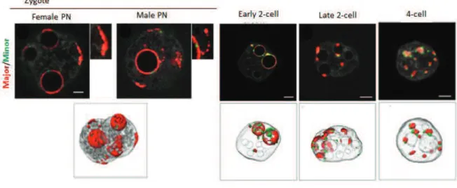
L’hétérochromatine centrique et péricentrique se compose respectivement de répétitions de séquences satellites mineurs et majeurs qui ont une organisation spatio! temporelle spécifique au cours du développement précoce. Chez le zygote, les répétitions péricentriques se concentrent autour des NLBs (Nucleolar!like Bodies) ! les précurseurs des nucléoles ! formant des structures en forme d'anneau dans les noyaux. Après la première division une organisation en "chromocentres" semblable aux cellules somatiques est progressivement initiée de telle sorte qu’au stade 4 cellules les structures en forme d’anneaux ne sont plus présentes. La répression de ces régions pourrait se produire en même temps du fait qu’un pic dans leur transcription est seulement détecté à la fin du stade zygote et dans les embryons au stade 2 cellules. De plus, de précédents travaux dans le laboratoire suggèrent qu’il pourrait y avoir un lien entre la localisation spécifique des répétitions péricentriques dans le noyau et leur répression. En effet, dans les embryons exprimant la mutation H3.3K27R la formation des chromocentres est perturbée, la transcription des satellites majeurs augmente et la protéine d’hétérochromatine HP1" est délocalisée. Ainsi, le premier objectif principal de cette partie de mon travail était de déterminer si dans le noyau la localisation spécifique de la chromatine péricentrique est importante pour sa répression. Pour y répondre, j’ai réalisé des expériences dans lesquelles j’ai artificiellement perturbé la localisation des régions péricentriques au cours du développement précoce pour forcer leur délocalisation des NLBs vers la membrane nucléaire. Ces expériences montrent que l’organisation spatiale spécifique des domaines péricentriques est essentielle pour leur répression ainsi que pour leur organisation correcte. De plus, mes résultats suggèrent que les défauts d’organisation de l’hétérochromatine conduisent à des défauts de division cellulaire et de prolifération.
II. L’hétérochromatine sur les séquences répétées
Environ la moitié du génome des mammifères est composée d’éléments répétés transposables (Transposable Elements : TE) qui sont regroupés en deux classes : les rétrotransposons et les transposons à ADN. Bien que proportionnellement les TEs représentent une grande partie du génome, seulement une faible proportion de ces éléments est capable de « sauter et se coller » ; ce qui est le cas des LINE!1 sans LTR (Long Interspersed Nuclear Elements L1) qui seraient les TEs les plus actifs chez la souris et pour lesquels les insertions semblent être les plus récentes dans le génome murin. Deux familles de L1, les A et F, ont été confirmés comme étant les éléments L1 les plus jeunes et plus abondants du
! 9 !
génome murin ; des essais de rétrotransposition ayant aussi démontré qu’ils seraient toujours actifs8. Cette activité est l’une des raisons principale pour laquelle les TEs, et en particulier les éléments L1, représentent une grande menace pour la stabilité du génome, leur dérégulation étant fréquemment observée dans de nombreux types de cellules cancéreuses. Des études approfondies dans des cellules somatiques et des cellules souches ont révélé certains mécanismes moléculaires les régulant et réprimant leur activité transcriptionnelle, notamment via la méthylation de l’ADN, les modifications d’histone et les voies à ARN. Cependant, la régulation des éléments L1 pendant la période de reprogrammation épigénétique et les divisions cellulaires rapides qui ont lieu au cours du développement reste encore indéterminée. De manière intéressante, des études récentes montrent que les L1 sont réactivés après la fécondation, leur activité transcriptionnelle diminuant ensuite progressivement. Du fait que le niveau de méthylation des L1 reste faible dans les embryons précoces de mammifère11, leur mode d’expression particulier après la fécondation ne peut s’expliquer par un effet secondaire de l’activation générale du génome mais pourrait plutôt avoir un intérêt fonctionnel. Quel est cette fonction si il y a, et comment les L1 sont régulés dans les embryons préimplantatoires, restent indéterminés et constituent ainsi le second objectif principal de ma thèse.
Pour y répondre et étudier l’importance et la possible fonction et régulation des éléments L1, j’ai décidé d’utiliser une approche expérimentale basée sur les TALEs (Transcription Activator–like Effectors) qui sont des protéines liant l’ADN sur des séquences spécifiques12, récemment découvertes chez Xanthomonas sp. Plus précisément, j’ai ciblé les éléments L1 avec des TALEs spécifiques fusionnés à des protéines modifiant l’activité transcriptionnelle. Cette approche m’a permis d’étudier en détail leur processus d’activation et de répression. Ainsi, dans la seconde partie de ma thèse, j’ai conçu et généré les outils adéquats pour répondre à ces questions et vérifier leur expression, localisation, capacité de liaison à l’ADN, et leur habilité à activer les L1 dans les cellules ES de souris. Ces résultats ont ensuite été utilisés pour choisir les meilleurs candidats pour effectuer les expériences dans les embryons de souris. La correcte localisation nucléaire et la capacité à activer les L1 in vivo ont été vérifiés dans les embryons pour les trois TALE!L1 les plus prometteurs. Du fait du mode d’expression connu des L1s dans l’embryon, avec une activation de transcription initiée au stade zygote tardif et une diminution à partir du stade 8 cellules, nous étions particulièrement
! 10 !
intéressés de voir si la répression des L1s est nécessaire pour le développement, et ainsi nous voulions prolonger leur phase d’activation au!delà du stade 2 cellules et observer le phénotype. Ainsi, après micro!injection des activateurs de L1, nous étions capables de détecter un signal plus important des transcrits de L1 dans l’embryon au stade 4 cellules, comparé aux embryons contrôles non!injectés ou injectés avec des TALE!L1 manquant le domaine d’activation. Le groupe expérimental montrait aussi un niveau d’expression plus élevé d’Orf1p codée par les L1, suggérant que nous avions réussi à moduler la traduction des L1 dans l’embryon. De plus, quand les trois groupes ont été cultivés en parallèle, seulement 50% des embryons présentant un niveau plus élevé de L1 ont atteint le stade blastocyste, alors qu’environ 90% des embryons contrôles se sont développés normalement. Ces résultats suggèrent que lors de la perturbation du mode spécifique d’expression des L1, les embryons ne sont plus capables de se développer normalement. Ceci suppose qu’un possible rôle régulateur des éléments L1 pourrait avoir lieu à ce moment, c’est à dire en activant/réprimant la transcription des régions voisines ou de manière plus générale en agissant sur l’organisation du génome, leur surexpression conduisant à une chromatine plus ouverte. En conclusion, ceci représente la première tentative pour élucider la biologie des éléments L1 dans l’embryon précoce de souris par l’utilisation de modificateurs de transcription ciblés spécifiquement, montrant que la sous régulation des éléments L1 est nécessaire pour le développement.
En conclusion, associé à la première partie de mon projet sur l’hétérochromatine péricentrique, j’espère que mon travail contribue à la compréhension des mécanismes responsables du contrôle de l’intégrité du génome.
! 11 !
I. Introduction
1. Characteristics of (hetero)chromatin in mammalian cells
1.1. Brief introduction to chromatin
In eukaryotic cells the genetic material is organized into a complex structure called chromatin which is composed of DNA and proteins and localized in a specialized compartment ! the nucleus (Fig.1). The nucleosome is the fundamental unit of chromatin, and is composed of ~147 base pairs of DNA wrapped around an octamer of four core histones: H3, H4, H2A, H2B. The core histones are predominantly globular with an exception of the N!terminal “tail” that can be chemically modified. Posttranslational histone modifications (PMTs) refer to the chemical changes occurring on the specific amino acid residues of histones and include acetylation, methylation, phosphorylation, ubiquitination, SUMOylation, and probably many others, less studied. The information about the patterns of histone modification across the genome offers insights into the regulatory state of promoters, genes, and other regions, as specific modifications can affect gene expression and chromatin compaction depending on the type of the modification, position of the amino acid and the number of modified residues (Kouzarides 2007). Several types of enzymes are known to catalyze addition or removal of histone modifications i.e. methylation of lysines and arginines is performed by histone methyltransferases (HMTs) or acetylation is catalyzed by histone acetyltransferases (HATs). Histones can be not only modified by PMTs by also replaced by their non!canonical variants which display different properties. Although most of the histones are synthesized during S! phase which allows their deposition behind the replication fork, replication!independent replacement of histones can also occur. Both pathways enable exchange of the canonical histones into non!canonical variants by histone chaperons, which may lead to the change of transcriptional state of chromatin. In addition, ATP!dependent chromatin remodeling enzymes mediate rearrangements of the chromatin as they restructure and slide nucleosomes, or eject histones, thereby regulating the dynamic properties of chromatin (Henikoff and Smith 2015).
! 12 ! Figure 1. Chromatin organization within the nucleus
Scheme depicting different aspects of chromatin regulation. PTM ! post!translational modification. Chromosome territories within the nucleus, shown in different colors, are composed of chromatin fiber, which, in turn, contain packed nucleosomes. (Modified from Rosa and Shaw 2013).
1.2. Euchromatin versus heterochromatin
In mammalian cells, chromatin is organized into two distinct domains known as euchromatin and heterochromatin. Euchromatin is the gene!rich part of the genome which is more accessible to the transcriptional machinery thanks to the ‘‘open state’’ and “flexibility” which leads to more permissive state and higher probability of expression (Fig. 2). Heterochromatin, on the contrary, is gene!poor and assembles into well compacted domains which have more ‘‘repressive’’ chromatin structure, thus, contain mainly transcriptionally silent regions (Fig. 2) (Jost, Bertulat, and Cardoso 2012). Moreover, heterochromatin shows a distinct pattern of replication, being replicated mostly at the late S!phase (Rhind and Gilbert 2013). Hence, as a general rule these diverse parts of the genome have different chromatin configurations which correspond to a functional state, and can be distinguished by distinct factors present on them i.e. histone modifications or DNA methylation.
! 13 !
Figure 2. Euchromatin and heterochromatin organization in mammalian cells
Scheme representing differences in heterochromatin and euchromatin organization depicting nucleosome position (in blue), modifications of histones’ tails (in purple and brown), transcription factors. (Modified from Grewal and Elgin 2007).
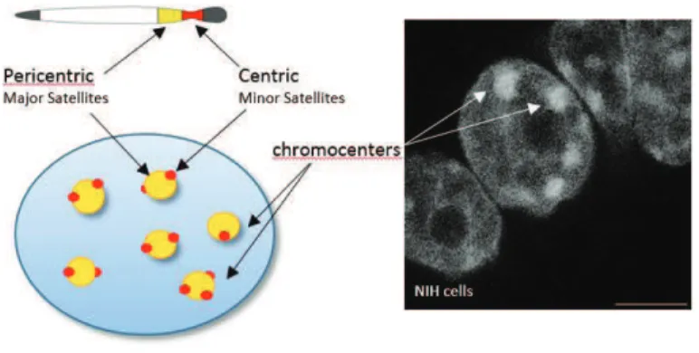
Although the characteristics of euchromatin and heterochromatin seem to be well defined, it is worth mentioning that there are some developmentally regulated loci, where the chromatin state can change in response to cellular signals and gene activity. These regions are referred to as “facultative heterochromatin” and are associated with proteins from Polycomb! group repressive complexes (PRC), distinct histone modifications like H3K27me3, and specific histone variants like macroH2A. “Constitutive heterochromatin” on the other hand, is marked by H3K9me3, H4K20me3, H3K64me3 and HP1", and contains high density of repetitive sequences and transposable elements. It is mostly found at telomeres and pericentromeric regions ! large blocks of chromatin flanking centromeres (Fig. 3). In the nucleus of interphase somatic cells pericentromers from several chromosomes cluster together into the foci called chromocenters that stain intensely by DNA dyes. The centromeres from the same chromosomes can be found around these regions, and are characterized by the presence of the centromere!specific histone H3 variant CENP!A (or Cen!H3) (Guenatri et al. 2004). In mice, centromeric and pericentromeric heterochromatin corresponds to minor and major satellite sequences, respectively (Fig. 3) (Probst and Almouzni 2008). Constitutive heterochromatin from telomeres, centromeres and pericentromeres remains silenced and condensed throughout the cell cycle as it is thought to enable the formation of structures that are essential for chromosomal function. Disruption of the establishment, condensation and/or silencing of pericentromeric chromatin can indeed cause centromere malfunction, incorrect chromosome segregation, and nuclear disassembly (Peters et al. 2001; Bouzinba!Segard, Guais, and Francastel 2006)
An additional feature of heterochromatin is its ability to propagate, and thereby influence gene expression in a region!specific, sequence!independent manner as it happens
! 14 !
during mammalian X!inactivation. This key feature of heterochromatin facilitates the control of the loci that are otherwise incapable of recruiting effectors by themselves. Thus, the heterochromatin can spread in cis and is coordinately regulated in trans and both these features make heterochromatin indispensable not only for genome organization but also because of its regulatory role (Grewal and Jia 2007).
Figure 3. Pericentric and centric repeats in mouse cells
Representation of chromosomal and nuclear location of pericentric (in yellow) and centric (in red) heterochromatin. Scale bar – 10 microns.
1.3. Maintenance and establishment of heterochromatin in somatic and stem cells
Chromatin can be modified and controlled by various epigenetic mechanisms at different levels of its organization, for example at the DNA itself, at the nucleosomes, or even at the higher!order structures which includes nuclear compartmentalization. All these events can lead to more open or closed chromatin configuration and result in on or off state of expression. Thus, while studying mechanisms responsible for chromatin organization, one has always bear in mind a complex picture of interactions whereby active and passive pathways operate in parallel to ensure proper control of chromatin state and gene expression to comply with cellular needs. Importantly, functionally distinct regions have to be regulated in different ways either allowing rapid and dynamic changes like in promoters or enhancers, or enabling complete transcriptional shut downs like in pericentromeric repeats. At the same time, the structure of the chromatin per se should stay flexible enough to enable proper progression of replication, which implies existence of tight regulation at many different levels of chromatin organization. In recent years many studies showed that the regulation of the heterochromatic
! 15 !
state is equally dynamic and complex as for gene!rich regions. Surprisingly, the mechanisms that initiate its formation and preserve its distinction from euchromatin, still remain elusive. Nevertheless, our current knowledge shows that maintenance and establishment of these repressive states are largely determined by a complex network that includes: enzymes able to modify DNA and histones tails; complexes with nucleosome remodeling activities; transcription factors (TFs); non!coding RNA; and nuclear organization (Fig.4). Each of the above mentioned pathways will be briefly discussed in this paragraph.
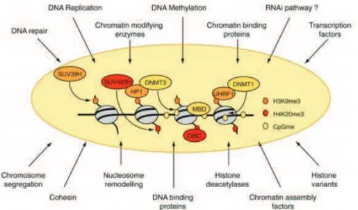
Figure 4. Complex network of factors controlling chromatin state
SUV39H is the responsible HTMase for H3K9me3 on pericentromeres, a histone mark recognized by HP1 proteins. HP1 proteins interact and recruit SUV420H and DNMTs, leading to H4K20me3 and DNAmethylation, respectively. These epigenetic marks function also as docking sites, like H4K20me3 for ORC (origin of replication complex) proteins and CpGme for MBDs (factors with a methyl!binding domain). An alternative for DNMT recruitment might be through UHRF1 that directly interacts with DNMT1 and might read the H3K9me3 mark. (Modified from Nehme Saksouk, Simboeck, and Dejardin 2015).
1.3.1. DNA methylation
DNA methylation refers to the methylation of cytosine residues in CpG dinucleotides, and is catalyzed by DNA methyltransferases (DNMTs): DNMT1 maintains DNA methylation, DNMT3A/3B exhibit both maintenance and de novo methylation activities, and DNMT3L which lacks the characteristic N!terminal catalytic domain but acts as a crucial activating cofactor of DNMT3A/B (Ooi, O’Donnell, and Bestor 2009). In general, DNA methylation is viewed as a final state of silencing and maintenance of a transcriptionally inactive state and is
! 16 !
one of the hallmarks of heterochromatin. Thus, hypermethylation of CpG dinucleotides is commonly present at genes!depleted regions including transposable elements or repeats, whereas hypomethylation is found on exones, intrones and intergenic regions. There are several mechanism how DNA methylation can impact transcription, for example, through interference with binding of transcriptional factors to imprinting loci like (Bell and Felsenfeld 2000). Another way occurs via 5!methyl cytosine binding proteins (MBP) which interpret methylation levels and recruit chromatin regulators that induce changes in chromatin state as in the case of HMTs recruitment by MBP1 (Sarraf and Stancheva 2004) or chromatin remodeler NuRD via MBP2. Interestingly, even though DNA methylation can be seen as a crucial guardian of gene expression in differentiated cells, its function in heterochromatin establishment during development, remains questionable. Upon depletion of all three DNMT enzymes in mESC (TKO), some heterochromatic regions indeed display transcriptional up! regulation, however, global gene expression is not affected, cells proliferate normally and global pattern of histone marks remains unchanged (Tsumura et al. 2006). Moreover, mice deficient for de novo DNMTs display no phenotype prior to implantation (Okano et al. 1999). In fact, alternative factors, described later in this chapter, were shown to compensate lack of DNA methylation implying that it is not a crucial player for heterochromatin establishment in undifferentiated cells.
1.3.2. Histone modifications and related pathways
In fission yeast and higher eukaryotes, histones in heterochromatin are hypoacetylated and selectively methylated at lysine 9 of histone H3. Lysine methylation can exist in three flavors: mono!, di, and tri!methylation, which are catalyzed by different enzymes: Prdm3 and Prdm16 that monomethylate H3K9 in the cytoplasm, which is then converted in the nucleus by the Suv39h1/2 enzymes (also called KMT1A/B) to H3K9me3 (Pinheiro et al. 2012). Other histone methyltransferases are also involved in that conversion i.e. SETDB1 (ESET) that can trimethylate H3K9 (Dodge et al. 2004), and G9a, which acts to mono! and di! methylate H3K9 (Tachibana 2002). However, their activity is mostly restricted to euchromatin, whereas Suv39h1/2 operates on heterochromatin (Martens et al. 2005). Suv39h1/2 role in the maintenance of heterochromatin has been mostly linked to the recruitment of multiple silencing factors i.e. HP1 proteins, Suv4!20h1/h2 enzymes, and DNMT1, which are all lost from pericentromeric chromatin in dn Suv39h1/2 mESC (Schotta et al. 2004). Additionally,
! 17 !
Suv39h1/h2 are required for proper mitosis as AURORA B, a major component of CPC (chromosomal passenger complex) that controls chromosomal segregation, is depleted from pericentromeric regions in the absence of H3K9me3 (Saksouk et al. 2014). Thus, a pathway in which Suv39h enzymes induce H3K9me3 which is then bound by HP1, arises as a hallmark of mammalian heterochromatin (Bannister et al. 2001). Given the ability of HP1 to bind to numerous proteins that are implicated in heterochromatin formation, including HDACs,it has been suggested that HP1, when bound to methylated H3K9, serves as an assembly platform. Indeed, HP1 mediates the recruitment of Suv4!20h HMTs to pericentromers where they catalyze di! and tri!methylation of H4K20 (Schotta et al. 2004). H4K20me3 is highly enriched at pericentric heterochromatin, telomeres, imprinted regions and repetitive elements where it is probably involved in transcriptional silencing. At pericentromeric regions, however, Suv4! 20h1/2 activity seems to be engaged also in chromocenters’ condensation and proper mitosis as cells deficient for these enzymes display chromosome segregation defects that coincide with reduced sister chromatid cohesion (Hahn et al. 2013). Thus, the recruitment of Suv4! 20h1/2 to pericentromers might be more related to heterochromatin organization and global nucleus arrangements than silencing per se.
Interestingly, when H3K9me3 activity is impaired, “rescue mechanisms” take over, mainly the Polycomb group proteins (PcG) which are recruited to compensate and maintain heterochromatic, silencing environment. In mouse ES cells, in which Suv39h1/h2 genes have been knocked out this alternative mechanism is switched on and levels of H3K27me3 become elevated (Martens et al. 2005). Similar situation has been observed in Dnmt TKO mESCs where the loss of constitutive heterochromatin marks on pericentromeric repeats was accompanied by the acquisition of factors belonged to Polycomb group, together with the enrichment of H3K27me3 (Saksouk et al. 2014). In general, PcG are hallmarks of facultative heterochromatin and are known to maintain cell fate by repressing hundreds of genes through the activity of two main polycomb repressive complexes (PRC): PRC1 and PRC2. PRC2 contains the H3K27 methyltransferase EZH2 (also called KMT6A), as well as EED, SUZ12, RbAp46/48 (RBBP4/7), and JARID2 (Jumonji/ARID domain!containing protein). The latter, together with Polycomb! like family (Pcl) proteins, have been suggested to be responsible for the recruitment of PRC2 to target genes in mammalian cells. PRC1 contains the E3 ubiquitin ligases RING1A and B (RNF1 and 2) that mediate H2AK119Ub. Canonical PRC1 complexes also contain Cbx ! proteins that
! 18 !
recognize and bind H3K27me3 through their chromodomain, leading to coexistence of both PRC2 and PRC1 on the same loci. Non!canonical PRC1 complexes contain Rybp (together with additional proteins, such as L3mbtl2 or Kdm2b) rather than the Cbx proteins, thus, their recruitment to target genes is mostly independent of H3K27me3 (Aloia, Di Stefano, and Di Croce 2013). Lysine demethylase Kdm2b has been proposed as one of the factors that enable binding of the non!canonical PRC1 to unmethylated DNA (Wu, Johansen, and Helin 2013). Surprisingly, this PRC1 complex can also recruit PRC2 in the H2AK119Ub!dependent manner which leads to the deposition of H3K27me3 mark on unmethylated promoters (Blackledge et al. 2014). In addition, PRC1 and PRC2 complexes have been shown to associate and form nuclear foci on their targets which stabilizes gene silencing and suggests another role for Polycomb ! in 3D chromatin organization (Cavalli 2015).
1.3.3. Histone variants
Specific histone variants determine the structure of distinct regions of heterochromatin and play a role in their maintenance. For example, when centromere!specific histone H3 variant CENP!A is not present, cells show severe defects in chromosome segregation suggesting its relevance for the integrity and function of kinetochores. In addition to CENP!A, in higher eukaryotes there are three other H3 variants: H3.1 and H3.2 which are mainly expressed in S!phase and deposited by the CAF1 complex, and H3.3 expressed throughout the cell cycle with replication!independent deposition by the HIRA and (Tagami et al. 2004) DAXX/ATRX complexes (Lewis et al. 2010; Drané et al. 2010). HIRA is required for localization of H3.3 to actively transcribed regions, while ATRX is essential for H3.3 incorporation at silent regions such as telomeres, and has been shown to play a role in heterochromatin formation and chromosome segregation during mitosis and meiosis (Rabindranath De La Fuente et al. 2004). DAXX is also present on repetitive regions and its deletion leads to disruption of chromocenters, thus, it is thought to be involved in the structural organization of the nucleus (Rapkin et al. 2015). The specific role of DAXX and ATRX chaperons at pericentromeric regions, however, is unclear but it might be linked to transcription of the locus as in Daxx !/! MEFs and in ATRX and H3.3 siRNA depleted cells levels of major satellite transcripts are lower (Drané et al. 2010). ATRX/DAXX complex can also replace the canonical H3.1/H3.2 with the H3.3 variant at specific classes of transposable elements ! class I and class II ERVs. It has been previously shown that these TEs are silenced
! 19 !
through H3K9me3 deposited by SETDB1 and its co!repressor complex containing KRAB! associated protein 1 (KAP1) (Rowe et al. 2010; Karimi et al. 2011). Elsässer and collegues suggest a role for H3.3 in this process as well because H3K9me3 and KAP1 occupancy is reduced at some class I and II ERVs upon H3.3 deletion. Thus, the authors draw a link between ERV!associated H3K9me3 and H3.3 deposition, and suggest that the recruitment of KAP1 and H3.3 by DAXX is co!dependent and occurs upstream of the recruitment of SETDB1 (Elsässer et al. 2015).
Exchange of another core histone ! H2A ! also takes part in the formation of heterochromatin as H2A.Z has been demonstrated to be involved in the recruitment of HP1 at pericentromeric loci, and direct binding to the pericentric heterochromatin!binding protein INCENP (Fan et al. 2004). These interactions facilitate folding of chromatin into high order structures but also play a role in the chromosome segregation (Rangasamy, Greaves, and Tremethick 2004). Although H2A.Z is present on constitutive heterochromatin in some developmental stages, in general it is found on facultative heterochromatin i.e. the inactive X chromosome, where it becomes monoubiquitylated by PRC1 (Sarcinella et al. 2007). Interestingly, its occupancy has been also correlated with the lack of DNA methylation and with H3K4me3/H3K27me3 loci which suggest that H2A.Z brings a dynamic instability to chromatin structure increasing access to chromatin!modifiers. Another variant of H2A ! macroH2A is also implicated in heterochromatin regulation as it is mainly present at discrete regions of facultative heterochromatin within the inactive X chromosome that alternate with regions of constitutive heterochromatin (Henikoff and Smith 2015).
1.3.4. DNA binding factors
Heterochromatin function can be also affected by various DNA binding factors which interact with chromatin remodelers and trigger changes in chromatin organization. One of the examples comes from the experiments in mESC where in the absence of H3K9me3 or DNA methylation, (dnSuv30h1/h2 or TKO) the levels of methylation!sensitive DNA binding protein BEND3 arise. It leads to the recruitment of PRC2 via MBD3/NurD chromatin remodeler complex and, only in the case of TKO, PRC1 components, presenting a mechanism which facilitates silencing and operates on unmethylated loci (Saksouk et al. 2014). NoRC is another chromatin remodeler which is known to establish repressive chromatin structure via interaction with DNA binding proteins. The biggest subunit of NoRC ! TIP5 ! is recruited to
! 20 !
distinct genome loci by specific chromatin!associated proteins, for example, to rDNA by TTF! I, to centromeres by CENP!A and to telomeres by the shelterin complex, and then interacts with HP1# and with histone!modifying enzymes to mediate higher!order chromatin structure. Knockdown of NoRC leads to de!condensation of heterochromatin, abnormalities in mitotic spindle assembly and impaired chromosome segregation (Postepska!Igielska et al. 2013). LSH (lymphoid specific helicase) is another factor involved in the formation of normal heterochromatin structure as it has been shown to maintain nucleosome density leading to more compacted state of the chromatin. Moreover, it plays a specific role in acquisition of de novo DNA methylation at transposable elements and pericentromeric repeats (Huang et al. 2004). Its ATP!binding site seems to be crucial for the stable association of Dnmt3 to DNA (Ren et al. 2015) and upon its depletion cells display lower levels of DNA methylation and upregulation of major satellites (Huang et al. 2004).
Recent discoveries in MEF cells have implicated a role for sequence!specific transcription factors in heterochromatin formation/maintenance. These TFs have been shown to use distinct binding sites as targets and their recruitment lead to heterochromatin silencing most likely by RNA dependent pathways (Bulut!Karslioglu et al. 2012). For example Pax3 and Pax9 transcription factors are components of mouse heterochromatin as they localize to DAPI dense regions and can bind to HP1 and KAP1. Upon their depletion accumulation of major satellite transcripts from both strands occurs, and reduction in H3K9me3 and H4K20me3 takes place which, in consequence, leads to genome instability. The putative role of these transcription factor binding sites might be to serve as a base for bidirectional transcription, which is then silenced by heterochromatin formation (Bulut!Karslioglu et al. 2012). This is not such a new concept as the establishment of higher!order chromatin structure at repetitive elements has been already shown to depend on specific noncoding RNAs, including pRNA, TERRA (Postepska!Igielska et al. 2013) and major satellite RNA (Almouzni and Probst). Moreover, the intersection of the RNA!interference (RNAi) pathway and heterochromatin formation has been well documented in Schizosaccharomyces pombe where small interfering RNAs generated from flanking outer repeat of a centromere direct histone H3 lysine 9 methyltransferase Clr4 to homologous loci which then recruit Swi6 (homologous of HP1). H3K9me and Swi6 are required to establish CENP!A which further leads to spreading of heterochromatin (Folco et al. 2008). Interestingly, siRNA seem to be important only for the
! 21 !
recruitment of Clr4 as heterochromatin can be still formed without operating iRNA pathway but with artificial tethering of Clr4 to the loci (Kagansky et al. 2009).
1.3.5. Small RNAs and RNAi pathway
Although RNA silencing pathways in mammals work mainly as a post!transcriptional gene silencing (PTGS) mechanism, they can also alter chromatin structure and silence genes at the transcriptional level (Morris et al. 2004). The core machinery includes small RNAs (sRNA) that are complementary to target genomic sequences, which form complexes with the proteins from the Argonaute family (Ago). Depending on the biogenesis of sRNA and the mechanism of silencing, 3 main pathways can be distinguished: RNA interference (siRNA), microRNA (miRNA) and piRNA pathways. siRNA and miRNA require Dicer activity whereas piRNA rely on Piwi proteins: Mili, Miwi1, and Miwi2 (in mouse). Whereas piRNAs are known to function in the male germline to maintain genomic integrity by epigenetic mechanisms including de novo DNA methylation of transposable elements (Aravin et al. 2007), siRNAs and miRNAs mainly mediate PTGS through degradation of mRNA and/or inhibition of mRNA translation in the cytoplasm. Nevertheless, they have been also reported to silence heterochromatin and enable acquisition of heterochromatin!specific chromatin modifications such as H3K9me2/3, H3K27me3 or DNA methylation (Li 2014). Moreover, in mouse cells RNA might have a structural role in heterochromatin, as cells treated with RNaseA lose heterochromatic marks (Maison et al. 2002) and RNase treatment of chicken liver cells revealed a role for RNA in the high!order chromatin structure and compaction (Rodríguez! Campos and Azorín 2007). Another evidence coming from human cell lines supports the hypothesis that RNA may have a general structural role in chromatin organization as RNase treatment results in both loss of CENP!C and chromosomal passenger complex (CPC) components from centromeres. This phenotype can be rescued by reintroduction of CENP!C, INCEP and centromeric RNA (Wong et al. 2007). Moreover, loss of Dicer, the main player in the production of siRNA and miRNA, leads to up!regulation of transcription from pericentric heterochromatin and loss of HP1" and H3K9me2/3 from these regions (Kanellopoulou 2005). All of these provide strong argument that RNA is required for silencing and structural integrity of the pericentromeres.
! 22 !
1.3.6. Nuclear organization
The three!dimensional organization of the genome and its importance for the normal functioning of the cell and, in particular, for (hetero)chromatin regulation, attained more attention in recent years. According to the definition by Nature nuclear organization refers to “the spatial distribution of nuclear contents and components in a way that it reflects or facilitates their activities”. Hence, specific regions of the genome tend to localize in the particular nuclear compartments in order to be properly regulated because distinct regions of the nucleus can regulate chromatin in a different way. One of the most striking examples come from the nuclear periphery and nucleolus that, already in the 1950s, have been associated with more dense chromatic regions, most likely heterochromatin.
The nuclear periphery can be separated into two main sub!compartments: nuclear pores and nuclear lamina. Whilst localization to the nuclear pores has been suggested to be correlated with transcriptional activation of genes, the nuclear lamina is most likely associated with heterochromatin and linked to the repression of genes and repetitive sequences (Akhtar and Gasser 2007). Recent advances in genome!wide high!throughput sequencing confirmed these observation and led to the discovery of lamin!associated domains (LADs) which in general are gene!poor and enriched for heterochromatic silencing marks such as H3K9me2 (Kind et al. 2013). Therefore, perinuclear localization appears to regulate chromatin function. Nevertheless, one has to keep in mind that not all genes have the same ability to be modulated by proximity to the nuclear envelope (Finlan et al. 2008) and detachment from the nuclear lamina does not always lead to transcriptional activation as it happens at some regions during ES cells differentiation (Kind et al. 2013). Moreover, even changes in chromatin condensation, without entailing transcriptional activation, are enough to re!localize some genes from the periphery towards the nuclear interior (Therizols et al. 2014). All of the above suggests that in somatic cells, the nuclear periphery functions more as a non!permissive compartment where unused regions are stored, rather than the actual main inhibitor of transcription.
LADs are not the only domains found to be associated with distinct nuclear compartments as the existence of nucleolus!associated domains (NADs) containing repetitive sequences i.e. centromeres, has been also documented (Németh et al. 2010). In general, the nucleolus is an organelle present in most mammalian cells with the major function related to the synthesis of ribosomal RNA and biogenesis of many ribonucleoprotein particles (RNPs).
! 23 !
The nucleolus most likely plays additional role in the establishment of kinetochores and heterochromatin structure as numerous studies have shown accumulation of many centromere!related proteins within its structure i.e. CENP!C or INCENP (Gent and Dawe 2012). Moreover, the alpha!satellite RNA seems to be required for the assembly of centromere! associated nucleoprotein components at the nucleoli and kinetochores in human cells (Wong et al. 2007). The presence of repressive chromatin marks on the NADs together with tethering experiments which showed that proximity to nucleolus reduces gene expression, support the notion that the nucleolus is an important nucleolar compartment involved in silencing and heterochromatin maintenance (Matheson and Kaufman 2015).
Importantly, nuclear architecture is orchestrated by chromatin modifiers similarly to other epigenetic mechanisms as they are all functionally related. Because regions that are in close proximity to the nuclear lamina are irreversibly bound to its components, change in chromatin state or lamina composition often leads to nuclear re!organization. Lamin proteins, but also lamin!associated transmembrane proteins like Emerin or LBR, are the main structural components of lamina that play a role in anchoring of the chromatin. LBR, for example, has been shown to bind HP1 through H3K9me2/3 (Ye and Worman 1996) in HeLa cells. Experiments performed in C.elegans where the localization of repetitive gene arrays which recapitulate endogenous heterochromatin behavior were monitored under genome!wide siRNA screen, demonstrate that the peripheral localization of arrays depends also on H3K9 methylation (Towbin et al. 2012). Moreover, the depletion of H3K9 methylation in Prdm3 and Prdm16 deficient mouse embryonic fibroblasts results in disintegration of pericentric heterochromatin and the destabilization of the nuclear lamina (Pinheiro et al. 2012). Another example comes from TKO cells where in the absence of DNA methylation the recruitment of pericentromeric heterochromatin to the nuclear lamina is impaired, as shown by the loss of binding of LBR (Lamin B Receptor), Lamin B1 and B2 to major satellites (Saksouk et al. 2014). The loss of interaction with lamina proteins is sufficient to reorganize the position of major satellites into large aggregates surrounding the nucleoli, which is strikingly similar to the phenotype observed in the LBR/Lamin A/C double!deficient cells (Solovei et al. 2013). Interestingly, the latter work shows that the LBR is the crucial tethering site in early stages of mouse cell differentiation, whereas lamin A/C replaces or supplements its role in terminally
! 24 !
differentiated cells. Mechanisms anchoring repetitive gene arrays to the lamina in C.elegans embryos also display a switch in later developmental stages (Towbin et al. 2012).
1.4. Concluding remarks
The above!mentioned studies imply the multilayered regulation of heterochromatin, with various epigenetic pathways shown to play a crucial role. Although heterochromatin formation and maintenance in different organisms is a common theme, the molecular details are difficult to pinpoint because of the complexity and redundancy of the pathways ensuring heterochromatin robustness. Additionally, there is a difficulty in unravelling the biochemical characteristics in vivo, and difficulty in combining in vitro results with in vivo events. Moreover, most of the data derives from models in which heterochromatin is already established, like in somatic cells which already reached their final epigenetic status, or ES cells that, although undifferentiated, have already acquire many of the hallmarks of heterochromatin. As a consequence, it is difficult to elucidate what is the order of events, and which factors are necessary for the formation versus the maintenance of heterochromatin. However, during early development, the epigenetic program has to be erased and reacquired in order to form a new organism. This makes it an ideal time!frame to look into the mechanisms responsible for de novo formation of heterochromatin and molecular pathways underlying its establishment.
2. Characteristics of heterochromatin during mouse development
2.1.
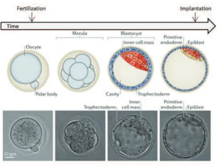
Mouse development at a glanceThe life cycle of an organism is an extremely complex process, which starts with two distinct identities – the oocyte and the sperm ! that come together to form a zygote. Within the subsequent 2 days, the zygote divides twice without changing its size and reaches the 8! cell stage with 8 blastomeres of equal volume. Then, individual cells lose their distinctive outlines and maximize intercellular contacts in a process called compaction. After one more day, at the early 32!cell stage, fluid begins to accumulate between cells and creates a cavity – the embryo reaches the blastocyst stage. At one end of the cavity lies a cluster of pluripotent cells known as the inner cell mass (ICM) surrounded by a thin layer of cells that form the polar
! 25 !
trophectoderm. On the opposite side, mural trophoblast cells close the blastocyst cavity (Fig. 5). The trophoblast cells or trophoectoderm (TE) will form the chorion, which is an embryonic part of the placenta, whereas the ICM will give rise to an embryo and its associated yolk sac, allantois, and amnion. These distinct cell types of blastocyst are demarcated by the expression of key tissue!distinctive transcription factors i.e. Cdx2 limited to TE or Oct4 and Nanog associated to the pluripotent ICM. By the 64!cell stage the TE and ICM are completely separate layers becoming the first cell fate specification event in development. The first segregation of ICM cells that happens afterwards gives rise to hypoblast (or primitive endoderm) and the epiblast, thought to contain all the cells that will generate the actual embryo.
Figure 5. Preimplantation development in mouse: from fertilization to implantation
Scheme depicting different stages of mouse preimplantation development with corresponding bright!field images.(Modified from Wennekamp et al. 2013).
2.2. Formation of gametes
2.2.1. Primordial germ cells specification
Germ cells are highly specialized cells that reached the final step of differentiation after many cell divisions and transcriptional, epigenetic and morphological changes. In the same time, they are the only cell types that, in vivo, have the capacity to reset their identity through reprogramming. The long process during which they are formed starts already at the early
! 26 !
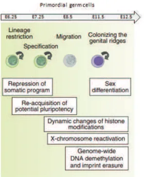
gastrulation stage (Fig. 6). Primordial germ cells (PGCs) are first specified in a response to bone morphogenetic protein 4 (BMP4) from the extra!embryonic ectoderm, which induces signaling pathway leading to expression of BLIMP1 (also known as PRDM1)(Vincent et al. 2005) and PRDM14 (Yamaji et al. 2008). As a consequence, a group of ~40 cells with high levels of alkaline phosphatase activity cluster and begin to shut down their somatic transcriptional program. At ~E7.5 PGCs start their migration from the posterior end of the embryo through the hindgut to the developing gonads. Once in gonads, PGCs continue dividing mitotically to accumulate their number before facing the last division – meiosis – in which they reduce their DNA content to 1n.
During migration and mitotic divisions PGCs undergo many changes in order to silence their somatic program (Fig. 6). In early PGCs, for example, levels of DNA methylation (Popp et al. 2010) and H3K9me2 (Seki et al. 2007) decrease, whereas H3K4me2, H3K4me3 and H3K9 acetylation (H3K9ac) together with H3K27me3 accumulate (Seki et al. 2007). Also transcription factors that are characteristics of pluripotent state, such as Sox2, OCT4, Nanog, Stella, start to be expressed. In later PGCs ~11 days after fertilization, the second wave of DNA methylation starts in which erasure of imprints begins (Popp et al. 2010)and further reprogramming takes place: linker histone H1 is removed leading to more loose chromatin configuration and enlargement of nuclei; chromocenters are lost; many repressive histone modifications are removed i.e. H2A/H4 R3 methylation (Ancelin et al. 2006), or heterochromatin specific H3K9me3 and H3K27me3; proteins associated with facultative heterochromatin or constitutive heterochromatin like heterochromatin protein 1# (HP1#), HP1" and HP1$, ATRX and CBX2 are redistributed and/or disappear (Hemberger, Dean, and Reik 2009). In short words, the epigenome undergoes profound changes. Worth mentioning is the fact that some of these changes are transient, and thought to serve a purpose on facilitating global DNA demethylation. Indeed, soon after reprogramming happens at E12.5, many chromatin marks are reversed i.e. heterochromatic H3K9me3 and H3K27me3 levels go up together with re!clustering of chromocenters (Hajkova et al. 2008).
! 27 !
Figure 6. A schematic representation of primordial germ cell formation in mice
A brief outline of germ cell development in mice is shown schematically. Key events associated with each stage of germ cell development are also shown. (Modified from Saitou, Kagiwada & Kurimotu, 2012).
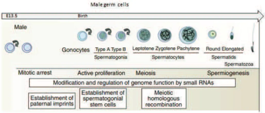
2.2.2. Formation of sex specific gametes
Male gametes
Following epigenetic reprogramming, remarkable differences are observed in the way that male and female gametes are formed after the sex determination has started at ~E12.5 (Fig. 7 & Fig. 8). In males, PGCs enter into mitotic arrest upon entry into the genital ridges, and stay in the G0/G1 phase of the cell cycle for the remaining embryonic period (Western et al. 2008). Only around day 5 postpartum (P5), spermatogenesis begins and many of PGCs resume active proliferation while others are recruited as spermatogonial stem cells (SSCs). Cells that resume mitotic divisions proliferate rapidly until they become primary spermatocyte – product of last mitotic division. They subsequently undergo meiosis I to produce two haploid secondary spermatocytes, which will later divide once more into haploid spermatids and start spermiogenesis in order to become fully mature spermatozoa (Rathke et al. 2014) (Fig. 7).
! 28 !
One of the most prominent changes during this time can be observed on the level of de novo DNA methylation. Re!methylation initiates at E14.5 in prospermatogonia and, during G0/G1 quiescence, male PGCs attain global DNA methylation. As a consequence, at birth, male specific methylation pattern is fully established and does not change throughout concomitant mitotic divisions but only when cells enter meiosis (Henckel et al. 2009). The importance of this de novo methylation pattern in male gametes has been shown by many studies, i.e. knock! outs of DNA methyltransferases (Bourc’his and Bestor 2004) or disruption of piRNA pathway by knock!outs of PIWI proteins (Aravin et al. 2007), with both suggesting its role in imprinting and meiosis (Sasaki and Matsui 2008). Other factors that seem to be crucial for proper meiosis and sperm formation include the acquisition of some histone modifications (e.g. H3K9me2/3) and an unusually high number of histone variants (e.g. TH2A, TH2B, TH3, H3.3A, H3.3B and HT1) which are incorporated in the nucleosomes of spermatogonia and/or spermatocytes (Boškovi% and Torres!Padilla 2013). Interestingly, the most dynamic process during specification of male gametes, is the global change in the chromatin structure through histone exchange. This transition starts in post!meiotic spermiogenesis and takes place during elongation and condensation of spermatids. At that phase of maturation, DNA is stripped of most of its nucleosomal packaging and becomes wrapped around so!called transition proteins (TPs) which are thought to facilitate chromatin remodeling (Boškovi% and Torres!Padilla 2013; Rathke et al. 2014). Moreover, the hyperacetylation of histone H4 takes place in elongating spermatids, just prior to histone removal (Grimes and Henderson 1984; G W van der Heijden et al. 2006), which, most likely, also induce histone displacement by creating more open chromatin structure. Interestingly, transcription does not occur during the elongation process of spermatid maturation, suggesting that core histone acetylation in these cells is not related to gene expression but indeed functions as a signal for the eviction of histones (Boškovi% and Torres!Padilla 2013). In late spermiogenesis TPs are exchanged for protamines ! small, highly basic proteins that bind DNA with high affinity and wrap it in a toroidal structure. This transition is essential for the formation of proper spermatozoa as defects in the process can lead to infertility. The incorporation of protamines into sperm chromatin induces global DNA compaction which is believed to provide safe environment for the genome. Moreover, as a consequence of histones!to!protamines exchange, most of the heterochromatin associated histone modifications (i.e. H3K9 or H4K20) together with HP1, are not present in the fully grown spermatozoa (G W van der Heijden et al. 2006). This leads to extremely rare
! 29 !
phenomena where in in vivo system heterochromatin associated marks are not present, thus have to be acquired de novo. However, an interesting twist is that mammalian sperm chromatin retains some of the spermatid histones, i.e. H4K8ac or H4K12ac, and some of the nucleosomal proteins, most likely at the constitutive heterochromatin (G W van der Heijden et al. 2006). Because nuclear organization of the sperm genome is well defined, with the telomeres positioned at the outer membrane and centromeric heterochromatin in the center of the nucleus forming the chromocenter (Haaf and Ward 1995), the colocalization of the remaining modified histones with chromocenter suggests that they may play a role in a subsequent reorganization of the male genome.
Figure 7. A schematic representation of male germ cell formation in mice
A brief outline of germ cell development in mice is shown schematically. Key events associated with each stage of germ cell development are also shown. (Modified from Saitou, Kagiwada & Kurimotu, 2012).
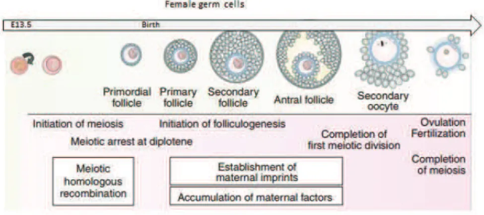
Female gametes
In females, contrary to males, PGCs continue to proliferate until E13.5. Then, they enter into the prophase I of meiotic division which arrests at the diplotene stage, until female reaches puberty (Speed 1982). This prolonged arrest is maintained by the signaling from somatic follicular cells which surround oocytes creating primordial follicle (Fig. 8). During the storage, oocytes grow and by selectively regulating gene expression, accumulate organelles and macromolecules indispensable for their further fate. In adult females, signal from follicle stimulating hormone (FSH) recruits groups of primary follicles that start to grow and, in consequence, become antral follicles containing fully grown oocytes. Then, upon hormonal
! 30 !
stimulation from luteinizing hormone (LH), groups of fully grown oocytes called germinal vesicles (GV) undergo germinal vesicle break down (GVBD) and complete first meiotic division with concomitant extrusion of the first polar body. The GV oocytes undergo an additional transition in their chromatin organization. Initially, fully grown oocytes contain a ‘Non Surrounded Nucleolus’ (NSN) which is transcriptionally active and contains less condensed chromatin displaying heterochromatic DAPI!rich regions in several clusters in the nucleoplasm, similarly to somatic cells. At later stages of oogenesis, the so called Surrounded Nucleolus (SN) oocytes form, which are transcriptionally inactive and contain more condensed chromatin of which the majority is wrapped around the nucleolus forming a characteristic ‘‘rim’’. Interestingly, only SN oocytes are able to resume maturation whereas others remain unresponsive to LH stimulation (Debey et al. 1993). As a consequence, only SN oocytes undergo the maturation process involving haploidization which is completed upon arrest at metaphase II of meiosis. Termination of meiosis, however, awaits fertilization. Only after fertilization with a haploid spermatozoon, oocytes can complete the second meiotic division, extrudes the second polar body and give rise to a totipotent zygote.
As mentioned before, acquisition of such a potency is possible due to dramatic changes and full reset of somatic program while PGCs are formed, but also afterwards. As a continuation of the reprograming, in female germ cells, DNA methylation levels remain low (at E16.5) which leads to i.e. further erasure of genomic imprinting and reactivation of the inactive X chromosome. The initiation of DNA methylation, leading to female specific imprints, begins in the growing oocytes after birth, and the de novo methylation process is complete by the time oocytes are supposed to resume meiosis. Hence, in the fully grown oocyte heterochromatic DNA is hypomethylated, however, some of the specific regions, like major satellites or LINE!1 transposones, remain hypomethylated (Arand et al. 2015). Changes in other epigenetic marks and associated proteins also take place but are not as profound as those observed in the male gametes. For example, some histone modifications change rapidly ! histones H3 and H4 are generally acetylated at prophase I of meiosis, but they rapidly become deacetylated at metaphase I by HDACs. On the other hand, levels of other histone modifications stay stable like in case of H3K9me2 which remains dispersed throughout the nucleus, or H3K9me3 that appears concentrated at condensed heterochromatic regions (Meglicki, Zientarski, and Borsuk 2008). Also some heterochromatin associated proteins
! 31 !
display diverge patterns of expression i.e. HP1" protein is present in the primordial oocytes and remains bound to heterochromatin regions of fully!grown oocytes disassociating soon after GVBD. In contrast, HP1# can be detected for the first time in the oocytes at the beginning of their growth phase and dissociates from the chromatin of fully grown oocytes during their transition from NSN to SN type (Meglicki, Zientarski, and Borsuk 2008). Moreover, during meiotic prophase I somatic histone H1 is replaced by an oocyte!specific variant H1FOO, which in consequence leads to a less condensed state of chromatin (Hayakawa et al. 2014). Although H1FOO is indispensable for GV oocyte and early embryogenesis (Furuya et al. 2007), soon after the second cleavage canonical H1 is reacquired.
Figure 8. A schematic representation of female germ cell formation in mice
A brief outline of germ cell development in mice is shown schematically. Key events associated with each stage of germ cell development are also shown. (Modified from Saitou, Kagiwada & Kurimotu, 2012).
In conclusion, an oocyte arrested at metaphase II and awaiting fertilization has very distinct epigenetic landscape from a somatic cell with some of the heterochromatic features being already erased (e.g. HP1#), and others still present on the chromatin (such as H4K20me3 and H3K9me3). Also gene expression pattern differs drastically because oocyte is not transcriptionally active during the last phase of maturation, and it relies only on the factors accumulated during its growth and maturation. As mentioned earlier, the sperm undergoes even more drastic changes with its histones!to!protamines transition leading to extreme compaction of DNA. Why all these changes are important, and how exactly they are regulated has been under deep investigation for many years and still leaves many unanswered
! 32 !
questions. What can be concluded, however, is the crucial role of epigenetic reprogramming and global chromatin reorganization in both gametes for the formation of the “first cell of an organism” – the zygote. This cell has to establish de novo its entire developmental program, which includes heterochromatinization of some regions and activation of others. How does the newly formed embryo distinguish these specific parts of a genome, acquires the correct histone modifications and reorganizes nuclear architecture, has become one of the main questions in cell biology. In the next chapter, I will provide a more detailed description of our current knowledge of these processes.
2.3. Preimplantation development
2.3.1. Most important events in preimplantation embryos
Development in mammals commences with the fertilization of an oocyte by a sperm, which results in the formation of a zygote that after many divisions give rise to embryonic and extra!embronic tissues (Fig. 5). This capacity is defined as a totipotency, in opposition to pluripotency that leads to the formation of embryonic tissues only i.e. stem cells, or multipotency which enables some differentiation but only according to already taken pathway, for example in neurons. Until now, totipotency has been observed only in 1!cell stage embryos and in each blastomere of 2!cell stage embryos, which after separation can also form a mouse. How such a potency is retained in a zygote and what are the mechanisms of its regulation, remain unknown, however, some of the molecular pathways and events/chromatin changes happening during preimplantation development seem to be indispensable for this process.
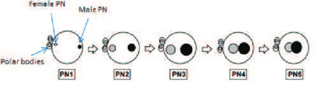
Epigenetic reprogramming of parental genomes is a crucial event which results in the creation of a totipotent cell from two differentiated ones. Not surprisingly, if one keeps in mind their morphology at the fertilization time point, maternal and paternal sets of chromatin behave in a distinct way during reprogramming process, showing different chromatin signatures, histone marks, and replication and transcription timings. One of the main reasons lays in the fact that in sperm protamines have to be exchanged for histones which results in a rapid decondensation of the paternal chromatin, whereas female chromatin does not have to undergo such an extensive remodeling. Moreover, the maternal and paternal pronuclei (PN) remain separate nuclear entities throughout the first cell cycle. Based on the