HAL Id: tel-02339364
https://tel.archives-ouvertes.fr/tel-02339364
Submitted on 30 Oct 2019HAL is a multi-disciplinary open access archive for the deposit and dissemination of sci-entific research documents, whether they are pub-lished or not. The documents may come from teaching and research institutions in France or abroad, or from public or private research centers.
L’archive ouverte pluridisciplinaire HAL, est destinée au dépôt et à la diffusion de documents scientifiques de niveau recherche, publiés ou non, émanant des établissements d’enseignement et de recherche français ou étrangers, des laboratoires publics ou privés.
Genetic Basis of Extra-Ocular Pigmentation in
Semi-Aquatic Embryos
Aidamalia Vargas Lowman
To cite this version:
Aidamalia Vargas Lowman. Genetic Basis of Extra-Ocular Pigmentation in Semi-Aquatic Embryos. Invertebrate Zoology. Université de Lyon, 2019. English. �NNT : 2019LYSEN042�. �tel-02339364�
Numéro National de Thèse : 2019LYSEN042
THESE de DOCTORAT DE L’UNIVERSITE DE LYON
opérée au sein de
l’Ecole Normale Supérieure de Lyon
Ecole Doctorale N° 340
Biologie Moléculaire, Intégrative et Cellulaire (BMIC)
Discipline : Sciences de la Vie
Soutenue publiquement le 24/09/2019, par :
Aidamalia VARGAS – LOWMAN
Les bases génétiques de la pigmentation dans les embryons de punaise
d’eau
Genetic Basis of Extra-Ocular Pigmentation in Semi-Aquatic Embryos
Devant le jury composé de :
Dr. NADEAU, Nicola DR University of Sheffield Rapporteure Dr. BELDADE, Patricia DR Instituto Gulbenkian de Ciência Rapporteure Dr. GIBERT, Patricia DR Université Claude Bernard Lyon 1 Examinatrice Dr. JORON, Mathieu DR Centre d’Ecologie Fonctionnelle & Evolutive
Examinateur
MERABET, Samir DR ENS de Lyon Examinateur
Acknowledgments
I can honestly say that no single step of this journey was taken alone and there are many people that I would like to acknowledge and give my sincerest gratitude to. I would like to thank Dr. Abderrahman Khila, my thesis advisor to give me the opportunity to work alongside him and shown me the amazing potential that Gerromorpha have as a model. I would like to extend my gratitude to Dr. François Bonneton, his guidance and feedback lit my path, thanks for the discussion and encouragement.
I must express my affection to my husband and kids for being my support and inspiration, special gratitude to my family and friends in Panama.
Thanks to my committee members, Dr. Michalis Averof and Dr. Alistair McGregor for your insightful comments and suggestions that gave another positive view to the project. I also thank the member of my jury, Dr. Nicola Nadeau, Dr. Patricia Beldade, Patricia Gilbert, Dr. Nicola Nadeau, Dr. Patricia Beldade, Dr. Patricia Gilbert, Dr. Mathieu Joron and Dr. Samir Merabet who agreed to evaluate this work.
Additionally, I would like to thank each and every present and past member of the Khila Lab for his or her feedback and insights into my project: Antonin, Séverine, William, Augustin, Vitoria, Emilia, Amélie, Cédric, Mohamed and Antoin. In particular, I would like to thank “Señor” Armisen, not only for being patient with me during my process to learn bioinformatics, but also for all the help that you and Ana offered me since the first step that I putted in Lyon, my sincere friendship.
I would like to acknowledge to the people that were working directly with me in this project during these years: Marie, Mathilde and Kahina. Thank you for your professionalism and commitment.
I would like to thank the administrative department of IGFL, in particular Fabienne and Martine, I know that my “dossier” was a little difficult to understand at the beginning. Also, I extend my gratitude to Christian, Laurent and Michel for your help.
4 I am immensely grateful to my colleagues in Panama for their support from the distance, in special to Professor Ovidio Madrigal for his unconditional friendship.
Thanks to all the amazing people that I had the fortune to meet during this adventure, “mis sobrinos de SFERE 2015”, Jazmín Arguello, Jenny Quintero and my beautiful people of FA. Thanks for the unforgettable moments.
I acknowledge La Secretaria Nacional de Ciencia y Tecnología (SENACYT) Panamá for providing support and my fellowship. I would like to thank to Université de Lyon, Lyon 1, Ecole Normale Supérieure de Lyon and the Institut de Génomique Fonctionnelle de Lyon (IGFL) for the reception during this four years.
6
List of figures
Figure 1. Endogenous pigments in insects. ... 18
Figure 2. Molecular phylogeny of Hemiptera based in mitochondrial genome sequences. ... 24
Figure 3. Correlation of the modes of oviposition in semiaquatic bugs (Gerromorpha) with the colour of the chorion and embryo. ... 33
Figure 4. Evolution of extra-ocular colouration in embryos of Hemiptera. ... 35
Figure 5. Evolutionary origin of BvMYB1 and betalain pathway. ... 37
Figure 6. Expression of the gene CYP2J19 in red feathers and red beaks of birds. ... 38
Figure 7. Phylogeny of the semiaquatic bugs. Single most parsimonious tree resulting from a simultaneous phylogenetic analysis of all morphological and molecular data and with gaps scored as a fifth character state. ... 41
Figure 8. Hypothesis to explain the different evolutionary process of extra-ocular pigmentation in Gerromorpha embryos. ... 112
Figure 9. Hypothesis on gene regulatory evolution of the pteridine pathway. ... 113
7
Table of contents
Abstract ... 8
Résumé ... 10
Foreword ... 12
Objective of the thesis ... 15
CHAPTER 1 ... 16
General introduction and literature review ... 16
Colour in nature ... 17
Biology and evolution of extra-ocular colour in Hemiptera embryos ... 22
Some examples of genetic co-option using pigmentation as a trail. ... 35
CHAPTER 2 ... 43
Co-option of the pteridine biosynthesis pathway underlies the diversification of embryonic colours in water striders ... 44
CHAPTER 3 ... 55
Spatial regulation of colour patterns in Limnogonus franciscanus embryos. ... 56
Introduction ... 57
Methods ... 58
Results ... 60
Discussion and perspectives ... 62
CHAPTER 4 ... 73
Collaborations on side-projects ... 73
CHAPTER 5 ... 109
Discussion and perspectives ... 110
References ... 117
8
Abstract
Understand how the re-use of pre-existing genetic pathways control the wonderful diversity in nature is a major challenge in evolutionary biology. The diversity of colours present in Gerromorpha (semi-aquatic bugs) during the embryonic development is an excellent model to achieve this objective. In order to explore this question, I present a review on the different functions of colouration in nature and on the mechanisms known to produce colours. I also introduce the concept of genetic co-option and how pigmentation traits have been used to understand the evolutionary recruitment of pre-existing genes or pathways to produce novelty. The principal aim of this doctoral thesis was to understand the genetic basis for the diversification of the extra-ocular pigmentation in Gerromorpha embryos. Most of the semi-aquatic bugs develop a variability of yellow or red colours patterns in legs and antennae during the embryonic stage. Since the red colour in appendages was similar to the one present in the eyes, we hypothesized that the extra-ocular colours could be produced by the co-option of the eye pigments biosynthesis pathway. First we inferred the evolutionary history of this trait based on its presence or absence in embryos of 34 species. We found that the ancestral state of the trait in Gerromorpha was yellow and that six independent lineages evolved bright red colour, while two lineages lost the colour. Using RNAi and in situ hybridisation on homologous genes from the pteridine and ommochrome biosynthesis pathways, we described the genetic pathway involved in the production of pigments in eyes and extra-ocular tissues in
Limnogonus franciscanus embryos. After that, we performed a screening of three genes from
this pathway in five other species with different extra-ocular colours and patterns. We discovered that the same pathway was recruited once to produce the diversity of patterns in Gerromorpha. Furthermore, we identified by UPLC-HRMS that xanthopterin and erythropterin pigments produce the variability of colours and patterns in different species. Our next step aimed to understand how the recruitment of a conserved pathway could produce this striking diversity of colour patterns. Using RNA-Seq technology and bioinformatics tools, we identified 167 transcription factors that are co-expressed in eyes, antennas and legs of embryos in Limnogonus franciscanus. These proteins might be involved in the regulation of genes responsible for the different colour patterns. We have started an RNAi screen of these transcription factors. This project is still ongoing but in this thesis I will present the preliminary results and conclusions.
9 Finally, I had the opportunity to participate in two important collaborative projects led by the team. I annotated genes in the first genome sequence for a Gerromorpha (Gerris buenoi). The manual annotation of this genome uncovered a number of local gene duplications and expansions of gene families known for their importance in a variety of processes associated with morphological and physiological adaptations to a water surface lifestyle. I also worked on the most actualized phylogeny from Gerromorpha prepared with transcriptomic data from 100 species. This new phylogeny enabled us to reconstruct ancestral traits that were important for the adaptation of Gerromorpha to water surface.
In conclusion, the pigmentation of semi-aquatic bugs during the embryonic stage is a good model to understand the co-option of pre-existing genetic pathways and underlying the question of how a conserved pathway could be regulated to produce diverse morphological phenotypes.
Keywords: co-option, diversification, pigments, pteridine network, phylogenomic.
10
Résumé
Comprendre comment la réutilisation de voies génétiques préexistantes contrôle la biodiversité est un défi majeur de la biologie évolutive. La diversité de couleur embryonnaire des Gerromorphes (punaises semi-aquatiques) est un excellent modèle pour atteindre cet objectif. Afin d’explorer cette question, je présente une synthèse sur les différentes fonctions de la coloration dans la nature, ainsi que sur les mécanismes de synthèse de ces couleurs. J’introduis également le concept de co-option et comment les traits de pigmentation ont été utilisés pour comprendre le recrutement évolutif de gènes ou de voies préexistantes dans la production de la variabilité.
Le but de ce doctorat était de comprendre les bases génétiques de la diversification de la pigmentation extra-oculaire chez les embryons des Gerromorphes. La plupart des punaises semi-aquatiques présentent une variabilité de pattern de couleur jaune ou/et rouge dans les pattes et les antennes au stade embryonnaire. La couleur rouge observée dans les appendices étant similaire à celle présente dans les yeux, nous avons émis l'hypothèse que les couleurs extra-oculaires pouvaient être produite par la co-option des voies de synthèse des pigments des yeux. Nous avons d'abord déterminé l'histoire évolutive de ce trait à partir de sa présence ou de son absence dans les embryons de 34 espèces. Nous avons découvert que l'état ancestral du trait chez les Gerromorphes était le jaune, que six lignées indépendantes ont acquis la couleur rouge, alors que deux autres lignées ont totalement perdu toute pigmentation extra-oculaire. Grâce à l'analyse génétique par interférence ARN et hybridation in situ, nous avons identifié les voies impliquées dans la pigmentation des yeux et des organes extra-oculaires dans l'espèce Limnogonus franciscanus. Nous avons ensuite testé par interférence ARN (ARNi) et hybridation in situ trois gènes de la voie ptéridine dans cinq autres espèces de Gerromorphes présentant des colorations extra-oculaires différentes. Les résultats suggèrent que la même voie a été recrutée une seule fois pour produire la diversité de pattern. De plus, grâce à une analyse chimique par ultra-chromatographie couplée à de la spectrométrie de masse, nous avons identifié que la xanthopterin et l’erythropterin sont les deux pigments responsables de la couleur chez différentes espèces. Nous nous sommes aussi demandé comment le recrutement d'une seule et même voie conservée pouvait produire une telle diversité de pattern. En utilisant la technologie de transcriptomique du RNA-seq, nous avons identifié 167 facteurs de transcription co-exprimés dans les yeux, les antennes et les pattes des
11 embryons de Limnogonus franciscanus. Ces protéines pourraient intervenir dans la régulation des gènes impliqués dans la formation des patterns de couleur embryonnaire. Nous avons initié un crible ARNi de ces facteurs de transcription. Ce projet est toujours en cours et nous présenterons dans cette thèse les résultats et conclusions préliminaires.
Enfin, j'ai eu l'opportunité de participer à deux importants projets collaboratifs de l’équipe. J’ai annoté plusieurs gènes sur le premier génome de Gerromorpha (Gerris buenoi). L’annotation manuelle de ce génome a permis de découvrir un certain nombre de duplications de gènes locaux et d’expansions de familles de gènes connues pour leur importance dans une variété de processus associés à des adaptations morphologiques et physiologiques afin de pouvoir vivre à la surface de l’eau. J’ai également travaillé sur la phylogénie la plus actualisée des Gerromorpha préparée avec des données transcriptomiques de 100 espèces. Cette nouvelle phylogénie nous a permis de reconstituer des traits ancestraux importants pour l’adaptation des Gerromorphes à la surface de l’eau. .
En conclusion, la pigmentation des punaises semi-aquatiques au stade embryonnaire est un bon modèle pour comprendre la co-option des voies génétiques et la question sous-jacente de la façon dont une voie conservée pourrait être réglementée pour produire divers phénotypes.
12
Foreword
I am laboratory professor since 2007 in the Genetics Department of the University of Panama (See CV in the annexes). I had the opportunity to obtain four years Panamanian fellowship to obtain a PhD in the Functional Genomics Institute of Lyon, with Abderrahman Khila and François Bonneton as advisors.
At the beginning I started my thesis as part of the Convergenomix project granted by ANR. This consortium was trying to identify the types of genomic changes that underlie convergent phenotypic evolution by studying three distant animal groups: Aselidea isopods, rodents and Gerromorpha.
The aim of The Convergenomix project is to understand the genomic causes of convergent phenotypic evolution and to provide molecular and bioinformatics methods, focusing on gene sequences, on gene expression levels, and gene expression timelines.
My collaboration as part of the thesis project was to understand the genetic basis underlying the transition from freshwater to marine habitat and the convergent evolution of this trait in Gerromorpha. Gerromorpha or semi-aquatic bugs, which initially specialized to live on the surfaces of fresh water zones, have been successful in colonizing marine habitats, including the open ocean (ANDERSEN 1982). Ancestral character reconstruction revealed that out of the 18 known marine lineages, 14 transitions were completed independently from each other in the group. The other 140 genera remained in fresh water zones. The marine versus freshwater preference covers both micro- and macro-evolutionary scales (ANDERSEN 1999). Figure 1 in the annexes shows examples of marine vs. freshwater habitat preference and the lineages that were sampled as part of the project. As is shown in Annexes Figure 1, Mesovelia is one of the genera that have representative species in marine and freshwater habitats. We designed an experiment with the species Mesovelia mulsanti that consisted in increasing gradually the salinity concentration until obtaining a population with the maximum tolerance. After one year I could maintain several populations, the increasing range of salinity was from 0% to 1%. Samples of females, males, nymphs and embryos were taken and are ready to be used for differential expression analysis. Sadly, because of the difficulty to keep alive some of the salinity species in the laboratory and my limited bioinformatics knowledge at that moment, my advisors proposed me to change the convergent project to studying the genetic basis of pigmentation during the embryonic stage of Gerromorpha. All that time was useful to learn
13 more about bioinformatics tools and evolutionary concepts as convergence, homology and parallelism, that until that moment I was unfamiliar with.
As mentioned above, I came thanks to a fellowship granted by the government of my country. The principal aim of that fellowship was to acquire knowledge in bioinformatics and Next Generation Sequences (NGS) analysis. Luckily, I was part of different projects in the laboratory where I could improve my knowledge in those areas. For example, I participated in the gene annotation of Gerris buenoi genome. The bioinformatics skills acquired, also were useful to make a contribution in the reconstruction of a new phylogeny of Gerromorpha group.
15
Objective of the thesis
The principal objective of my thesis was to understand the genetic basis of pigmentation during embryonic development in the Gerromorpha. Most of work previously performed to understand the genetic bases of colour diversification was focused on the adult stage on Holometabolous insects (Bodo et al. 2017, P.J. Wittkopp, P. Beldade 2009 and Gautier M. et al. 2018). The extra-ocular pigmentation in semi-aquatic insects is a new model system to understand the evolution of colour and diversification. Therefore, in chapter one, I present an introduction about the nature of colours, I explain the evolutionary history of the trait and how we could test our hypothesis of eye pigment pathway co-option.
In chapter two, I describe a transcriptomic analysis of different pigmented organs from the species Limnogonus franciscanus to identify transcription factors that could be involved in the regulation of the production of the different colours and patterns.
Chapter three explains different studies where I was involved during my time as a PhD student: The Gerris buenoi genome from Gerromorpha group, the reconstruction of a novel and more complete Gerromorpha phylogeny and in the study of sexually antagonistic coevolution in a genus of water striders called Rhagovelia. Finally, I discuss the results obtained throughout the doctoral work, along with brief suggestions for future directions and perspectives.
16
CHAPTER 1
General introduction and literature review
To understand the genetic basis of extra-ocular colour during the embryonic development of semi-aquatic bugs (Gerromorpha), I consider necessary to first introduce what the sources of biological colours in nature are. Also, it is important to discuss what we know about extra-ocular colour, not only in Gerromorpha but also in the entire order of Hemiptera. Additionally, other subjects covered in this introduction are co-option as evolutionary process to produce diversity and how pigmentation has been used as a trait to study genetic recruitment.
The evolution of colour in nature is one of the most exiting fields in biology. In this chapter, I discuss about biological mechanisms that have been discovered to produce the diversity of colour and colour patterns. Even if my thesis is centred in pigmentation, I found important to provide a brief overview and examples of how structural colours are produced by light interference.
Until now, little is known about the pigmentation, colours and colour patterns in Hemiptera embryos. Given the scarce information, I gathered all the information that I could access to infer about the embryonic extra-ocular colour and their historical evolution.
Genetic co-option, the re-use of pre-existing genes and/or pathways, even if it is an important evolutionary process to produce phenotypic diversity, is not really well understood. The third section of the introduction is a summary of several examples where recruitment of genes for pigment pathways might have generated diversification. Lastly, I introduce Gerromorpha, an emerging model that has been used in the recent years in the field of evolution and developmental.
17
Colour in nature
During the 19th century, naturalists were interested in classifying and understanding the function of colouration and colour patterns on animals and plants and made a correlation between this diversity and natural selection (WALLACE 1878; BATES 1879; POULTON 1890; THAYER AND THAYER 1909; CARO 2017).
Today, we know that colouration mediates the relationship between an organism and its environment in important ways, including social signalling, antipredator defence, parasitic exploitation, thermoregulation, and protection against UV, microbes, and abrasion (CUTHILL
et al. 2017).
Two main sources are known to produce colours in nature: structural colours (schemochromes) and pigments (chemochromes).
Structural colour, which is the colour produced by the organization of micro- or nano-structures (keratin, melanin, chitin or guanine) with the interaction of air and light scattering (BAGNARA T.T. et al. 2006; DOUCET et al. 2006; SUN et al. 2013) have been well documented
in animals and plants. Examples include the blue colouration in many amphibians, the iridescent colour pattern in wings of flies or butterflies and the “white” colour in some plants (VIGNERON et al. 2005; BAGNARA T.T. et al. 2006; SHEVTSOVA et al. 2011).
In biology, pigment is defined as any material that produces colour in plant or animal cells, and in the result of selective absorption (BANDARANAYAKE 2006). Chemochromes are
normally products of complex biosynthetic pathways involving numerous enzymes (BANDARANAYAKE 2006). Pigments can be classified according to their origin as natural, synthetic or inorganic pigments. Also, natural pigments can be classified by their structural characteristics (DELGADO-VARGAS et al. 2000). Delgado-Vargas and collaborators, made a
review of characteristics and biosynthesis of pigments in nature. Moreover, they presented the function of six of the groups most important pigment in nature and indicated examples of organisms where can be identified, this information is summarised in Table 1.
Pigment present in insects
Insects are the most diverse and abundant group among the animal kingdom with 1 to 10 million species (MORGAN 2010). The physiology of insects is important for adaptation and competition with other species, due to their ability to synthesize or acquire an extremely diverse array of compounds for defence, attack or communication, which includes a great
18 diversity of secondary metabolites (EISNER et al. 1994). Colouration is an important trait to
enhance the adaptation and competitiveness of insects in nature and plays several roles in many biological activities, such as camouflage, mimicry, aposematism or warning, selection for sex, and communication by signalling (ALCOCK 1998). Furthermore, pigments are known to have function in insect vision (SHOUP 1966).
The diversity of colour observed in insects is due either to schemochromes (VUKUSIC et al.
1999) or chemochromes (SHAMIM et al. 2014), or to both (RUTOWSKI et al. 2005).
Understanding the biosynthesis and biochemistry of pigments has been the subject of many studies, especially for those that are widespread in nature, such as melanins and carotenoids (DELGADO-VARGAS et al. 2000).
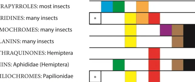
Another manner to classify pigments in insects is based on their origin, with those that can be synthesised endogenously by the insects, such as anthraquinones, aphins, pterins, tetrapyrroles, ommochromes, melanins and papiliochromes; and those that are sequestered from their host plants, the anti-oxidative carotenoids and water-soluble flavonoids (SHAMIM et al. 2014). Many endogenous pigments may have a large range of colours and can be identified in different groups, for example melanins (Figue 1). Others pigments are restricted to a family, such as the papiliochromes in Papilionidae (Lepidoptera) or aphins in Aphididae (Hemiptera) (Figure 1).
Figure 1. Endogenous pigments in insects. Certain pigments synthetized by insects and the variability of colour that are identified. Based on (SHAMIM et al. 2014). The boxes represent the colours observed in each pigment. (*) when is colourless.
19 Table 1. Principal function of natural pigments. Major groups of organic pigments groups in nature and there function. Based on (DELGADO
-VARGAS et al. 2000).
Structural characteristic Group of pigments Pigments Organisms Function
Tetrapyrrole derivatives
Chlorophylls and heme colors
Phytochrome Algae, plants Signal-transducing photoreceptorion Heme group:
hemoglobin and myoglobin
Animals Transport and storage of oxygen
Chlorophyll Algae, plants Photosynthetic process Bilins Invertebrates, cyanobacteries and some algae Integuments of many invertebrates, accessory pigments in light harvesting Isoprenoid derivatives
Carotenoids and iridoids
Carotenoids photosynthetic and nonphotosynthetic organisms Required in photosynthesis and colour of fruits, vegetables, fungi, flowers, also present in birds, insects, crustaceans, and trout.
20 Iridoids Plants Not particularly
important as colorants N-heterocyclic compounds different from
tetrapyrroles
Purines, pterins, flavins, phenazines, phenoxazines, and betalains
Purines Living organisms, plants
Structural
components in DNA and RNA
Pterins Mammals, birds, fish, insects, bacteria
Colour in some insects, in vertebrate eyes, human urine, and bacteria. Some pterins are growth factors (Ex. folic acid)
Benzopyran derivatives (oxygenated heterocyclic compounds)
Anthocyanins and other flavonoid pigments
Anthocyanins Vascular plants Attract pollinators. Antioxidant Flavonoid pigments: aurones, chalcones and flavonols and flavanones Contribute to the pollinating process together with anthocyanins. Photoprotection, defence mechanism in plants
Quinones Benzoquinone, naphthoquinone, Plants, algae and
Echinodermata
Echinodermata family, contribute to
21
anthraquinone family the pigmentation of
spines, shell, ovaries, and eggs. Bronze colour in Polyporus
rutilans (fungus) and
bacteries. Melanins
Eumelanins Black, gray, and
brown colorations of animals, plants, and microorganisms Vertebrate and invertebrate animals Photoprotection
Phaemelanins Mammals and
birds
Photoprotection
Allomelanins seeds, spores, and
fungi, and esclorotins in arthropods
22
Biology and evolution of extra-ocular colour in Hemiptera embryos
Evolution of Hemiptera.
Hemiptera is a hemimetabolous order of insects with unique morphological adaptations for piercing and sucking liquids from plants or other insects, due to modification of the mouthpart into elongated rostrum and other parts such as the maxilla and mandibles into long point stylets (GOULA AND MATA 2015). Bugs can be found in terrestrial and freshwater habitats;
some are even marines. More than 100,000 species of hemipterans have been described in 104 families worldwide, and this number corresponds to about 10% of known insects (BARNARD
2011; JOHNSON et al. 2018).
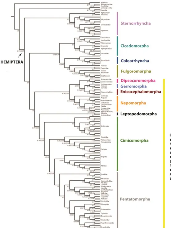
Traditionally, hemipterans were divided in two groups, “Homoptera” and “Heteroptera” based on morphological classification (MYERS AND CHINA 2009). After molecular analysis, Homoptera was divided into three groups (Cicadomorpha, Fulgoromorpha, Sternorrhyncha), while Heteroptera is a monophyletic group (SONG et al. 2012). Recent molecular data divide
the Hemiptera order into five groups: Sternorrhyncha, Cicadomorpha, Fulgoromorpha, Coleorrhyncha, and Heteroptera. Auchenorrhyncha (suborder of the Hemiptera containing most of the familiar members called the “Homoptera”) is paraphyletic, with Cicadomorpha forming the sister group to (Fulgoromorpha + Coleorrhyncha). Within Heteroptera, all infraorders were recovered as monophyletic, as previously shown (Figure 2.) (LI et al. 2017).
The oldest Homoptera fossil was registered since the Early Permian, a period marked with an expansion of Mesophytic gymnosperm groups, around 298.9 million years ago (Mya). A rapid diversification of homopterans occurred during the Late Permian and a considerable extinction took place at the Permo-Triassic boundary (SHCHERBAKOV 2000). Moreover, the
oldest Heteroptera reported is a water bug Arlecoris louisi, from the earliest Middle Triassic of France (approximately 240 Mya) ((SHCHERBAKOV 2010). The true bugs became even more
numerous and diverse during the Mesozoic period, especially in the Late Jurassic and Early Cretaceous (YUR I et al. 2011).
Currently, Heteroptera species have been a focus of interest because of their potential as biological pest control agents. For example, the flower bugs of the genus Orius have been used to decrease the population of bean thrips (Megalurothrips usitatus, Thysanoptera: Thripidae) on red beans (WANG 2001). On the other hand, stink bugs such as Halyomorpha
halys and Homalodisca vitripennis are considered as pests for the damage caused to crops
while Rhodnius is a vector of Chagas disease (DIAS et al. 2002; GRANDGIRARD et al. 2008;
PANIZZI 2015). Conversely, hemipterans can also be used as ecological and environmental indicators in forest ecosystems (M.L.MOIR 2007).
23
Phenotypic variability in eggs of Hemiptera
Most of the scientific works done on the embryonic stages of Hemiptera have been directed to morphological comparisons for establishing classification of the different groups (SOUTHWOOD 1956; COBBEN 1968; PANIZZI AND GRAZIA 2015). Studies based on detailed
description of embryonic stages and a few mutants were also performed with Oncopeltus
fasciatus in the early seventies (BUTT 1949; SPRINGER 1964; LAWRENCE 1970; LAWRENCE
1973). The advent of developmental genetics in the early eighties pushed aside most of the studies with insects, including hemipterans. It was not until the evo-devo revolution of the late 1990s and the development of protocols in molecular tools, that the Hemiptera start to be used as a models to understand the molecular mechanisms regulating the evolution of specific traits (CHIPMAN 2017). Also, transcriptomic data and complete genomes of several species have
been recently published (ARMISÉN et al. 2018; PANFILIO AND ANGELINI 2018; PANFILIO et al.
2019)
In order to study the evolution of colouration in the embryos of Hemiptera, I performed a bibliographic research to obtain information concerning the colouration of the eggs, description of embryonic development and ecological information on different groups (Table 2).
Oviposition sites
Southwood (SOUTHWOOD 1956) classified Hemiptera eggs in four classes depending on the place where they are laid : 1) exposed, 2) semi-exposed, 3) Embedded in dead plants 4) Embedded or associated with living plants. As an example of class 1, females of Pentatomomorpha lay their embryos on the surface of different substrates (V. MATESCO 2009). Also, Adomerus variegatus and Canthophorus niveimarginatus (Pentatomorpha: Cydnidae) females lay a cluster of eggs on the ground and show a maternal care during embryogenesis and first nymphal stage (FILIPPI et al. 2009; MUKAI et al. 2010). The
Belostomatidae (Nepomorpha) females lay embryos on the dorsum of the males (ICHIKAWA
1988; RODRIGUES AND MOREIRA 2005). The species Dysdercus albofasciatus (Pyrrhocoridae)
is an example of class 2. The eggs are found semi- exposed in cracks or between litters (STADLER et al. 1987). As example of class 3 and 4, the glassy-winged sharpshooter
(Homalodisca vitripennis) is considered a pest because the females lays her eggs in groups just under the epidermis layer of leaves of holly, sunflower and citrus (AL-WAHAIBI AND
MORSE 2009). In the specific cases of aquatic (Heteroptera: Nepomorpha) and semi-aquatic (Heteroptera: Gerromorpha) bugs, females lay eggs embedded in plant tissues, on the surface
24 of submerged plants or stones attached by a gelatinous substance or with a pedicel (ANDERSEN 1982; PAPACEK 2001).
Figure 2. Molecular phylogeny of Hemiptera based in mitochondrial genome sequences.
Phylogeny of Hemiptera as inferred from PhyloBayes analyses of the PCGRNA and PCG12RNA datasets under the CAT þ GTR mixture model (modified from (LI et al. 2017)).
25 Table 1. Bibliographic review of Hemiptera embryos. Summary of colouration of eggs and embryos in different species of hemipterans, with additional information about the sites where the eggs are laid and possible predators during the embryonic development. Nd: Not determined. *: transparent chorion.
Infraorder Family Species Egg-layig site Colour of eggs * Description of embryonic development
Egg parasites References
Cicadomor pha
Cercopidae Callitettix versicolor
Laboratory condition: eggs inserted singly in filter paper
Milky white when laid and became brown Nd Nd (CHEN AND LIANG 2012) Membracida e Guayaquila projecta
White when laid.
The chorion was translucent and smooth Nd Nd (M.A.LINARES et al. 2010) Membracida e Sextius virescens Yellowish-white Nd Nd (KITCHING 1974) Membracida e Ceresa nigripectus
Stems of alfalfa plants. Inserted and hidden.
Nd Nd (PEREZ GROSSO
et al. 2014)
Cicadellidae Homalodisc a vitripennis
Below the epidermis of leaves as a cluster
White-yellowish yes Gonatocerus ashmeadi and Gonatocerus triguttatus (AL-WAHAIBI AND MORSE 2009) Cicadellidae Kolla paulula
Inserted in host plant tissue. White eggs; transparent chorion Nd Hymenoptera: Mymaridae and Trichogrammati dae (TRIAPITSYN AND SHIH 2014) Fulgoromo rpha Dictyopharida e Phylloscelis pallescens Cream white: chorion translucent Nd Nd (MCPHERSON AND WILSON 1995)
26 Delphacidae Megamelus
bellicus
Into the parenchyma of the petiole
Milky white when laid, turning yellowish white before hatching. Chorion translucent, smooth. Nd Aprostocetus (O otetrastichus) sp
(A.J.SOSA et al.
2005; MARIANI et al. 2007) Dictyopharida e Nersia Florens
Pale yellowish * Nd Nd (MCPHERSON
AND WILSON 1995) Dictyopharida e Dictyophara europaea
Soil surface. Female cover the eggs with soil particles
Yellowish-green * Nd Nd (KRSTIĆ et al.
2016) Dictyopharida
e
Taosa (C.) longula
Inserted in the petioles. Milky white * Nd Nd (LENICOV et al.
2012) Ricaniidae Ricania
speculum
Inserted into the woody tissue of small twigs of different host plants.
Milky white Nd Aprostocetus
(Ootetrastichus) crino (Hymenoptera Eulophidae), Chaetostricha similis, (Hymenoptera Trichogrammati dae) and Polynema sp. (Hymenoptera Mymaridae) (ROSSI AND LUCCHI 2015; S. LAUDONIA et al. 2017)
27 Sternorrhy
ncha
Psyllidae Retroacizzia mopani
Randomly laid on both the adaxial and abaxial surfaces of mature green and senescent leaves.
Egg clusters appeared to the naked eye as black spots on the leaflet. However, under the light microscope, the eggs were black with smooth shiny grey spots closely spaced near the basal end of the egg.
Nd Nd (OPPONG et al.
2009)
Triozidae Bactericera lyrata
In upper and lower sides of leaves
Freshly laid eggs whitish, later becoming orange-yellow, with brownish pedicel Nd Nd (SELJAK AND MALENOVSK Ý 2014) Aleyrodidae Aleurocanth us woglumi
Laid in spiral and fixed, through a stalk, in the inferior face of the leaf
yellow-clear becoming yellow-darkness when close to the appearance of the nymphs. Nd Nd (RONCHI-TELES et al. 2009) Psyllidae Acizzia jamatonica
On abaxial surfaces and edges of leaflets
yellow Nd Nd (WHEELER AND
HOEBEKE 2009) Nepomorp ha Corixidae Sigara (Tropocorix a) santiagiensi Yellow. Surface ornamented by irregular interlocking smooth hexagons Nd Nd (KONOPKO 2014) Nepidae Laccotrephe s
Under the sand grains in bunches
28 Nepidae Ranatra Inside the soft parts of
floating vegetation of the fresh water habitats
White (from internet pictures) Nd Nd (HINTON 1961; AKILAN et al. 2017)) Belostomatida e Lethocerus medius
In the back of males White when laid and then become dark
Nd Nd Christine L.
Goforth, from the University of Arizona (video) Notonectidae In plant tissues or on
the surface of submerged plants or stones. White (from internet pictures) Nd Nd (PAPACEK 2001)
Corixidae Attached and
embedded in cement by a disc or pedicel on the substratum.
Nd Nd Nd
Pleidae In slits in the tissues of submerged aquatic plants.
Nd Nd Nd
Helotrephidae On the surface of water plant leaves in clusters.
Nd Nd Nd
Aphelocheirid ae
On the surface of various objects on the bottom, on the cuticle of crustaceans, and shells of Bivalvia. Nd Nd Nd Gelastocorida e On the surface of various objects. Nd Nd Nd
Ochteridae On the surface of sandy grains, plants, or roots of plants.
Nd Nd Nd
29
morpha ae us uagans sphagnum
Gerromor pha
Mesovelidae Mesovelia mulsanti
Yellow * Nd Nd (Vargas et al.
Submitted 2019) (M.SOUSA 2009) Velidae Oiovelia cunucunum ana White,* Nd Nd Velidae Paravelia conata White * Nd Nd Velidae Rhagovelia antilleana Yellow * Nd Nd Gerridae Gerris buenoi Yellow * Nd Nd Gerridae Limnoporus dissortis Yellow, dark chorion Nd Tiphodytes gerriphagus Gerridae Limnogonus franciscanu s
Yellow with red * yes Nd
Gerridae Neogerris magnus
Yellow with red * Nd Nd
Leptopodo morpha
Inserted into the stems
of plants near the soil surface or into an algal layer Nd Nd Nd Cimicomo rpha Miridae Helopeltis antonii Embedded in plant tissue. Operculum and extra-chorionic
processes exposed.
White and after two days change to orange with a distinct dark orange band at the distal region.
30 Miridae Lygus
lineolaris
Inserted into plant tissue with only the cap exposed.
Eggs creamy white * Nd mymarid wasp Anaphes ovijentatus (DIXON 1989; ALLEN 2013) http://jenny.tfrec. wsu.edu/opm/dis playSpecies.php? pn=180 Reduviidae Rhynocoris albopilosus
Arranged in lines along the stem of the plant.
Egg body yellowish to light brown.
yes Nd (KWADJO et al.
2008) Miridae Lygus
hesperus
Inserted in plants Red pigmentation of the distal antennal segments and eyes was first observed.
yes Anaphes iole
(Hymenoptera: Mymaridae) (CONTI et al. 1997; COOPER AND SPURGEON 2012) Miridae Helopeltis bradyi
Inserted in fruit. White, shiny Nd Nd (MELINA et al.
2016) Tingidae Leptoypha
hospita
Inserted along leaf margins and petioles, although some were laid singly close to the leaf midrib.
Yellowish Nd Nd (ZHANG et al.
2011)
Tingidae Tanybyrsa cumberi
Inserts eggs into Astelia leaves.
Yellowish Nd Eggs are
parasitised by a mymarid wasp, at present unidentified. (M.MAY 1977) Pentatomo rpha Pentatomidae Edessa meditabund a Deposited vertically, with the posterior pole of embryo fixed to the substratum by a female sustance
Light green * yes Nd (MATESCO et
31 Pentatomidae Genus
Euschistus
Deposited vertically, with the posterior pole of embryo fixed to the substratum, and, in clusters, attached to each other and to the substratum through an adhesive material secreted by the female Attached to the surface by an unknown structure. Before hatching, eggs milky-white in E. hansi and E. picticornis to yellow in E. convergens *
yes Nd (RODRIGUES AND
MOREIRA 2005; MATESCO et al. 2009) Pentatomidae Genus Chinavia Chorion surface reticulated, light brown. A light brown colour of the eggs is due to a pigmented chorion.
yes Nd
Pentatomidae Chinavia musiva
Egg green *. With the development of the embryo, the egg becomes dark green and with dark red eyes, two median red strips.
yes Nd
Pentatomidae Grazia tincta
Pigmented chorion white with two red round bands Nd Nd Pentatomidae Pallantia macunaim Light brown to greenish* Nd Nd Coreidae Holymenia clavigera
Gold colour just after deposition, turning to brown later in the development.*
32 Coreidae Anisoscelis
foliacea marginella
Attached to the surface by an unknown
structure.
Gold colour just after deposition, turning to brown later in the development*. Nd Nd (RODRIGUES AND MOREIRA 2005) Cydnidae Adomerus variegatus In shallow cracks on the soil surface
White with red eyes after 14 days* Nd Nd (MUKAI et al. 2010) Cydnidae Canthophor us niveimargin atus
On the ground, under stones or in a slight depression in the soil. Maternal care
behaviour.
Creamy colour eggs gradually turned a deep pink Nd Nd (FILIPPI et al. 2009) Pyrrhocoridae Dysdercus albofasciatu s
Wet places, cracks in the soil or between litter
Initially ivory colour, after 48 hours the embryo become red.
Nd yes (STADLER et al.
1987) Pentatomo rpha Lygaeoidea Anochrosto mus formosus Laboratory condition: on wet wipes, arranged in aligned masses and sometimes under the sunflower seeds.
Light orange when deposited, turning red with the
development of the embryo. yes Nd (CERVANTES -PEREDO AND ELIZALDE -AMELCO 2007) Pentatomo rpha Lygaeoidea Lygaeus reclivatus reclivatus Laboratory condition: on the wet wipes.
Yellowish white when deposited and later orange. yes Nd Pentatomo rpha Lygaeoidea Oncopeltus sexmaculatu s Laboratory condition: on wet wipes, within fruits or under sunflower seeds.
Pale yellow when deposited, becoming reddish with the development of the embryo. yes Nd
33
Colour evolution in Hemiptera eggs
The different ways that Gerromoprha females lay eggs, depending on the species, could be correlated with the presence or absence of the extra-ocular pigmentation during the embryonic stage. In species with faint or without extra-ocular colour, such as Mesovelia mulsante,
Oiovelia cunucunumana or Paravelia, the embryos are found embedded in the substrate
(Figure 3A). Instead, embryos with yellow-red colour in the body with transparent chorion are attached with a gelatinous substance and exposed in the water, as is the case of our biological model, Limnogonus franciscanus or Neogerris magnus (Figure 3B). Also, in some species (Gerris buenoi and Hydrometra) eggs are exposed but the chorion has a dark colour (Figure 3C). In several species of Pentatomorpha, Lygaeoidea and Pyrrhocoridae families, the eggs that are exposed or semi-exposed and have either a red extra-ocular colour or a dark chorion (STADLER et al. 1987; RODRIGUES AND MOREIRA 2005; CERVANTES-PEREDO AND ELIZALDE -AMELCO 2007; V.MATESCO 2009). Overall, the red colour present in the appendages of embryos or the dark chorion could be a strategy to avoid predators either by aposematism or camouflage, while species that are embedded in subtrates do not need to produce colours to avoid predation.
I wondered if the presence of yellow and/or red colours in the eggs or embryos is common between the different groups of bugs. Hemipterans developed different strategies to lay eggs
Figure 3. Correlation of the modes of oviposition in semiaquatic bugs (Gerromorpha) with the colour of the chorion and embryo. A. Egg unexposed, embedded in substrate (Mesovelia mulsanti and Oiovelia cunucunumana), the embryos have transparent chorion without red pigmentation in the appendage. Exposed embryos: B. Egg deposited superficially and upright on substrate (Hydrometra stagnorum), C. Egg deposited superficially (but sheltered), examples: Microvelia americana,
Stridulivelia tersa. D. Egg deposited superficially, either on upper or lower side of floating object (Neogerris magnus, Limnoporus canaliculatus). The embryos that are exposes present dark chorion or red extra-ocular pigments. (Based on:
34 and the presence or absence of colours in the eggs is documented in a few species (KWADJO et
al. 2008; AL-WAHAIBI AND MORSE 2009; P.L. SAROJ et al. 2016). This lack of information might be attributed either to the insertion of eggs into substrates or to the opacity of the chorion. I prepared a cladogram using the most recent phylogeny of Hemiptera (LI et al.
2017) (Figure 4). Most of the pictures were found in the literature and in web pages of people interested by hemipterans eggs.
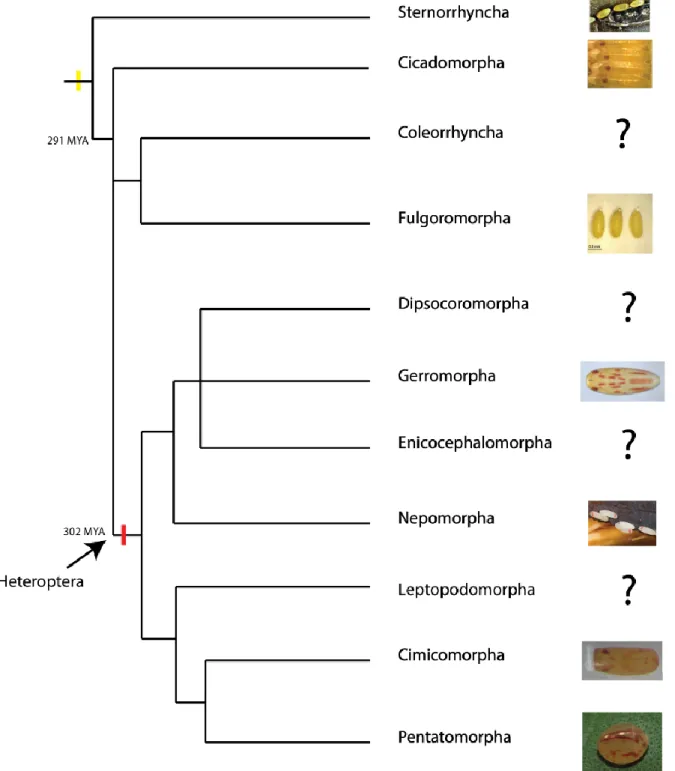
This bibliographical analysis revealed that in the previously called suborder “Homoptera”, eggs are coloured uniformly with tones that can be totally white, cream, brown or yellow- orange and a transparent chorion (Figure 4, Table 2). In the monophyletic suborder Heteroptera, the colour of the eggs, embryos and chorion is more diverse. I could identify 1) eggs without extra-ocular colours with transparent chorion, 2) eggs with dark chorion and 3) eggs with yellow/red extra-ocular colour (Figure 4, Table 2).
This brief overview suggests that yellow could be the ancestral colour of embryos in Hemiptera and that the acquisition of red colouration was specific to the sub-order Heteroptera (Figure 4). Also, most of the basal species of Hemiptera lay their eggs inside substrates or hide them after deposition. By contrast, the majority of eggs from Heteroptera species are exposed, suggesting that the red embryonic colour and opaque chorion may be an adaptation to avoid predators as aposematic signal or camouflage.
Of course, more intensive work should be done to establish relation between the colouration of the eggs, chorion and extra-ocular colour of embryos. In addition, whether the colouration present in the basal species and in the derived species is due to the same pigments is another question that needs to be clarified. In addition, the loss of extra-ocular colour in some Gerromorpha (personal observation) and Nepomorpha species (webpage photo), shows that the extra-ocular colour could have been gained or lost several times during evolution.
35 Figure 4. Evolution of extra-ocular colouration in embryos of Hemiptera. Embryos from the basal suborder “Homoptera” (show this paraphyletic group in the figure) are yellow; no evidence of red extra-ocular eggs could be identified. The red extra-ocular colour was found in the monophyletic suborder of Heteroptera. In Nepomorpha, we found images of embryos with or without extra-ocular colour, suggesting a possible loss of this character in some species of this group. The question marks indicate a lack of information.
Some examples of genetic co-option using pigmentation as a trail.
Understanding how novel traits arise and contribute to species diversification has long been a major question in biology. Recent advances in evo-devo established co-option of existing genes and networks as one of the mechanism involved in the evolution of morphological and
36 physiological novelties (TRUE AND CARROLL 2002; VOPALENSKY AND KOZMIK 2009;
GLASSFORD et al. 2015; MARTINSON et al. 2017; HU et al. 2018).
Pigmentation is a trait that has been used in evo-devo field to try to understand the evolutionary events underlying diversification. Hereafter, I provide a summary of studies where genetic co-option underlies the evolution of phenotype diversity.
Alternative pigments in plants by the co-option of MYB gene
The myeloblastosis protein (MYB) DNA-binding domain defines a class of transcriptional regulators found in animals and plants. In animal, the three proteins - c-MYB, A-MYB and
B-MYB play roles in cell proliferation (LIPSICK 1996). In plants, MYB gene family expanded dramatically with hundreds of genes in Arabidopsis, maize and soybeans (MARTIN AND PAZ -ARES 1997; DU et al. 2012a; DU et al. 2012b). MYB genes control various processes such as
responses to biotic and abiotic stresses, development, differentiation, metabolism and defence (AMBAWAT et al. 2013).
In plants the MYB proteins contain two imperfect MYB repeats corresponding to the R2R3 Myb repeats of the three repeat R1R2R3 in animal MYB proteins. The R2R3-MYB proteins interact with two other large gene families, the basic helix-loop-helix (bHLH) and WD40-repeat (WDR), all together known as the MBW complex (RAMSAY AND GLOVER 2005;
FELLER et al. 2011). The MBW complex is well characterized in regulating differences in
anthocyanin pigment intensity.
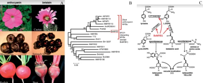
Flavonoids represent a large class of secondary plant metabolites, of which anthocyanins are the most conspicuous class, due to the wide range of colours resulting from their synthesis. Synthesis of anthocyanins in petals is undoubtedly intended to attract pollinators, whereas anthocyanin synthesis in seeds and fruits might aid in seed dispersal. Betalains are red and yellow pigments that are produced in some families of the Caryophyllales order. Betalains are mutually exclusive with the anthocyanins and they appear to provide all of the functions of anthocyanins, pigmenting flowers and fruits and responding to the same environmental stimuli (HATLESTAD et al. 2015; LLOYD et al. 2017) (Figure 5A). Anthocyanins and betalains
are chemically unrelated, while the precursors for the synthesis of all flavonoids, including anthocyanins, are ultimately phenylalanine, betalains compounds derive from tyrosine (TANAKA et al. 2008).
Until a few years ago it was thought that the regulation of betalains was achieved by similar conserved anthocyanin MBW complex. Hatlestad and collaborators have shown that a
37 previously uncharacterized anthocyanin MYB-like protein, Beta vulgaris MYB1 (BvMYB1), regulates the betalain pathway in beets. BvMYB1 is a typical protein R2R3-MYB containing the R2 and R3 MYB domains and a C-terminal activation domain. Phylogenetic analysis using the R2R3 domains showed that BvMYB1 forms a monophyletic clade with dicot anthocyanin MYBs (HATLESTAD et al. 2015) (Figure 5B).
However, BvMYB1 has lost the ability to interact with bHLH members of the MBW complex. Furthermore, BvMYB1 cannot up-regulate the anthocyanin pathway in Arabidopsis, and the Arabidopsis PAP MYBs cannot regulate betalains in beet. Hatlestad and collaborators established that BvMYB1 and anthocyanin MYBs probably derived from a common ancestor. The BvMYB1 gene acquired new transcriptional targets, allowing it to regulate betalain biosynthesis (Figure 5C). In conclusion, the mutual exclusion of both pigment suggest that after the co-option of BvMYB1, some Caryophyllales plants kept one of the biosynthesis gene pathway to avoid redundancy between pigments.
Figure 5. Evolutionary origin of BvMYB1 and betalain pathway. A) The six left panels are representative flowers, fruits and roots, with the far left panels pigmented with anthocyanins and the right with betalains. B) Neighbor-joining tree of R2R3 MYB domains of BvMYB1 and other MYBs. Scale represents amino acid changes per position. BvMYB1 clusters with dicot anthocyanin MYBs within the larger bHLHinteracting cluster. Beta vulgaris is a dicot. C) The betalain pathway is, with the enzymatic steps regulated by BvMYB1 indicated with arrows. Extracted from (HATLESTAD et al. 2015; LLOYD et al. 2017).
Co-option of CYP2J19 by possible cis- regulatory changes and gene duplication in birds
Carotenoid ketolation is an enzymatic reaction that converts yellow dietary carotenoids (xanthophylls) to red C4 ketocarotenoid pigments such as astaxanthin or canthaxanthin (BRITTON 1995). The ketarocatenoid reaction is an important key innovation in avian evolution and colour diversification (PRAGER AND ANDERSSON 2010; THOMAS et al. 2014).
38
CYP2J19 is the gene encoding the ketolase avian enzyme involved in the production of
ketocarotenoid pigment (LOPES et al. 2016; MUNDY et al. 2016)
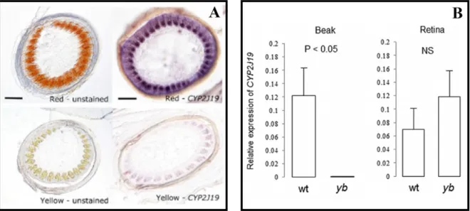
The CYP2J19 is expressed in retina of all canaries but only in the feather follicles of red canaries and not in the yellow ones (Figure 6A) (LOPES et al. 2016). This result suggested that
the differential expression of CYP2J19 in the skin of red birds to produce the ketocarotenoid pigments could be due to cis- regulatory differences. Furthermore, this is not the only way that this gene evolved to generate diversification in birds. The wild-type zebra finch has a red beak due to ketocarotenoids, while the yellowbeak mutant (mutation only described for captive birds) does not contain these pigments (MUNDY et al. 2016). The CYP2J19A gene is
involved in the production of ketocarotenoids in eyes, while the gene CYP2J19B is expressed in both eyes and beak, suggesting that after gene duplication, one of the paralog was co-opted to produce red pigments in the beak without losing its function in the eyes (Figure 6B). Additionally, in the yellowbeak birds the CYP2J19 contains one mutation (CYP2J19yb) that prevents the ketarocatenoid reaction in the beak (MUNDY et al. 2016).
Today we know that the ancestral function of CYP2J19 was colour vision, specifically in the production of ketocarotenoids in red retinal oil droplets of the archelosaurs (archosaurs and turtles) (TWYMAN et al. 2016; TWYMAN et al. 2018), showing that red extra-ocular
pigmentation was recruited later during the evolution of some birds and turtles.
Figure 6. Expression of the gene CYP2J19 in red feathers and red beaks of birds. A) Unstained sections and in situ
hybridization of regenerating feather follicles (10 days post-pluck) of red and yellow canaries. Reddish-orange ketocarotenoid pigmentation is evident in the developing barb ridges of red canary feather follicles, whereas fainter yellow carotenoid-based pigmentation is evident in the yellow feather. B) Expression level of CYP2J19 using qPCR with primers that amplify all copies (CYP2J19A, CYP2J19B, and CYP2J19yb) in the beak and retina of wildtype (wt) and yellowbeak (yb) zebra finch. Extracted from (LOPES et al. 2016; MUNDY et al. 2016).
39 The evolution of some morphological novelties is correlated with the co-option of regulatory circuits or sets of regulatory genes (BRAKEFIELD et al. 1996; BELDADE et al. 2002; ÖZSU AND
MONTEIRO 2017; DESHMUKH et al. 2018).
In butterflies, several genes implicated in the colour patterns of wingeyespots have been identified and it is clear that many developmental genes have been co-opted to produce phenotypic diversity. However, a debate still exists to explain the interaction between these genes and how they were recruited (KEYS et al. 1999; MONTEIRO AND PODLAHA 2009;
OLIVER et al. 2012a; CALLIER 2018). Previous research suggested that nymphalid butterfly
eyespots originated once via the co-option of pre-existing gene networks (OLIVER et al.
2012b; OLIVER et al. 2014). Today, the twelve genes that have been associated with eyespots
development in several species are involved in different gene regulatory pathways and/or circuits (WEATHERBEE et al. 1999; MONTEIRO et al. 2006; SAENKO et al. 2011). Recent
investigation using RNA-Seq differential expression analysis predicted the association of at least two different pathways involved in the production of eyespots and different colour patterns (ÖZSU AND MONTEIRO 2017).
However, a number of hypotheses have arisen in relation to the interpretation of those findings. Developmental regulatory genes may have been involved either by co-option of pre-existing regulatory pathways, followed by modifications (OLIVER et al. 2012a) or by the
co-option of individual genes from independent regulatory networks that shaped the new regulatory pathway.
Gerromorpha
Gerromorpha also known as semi-aquatic bugs is a cosmopolitan group of insects that live on the surface of water (ANDERSEN 1982). In the recent years, the semi-aquatics bugs have been used as models in the evo-devo field to study the genetic basis of morphological innovations (SANTOS et al. 2017; FINET et al. 2018) (KHILA et al. 2009; REFKI et al. 2014; ARMISEN et al. 2015; CRUMIÈRE et al. 2016) and evolution of sexual traits (TOUBIANA AND KHILA 2019). Gerromorpha is a good model lineage to understand the genetic basis of ecological and developmental processes and their evolution.
The most recent phylogeny was built using parsimony analysis based on 64 morphological characters and three DNA markers (Figure 7) (DAMGAARD 2008). In this phylogeny, only four out of eight families were well supported (Mesoveliidae, Hebridae, Hydrometridae and Gerridae). Several relationships were not resolved, as for example the sister group relationship
40 between Gerridae and (Haloveliinae + Microveliinae) or the relation between “Gerroidea” and the “Hydrometroidea” super-families (Figure 7). The aim of our team and collaborators was to build a new phylogeny in order to resolve the paraphyletic classification of some clades and to have a better framework to understand the evolution of different traits.
In the last chapter of this thesis, I will also present the sequencing and manual annotation of the Gerris buenoi (G. buenoi) genome; the first water strider genome to be sequenced and published (ARMISÉN et al. 2018).
41 Figure 7. Phylogeny of the semiaquatic bugs. Single most parsimonious tree resulting from a simultaneous phylogenetic analysis of all morphological and molecular data and with gaps scored as a fifth character state. Numbers in bold above branches refer to Bremer support, and numbers below branches refer to bootstrap support values.
43
44 Co-option of the pteridine biosynthesis pathway underlies the diversification of
embryonic colours in water striders
Synopsis of the paper:
Animal colours and colour patterns in nature have an important influence on fitness. In
general, coloration related-traits are involved in sexual selection, UV protection and signalling communication. Colour-related traits are well spread in nature but our understanding of how they originate and diversify remains limited. A first important step to understand colour diversity is to study the genetic and developmental basis of colouration and the evolutionary mechanisms at the origin of their emergence across lineages.
Genetic basis of colour has been widely studied at the adult stage in different groups of insects but, we know little about colour patterns in the embryo. Here we study a new trait consisting of colouration that is specific to the embryo and absent from post embryonic stages in semi-aquatic bugs (Gerromorpha). By combining developmental genetics with chemical and phylogenetic analyses across a broad sample of species, we uncovered the mechanisms underlying the emergence and diversification of embryonic colours in this group of insects.
Summary of results:
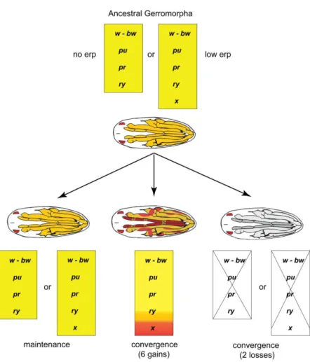
We demonstrate that the entire pteridine biosynthesis pathway, which ancestrally produces red pigment in the eyes, has been recruited during embryogenesis in various extra-ocular tissues including antennae and legs. In addition, we discovered that this co-option is common to all water striders and initially resulted in the production of yellow extra-ocular colour.
Subsequently, six lineages evolved bright red colour and two lineages independently lost all extra-ocular colour. Despite the high diversity in colours and colour patterns, we show that the underlying biosynthesis pathway remained stable throughout the 200 million years of
Gerromorpha evolutionary time. Finally, we identified erythropterin, xanthopterin and chrysopterin as the pigments responsible for these colours in the embryo. These findings demonstrate how novel traits can emerge through the activation of a complete pathway in new developmental contexts.
Conclusion:
Our results confirm the complete co-option of ancestral pathway through Gerromorpha group to produce diversification during the embryonic stage. Additionally, we propose colouration
45 trait in Gerromorpha as an excellent model to understand how the same pathway is regulated to produce phenotypic variability.
Author contribution:
Aidamalia Vargas-Lowman, first author: extraction of RNA, gene cloning, reconstruction of ancestral states and analysis, in situ hybridization, RNA interference, design of the study, analysis of the data and redaction of the paper, with major contribution in terms of results, methodology, discussion and figure-making.
55 CHAPTER 3
56 Spatial regulation of colour patterns in Limnogonus franciscanus embryos.
Author: Aidamalia Vargas-Lowman1, Kahina Saker1, François Bonneton1, Abderrahman Khila1.
Affiliation:
1-Developmental genomics and evolution team; official affiliation from IGFL web site (no accent): Institut de Genomique Fonctionnelle de Lyon, Univ Lyon, CNRS UMR 5242, Ecole Normale Superieure de Lyon, Universite Claude Bernard Lyon 1, 46 allee d'Italie F- 69364 Lyon, France.
Abstract:
Determining the regulatory processes underlying effector genes of morphological trail is essential to understand the evolution and maintenance of phenotypic diversification. Here, we showed a preliminary study to identify transcriptional regulators involved in the recruitment of the pteridin network in semi-aquatic embryos. We performed RNA sequencing, comparative transcriptomics, and gene function assays to identify possible transcription factors involved in the production and regulation of pteridine pigments in eyes and appendages. A total of 135 transcription factors were found to be in common between pigmented organs. From the 80 genes tested, we failed, so far, to identify any that were involved in the spatial regulation of colour patterns in the appendages of L. franciscanus embryos.
Key words: Colour, transcription factors, pteridine pathway, diversification, Gerromorpha, co-option, semi-aquatic insects, embryos.
57 Introduction
Conservation in coding regions across species indicates that evolutionary novelties may arise through combinatorial processes, such as changes in gene regulation and the co-option of novel genes into existing regulatory gene networks (SANETRA et al. 2005). Studies in
butterflies suggest that not more than four genes (optix, wntA, cortex and doublesex) play causative roles in the formation of wing colour patterns (REED et al. 2011; MARTIN et al.
2012; KUNTE et al. 2014; NADEAU et al. 2016). These genes are characterized as “adaptive
hotspots,” which repeatedly drive morphological evolution across different linages (PAPA et
al. 2008; MARTIN AND ORGOGOZO 2013). However, pigmentation traits in Drosophila have
tended to be caused by differences in cis-regulatory elements of the pigmentation enzymes themselves (ORDWAY et al. 2014).
In Gerromorpha (semi-aquatic bugs bugs), embryos have red-dark colour in the eyes, while the appendages and abdominal parts can be yellow, yellow-red or colourless, depending on the species (Figure 1). We have shown that the colour diversification of semi-aquatics embryos is produced by the co-option of the same pteridine pathway
involved in the production of red pigments in the eye (VARGAS-LOWMAN et al. 2019).
Additionally, this co-option is common to all Gerromorpha and the shift from yellow to red Figure. 1. Diversity of colour and patterns in Gerromorpha embryos. A-E) different species of semi-aquatic bugs xx hours after lay. F) Limnogonus franciscanus embryo without chorion, 64 hours after laying. Dotted lines represent the tissues that were dissected. A. distal part of the antennae, b. eye and c. second spot of the second leg. Arrow heads signalling the eyes. Scale bar 100 µ.