DISCLAIMER OF QUALITY
Due to the condition of the original material, there are unavoidable flaws in this reproduction. We have made every effort possible to
provide you with the best copy available. If you are dissatisfied with this product and find it unusable, please contact Document Services as soon as possible.
Thank you.
Pages are missing from the original document.
Synovectomy and Use of the l°B(n,a) Nuclear
Reaction to Examine the Pathology of
Rheumatoid Arthritis
by
Leigh Scott Johnson, S.M.
Submitted to the Department of Nuclear Engineering
in partial fulfillment of the requirements for the degree of
Doctor of Philosophy in Nuclear Engineering
at the
MASSACHUSETTS INSTITUTE OF TECHNOLOGY
May 1994
) Massachusetts Institute of Technology 1994. All rights
Author ...
I-.
..
~ '" 1 .-.. '~
~
P""' I".o, ,..reserved.
Science
MASSACHlUSETTS INSTITUE OF TECHNOLOGY [rJUN 3 0 1994 LIBRARIESDepartment of Nuclear Engineering
May 12, 1994
Certified
by ...
.
Jacquelyn C. Yanch
Associate Professor
Thesis Supervisor
Certified
.- v .' '. ..- ; .Sonya Shortkroff
Thesis Reader
Accepted by ...
...
Allan F. Henry
Chair, Departmental Committee on Graduate Students
--
--. ..~ ; ;... .. ..
on May 12, 1994, in partial fulfillment of the requirements for the degree of
Doctor of Philosophy in Nuclear Engineering
Abstract
Beta-particle dosimetry in radiation synovectomy, which is a radiation therapy to treat rheumatoid arthritis, was evaluated using Monte Carlo radiation transport sim-ulation in a mathematical model of the treated joint and using experiments in joint phantoms and the knees of cadavers. Radiachromic film dosimeters and reactor-produced radionuclides were used in the experiments. Results of the dosimetry eval-uation are presented as absorbed dose factors (cGy-cm2/MBq-s) at depth in bone,
articular cartilage, the fluid-filled joint capsule, and synovium, and can be used to ex-trapolate beta dose distributions in treated joints. Next, use of the 1°B(n,a) nuclear
reaction as a probe to examine the pathology of rheumatoid arthritis was developed. The approach is to use the nuclear reaction to determine whether killing the surface layer of synovial lining cells alone is sufficient to arrest the progression of RA in the joint, or whether killing the entire synovium is required. While the question was not answered here, preliminary studies of 10B uptake in excised samples of human
rheumatoid synovium were completed. Prompt gamma neutron activation analysis was used to demonstrate and quantify the temporal distribution of boron particulate and boric acid uptake in the samples; substantial uptake was seen to occur within the first hour of incubation. Neutron-induced alpha-track autoradiography was used
to in an attempt examine the spatial distribution. Although the autoradiography
experiments did not reveal the desired spatial distribution, other information was obtained.
Thesis Supervisor: Jacquelyn C. Yanch Title: Associate Professor
Thesis Reader: Sonya Shortkroff
MIT Nuclear Dept. Engineering LS Johnson~~~~~~~~~~~~~~~~~~~~~~~~~~ I MIT N~uclear Engineering Dept LS Johnson I
There are a number of individuals whose time, assistance, and support I would like to acknowledge. In particular, I would like to thank the three members of my thesis committee, Professors Jacquelyn Yanch, Otto Harling, and Sidney Yip, for the oppor-tunities, instruction, and guidance each of them has provided me during my years as a graduate student at MIT. For always treating me with respect and for challenging ;and inspiring me, not only to do my best, but also to strive to improve, I thank you.
Next, I would like to acknowledge the contributions of my collaborators at Brigham and Women's Hospital, Dr. Clement Sledge, Sonya Shortkroff, and Christina. With-out their help in planning and conducting the cadaver and in vitro experiments, much of the work in this thesis would not have been possible. Thank you for sharing your :facilities, time, and experience. I look forward to continuing to work with you in the
future.
I also would like to acknowledge the assistance of a number of other individuals: Dr. Guido Solares for helping me to complete the autoradiography experiments, Dr. Ronald Rogus for demonstrating the prompt gamma facility, Dr. Naengnoi Limpa-Amara for sharing her laboratory space, Brett Mattingly for the scanning electron micrographs, and my two undergraduate research assistants, Ruth Lim and Bridget Hanser, for their help in a variety of tasks.
Finally, I would like to acknowledge the support, humor, and patience of my friends, both at MIT and at home. Lisa, thank you for your help and encouragement over the past few weeks. Chris, Joe, Jason, and Andrew thank you for laughing at my jokes, putting up with my attitude, and keeping me in line. Because of you four, I know that, in the very near future, I will look back on this time in Boston and miss
it dearly.
LS Johnson
1.1.2 Other methods . . . .
1.2 Use of the 10B(n,a) Nuclear Reaction to Examine the Pathology of
Rheumatoid Arthritis ...
1.3 Conclusion. ...
2 Rheumatoid Arthritis and Radiation Synovectomy
2.1 Introduction. ... 2.2 Synovial Joints . . . . 2.2.1 Anatomy . ... 2.2.2 Synovium ... 2.3 Rheumatoid Arthritis ... 2.3.1 Epidemiology ... 2.3.2 Pathology ... 2.3.3 Treatment ... 2.4 Radiation Synovectomy. ... 2.4.1 Radionuclides of interest . . . .
2.4.2 Beta-particle dosimetry in radiation synovectomy ... 2.4.3 What thickness of synovium should be removed? ... 2.5 Conclusion. ...
3 EGS4 Monte Carlo Transport of Electrons
3.1 Introduction. ...
3.2 Monte Carlo method ...
3.2.1 Sampling random variables ... 3.3 EGS4 code. ...
3.4 Condensed History Electron Transport . . . 3.4.1 Transport step sizes ...
MIT Nuclear Dept. Engineering LS Johnson~~~~~~~~~~~~~~~~~~~~~~~~~~~~~~~~~~~~~~~~~~~~~~~~~~~~~~~
17 18 19 20 20 20 21 21 23 24 24 26 28 30 33 35 35 37 37 38 38 43 44 45 LS Johnsoni
...
...
. .. . . . .. . . .3.4.2 Electron transport logic . . .
3.4.3 Multiple scattering ... 3.4.4 Continuous energy loss . . 3.4.5 Subtep size restrictions ....
3.5 Major Interactions.
3.5.1 Bremsstrahlung production 3.5.2 Knock-on electron production 3.6 Boundary Crossing and Scoring . . . 3.7 Conclusion. ...
4 Monte Carlo Estimates of Beta Dosimetry
4.1 Introduction. ...
4.2 Materials and Methods ...
4.2.1 Mathematical joint model ... 4.2.2 Tissue cross sections ... 4.2.3 Radionuclide spectra .
4.2.4 Computer simulations . 4.2.5 Dose estimates.
4.2.6 Dose rate estimates ...
4.2.7 Backscatter contributions to absorbed dose . 4.2.8 Effect of disease progression on dosimetry ... 4.2.9 Simulation parameters ...
4.3 Results. ...
4.3.1 Absorbed dose factors versus distance ...
4.3.2 Backscatter ...
4.3.3 Dosimetry and progression of rheumatoid arthritis . 4.4 Discussion ...
4.4.1 Backscatter calculations and absorbed dose to bone 4.4.2 Dosimetry and rheumatoid arthritis progression . .
4.4.3 Therapeutic range . . . .
4.5 Conclusion. ...
5 Experimental Beta Dosimetry
5.1 Introduction. ...
5.2 Experiments in Phantoms 5.2.1 Materials and methods 5.2.2 Results ...
5.2.3 Discussion ...
5.3 Experiments in Cadavers . . . 5.3.1 Materials and methods
MIT Nuclear Engineering Dept. LS Johnson I
48 50 53 55 56 57 59 60 61 63 63 64 64 66 67 67 76 77 78 78 79 80 89 89 90 90 92 93 94 95 96 96 97 97 106 110 112 113 LS Johnson i
...
...
...
...
...
...
...
...
...
. . .. . . . . . . ....
. . .. . . . . . . . .. . . . . . .. . . .6.2 Is Removal of the Lining Cells Sufficient? ... 6.3 °B(n,a) 0 Nuclear Reaction ...
6.3.1 Two Boron Preparations . . . .
6.4 Conclusion. ...
7 Temporal and Spatial Distribution of 'B Uptake1
7.1 Introduction. ...
7.2 Prompt Gamma Neutron Activation Analysis ... 7.2.1 Materials and Methods ...
7.2.2 Results . ...
7.3 Neutron-induced Alpha-track Autoradiography ... 7.3.1 Materials and Methods ...
7.3.2 Results . ...
7.4 Discussion ...
7.5 Conclusion. ...
8 Conclusion
8.1 Beta-particle Dosimetry in Radiation Synovectomy ...
8.1.1 Monte Carlo calculations . . . . 8.1.2 Experimental dosimetry . . . .
8.2 Use of the 1°B(n,a) Nuclear Reaction to Examine the Pathology
Rheumatoid Arthritis ...
8.3 Recommendations for Future Work . . . .
8.3.1 Examining the pathology of rheumatoid arthritis ... 8.3.2 Procedure ... of M 120 122 123 125 126 126 126 128 133 136 137 139 139 141 142 143 143 144 145 145 146 149 LS Johnsoni
2-1 Diagram of a simple synovial joint showing the ends of the two ar-ticulating bones separated from each other by synovial fluid which is contained within the joint capsule. ... 22 2-2 Phagocytosis refers to the process in which a cell's plasma membrane
infolds around a large particle and forms a vesicle which then moves into the cell. The ingested particles typically are digested and expelled. 23 2-3 In radiation synovectomy, a beta-emitting radionuclide is injected
di-rectly into the joint capsule in order to kill rheumatoid synovium. .. 29 3-1 Graph of a probability density function, above, and its corresponding
cumulative density function, below ... 40 3-2 Graphs of a difficult probability density function f(x) and an easier
probability density function g(x) which approximates f(x). The ratio
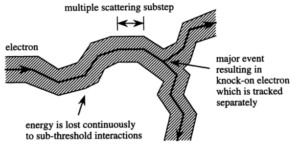
h(z) = g(x)/f(z) is plotted in the lower figure. Here h,ma is any finite upper bound for h(x). ... 42 3-3 Typical electron track as simulated by EGS4. The electron's path is
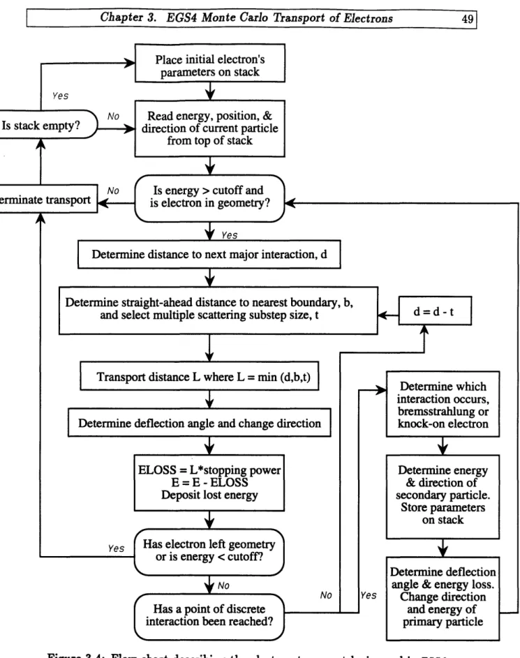
broken into a number of small multiple scattering substeps. Along each substep, the electron is assumed to travel in a straight line. At each substep's end, changes in the electron's energy and direction due to sub-threshold interactions are determined using probability density functions for multiscattered electrons. When a point of catastrophic interaction is reached, it is treated separately by conventional random sampling according to the single-scattering cross sections ... 47 3-4 Flow chart describing the electron-transport logic used in EGS4. ... 49 3-5 A single multiple scattering substep of a real electron. In EGS4 only the
straight-ahead portion of the substep is simulated; the lateral deflection is ignored. It is important to note, however, that by breaking the transport history into small multiple scattering substeps and deflecting the electron at the end of each substep, lateral transport of the electron is effectively accomplished. ... ... ...51
LS Johnson I MIT Nuclear Engineering Dept.
Beta spectrum Beta spectrum Beta spectrum Absorbed dose Absorbed dose Absorbed dose Absorbed dose Absorbed dose Absorbed dose Absorbed dose Absorbed dose
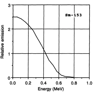
for 86Re. Adopted from Hogan et al. . . . for 188Re. Adopted from Hogan et al ... for 198Au. Adopted from Hogan et al. . . . factors versus distance in the joint model for factors versus distance in the joint model for factors versus distance in the joint model for factors versus distance in the joint model for factors versus distance in the joint model for factors versus distance in the joint model for factors versus distance in the joint model for factors versus distance in the joint model for
. . . 73 . . . 74 . . . 75 32p
.
. . 81 oy ... . 82 1 53Sm. . 83 16 5Dy.. . 84 166Ho.. 85 186 Re... 86 18 8Re... 87 1 9 8AU. . 885-1 Phantom used to experimentally measure radiation absorbed dose pen-etration in synovial joints treated with radiation synovectomy. Dis-tances are defined in relation to the leftmost solid water-radioactive source interface. ...
5-2 Block diagram of a simple radiachromic film dosimeter reader.
5-3 Calibration curve for the radiachromic film dosimeters used in these
experiments . . . .
5-4 Comparison of the absorbed dose factors derived from measurements obtained using planar 165Dy radioactive sources in the joint phantom with those obtained using Monte Carlo radiation transport simulation in a mathematical model of the joint. ...
5-5 Comparison of the absorbed dose factors derived from measurements obtained using a planar ls6 Ho radioactive source in the joint phantom with those obtained using Monte Carlo radiation transport simulation in a mathematical model of the joint. ...
5-6 Comparison of the absorbed dose factors derived from measurements obtained in the knees of cadavers with the estimates of absorbed dose factors versus depth obtained using Monte Carlo radiation transport simulation in a mathematical model of the joint...
MIT
Nuclear
Engineering
Dept.
LS
Johnson~~~~~~~~~~~~~~~~~~~~~~~~~~~~~~~~~~~~~~~~~~~~~~~~~~~~~~~~~~~~~~~~~~~~~~~~~~
4-7 4-8 4-9 4-10 4-11 4-12 4-13 4-14 4-15 4-16 4-17 102 105 107 109 111 116
7-1 Schematic diagram of the prompt gamma neutron activation analysis facility operated at MIT. The facility uses a diffracted thermal neutron beam to reduce the numbers of fast neutrons and gamma-rays at the
sample position ... 129 7-2 Scanning electron micrograph of the boron particulate. Magnification
165x. A 60 pm bar at the bottom of the micrograph can be used to estimate the size distribution of the particles. ... 130 7-3 Scanning electron micrograph of the boron particulate taken at a higher
magnification 1000x. ... 131 7-4 A sample calibration curve for the analysis of bulk 10B content at the
MIT prompt gamma neutron activation analysis facility. ... 134 8-1 Mathematical joint model which could be used to evaluate various
irradiation parameters required to deliver therapeutic synovial doses in as short an irradiation time as is possible ... 148
LS Johnson
IMIT Nuclear Engineering Dept.
4.1 Elemental compositions and densities of the four absorbing media used
in the joint model. Adopted from ICR U-44. . . . . . . 66
4.2 Parameter values used in PEGS4 to compute tissue cross section data. 67 4.3 Characteristics of the three stages of advancing rheumatoid arthritis
used in the computer simulations ... 79 4.4 Parameter values used in the EGS4 simulations. .. .... . 79 4.5 Effect of advancing rheumatoid arthritis on absorbed dose deposition
in radiation synovectomy. ... 90 4.6 Bone surface (m) beta dose expressed as a percentage of the beta dose
imparted to the lining cells ... 93
4.7 Therapeutic and maximum ranges of the 8 radionuclides investigated. 94 5.1 Electron mass stopping powers (MeV-m2/kg) and mass scattering
pow-ers (Radian2-m2/kg) for BONE. Adopted from ICR U-44. . . . . ... 98
5.2 Electron mass stopping powers (MeV-m2/kg) and mass scattering
pow-ers (Radian2-m2/kg) for ARTICULAR CARTILAGE. Adopted from
ICR U-44. . . . . . . . . . ..... 99
5.3 Electron mass stopping powers (MeV-m2/kg) and mass scattering
pow-ers (Radian2-m2/kg) for SOFT TISSUE. Adopted from ICRU-44. .. 99
5.4 Electron mass stopping powers (MeV-m2/kg) and mass scattering
pow-ers (Radian2-m2/kg) for ALUMINUM. Adopted from ICR U-44. . .. 100
5.5 Electron mass stopping powers (MeV-m2/kg) and mass scattering pow-ers (Radian2-m2/kg) for SOLID WATER. Adopted from Constantinou
et al ... 100
5.6 Composition of solid water. Adopted from Constantinou et al. .... 101
MIT Nuclear Engineering Dept. LS Johnson LS Johnsoni
I~~~~~~~~~~~~~~~~~~~~~~~~~~~~~~~~~~~~~~~~~~~~~~~~~~~~~~~~~~~~~~~~
5.7 Results of the depth-dose measurements for 165Dy obtained using the joint phantom. The results are expressed as absorbed dose factors (cGy-cm2/MBq-s) at depth in the phantom. ... 109 5.8 Results of the depth-dose measurements for 166Ho obtained using the
joint phantom. The results are expressed as absorbed dose factors
(cGy-cm 2/MBq-s) at depth in the phantom. . . . .... 110
5.9 Joint locations at which radiachromic dosimeters were positioned to measure absorbed dose in the knees of cadavers ... 114 5.10 Results of the absorbed dose measurements made at several strategic
locations in the knees of fresh, human cadavers. Each of the knees was injected with a therapeutic dose of 165Dy ferric hydroxide
macroaggre-gate. Results are listed as absorbed dose factors (cGy-cm2/MBq-s). . 115 6.1 Thermal neutron capture cross section values of various nuclides.... 122 6.2 Thermal neutron capture cross section values of elements normally
found in the body's tissues. ... 124 7.1 Temporal distribution of bulk BORON PARTICULATE uptake in
hu-man rheumatoid synovium. ... 135 7.2 Temporal distribution of bulk BORIC ACID uptake in human
rheuma-toid synovium. ... 135
MIT.~~~~~~~~~~~~~~~~ Dept. Nuclear Engineering LS Johnson,~~~~~~~~~~~~~~~~~~~~~~~~~~~~~~~~~~~~LS Johnsoni
tomy, which is a radiation therapy to treat rheumatoid arthritis, using Monte Carlo radiation transport simulation, experiments in joint phantoms, and experiments in the knees of cadavers; to provide a rationale for the use of the l°B(n,a) nuclear reaction to examine the pathology of rheumatoid arthritis; and to determine the temporal and spatial distribution of 10B uptake in excised samples of live, human rheumatoid syn-ovium. Presentation of the thesis is organized as follows. In this chapter, background information regarding motivation for the work is provided, and theoretical and ex-perimental techniques used to complete it are introduced. In Chapter 2, rheumatoid arthritis and radiation synovectomy are reviewed. Rheumatoid arthritis is a painful degenerative disease affecting the synovial joints. Treatments for the disease, includ-ing radiation synovectomy, are described, as are the synovial joints and the effects of rheumatoid arthritis on those joints.
Evaluating beta-particle dosimetry in radiation synovectomy is a difficult problem for a variety of reasons (see below). The approach taken in this thesis was to evaluate the dosimetry in 3 different ways: through Monte Carlo simulation of radiation trans-port in a mathematical model of the rheumatoid joint, through experiments in joint phantoms using radiachromic film dosimeters and reactor-produced radionuclides, and through experiments in the knees of fresh, human cadavers using radiachromic
film dosimeters and reactor-produced 1 65Dy.
In Chapter 3, the Monte Carlo computer code used to simulate radiation transport in a mathematical joint model is introduced. EGS4 is the code which was used, and a complete description of the code's approach to electron transport simulation is presented. In Chapter 4, use of EGS4 as a tool to evaluate the dosimetry of 8 beta-emitting radionuclides of interest in radiation synovectomy is described. The results of the computer simulations can be used to estimate absorbed dose distributions in
treated joints.
M
In Chapter 5, experiments aimed at verifying the accuracy of the Monte Carlo predictions of beta-particle dosimetry in radiation synovectomy are presented. As mentioned above, experiments were conducted in both joint phantoms and the knees of cadavers using radiachromic film dosimeters and reactor-produced radionuclides. Comparisons of the experimental results with the EGS4 results are made.
In the remainder of the thesis, the focus is shifted from beta-particle dosimetry in radiation synovectomy to use of the °B(n,a) nuclear reaction to examine the pathol-0
ogy of rheumatoid arthritis. Chapter 6 is devoted to presenting data which support the hypothesis that killing the surface layer of synovial lining cells in rheumatoid joints is sufficient to temporarily arrest the progression of rheumatoid arthritis in the joint and provide patients with symptomatic relief of the disease. Use of the l°B(n,a) nuclear reaction as a probe to test the hypothesis is developed. The 10B(n,a) nuclear
reaction was chosen specifically because the range of the emitted particles is short, on the order of 10 /Sm. The reaction thus can be used to selectively damage and kill only boronated cells and their nearest neighbors. While the hypothesis itself is not tested in this thesis, preliminary studies of 10B uptake in excised samples of live, human rheumatoid synovium are presented in Chapter 7. Prompt gamma neutron activation analysis is used to quantify the temporal distribution of 10B uptake in the samples, while neutron-induced alpha-track autoradiography is used in an attempt to
determine the spatial distribution.
Finally, in Chapter 8 important conclusions drawn from the work of this thesis are reviewed and recommendations for future research are made. The recommendations include descriptions of in vitro and in vivo experiments aimed at determining the thickness of synovium which must be killed to arrest the progression of rheumatoid arthritis and provide patients with symptomatic relief of the disease, i.e., is it sufficient to kill only the lining cells or must the entire synovium be killed. The possible development of neutron capture therapy as novel treatment for the disease also is presented.
1.1 Beta-particle Dosimetry in Radiation
Synovec-tomy
Radiation synovectomy is a radiation therapy to treat rheumatoid arthritis, a painful, degenerative disease affecting the synovial joints. The procedure consists of the in-jection of a short-lived, low-energy beta-emitting radionuclide into the joint capsule where it comes into direct contact with the diseased tissue (rheumatoid synovium) [1]. Phagocytic cells along the tissue surface quickly absorb radioactive particulate from the joint capsule [2, 3, 4]. Then as the radionuclide decays, radiation absorbed dose is imparted to the rheumatoid synovium. Provided the amount of radioactivity injected
LS Johnson|
100% of patients treated with radiation synovectomy are reported to experience relief of pain, reduced joint swelling, and increased joint mobility at 6 months, 1 year, and 2 years after injection [5]. Improvement however is dependent on a variety of factors, including the condition of the joint before treatment and whether or not a therapeutic radiation dose is delivered to all the rheumatoid joint tissue. At the same time, one wants to ensure that other, healthy, joint components located nearby (e.g., bone and articular cartilage) are not subjected to an unnecessary radiation hazard. It is for these reasons that beta-particle dosimetry in radiation synovectomy is of interest. Before discussing approaches to beta-particle dosimetry however, it is instructive to review the concept of absorbed dose and the role of dosimetry in radiation therapy.
Concept of absorbed dose
The ultimate radiobiological effects of beta-emitting radionuclides injected into rheuma-toid joints are initiated by processes in which atoms and molecules of the joints' tis-sues are ionized or excited. Some of the ionization events result in the production of chemically active species, i.e., free radicals, in regions near the paths traversed by the emitted particles. Free radical production is the direct result of energy deposition in the tissues and their presence is recognized as playing a central role in the generation of a wide variety of biological damage, and other changes, from which subsequent ra-diobiological effects evolve. In general, as the number of ionization events increases, so does the production of free radicals, and therefore the production of biological damage.
Over a wide range of conditions, it is considered an excellent approximation to as-sume that the average number of ionizations and excitations in the volume of interest is proportional to the amount of energy deposited in the volume [6]. The absorbed dose, i.e., the average amount of energy imparted per unit mass, therefore is of cen-tral importance for the production and evaluation of the radiobiological effects of radiation synovectomy.
Role of dosimetry in radiation therapy
The role of dosimetry in radiation therapy is to provide quantitative information regarding absorbed dose distributions in patients. In particular, it is of interest to know accurately the absorbed dose imparted to target tissues and whether or not
LS Johnson[
the incidental irradiation of other, "non-target," tissues subjects the patient to an unnecessary or undesirable radiation hazard. In beta-particle dosimetry in radiation
synovectomy, the target tissue is rheumatoid synovium, and the other, non-target, tissues to be considered include bone and articular cartilage.
In general, there are 3 methods for estimating absorbed dose distributions in radiation synovectomy or any other radiation therapy. These include: (1) direct measurement in humans, (2) extrapolation from measured data in animals, phantoms, and cadavers, and (3) calculations based on mathematical models. Estimates of absorbed dose distributions also can be extrapolated from clinical results, but this "empirical" method lacks scientific rigor and is not considered here.
1.1.1
Calculations based on mathematical models
Typically in radiation dosimetry, the ranges of beta particles emitted from internally distributed radionuclides are small compared to the size of the tissues or organs containing the radioactivity. When this is the case, it has been considered permissible to assume that the entire kinetic energy of the emitted particles is deposited in the source [6]. This clearly is not a reasonable assumption in radiation synovectomy, however, since the ranges of the emitted beta particles (up to 10 mm depending on the radionuclide [1, 7, 8]) is large compared to the size of the source (less than 1 mm [9, 10, 11]). In addition, the radiation absorbed dose to other, "non-source," regions of the joint (e.g., synovium at depth, bone, and articular cartilage) also are of interest in radiation synovectomy. Alternative approaches therefore must be used.
For beta-particle sources located at or near target tissues, beta dose point kernels have been developed to represent the distribution of absorbed dose in a tissue medium as a function of distance from a point-isotropic beta-emitting source. By suitable integration over the point kernels, dose distributions can be readily determined for a wide variety of source-to-target configurations.
The approach is best illustrated with the aid of an example. In 1973, Husak et
al. [9] used the beta dose point kernel method to calculate beta-particle dosimetry
in radiation synovectomy. Their approach was to assume that radionuclides injected into the rheumatoid joint are absorbed by the synovial surface and are uniformly distributed over it so that they form a thin plane source of activity.
The absorbed dose rate R (rad/s) at a distance s from a thin plane source of finite thickness d then is calculated as:
1 f +d
R(s) = d. PR(z)
dz,
(1.1)
where R4(z) is the absorbed dose rate at distance z from an infinitely thin plane
LS Johnsonl
energy of the emitted beta-particles (MeV), and R,=. is the penetration range in tissue of the most energetic beta-particle emitted (cm). The symbol ,(x) represents the point isotropic specific absorbed fraction, i.e., the fraction of the energy emitted from a point isotropic source that is absorbed at distance z per unit mass of tissue.
The specific absorbed fraction can be calculated using tabulated values of the dimensionless scaled absorbed dose distribution function F(~) [12]. The function
F,(~) and the specific absorbed fraction are related as follows:
F(~) = 47rpX2z9oO(z), (1.3)
where = z/xx 9o, 90o is the distance from the point isotropic source at which 90% of
the emitted energy is absorbed (cm), and p is the density of the medium (g/cm3).
Now, if the expression 2xzx(x) in Eqn. 1.2 is replaced with Fp()/2pxx 9o, the
re-sulting expression is as follows:
~Rp(z)
nkE.fg
=
Ff)dz.
(1.4)
This equation can be solved by numerical integration using tabulated values of the function Fp(~) [12].
Husak et al. have solved the equation. Their results present the dependence of absorbed dose in tissue on distance (up to 0.6 mm) from thin plane sources of
s
98Au and 90Y, two radionuclides which have been used extensively in clinical trials
in radiation synovectomy (see Chapter 2).
But the point kernel method has been developed for homogeneous media only. As a consequence, the approach taken by Husak et al. was to approximate the joint as a homogeneous soft tissue volume and to neglect the incidental irradiation of other joint components, such as bone and articular cartilage. Doses to these other, "non-target," joint components were not calculated. As mentioned above however, one of the roles of dosimetry in radiation therapy is to provide the quantitative information necessary for radiotherapists to ensure that non-target tissues are not subjected to an unnecessary or undesirable radiation hazard. Doses to bone and articular carti-lage in radiation synovectomy therefore must be considered. Absorbed dose to the bone surface (10 pm) is particularly of interest since the International Commission on Radiation Protection (Vienna, Austria) has identified this tissue region as one
LS Johnson i
of two principal radiosensitive targets to be protected in procedures involving bone
irradiation [13].
In addition, the tabulated beta dose point kernels referenced above and used by Husak et al. to calculate beta dosimetry in radiation synovectomy are based on a model which neglects the production and transport of secondary electrons and the fluctuation of energy loss with distance [14, 15]. Prestwich et al. have reported Monte Carlo calculations of beta dose point kernels that do include the transport of secondary electrons and the effects of energy fluctuations [14]. Their approach shows a dose distribution for 90Y in water that is 7% lower at the origin than that predicted
by the above methods and significantly higher at distances greater than 5 mm from the source. While these results have shown improved agreement with experiment, a systematic error in the energy-loss straggling algorithm has been revealed [15], indicating yet another limitation of the accuracy of the point kernel method.
1.1.2 Other methods
As mentioned above, other methods for estimating absorbed dose distributions in radiation therapy are available. These include direct measurements in humans, which often can be difficult to obtain (see Chapter 5), and extrapolation from measured data in animals, phantoms, and cadavers. Other mathematical methods, e.g., the Monte Carlo method, also can be used. The approach taken in this thesis was to evaluate beta-particle dosimetry in radiation synovectomy 3 different ways.
First, Monte Carlo simulations of radiation transport in a mathematical model of the rheumatoid joint were completed for 8 radionuclides of interest in radiation synovectomy. Each of these radionuclides either has been used in clinical trials or is of interest due to its favorable half-life, emission energies, and radiochemistry. Sim-ulation begins by choosing a starting position, direction, and energy for the emission of a single beta-particle from within the radiation source region. The emitted particle is transported through the mathematical joint model by the Monte Carlo computer code. At each point in the particle's "life" where a choice of interaction type, scatter-ing direction, distance to the next interaction, etc. has to be made, a random number is generated by the code and used to determine the event's outcome from known prob-ability distributions which describe the likelihood of occurrence of each of the possible outcomes. Simulated interactions include all the major events, such as the produc-tion of secondary "knock-on" electrons and bremsstrahlung photons. "Continuous" energy loss and multiple scattering through minor interactions with atomic electrons
also are accounted for. Simulation ends when the emitted beta-particle and all its secondaries no longer have enough energy to excite the absorbing media. At this point, a new beta-particle is "emitted" from within the radiation source region, and the process is repeated. The simulations are described in detail in Chapter 4.
LS Johnsoni
[MIT Nuclear Engineering Dept.
I
Planar radioactive sources of uniform activity (similar to those assumed by Husak et
al. to exist in joints treated with radiation synovectomy) were made by soaking filter
papers in liquid solutions of Dy203 (stable, natural Dy) and Ho203 (stable,
natu-ral Ho) dissolved in nitric acid. The wet filter papers then were dried, stacked and stitched together, and irradiated at the MIT Nuclear Reactor Laboratory, yielding thin plane sources of 165Dy and 166Ho which were inserted into the joint phantoms and
allowed time to decay. Radiachromic film dosimeters were used to measure depth-dose data in the phantom. The experiments, dosimeters, and results are explained in detail in Chapter 5.
Third, experiments were conducted in the knees of fresh, human cadavers using radiachromic film dosimeters and reactor-produced 165Dy. The knees were surgically
opened and several dosimeters were stitched into strategic locations throughout the joints. The knees then were sewn tight and injected with a therapeutic dose of ra-dioactivity. After allowing time for decay, the knees were reopened and the dosimeters were removed for analysis. These experiments also are explained in detail in Chapter
5.
1.2 Use of the
10
B(n,a) Nuclear Reaction to
Ex-amine the Pathology of Rheumatoid
Arthri-tis
In Chapter 6, the focus of the thesis is shifted from beta-particle dosimetry in radia-tion synovectomy to the use of the '°B(n,a) nuclear reacradia-tion to examine the pathology of rheumatoid arthritis. For the most part, radiation synovectomy can be described as attempting to kill the entire synovial membrane. Existing data suggest, however, that removal of only the surface layer of lining cells may be sufficient to arrest the pro-gression of rheumatoid arthritis and provide patients with temporary, symptomatic relief of the disease. These data are presented in Chapter 6, along with a series of experiments aimed at testing the above hypothesis.
The proposed approach is to selectively irradiate and kill either the lining cells alone or the entire synovium using the 'OB(n,a) nuclear reaction. This reaction was chosen specifically because the range of the high-LET particles emitted is very short,
LS Johnson
I
IMIT Nuclear Engineering Dept.
on the order of 10 /im. As a consequence, the resulting radiation damage is very cell-specific, restricted only to boronated cells and their nearest neighbors. While these experiments have not been attempted in this thesis, preliminary studies of 10B uptake in excised samples of live, human rheumatoid synovium were completed.
Samples of rheumatoid synovium obtained from the operating room were incu-bated with either boron particulate or boric acid for varying periods of time. The boron particulate is hypothesized to be absorbed by the surface layer of lining cells only. These lining cells are phagocytic and have been shown to actively absorb a wide variety of particulate matter injected into synovial joints [2, 3, 4]. The boric acid, on the other hand, is a water soluble compound; and like other water soluble compounds, it is expected to diffuse freely and rapidly throughout the entire synovial membrane. Prompt gamma neutron activation analysis was used to quantify the tem-poral distribution of bulk 10B uptake by human rheumatoid synovium, for both boron preparations. Neutron-induced alpha-track autoradiography was used to determine the spatial distribution. The experiments and results are presented in Chapter 7.
Finally, in Chapter 8 important conclusions drawn from the thesis are reviewed and recommendations for future work are made. In particular, in vitro and in vivo experiments aimed at determining the thickness of rheumatoid synovium which must be removed for successful therapy are presented. The possible development of neutron capture therapy as a novel treatment for rheumatoid arthritis also is described.
1.3 Conclusion
The aims of this thesis are: to evaluate beta-particle dosimetry in radiation synovec-tomy using Monte Carlo radiation transport simulation, experiments in joint phan-toms, and experiments in the knees of cadavers; to provide a rationale for the use of the '°B(n,a) nuclear reaction as a probe to examine the pathology of rheumatoid arthritis; and to determine the temporal and spatial distribution of 10B uptake in excised samples of human rheumatoid synovium.
In this chapter, the framework of the thesis was outlined, and the theoretical and experimental techniques used to complete it were introduced. In the next chapter, rheumatoid arthritis and radiation synovectomy are reviewed.
2.1 Introduction
One of the oldest diseases known to affect humans, rheumatoid arthritis is a chronic, autoimmune disease characterized by recurrent swollen, inflamed, and painful joints. Although rheumatoid arthritis has many clinical manifestations, its distinguishing characteristic is a persistent inflammation of the tissue (synovium) which lines the joint. Treatment is generally aimed at relieving the inflammation. In some patients, however, the inflammation may prove unresponsive to the conventional modes of treatment; and the synovium instead may be removed, or killed. Radiation syn-ovectomy has been developed as an effective, quick, and low-cost means of killing rheumatoid synovium.
In this chapter the effects of rheumatoid arthritis on synovial joints are described. Conventional treatments for the disease are reviewed, and radiation synovectomy is introduced. Discussions of the importance of understanding beta dosimetry in radiation synovectomy and determining the thickness of tissue which must be removed for successful treatment also are included.
2.2 Synovial Joints
The joints of the human body often are classified into two main functional groups: synovial and non-synovial. The synovial joints comprise the majority of the body's articulations and are characterized by the extensive, almost frictionless movement of bones upon one another. Examples of synovial joints include the knee, shoulder, and hip. The non-synovial joints, e.g., the suture joints of the skull, provide only limited
MIT Nuclear Engineering Dept. LS Johnson
IMIT Nuclear Engineering Dept
l LS Johnsoni
movement, if any.
2.2.1 Anatomy
In the synovial joint, the ends of the two articulating bones are ensheathed by a strong fibrocollagenous capsule and are separated from each other by a capillary thin layer of a highly viscous fluid, the synovial fluid. As shown in Fig. 2-1, the fluid is contained within a capsule that is defined by a specialized secretory epithelium, the synovium. Synovium lines the capsule everywhere except over the ends of the articulating bony
surfaces [16].
The ends of the two bones are covered with a thin layer (0.5 to 3 mm depending on the size of the joint [17, 18]) of smooth, spongy articular cartilage. The articular cartilages are the actual load bearing surfaces of the joint and thus are designed to resist the repetitive rubbing and considerable deformation to which they are subjected over the years. Synovial fluid serves as a lubricant for the cartilages.
Structures around the joint, which provide strength, support, and stability, include collagenous ligaments and tendinous muscle attachments. In some complex joints
(e.g., the knee) there are also internal ligaments to prevent overstretching or twisting
and fibrocartilaginous menisci to stabilize and guide gliding movements.
2.2.2 Synovium
Principal among structures found in synovial joints, the synovium performs at least three main functions. It provides an unobtrusive, low-friction lining for the joint. It is responsible for secreting and maintaining the lubricating synovial fluid. And it plays an important role in maintaining joint stability.
Synovium is composed of collagen fibers, elastic fibers, and synovial lining cells [19]. The collagen and elastic fibers form a wide-meshed network along the synovial surface and continue into the interior of the membrane where they are connected to underlying, loose connective tissues. Cauliflower-like protrusions of synovial lining cells near the surface emerge from beneath the superficial fibers.
Synovial lining cells often are divided into two major cell types, the macrophage-like type-A cells and the fibroblast-macrophage-like type-B cells [19]. Type-A cells are phagocytic,
i.e., they have the ability to engulf and destroy particulate matter, as shown in
Figure 2-2. These cells have elongated bodies and send processes mostly toward the joint capsule where they emerge and are available to remove waste from the synovial
fluid. Type-B cells are secretory. They also send processes toward the joint capsule, and evidence has shown that these cells secrete proteoglycans and hyaluronic acid which play important roles in the metabolism of synovium and in the formation of synovial fluid. In some areas, both type-A and type-B lining cells may be absent.
MI Nula nierngDp.L
LS Johnsoniono
ligament
;ule
cartilage
bone ends
Figure 2-1: Diagram of a simple synovial joint showing the ends of the two articulating bones separated from each other by synovial fluid which is contained within the joint capsule.
MIT Nuclear.Engineering Dept. LS Johnson
joint c
filled
fluid
large particle
· expulsion
Figure 2-2: Phagocytosis refers to the process in which a cell's plasma membrane infolds around a large particle and forms a vesicle which then moves into the cell. The ingested particles typically are digested and expelled.
At these points, the underlying, loose connective tissue is exposed directly to the synovial fluid.
Other cells found in the synovium include mast cells, a variable number of fat cells, and fibroblasts, which are cells capable of generating collagen fibers. In addition, an abundant supply of capillaries and nerve fibers runs through the mesh of collagen and elastic fibers and underlying connective tissue [19].
2.3 Rheumatoid Arthritis
Rheumatoid arthritis is a chronic, autoimmune disease affecting the synovial joints. While its precise etiology remains a mystery, rheumatoid arthritis is known to begin as a persistent inflammation of the synovium [20, 21]. If left untreated, the inflam-mation eventually leads to significant pain, deforinflam-mation, and disability. Despite its destructive potential, the course of rheumatoid arthritis can be quite variable. Some patients may experience only mild symptoms with minimal joint damage, while oth-ers may suffer relentless, progressive rheumatoid arthritis with marked deformity in several joints. Most patients will experience an intermediate course [20, 21].
MIT Nuclear Engineering Dept. LS LS JohnsoniJohnson I
patients developing rheumatoid arthritis between the ages of 35 and 50 [21]. A small proportion eventually become severely disabled.
The first criteria for the classification of the disease were published in 1958. These criteria were used heavily for 30 years and then revised in 1988 [22]. Designed pri-marily for epidemiologic purposes and not for the diagnosis of individual cases, the criteria are intended to be useful guidelines for identifying the disease. They include: morning stiffness in and around joints lasting at least one hour before maximal im-provement; swelling of the tissue around three or more joints; swelling of the proxi-mal interphalangeal (finger), metacarpophalangeal (hand), or wrist joints; symmetric joint swelling; subcutaneous nodules; a positive test for rheumatoid factor; and ra-diographic evidence of bone erosions. To make a diagnosis of rheumatoid arthritis, at least the first four symptoms must have been present for six or more weeks [22].
2.3.2
Pathology
The precise cause of rheumatoid arthritis is unknown. Indeed, it is probable that many different stimuli can activate the inflammatory response, and more is known about the genetic susceptibility of individuals to the disease than about causitive agents. In any event, rheumatoid arthritis is known to begin as an inflammation of the synovium. Inflammation is a defense mechanism which aims to protect the host tissue from foreign invaders. Synovial inflammation which is allowed to persist over time, however, leads to a number of destructive structural changes in the joint. Development of the disease can be summarized as follows.
Onset
The first cells to be involved in the immune response are antigen-presenting cells, e.g., macrophages or dendritic cells in the synovium. The antigen-presenting cells ingest, process, and present foreign protein antigens to the circulating T-lymphocytes of the immune system. The T-lymphocytes, in turn, initiate a cellular immune response and stimulate the differentiation of B-lymphocytes into antibody-secreting plasma cells.
MIT Nuclear Engineering Dept. LS LS JohnsoniJohnson I
Progression
As the immune response becomes organized, the increase in the number of T-cells, B-cells, and secreted antibodies evolves within an expanding scaffold of new blood vessels and an intensely active layer of highly proliferative synovial lining cells. The development of new blood vessels in the synovium is essential to the progression of rheumatoid arthritis, since they facilitate the delivery of nutrients to the rapidly increasing number of lining cells.
Each of the steps in the above progression is mediated by the presence of cytokines, which are small proteins secreted by the lining cells, immunocytes, and macrophages. While cytokines are known to have the ability to affect gene expression in cells, their activities are complex and poorly understood [21]. For example, one cytokine may stimulate the proliferation of cells under some conditions but inhibit its growth under others. Cells identified as targets of one cytokine also may be the cells responsible for secreting other cytokines. Different cytokines may serve similar functions, and their interactions may be synergistic, additive, or inhibitory. In spite of this complexity,
there is little argument that cytokines play a central role in the amplification and perpetuation of chronic synovial inflammation and, as a consequence, in the eventual
destruction of bone and articular cartilage [23].
Destruction
The precise mechanism by which bone and articular cartilage destruction occurs has yet to be completely resolved. Although the synovial fluid contains a number of enzymes potentially able to destroy bone and articular cartilage, the majority of the destruction typically occurs when the proliferating synovium becomes sufficiently organized to invade the affected joint. The organization is characterized by what often is referred to as an extravagant growth of synovial lining cells. The result is the development of an offensive front of tissue, which grows to form slender finger-like projections, or "pannus." Behaving much like a localized neoplasm, the developing pannus eventually grows to protrude into the joint capsule where it spreads to cover the articular cartilages [21].
Activated by the presence of various cytokines, the large number of synovial lining cells in the invading pannus begin to secrete high concentrations of enzymes capable of destroying articular cartilage and bone. It is important to realize that very little gross destruction of articular cartilage is necessary before its normal function is weakened to the point that progressive, irreversible disintegration follows in response to the normal movements of weight-bearing joints. Over time the disintegration can lead to the complete loss of articular cartilage in some areas, leaving the underlying subchondral bone exposed and vulnerable to erosion.
Once destruction of the articular cartilages is well underway, attempts to protect
LS Johnson
3 Synovial proliferation without pannus, 4 Formation of pannus,
degradation of cartilage by enzymes 5 Erosion of bone,
invasion of cartilage by pannus
mild stiffness and swelling Joint pain and swelling, morning stiffness,
malaise and weakness
Same as stage 3 Same as stage 3,
loss of function and early deformity
affected joint from progressive, irreversible degradation generally are considered to be futile [23]. Treatments thus are aimed at delaying the onset of articular cartilage destruction, primarily through the relief of synovial inflammation. The above stages of rheumatoid arthritis are reviewed in Table 2.1 [21].
2.3.3
Treatment
The goals of treatment of rheumatoid arthritis are: (1) relief of pain, (2) reduction of inflammation, (3) preservation of joint function, (4) inhibition of the pathologic process, and (5) facilitation of healing [23]. There is a wide variety of medications available which are capable of providing pain relief and some reduction in inflamma-tion in most patients. None of the medicainflamma-tions are curative, and therefore all must be viewed as palliative, i.e., aimed at relieving the signs and symptoms of the disease.
Medical management of rheumatoid arthritis involves 3 general approaches. The first is the use of aspirin and other nonsteroidal antiinflammatory drugs (e.g., ibupro-fen, fenoproibupro-fen, naproxen, and sulindac) to control the local inflammatory process. While these drugs are rapidly effective at alleviating the signs and symptoms of rheumatoid arthritis, they appear to have little effect on the progression of the dis-ease. They also are associated with a wide spectrum of toxic side effects. Elderly patients are particularly at risk for certain toxic effects. None of these drugs has been shown to be more effective than aspirin in the treatment of rheumatoid arthritis [23].
The second approach is the use of disease-modifying drugs which appear to have the capacity to decrease elevated levels of certain reactants in treated patients and
MIT Nuclear Engineering Dept. LS Johnson
thus are thought to modify the destructive capacity of the disease. This group in-cludes gold compounds, D-penicillamine, antimalarials, and sulfasalazine. Unfortu-nately, each of these drugs is associated with considerable toxicity and, as a result, require careful patient monitoring. In addition, these drugs appear to have mini-mal antiinflammatory effects and therefore must be used in combination with the first group of drugs. It has been reported that as many as two-thirds of patients develop some clinical improvement after therapy with disease-modifying drugs [23]. Despite this result, there is minimal evidence that they actually delay the long-term development of bone erosions and the eventual loss of joint structure and function
[23].
The third approach is the use of immunosuppressive and cytotoxic drugs. These have been shown to be effective in some patients, yet no more so than the disease-modifying drugs. They also appear to predispose some patients to the development of malignant tumors [23], and as a consequence, these drugs have been reserved for patients who have clearly failed to respond to other forms of treatment.
At the onset of disease, it is difficult to predict the natural course an individual patient's illness will take. The usual approach thus is to attempt to relieve the patient's symptoms and synovial inflammation with nonsteroidal antiinflammatory drugs. Some patients may have mild rheumatoid arthritis that requires no additional treatment. Other patients however may have symptoms that cannot be controlled adequately with this group of drugs.
At some point in most patients' course of illness, the use of nonsteroidal antiin-flammatory drugs proves to be insufficient, and the use of disease-modifying drugs is entertained. It must be kept in mind however that all disease-modifying drugs es-sentially provide what can be described as an immuno-suppressive effect and require prolonged administration. Even with a successful response to the use of disease-modifying drugs, occasional synovial inflammation may persist in a limited number of joints in some patients; and problems associated with progressive loss of articular cartilage, bone erosions, or deformity may require additional treatment. As men-tioned above, use of the third group of drugs generally is reserved only for the most severe cases, in which the possible delay of complete joint destruction is believed to outweigh the risk of inducing cancer.
In patients whose joints already have been severely damaged by progressive rheuma-toid arthritis, drug treatments alone may prove insufficient. For relief of pain or modest functional improvement, some patients elect to have joint surgery. Total joint replacements, for instance, can be done on a number of joints, with the most successful procedures primarily carried out on hips and knees.
In addition, surgical synovectomy has proven to be useful in some patients with persistent rheumatoid arthritis in a single joint, especially the knee [24]. The proce-dure entails opening the joint to surgically remove the inflamed synovium and thereby MIT Nuclear Engineering Dept. LS Johnson I MIT Nuclear Engineering Dept. LS Johnsoni
2.4 Radiation Synovectomy
Radiation synovectomy has been developed as an effective, low-cost alternative to surgery for patients who have proven unresponsive to the traditional modes of treat-ment [7, 8]. The procedure consists of the injection of a short-lived, low-energy beta-emitting radionuclide directly into the joint capsule where it is free to mix with the synovial fluid and migrate throughout the joint, as shown in Fig. 2-3. Phagocytic lining cells along the synovial surface quickly absorb some of the radioactive partic-ulate from the capsule. Then as the radionuclide decays, kinetic energy is deposited in the rheumatoid synovium. Provided the amount of activity injected is sufficient to deliver a therapeutic dose to the tissue, it will be killed. Resulting histologic changes include regression of destructive pannus, reduced production of destructive enzymes and proinflammatory cytokines, and eventually sclerosis of the synovium [1]. The expectation is that the treated joint will be free of disease, at least temporarily, and the pain and symptoms of rheumatoid arthritis thereby temporarily alleviated.
Advantages over surgery include an increased likelihood of removing all the in-flamed synovium, a reduced potential for the formation of blood clots and infection, a requirement for local anesthesia only, and the fact that the radiation treatment is much less costly and time consuming (see Table 2.2). Results of clinical trials, in general, are equally as appealing, with improvement having been reported in up to 100% of treated joints [5]. As with surgery however, the treatment is not curative and is aimed instead at relieving the pain and symptoms of rheumatoid arthritis. Since the inflamed synovium itself is removed, rather than the offending agent, the symptoms of the disease typically are expected to return, often within 2 to 5 years [26]. At which time, patients can be reinjected [27].
Radiation synovectomy is regularly practiced in Australia and Canada, and ad-ditionally used in a few European centers. On a global basis, it is not extensively performed. And in the US, the technique is virtually nonexistent. Resistance gener-ally is held to be due primarily to concerns about excessive extraarticular irradiation due to leakage of radioactivity from the joint [7].
MIT Nuclear Engineering Dept.
LS Johnson I MIT Nucleatr Engineering Dept. LS Johnsoni
joint
radionuclide
injection needle
not to scale
Figure 2-3: In radiation synovectomy, a beta-emitting radionuclide is into the joint capsule in order to kill rheumatoid synovium.
Radiation synovectomy versus surgery. Adopted from Radiation synovectomy $2000-5000 Weeks to rehabilitate 2-5 y symptomatic relief Extraarticular irradiation, radiation damage
in healthy joint components
injected directly
Harling et al. Surgery
$20,000-30,000
Months with some hospitalization Same
Infection, hemorrhage, anesthesia
jMIT Nuclear Dept. Engineering LS Johnson~~~~~~~~~~~~~~~~~~~~~~~~~~~~~~~~~~~~~~~~~~~~~~~~~~~~~~~~~~~~~~~~~~ Table 2.2: Item Cost Time Relief Dangers -i
153Sm 0.80 46.8 165Dy 1.28 2.3 166Ho 1.85 27 186Re 1.07 90 8ss Re 2.12 16.8 19 8Au 0.96 64.8
2.4.1 Radionuclides of interest
Radionuclides of interest in radiation synovectomy are listed in Table 2.3. These 8 beta-emitters either have been used in clinical trials or are of interest due to their favorable half-lives, emission energies, and radiochemistry.
In the past, the "ideal" radionuclide for the treatment has been considered to be a short-lived low-energy beta-emitter with little or no coincident gamma-ray emission. Low-energy beta particles have been desirable because they typically deposit all of their kinetic energy within a few mm of tissue or less, a thickness which is on the order of the thickness of rheumatoid synovium. Short physical half-lives have been desirable since leakage of radioactivity from the joint requires time, and atoms which decay before leaking do not subject the patient to unnecessary extraarticular irradiation.
Before injection, these radionuclides usually are attached to larger molecules or particles to limit leakage of activity from the joint. The larger molecules or particles must be small enough to be phagocytized, but not so small as to allow rapid biological removal from the joint. The most appropriate size range is generally considered to be 2 to 5 psm [28]. In addition, the particles should be biodegradable and easily removed from the joint after radioactive decay.
In the past, the radiopharmaceuticals most commonly injected have been inorganic colloids. Unfortunately, leakage of these colloids from the joint has sometimes led to significant radiation doses to the liver, spleen, and draining inguinal lymph nodes [29]. Recent advances in radiopharmaceutical design and synthesis, however, could eventually lead to the generation of a new class of radiation synovectomy agents which will exhibit minimal leakage of activity from the treated joint [30, 31]. In the remainder of this section, previous clinical trials in radiation synovectomy using
1 9 8Au, 90y, 32
p, and le6 Dy are discussed.
LS JohnsonI
198Au
The first radionuclide to be injected into rheumatoid joints was 98Au (in Vienna, Austria in 1952) [32]. The decay of '19Au results in the emission of a beta-particle (maximum energy 0.96 MeV) 98.6% of the time. Emission of the particle is always
followed by a 411.7 keV gamma-ray.
In a later study (in London, England in 1963), Ansell et al. demonstrated im-provement in 23 of 30 patients who had been injected with 185 to 370 MBq (5 to 10 mCi) 19 8Au [33]. The patients were evaluated 12 months after injection, and im-provement was loosely defined as an increase in joint movement or a reduction in pain or swelling. Similar results were reported by Topp et al. (in Toronto, Canada in 1970) [3] with 12 of 18 patients showing complete disappearance of effusion (i.e., joint swelling due to an increased synovial fluid volume) at the end of one year and
also by Virkkunen et al. [34] with 67 of 85 patients improving.
Although these and other studies [32, 33, 3, 34, 35] indicated that radiation syn-ovectomy with 9 8Au showed promise in the management of rheumatoid arthritis, they also noted leakage of 198Au from the joint varying from 0-60% of the injected
activity. In a patient group that otherwise could be regarded as healthy, the result-ing extraarticular irradiation was considered to be unacceptable, and methods were
sought to reduce the leakage.
90y
Throughout the 1970s, 9 0Y gradually replaced 198Au as the radionuclide of choice in
radiation synovectomy. It is a pure beta-emitter (maximum energy 2.3 MeV) with a 64 h half-life. Because 90Y does not emit a coincident gamma-ray, it generally was held to be safer for the patient. In addition, the greater penetration of its beta particles was believed to increase the likelihood of removing the entire volume of rheumatoid synovium [36, 37]. And the injected sY radiocolloids (prepared as citrates, hyroxides, silicates, and resins) were found to be somewhat less likely to leak from treated joints
[8].
Oka and Hypen (in Finland in 1974) [38] reported 60% improvement in 48 knees at one year after injection with 220 MBq (6 mCi) 90Y resin colloid. Improvement again was loosely defined as an increase in joint movement or a reduction in pain or effusion.
A larger study, by Menkes et al. [39], included 1240 patients treated in a variety of synovial joints. They demonstrated a result similar to that of Oka and Hypen, with 50 to 60% of their patients showing improvement. In addition, Menkes et al. reported observing the best results in the knee.
Leakage of radioactivity from the joint remained a problem however. Although the overall leakage rates had been reduced, radiation doses to the draining lymph
MIT Nuclear Dept. Engineering LS Johnson~~~~~~~~~~~~~~~~~~~~~~~~~~~~~~~~~~~~~~~~~~~~~~~~~~~~~~ I MIT Nuclear Engineering Dept. LS Johnsoni
p chromic phosphate in colloidal suspension also has been used in clinical trials. It, too, is a pure beta emitter (maximum energy 1.71 MeV) but has a much longer half-life of 14.3 d. Such a long half-life would seem to preclude the use of 3 2p in
radi-ation synovectomy, since it would appear to provide ample opportunity for injected radioactivity to leak from the joint before decay. In practice however research has indicated that the colloid tends to remain in the joint and is preferentially absorbed by the synovial lining cells. Because of the longer half-life, the amount of injected activity also is reduced, generally down to 37 to 110 MBq (1 to 6 mCi) 32p.
In a study of 112 knees of patients at varying stages of rheumatoid arthritis, Onetti et al. [41] reported a good result in 73% of their patients treated at the early stages and in 31% of those treated at the advanced stages. Biopsies collected from 8 patients revealed decreases in the number of lining cells and lymphocytes in the synovium after treatment.
16s5Dy
Finally, use of '65Dy currently is in clinical trials under the direction of Sledge at Brigham and Women's Hospital (Boston, MA). 165Dy has a 2.3 h half-life and
de-cays by beta-particle emission (maximum energy 1.3 MeV) with a coincident 95 keV gamma-ray emitted in 3.6% of the decays.
In a study of 138 knees evaluated at 12 months after injection of 11 GBq (300 mCi) 16sDy, Sledge et al. reported a good result in 63% of the patients, a fair result in 24%, and a poor result in 13% [7]. Patients with a good result were reported to have complete, or almost complete, relief of pain in the treated joints; little or no effusion; and an improved or maintained range of motion. Patients with a fair result were reported to have partial relief of pain; a diminished, or small, effusion; and to have maintained their pretreatment range of motion. Patients with a poor result were reported to have derived no benefit from the treatment, as evidenced by continued pain, effusion, and a limited range of motion. A second injection was given to the 13% of the patients who did not demonstrate improvement after the first injection. Of those who returned for follow-up, 54% demonstrated improvement after the second
injection [27].
The radiopharmaceutical used in clinical trials by Sledge et al. is 165Dy ferric
hydroxide macroaggregate. The 165Dy is produced at the MIT Nuclear Reactor
Lab-oratory (Cambridge, MA) by thermal neutron activation of natural dysprosium
ni-LS Johnson