Binding with Intent to Destroy:
RssB adaptor function in ClpXP-mediated proteolysis of sigmaS
by
Shamsah Ebrahim B.A. Biochemistry Swarthmore College, 1999
SUBMITTED TO THE DEPARTMENT OF BIOLOGY IN
PARTIAL FULFILLMENT OF THE REQUIREMENTS FOR THE DEGREE OF DOCTOR OF PHILOSOPHY IN BIOCHEMISTRY
AT THE
MASSACHUSETTS INSTITUTE OF TECHNOLOGY
JUNE 2007
C 2007 Shamsah Ebrahim. All rights reserved.
The authors hereby grant to MIT permission to reproduce and to distribute publicly paper and electronic copies of this thesis document in whole or in part
in any medium now known or hereafter created.
Signature of A uthor... . . ... Department of Biology August 20, 2007 I Certified by ... . . ... ... ... .. ... Tania A. Baker E. C. Whitehead Professor of Biology Thesis Supervisor
A ccepted by... ." ... ... ... Stephen P. Bell
ACHUS•iTS INSTITUTEM Professor of Biology
OF TECHNOLOGY Chair, Graduate Committee
SEP
17
2007
MtCHVES
LIBRARIES
MASSIBinding with Intent to Destroy: RssB adaptor function in ClpXP-mediated proteolysis of as
by
Shamsah Ebrahim
Submitted to the Department of Biology on August 20, 2007 in Partial Fulfillment of the Requirements for the Degree of
Doctor of Philosophy in Biochemistry
ABSTRACT
Severe stress results in global changes in the bacterial proteome. To respond effectively, new proteins must be synthesized, others destroyed. Coordinated changes in protein synthesis often result from the engagement of an alternate a subunit with the RNA core polymerase. The as subunit
of the RNA polymerase is a master stress response regulator in E. coli. Under satisfactory growth conditions, proteolysis keeps the levels of as low; upon stress, various mechanisms converge to raise the levels as. In this work we focus on the facilitated delivery of as to the ClpXP protease.
Proteolysis of as by ClpXP requires the accessory factor RssB. RssB is a two-component response regulator. Not surprisingly, its activity is positively regulated by phosphorylation of a
conserved aspartate in its receiver domain. Whereas most response regulators are transcription factors, however, RssB is an adaptor protein. RssB binds to as, promoting a conformational change in as that exposes its N-terminal ClpX recognition motif. RssB also contacts ClpXP itself, enabling the formation of a quaternary degradative complex. Following the degradation of as, RssB is released.
This work addresses two outstanding questions regarding RssB-mediated proteolysis of aS: 1) the requirement of RssB phosphorylation for as degradation; and 2) the mechanism of RssB interaction with ClpXP. Previous studies have shown that phosphorylation of RssB increases its affinity for as. Initially, phosphorylation of RssB was thought to be a pre-requisite for as binding;
more recently this has come under some debate. Our data demonstrate that phosphorylation is not strictly required for RssB function. Using both wild-type RssB and an unphosphorylatable variant, we show that the impact of phosphorylation on RssB activity is condition-dependent.
With regards to the interaction between RssB and ClpXP, we demonstrate that critical contacts are made between the N-terminal domain of ClpX and a ClpX-binding sequence located, not at the C-terminus of RssB as previously predicted, but within the inter-domain linker of RssB. This sequence motif is similar to those used by the other ClpXP adaptor proteins, all of which make contact with the ClpX N-domain. These results support a model in which adaptor proteins interact with a common binding site on ClpX. Indeed, we show that competition between SspB and RssB can occur in vivo and in vitro. We postulate that competition between adaptors is important in modulating substrate prioritization in times of stress.
Thesis supervisor: Tania A. Baker
ACKNOWLEDGMENTS
The personal, professional and intellectual growth that has accompanied the
production of this work would not have been possible without the advice, support, friendship and encouragement of numerous people.
First and foremost I would like to thank my family for their lifelong, unswerving faith in my ability to achieve, and for the many sacrifices they have made so that I could pursue this goal. I am also indebted to two very dear advisors, Frank Solomon and Kathy
Sweeney, who have been continuous sources of inspiration, guidance, wisdom and friendship. To my many friends at MIT -classmates, labmates, and colleagues- I will remember with fondness our intellectual musings, our lunch-room banter, rotations around the river, late night 'seminars', and more chocolate than can possibly be healthy. I am
grateful to my 'outside MIT' friends, my gratitude for realizing "When are you going to be done already?" is not a supportive question, and for making up with generous amounts of affection, humor, and edibles. Finally, I thank Salman Khawaja, for his untiring support and encouragement at the eleventh hour, and for giving me extra incentive to move on to a new phase of life.
I would like to acknowledge the members of my thesis committee, for providing helpful suggestions as my work unfolded over the years; and especially my co-advisor, Robert Sauer, for continuous and careful consideration of my work at every turn. Of course, none of this work would have been possible without the support and encouragement of my
advisor Tania Baker, who welcomed me into her fold in my fourth year of graduate school to engage with a new field entirely. For her guidance, and for unshakable enthusiasm and
TABLE OF CONTENTS
TITLE PAGE 1 ABSTRACT 3 ACKNOWLEDGEMENTS 5 TABLE OF CONTENTS 6 CHAPTER I: Introduction 9Induction of the Stress Response in Bacteria 10 An introduction to bacterial sigma factors 10
All in the Family 12
The Role of Proteolysis in Handling Stress 14
Good Housekeeping Under Stressful Conditions 14
Regulation of Stress Response Timing 14
Facilitated Proteolysis of SigmaS - Key Players 16
ClpXP - A AAA+ Proteolytic Machine 16
Structure and Function 16
Regulation of Substrate Selectivity 20
as - A Secondary Vegetative a Factor for Non-Optimal Conditions 23
The as regulon - a misnomer? 24
Regulating the regulator 24
Determinants of as Proteolysis 26
RssB - Proteolytic Regulator of as 27
RssB - an atypical two-component response regulator 28 Phosphorylation of RssB - location and effect 30 Regulation of RssB - An unsolved mystery 32
A General Model of Adaptor Function 36
RssB - the maverick adaptor protein 38
Implications of the Model - Competition for ClpXP 39
CHAPTER II: Adaptor Fight Club - A Uniquely Positioned ClpX-Binding Sequence
Within RssB Promotes Competition with SspB for ClpXP 43
Abstract 44
Introduction 45
Results 48
Essential components for ClpXP-mediated degradation of as 48
SspB antagonizes recognition of aoS by ClpXP in vivo 49
SspB and RssB compete for a common adaptor-binding site on CIpX 52 The C-terminus of RssB does not function as a ClpX-binding sequence 55 RssB's ClpX-binding sequence is located within an inter-domain linker region 56 RssBR117A is defective in interaction with the N-domain of ClpX 60
Discussion 62
RssB enhances the rate of as degradation approximately 100-fold 62
SspB and RssB compete for a common dockiing site on ClpX 62 Competition for ClpXP tunes the substrate profile to cellular conditions 63 RssB does not use its C-terminus to mediate contact with ClpX 64
Identification of a ClpX-binding sequence in RssB 66
Despite sequence similarity, SspBxB and RssBxB engage differently with ClpX 66
Implications of an internal ClpX-binding sequence 67
Comparing and contrasting the ClpXP adaptor proteins 68
Topics for further study 69
Materials and Methods 70
Appendices to Chapter II 75
Appendix I: Unexpected activity of an SspB-RssB chimera probes our
understanding of SspB function 76
Appendix II: How/Why does the SspBXB peptide affect proteolysis of
CHAPTER III: Structural and Functional Investigation into the Necessity of RssB
Phosphorylation Gives Hints as to the Mechanism of RssB Activation 83
Abstract 84
Introduction 84
Results 85
The importance of phosphorylation to RssB is condition-dependent 88 A Mg++ coordination mutant of RssB is unphosphorylatable 89 Activity of RssB-D 15K suggests a role for Mg++-binding in pre-activation 90 RssB phosphorylation mutants show differential activity during
exponential phase 94
Discussion 96
RssB phosphorylation is a dial for activity, not an on/off switch 96 Mg++ coordination likely plays a similar role for RssB and CheY 97
Multi-conformational mechanism of CheY activation 98
RssB results are consistent with a CheY-like activation mechanism 99 Conformation of the RssB activation Mechanism requires further study 101
Summary 102
CHAPTER IV: Conclusions and Discussion 103
Summary of Conclusions 104
Discussion 105
Characterization of the adaptor-binding site on the ClpX N-domain 106
Are there other ClpXP adaptor proteins? 107
Does the C-terminal domain of RssB regulate its interaction with ClpX? 108
What is the structure of... 109
RssB- as complex 110
Phosphorylated RssB 112
INDUCTION OF THE STRESS RESPONSE IN BACTERIA
Survival of a bacterium depends on its ability to adapt to extreme, rapidly changing and
damaging environmental conditions. Common environmental stresses encountered by bacteria
are extreme temperature, hyperosmolarity, UV-damage, oxidative stress, low pH, and nutrient
depletion. Growth under these non-optimal conditions requires the activation of stress
responses. These consist of programmed cascades of cellular events that modulate protein
synthesis and activity. The result of a stress response is stress resistance, elimination of the
stress agent and/or repair of cell injury (Giuliodori et al., 2007).
An introduction to bacterial a factors
A stress response is triggered when initial sensing of a change in environmental
conditions is transduced into a change in protein expression and/or activity. In many cases, an
effective stress response requires an alteration of the cellular transcriptional machinery. Unlike
eukaryotes, which have three different polymerases to mediate transcription, bacteria have only
a single core polymerase. Partnership of the core subunits (P'a 2co) with a dissociable a
component results in a holoenzyme is now competent for transcription initiation (Fig 1.1a).
Specificity of gene expression is achieved through interaction of the a component of the
holoenzyme with the promoter regions of particular sets of genes. During exponential growth,
the default a factor in most bacteria is a single, essential, 'housekeeping' a factor (Paget and
Helmann, 2003); in E. coli the primary factor is called RpoD (RNA polymerase subunit D), or
a70 after its molecular weight. Depending on the bacterial species, between 4 and 64 other a
factors are also encoded in a given genome (Bentley et al., 2002; Fassler and Gussin, 1996).
The regulation of a factor choice is complex and ultimately is resolved through
competition among a factors for the core polymerase. Thus, any process that upregulates the
levels of an alternate a factor -transcription, translation, post-translational processing or
-.
.
.
.
..-
-
m
-.
1%
Figure 1.1. a) Crystal structure of the RNA polymerase holoenzyme from Thermus thermophilus. Coloring depicts the deviation in the positions of the Ca atoms from the corresponding T. aquaticus crystal structure, after superimposition of the two b subunits (r.m.s.d. = 1.65 A over 700 Ca atoms). The
s subunit and other structural elements that are absent from the T7. aquaticus structure are shown in magenta and white, respectively, b) Ribbon structure of s70, color coded to match section c;
nonconserved regions are gray. c) b) Representation of s70 structural domains and conserved regions. a-helices are shown as pink boxes, coils as solid lines.and disordered structures as dotted lines. (From separate figures in Vassylyev et al., 2002)
by which the levels of various a factors are modulated are different for different a factors. Cross-species comparisons indicate that they can even be different for a factor homologs in different species (Ventura et al., 2006). However, the use of alternate a factors to alter the transcriptional profile of a cell in response to changing cellular needs suggests tight coordination in each case between environmental sensing and a factor-dependent cellular response.
All in the Family
a factors are divided into two families based on gene structure: the much larger a70
family, named after the primary E. coli a factor; and the functionally similar, but structurally different, 054 family (Paget and Helmann, 2003). Members of the a5 4 family are much more
rarely encountered. For example, in Streptomyces coelicolor, which encodes 65 varieties of a factor, all belong to the a70 family, and none to the a5 4 family (Bentley et al., 2002). E. coli also
does not express any a54 family members (Paget and Helmann, 2003). Since the work in this
thesis was done in E. coli, we will constrain our discussion to the a70 family.
The a70 family is divided into several major phylogenetic groups (Fig 1.2). Group 1
consists of primary a factors, like a70 itself (Fig 1.1b). These proteins are essential. Group 2
consists of non-essential but closely related 'global' a factors, like as (also a38 or RpoS) in E.
coli. as is discussed in more detail separately. Group 3 and Group 4 include more distantly
related a factors, each of which specifically controls the expression of one or more regulons.
The expression of genes within a regulon is precisely coordinated to allow efficient response to a
change in a particular environmental condition. For example, Group 3 includes the the heat
shock response a factors in E coli and C. crescentus as well as the sporulation a factors in B.
subtilis. Group 4, the largest and most highly divergent group of a factors, includes the
extracytoplasmic function (ECF) a factors; these are involved in responding to various types of
extracytoplasmic stress. Indeed, 49 of the 65 a factors in S. coelicolor belong to the
phylogenetic Group 4 (Paget and Helmann, 2003). As shown in Fig 1.2, Group 4 a factors are
02
- GruMp 3
EC o-aetm
Figure. 1.2. Phylogenetic relationships between the groups within the o
70-family (Figure
from Wosten, 1998).
THE ROLE OF PROTEOLYSIS IN HANDLING STRESS
Good Housekeeping Under Stressful Conditions
An effective response to severe stress requires rapid and coordinated alteration of the cellular proteome. In addition to modulating the de novo synthesis (transcription and
translation) and activity of stress response proteins, the cell also needs to eliminate existing proteins that have become unnecessary or even damaging to the cell under the new
environmental conditions. After the stress situation has been overcome, the cell must remove any stress response proteins that are no longer needed and may now be potentially harmful.
Several energy-dependent proteases function to enable bacteria to clear unwanted proteins during and after a stress response. The Lon, FtsH and ClpP protease families are widely conserved among bacteria; all are involved in protein quality control. FtsH degrades misfolded membrane proteins, contributing to membrane protein quality control. FtsH has also been implicated in proteolysis of the transcription factor SoxS (Griffith et al., 2004) and the heat shock a factor RpoH/ 32 under conditions where SoxS and RpoH activity are no longer needed
(Herman et al., 1995; Tomoyasu et al., 1995). Both Lon and ClpP protease complexes ensure efficient degradation of cellular garbage, i.e. damaged proteins and proteins that, due to transcriptional, translational or folding errors, did not reach their final native three-dimensional
structure. The central role of Clp proteases in cytoplasmic protein quality control (discussed further in the context of the ClpXP protease in E. coli), becomes even more important in stressed cells, which contain increased numbers of damaged or misfolded proteins. Indeed, in
gram-positive bacteria, expression of ClpP and most Clp ATPases is upregulated following heat shock
or other severe stresses (Frees et al., 2007).
Regulation of Stress Response Timing
Proteolysis is also intimately involved in the timing of stress responses. In addition to
initiation by controlling a factor activity. Below are several examples of how the
well-characterized Clp family of proteases in particular directly and indirectly regulates a factor
activity.
Under conditions of severe starvation, sporulation is induced in B. subtilis. Clp proteins
are involved at multiple steps during the sporulation process. ClpX, ClpC and ClpP ensure the
appropriate timing of sporulation initiation by aH (Frees et al., 2007). Later, the sporulation
factor aF is liberated from the anti-a SpoIIAB through the action of the anti-anti-a factor
SpoIIAA. Unpartnered SpoIIAB is recognized and degraded by ClpCP, thus
pushing the reaction toward unpartnering of more SpoIIAB. As this process only in the
forespore, ClpCP is involved in ensuring cell-specific aF activity (Pan et al., 2001).
In E. coli, stress-induced accumulation of misfolded proteins in the periplasm induces sequential cleavage of the anti-a factor RseA (Alba et al., 2002; Kanehara et al., 2002). The
result is release of the N-terminal domain of Rse-A into the cytoplasm with a newly exposed degradation signal at its new C-terminus. ClpXP efficiently recognizes this signal and
proteolyses N-RseA, freeing oE for initiation of the extracytoplasmic stress response (Flynn et al, 2004). Similarly, in B. subtilis, alkaline stress induces intramembrane cleavage of the transmembrane anti-a factor RsiW (Schobel et al., 2004). This event exposes a cryptic degradation signal; it also releases RsiW into the cytoplasm, where the newly exposed degradation signal is recognized by the cytopasmic proteases ClpXP and ClpEP (Zellmeier et al., 2006). Degradation of RsiW frees its binding partner SigW for interaction with core polymerase and initiation of the alkaline stress response program.
Finally, in at least E. coli and S. typhimurium, proteolysis by C1pXP is critical in limiting the cellular level of the master stress response regulator and starvation response factor as during exponential growth (Moreno et al., 2000; Schweder et al., 1996). Free as is not readily
recognized or degraded by ClpXP in the absence of its delivery co-factor, or adaptor protein, RssB (Muffler et al., 1996a; Pratt and Silhavy, 1996; Studemann et al., 2003; Zhou et al., 2001).
allowing accumulation of cellular a s and subsequent initiation of the downstream stress
response.
The work of this thesis is focused on this last stress response pathway, i.e.
RssB-mediated as proteolysis by ClpXP. We introduce each of the main players in this pathway in
more detail below. We conclude the chapter by considering what we currently know about
adaptor-mediated substrate delivery to ClpXP, and then building a model that incorporates existing knowledge about the functions of the adaptor proteins SspB and UmuD with new knowledge from our own work on the as adaptor protein, RssB.
FACILITATED PROTEOLYSIS OF os - KEY PLAYERS
ClpXP - A AAA+ Proteolytic Machine
Structure and Function
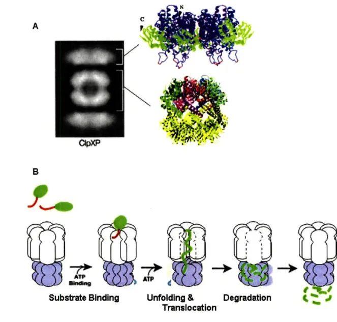
ClpXP is a highly conserved ATP-dependent protease that has been well-characterized in E.coli. As shown in Fig 1.3a, ClpXP is composed of ring-shaped subunits stacked on top of one another: the hexameric ATPase ClpX, a member of the AAA+ (ATPases associated with
variety of activies) family; and the tetradecameric serine peptidase CIpP (Grimaud et al., 1998; Maurizi et al., 1994). ClpP is itself a barrel-shaped double ring of heptamers stacked back to
back (Wang et al., 1997). In the absence of ClpP, ClpX functions as a molecular chaperone.
When partnered with ClpP, ClpX acts as a selective gatekeeper for regulated proteolysis (Fig
1.3b). ClpX recognizes and binds particular classes of substrates (Flynn et al., 2003). Through iterative cycles of ATP-powered pulling, it unfolds these substrates and translocates them into
the central cavity of ClpP (Kim et al., 2000; Ortega et al., 2000). This internal cavity is lined
with peptidase active sites all facing inward, constituting a degradation chamber that is
sequestered from the cytoplasm (Wang et al., 1997). Entry is by invitation only; protein
A
7~
B ATP shwweAT
Substrate Binding Unfolding & Degradation
4
J TranslocationFigure 13. a) Structure of CIpXP: Electron micrograph showing the barrel-shaped ClpP sandwiched between two hexameric rings of CIpX (From Ortega et al, 2000). Crystal structures of the hexameric HekiobacterpykoriClpX ATPase domain and E. coi CIpP tetradecamer from Kim, D. Y. et al. J. Biol. Chem. 2003 and Porankiewicz et al, 1999. respectively. In the latter structure, color is used to highlight each subunit of the top heptamer of CIpP. b) Cartoon showing the steps of ATP-powered proteolysis
by CIpXP.
p
r r II / I \-41
i - .- -isubunit associated with ClpP and fed processively through ClpP's narrow entry portal (Wang et
al., 1997). ClpP makes short work of polypeptide visitors, releasing them back into the
cytoplasm as a series of small peptides (Thompson and Maurizi, 1994).
Recently, attention has been directed toward the role of the zinc-binding N-terminal
domains of ClpX in the function of the ClpXP protease. There are six N-domains per ClpX
hexamer (Donaldson et al., 2003). Each N-domain is attached to the ATPase core by a flexible
linker region that is sensitive to protease digestion (Singh et al., 2001). The isolated N-domains
have been shown to form dimers in solution (Donaldson et al., 2003; Wojtyra et al., 2003). The
position of N-domain dimers relative to the ClpX hexameric core is still under debate (Fig 1.4).
One model places the dimers at the periphery, where they can interact with substrates or adaptor
proteins, but not with each other. Another model -one which seems to hold true for the
N-domains of the related AAA+ ATPase ClpA- proposes that a ring formed from a trimer of
dimers sits at the apical surface of ClpX (Donaldson et al., 2003; Ishikawa et al., 2004). As the
ClpX N-domain has a flexible connector sequence, it is also possible that the N-domain dimers
are mobile, spending some time at the periphery and some time as a ring atop ClpX.
The exact functions of the ClpX N-domain during substrate proteolysis also remain to be
elucidated. We know that the N-domain is not required for mediating ClpX-ClpP interaction, as
ClpX subunits lacking their N-domains still hexamerize and associate with ClpP. However, ClpXANP complexes exhibit a requirement for ATPase activity and decreas ed proteolysis
function, suggesting that the N-domain of ClpX may be involved in gating access of substrates
to the ClpX processing pore as well as in substrate selection and/or processing (Singh et al., 2001). Whereas the N-domain is dispensable for proteolysis of certain substrates, like
ssrA-tagged proteins, we have evidence that it plays a positive role in selection and/or degradation of
most other ClpXP substrates (Siddiqui, PhD thesis, MIT, 2004). For interactions between
ClpXP and its adaptor proteins, the N-domain is required (Bolon et al., 2004; Dougan et al., 2003; Neher et al., 2003b; Wojtyra et al., 2003).
B3a
A
C V;
Figure 1.4. a) Ribbon representation of the N-terminal homodimer of CIpX, with helices in blue, strands in red and Zn(ll) atoms shown as yellow spheres. The cysteines involved in chelating the Zn(1l) are drawn as yellow sticks. (From Donaldson et al, 2003). b) Placement of the N-domain homodimer at the periphery of the H., pylori ClpX ATPase domain, viewed from above c) Cartoon model of the N-domains at the periphery of CIpX. d) Trimer of dimers model, viewed from above from the side on top of the H pylori CIpX ATPase domain. e) Cartoon showing the trimer of dimers stacked above the CipX pore.
~ a
·d~-Z
Regulation ofsubstrate selectivity
ClpX selects proteins to be degraded by ClpXP by recognizing short peptide sequences
contained in potential substrates. These sequences are interchangeably called ClpX 'degradation
tags' or 'recognition motifs'. Experiments using a proteolytically inactive variant of ClpP to trap ClpXP substrates revealed at least three classes of N-terminal motifs and two classes of
C-terminal motifs. When representative sequences from each class were fused to model proteins, the fusion proteins were efficiently recognized and degraded by wild-type ClpXP (Flynn et al.,
2003). Like the fusion proteins, certain ClpXP substrates contain degradation tags that are
constitutively accessible. These substrates are extremely unstable. Not all proteins are targeted
for degradation immediately upon synthesis, however. Additional layers of regulation modulate
substrate choice by ClpXP (Fig 1.5). Below we discuss one class of proteins with a constitutively recognized degradation tag. We then explore the various mechanisms that
modulate the recognition and degradation of other substrates.
ssrA-tagged proteins constitute a class of ClpXP substrates that contain a constitutively
accessible degradation tag (Fig 5a). As described below, accessibility of the tag in these
proteins serves an important function in protein quality control. The ssrA tag is an 11
amino-acid sequence (AANEDENYALAA) that is appended to nascent polypeptide chains whose
translation machinery has stalled (Keiler et al., 1996). Ribosomes may stall during translation
for a number of reasons; perhaps the most common reason is the absence of a stop codon, due to
damaged mRNA, ribonuclease activity or premature termination of transcription. A stalled
ribosome is unable to release either its polypeptide chain or any sequestered translation factors.
The impotent complex is rescued through a process called trans-translation (Dulebohn et al., 2007). Briefly, an aminoacelyated tmRNA, which has properties of both a tRNA and an mRNA, is inserted into the empty A site of the ribosome. A switch in template occurs from the original
mRNA to the tmRNA encoding the ssrA-tag. As translation proceeds from the new template, the ssrA sequence is added to the polypeptide being synthesized. The ssrA sequence contains a
C-terminal tri-peptide sequence (LAA) that is readily recognized by ClpXP as a C-terminal class
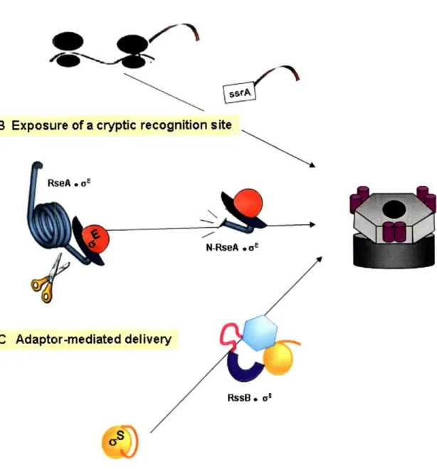
A Direct recognition of a degradation sequence
B Exposure of a cryptic recognition site
N-RseA * aE
Q~tv
C Adaptor-mediated
delih
RssB * ac
Figure 1.5. Regulation of substrate choice by CIpXP. a) Rescue of stalled ribosomes by
trans-translation results in ssrA-tagging of the nascent polypeptide. A tripeptide motif contained in the ssrA tag is directly recognized by ClpX and is sufficient to commit ssrA-tagged proteins to proteolysis. b) Intra-membrane processing following extracellular stress results in the cleavage of RseA and exposure of a cryptic CIpX-recognition sequence. Degradation of N-RseA liberates 0E. c) Proteolysis of os by ClpXP
requires the help of an accessory factor, RssB. Binding of RssB to as allows presentation of its buried CIpX recognition tag; in additon, RssB makes its own contacts with CIpX, enhancing the efficiency of ClpXP-substrate interaction.
machinery, the ssrA-tagged protein is efficiently recognized and degraded by ClpXP. The
ssrA-tagging process ensures that potentially harmful translation products, for example truncated
enzymes lacking their regulatory domains, do not accumulate in the cell. The spatial positioning
of ClpXP complexes close to translation complexes (P. Chien, personal communication), as well
as the use of an adaptor protein to further enhance the degradation of ssrA-tagged proteins
(discussed below) highlights the importance of constitutive proteolysis of these substrates by
ClpXP.
Many proteins, including regulatory proteins, become ClpXP substrates only under
certain conditions. These proteins have evolved mechanisms which render their degradation
motifs inaccessible to ClpXP under all other conditions. One such mechanism is the burial of a
cryptic recognition sequence in an internal region of the potential substrate (Fig 1.5b). In E.
coli, both the LexA repressor protein and the anti-c factor RseA require proteolytic processing
to reveal cryptic ClpX recognition tags at a newly created C-terminus (Flynn et al., 2004; Neher
et al., 2003a).
Another mechanism is the use of adaptor proteins to deliver substrates to ClpXP (Fig
1.5c; described in more detail below). For example, the N-terminal degradation tag of ys is too
weak on its own to commit the protein for degradation. Upon binding of its adaptor protein, RssB, Us becomes a substrate for ClpXP. Similarly, the activated SOS repair protein UmuD' is
not degraded by ClpXP in the absence of its delivery cofactor UmuD (Gonzalez et al., 2000;
Neher et al., 2003b). In each case, the adaptor protein makes its own contacts with ClpX,
thereby tethering its binding partner, the potential substrate, to ClpXP (Neher et al., 2003b; Zhou
et al., 2001). Adaptor-mediated increase in the effective concentration of a substrate around the
processing pore of ClpX facilitates the degradation of substrates with otherwise ineffective
degradation signals (McGinness et al., 2006). Regulation of the level and/or activity of the
adaptor protein results in condition-dependent proteolysis of ClpXP substrates.
Adaptor proteins are not only used by ClpXP to broaden substrate specificity. In fact,
most of what is known about the mechanism of adaptor-mediated substrate delivery to ClpXP is
enhance the degradation of ssrA-tagged proteins as well as the N-terminal cleavage product of RseA, N-RseA (Flynn et al., 2004; Levchenko et al., 2000). Both of these substrates are efficiently turned over even in the absence of an adaptor. This suggests that, under certain conditions, even the degradation of constitutive ClpXP substrates must be regulated. Use of the adaptor protein SspB to enhance degradation of ssrA-proteins seems to serve several purposes: 1) to direct flux of ssrA-tagged proteins away from the ClpAP protease to ClpXP by obscuring the ClpA recognition motif also contained within the ssrA-tag (Flynn et al., 2001) 2) to enhance the turnover rate of ssrA-tagged cellular garbage, allowing other substrates greater access to ClpXP (Levchenko et al., 2000); and 3) to make ssrA-tagged substrates 'weaker' with regards to ClpX interaction by creating steric hindrance for ClpX (Hersch et al., 2004); thus substrates that are able to compete with SspB for binding to ClpX may be able to gain priority over SspB-ssrA-protein complexes. Under certain conditions, competition by specific substrates for priority of degradation over cellular junk may be important for appropriate stress response and cell survival.
as - A Secondary Vegetative a Factor for Non-Optimal Conditions
oS/RpoS/ 38 is the master regulator of the general stress response in E.coli. It is structurally similar to the primary a factor, a70, shown in Fig 1.1. However, its expression and activity are regulated by a complex network of proteins and small RNAs. The result is a low level of as during exponential growth, and rapid accumulation in response to an array of environmental insults, including heat or cold shock, pH downshift, hyperosmolarity, or nutrient depletion/starvation (reviewed in Hengge-Aronis, 2002). This is in stark contrast to the ECF a
factors, which in general are induced by and counteract a single stress condition. Induction of as provides a kind of preventative medicine for the cell: activation of as-dependent genes results
besides the stress already being experienced. It is not surprising, then, that as is directly or indirectly involved in regulating up to 10% of the E. coli genome (Weber et al, 2005).
The as regulon - a misnomer?
Out of the approximately 500 genes found by microarray analysis to be as dependent, only about 25% behave like genes in a typical regulon. This core set of genes seems to be upregulated whenever as levels, and therefore as-containing RNA polymerase levels, rise in the cell (Weber et al, 2005). In contrast, the majority of as-dependent genes seem to require
additional activation or de-repression factors for expression. Many as-dependent genes are
co-regulated by secondary factors like CRP (cAMP receptor protein) and Lrp (leucine-responsive
protein) (Colland et al., 2000; Marschall et al., 1998; Weber et al., 2005). For some genes, as
dependence is even conditional. One example is a cluster of acid response genes which are
strongly as-dependent during entry into stationary phase, but become much less dependent on,
or independent of, as under conditions of acid stress (Weber et al, 2005). Such examples of secondary regulation of as-dependent gene expression suggest the existence of various as
-dependent stress response modules, some of which can be even temporarily recruited by other
stress-specific regulators under a70 control. Thus, as has been recently suggested to control not
one general stress regulon, but a dynamic and flexible network of stress-response modules that
are activated in various combinations, or may even be activated separately. as can be
considered to be a second vegetative a factor, one that allows the cell to grow under non-optimal
environmental conditions (Weber, 2005).
Regulating the regulator
As befits a master transcriptional regulator, as ' own expression is regulated by a signal
transduction network with a bewildering array of cross-signals, feedback loops, redundancies
and internal regulation. While this demonstrates a remarkable capacity for signal integration, it
also makes elucidation of the individual regulatory pathways much more difficult. Signals
mRNA; as activity (through competition for RNA core polymerase); or turnover (Fig 1.6; reviewed in Hengge-Aronis, 2002). We confine ourselves in this work to a discussion of os proteolysis by ClpXP, which has been argued to be the single most important modulator of as levels. In addition, regulation of as proteolysis provides the most rapid means of altering as levels in response to sudden environmental changes. It must be kept in mind that high as levels do not necessarily result in induction of the as-dependent stress response (Fredriksson et al., 2007). Elevated levels of the alarmone ppGpp (an indicator of non-optimal conditions) are required for successful competition of as with a70 for RNA polymerase (Jishage et al., 2002). Therefore ppGpp ultimately controls induction of as-dependent gene expression. Interestingly, since as is protected from association with RssB during interaction with RNA core polymerase (Zhou et al., 2001), an indirect effect of ppGpp is to stabilize as against proteolysis.
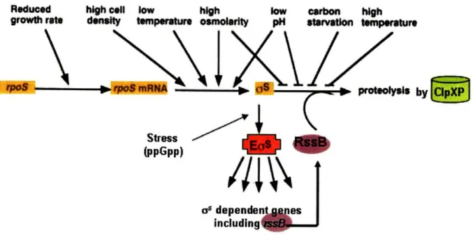
Reduced
growth ra high cll density temperatur low high osmwolarity low pH starvation temperaturearbon high
- I s proo s
2=7*
Stress (ppGpp) Sdepende as including, IFigure 1.6. Induction of RpoS expression through the influence of various environmental signals on the different levels of RpoS regulation. Stimulation of RpoS synthesis via rpoS transcription or mRNA
translation, or inhibition of RpoS proteolysis all lead to increased levels of RpoS and potential induction of its downstream genes. (Figure adapted from Hengge-Aronis, 2002 and Jenal & Hengge-Aronis, 2003)
lm Il
3Y
Determinants of as proteolysis
During exponential growth phase, the ClpXP protease is required to maintain a low cellular level of as (Schweder et al., 1996; Zhou et al., 2001). as carries an N-terminal Class 1 ClpX recognition tag (Flynn et al., 2003). Comparison of wild-type as degradation with the degradation of various as-lacZ fusion constructs, however, suggests that this recognition tag is
inaccessible to ClpX in the full-length free as. Additional evidence from the lacZ fusion constructs indicates that interaction of ClpX with this N-terminal tag is not sufficient to commit as for degradation (Studemann et al., 2003). We have found that RssB-independent degradation of as by ClpXP, although very slow, is even less efficient with ClpX that is lacking its N-terminal domain (Siddiqui, PhD thesis, MIT, 2004). Taken together, these results suggest that a secondary sequence in as, possibly within its C-terminal region, interacts with the N-domain of ClpX either to promote ClpXP activity or to stabilize the interaction of as with ClpXP during processive degradation.
The adaptor protein RssB is required for efficient proteolysis of as (Muffler et al., 1996a; Pratt and Silhavy, 1996). RssB binds to 'region 2.5' of as (for a view of the comparable region in o70, see in Fig 1. 1b), the same surface-exposed alpha helix that interacts, as part of the
RNA polymerase holoenzyme, with as-dependent promoters (Becker and Hengge-Aronis, 2001;
Becker et al., 1999). Within 'region 2.5', RssB makes critical interactions with Lys173 of as;
mutation of this residue to a glutamate, which is the corresponding residue in the vegetative a
factor a70, abrogates binding of RssB and prevents proteolysis of as (Becker et al., 1999). Binding of RssB has been suggested to alter the conformation of as, thus exposing the buried
N-terminal ClpX recognition tag and possibly a secondary ClpX interaction sequence (Studemann
et al., 2003). However, variants of as carrying an exposed N-terminus still require RssB for
ClpXP-mediated degradation; this suggests that RssB plays at least one other critical role in
facilitating as degradation. RssB has been shown to make its own contacts with ClpX (Moreno
to ClpXP, thereby enhancing the rate of aS recognition and processing. Whether RssB plays yet
another role in facilitating degradation -promoting ClpXP activity and/or assisting in the
transition from substrate recognition to substrate processing-remains to be seen.
RssB - Proteolytic Regulator of c'
The function of RssB, also called SprE (stationary phase regulator), was uncovered in
E.coli at the same time by two research groups. Both showed that RssB negatively regulates the
cellular level of as in vivo (Muffler et al., 1996a; Pratt and Silhavy, 1996). Indeed in growing
cells, the half-life of as is greater than 60 minutes in an rssB mutant, as compared to 1-4 minutes
in a wild-type strain (Lange and Hengge-Aronis, 1994; Muffler et al., 1996a; Takayanagi et al.,
1994). Use of appropriate rpoS-lacZ fusion constructs by both investigators revealed that the effect of RssB on as is post-transcriptional. Pulse-labeling experiments showed that the effect is also post-translational, i.e. not due to regulation of protein synthesis (Muffler et al., 1996b). Using complementary techniques, each group showed that RssB acts specifically to promote as turnover. The protease ClpXP was already known to be involved in proteolysis of as (Schweder et al., 1996). Use of genetic mutants in clpX, clpP and rssB placed RssB and ClpXP in the same pathway (Pratt and Silhavy, 1996). Several possible models for RssB function were posed by each group; one that has been borne out is a role for RssB in presenting as to ClpXP for degradation.
We know that proteolysis of a
sis inhibited by various environmental stresses, including
carbon starvation (Lange and Hengge-Aronis, 1994; Takayanagi et al., 1994), heat shock(Muffler et al., 1997), osmotic upshift (Muffler et al., 1996b) and pH downshift (Bearson et al., 1996). The placement of the negative regulator RssB within the degradation pathway makes it an attractive and likely point for signal integration. That RssB is also a two-component response regulator adds further weight to the supposition that input from one or more environmental
mechanism/s by which high level environmental signals influence the activity of RssB remain mysterious.
In the following discussion, we first place RssB within its structural family of two-component response regulators. We then discuss possible mechanisms by which the activity of
RssB, and therefore of rs, may be regulated.
RssB - an atypical two-component response regulator
Response regulators are modular proteins that, in conjunction with their cognate two-component histidine sensor kinases, function to initiate major changes in the bacterial proteome.
Sensor kinases are also modular proteins; canonically, they function to sense the extracellular environment and transduce signals back into the cytoplasm (for a review, see Stock et al., 2000). Figure 1.7 depicts the steps of communication between three representative sensor kinases and
their cognate response regulators. First, detection of signals occurs via a variable input domain.
Depending on the particular signal, activity of the kinase domain is either activated or inhibited.
If activated, the kinase domain hydrolyzes ATP to auto-phosporylate a histidine residue within a
conserved structural motif (the "H box"). This phosphate is transferred, sometimes through a
relay of several His-Asp phosphotransfer events, to a conserved aspartate residue in the receiver
domain of a response regulator. Phosphorylation activates the response regulator and its downstream cellular responses.
As far as response regulators go, RssB is considered a maverick in both structure and
function. Its N-terminal domain is a typical response regulator receiver domain, having extensive homology with the prototypical response regulator CheY, and sporting a conserved
aspartate (D58) that accepts regulatory phophotransfer (Bouche et al., 1998; Chapter Three).
However, RssB boasts an unusual C-terminal module. Across bacteria and archaea, 66% of response regulators bind DNA with their output domains; thus they can function as
transcriptional regulators that respond quickly and specifically to sensed changes in the
environment. Out of the remaining, 14% are like CheY and lack an output domain altogether;
Response Regulator a) E. coff Osmoregulation
b) E. co# Anoxic Redox Control
I-EnvZ ArcB 1" 0* " ATP U
c) E. co# Chemotaxis CheA CheB and CheY
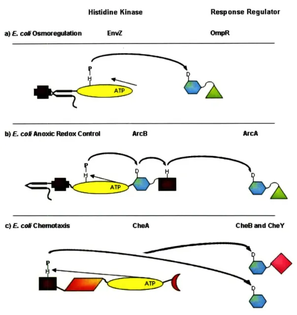
Figure 1.7. Depiction of the modular organization of the histidine kinases and their cognate response
regulators using three representative examples, a) Class I enzymes (a and b) like the prototypical EnvZ have variable input domains (
*)
which sense a variety of signals; at least putative transmembrane regions ( ); and a conserved transmitter/kinase core-domain ( 0 ). b) Hybrid kinaseslike ArcA additionally contain a phosphorylatable receiver module (
0)
and in some cases, as in the one shown, a phosphotransfer unit ( M ). c) Class II histidine kinases like CheA do not have their own trans-membrane domains; they are coupled to membrane receptors. Some sensor kinases additionally contain response regulator-binding domains ( A ) and/or regulatory domains ( ( ). Response regulators contain a conserved phosphorylatable receiver domain ( * ) that regulates the activity of a carboxy-terminal output module when present. The majority of output modules areDNA-binding domains ( A ); however some have enyzymatic activity (
*
) or regulatory activity (not depicted). (Adapted from Stock et al, 2000 and Foussard et al., 1997.)OmpR ArcA
4
H
ATP C=7I, r., Histidine Kinase:ATP>O
I
&x
not bind DNA, nor has it been found to possess enzymatic function; it is instead a delivery factor, mediating proteolysis of &s by ClpXP (Klauck et al., 2001; Zhou et al., 2001). Without additional information it seems sensible to conjecture that the unique C-terminal output domain of RssB reflects, and mediates, its unique function. On the basis of an alignment of the C-terminal residues of SspB and RssB, a ClpX-binding consensus sequence was proposed (Dougan et al., 2003). The notion that RssB uses its very C-terminus in an SspB-like manner to mediate interactions with ClpX rapidly became accepted in the field. Until now, it has never been tested. In Chapter Two we show that, in fact, this assumption is incorrect. Although the C-terminal output domain of RssB is indeed unique among response regulators, it does not mediate RssB's delivery function to ClpXP.
Phosphorylation of RssB - location and effect
The output function of a typical response regulator is activated by autophosphoryation of a conserved aspartate residue in its receiver domain (Fig 1.7). Upon phosphorylation, the receiver domain undergoes an allosteric conformational change (Fig 1.8). Depending on the particular response regulator, this promotes release of enzymatic inhibition by the receiver domain; dimerization or higher order oligomerizaton; or binding of the output domain to a new
protein or DNA partner (Stock et al., 2000). In the case of the close structural homolog, CheY, the phosphorylation-induced conformational change enhances affinity for its ligand, FliM
(Welch et al., 1993; Welch et al., 1994).
We know that RssB is phosphorylated on its conserved aspartate, Asp58; evidence thus
far indicates that Asp58 is the only site of RssB phosphorylation (Bouche et al., 1998; Chapter
Three). Initially, phosphorylation of RssB was thought to be required for its activity (Zhou et
al., 2001). Indeed, Asp58 mutants of RssB are impaired in regulating as turnover in vivo (Cunning and Elliott, 1999; Moreno et al., 2000). However, this is not true under all conditions
(Peterson et al., 2004). Our own work with wild-type RssB in vitro indicates that
phosphorylation is dispensable under certain conditions, for example when concentrations of all
B
cz
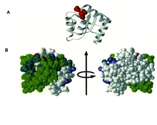
Figure 1.8. Phosphorylation of a response regulator receiver domain induces conformational changes
a) Ribbon diagram of CheY with the three conserved active-site aspartate residues shown in red. b) Space-filling models of the CheY structure above are used to highlight residues whose conformations are affected by phosphorylation. The two views are related by a 180 * rotation about the vertical axis. Residues that were shifted in the nuclear magnetic resonance (NMR) structures of the phosphorylated forms of NtrC, or CheY, versus the unphosphorylated forms are mapped in green, or blue, respectively, onto the corresponding surface of unphosphorylated CheY. Residues that are affected by
phosphorylation in both NtrC and CheY appear as cyan. (From Stock et al., 2000).
L
MMrl
wowlz
(Chapter Three). As for CheY, phosphorylation of RssB enhances its affinity for its substrate, as (Becker et al., 1999; Klauck et al., 2001; Chapter Two). We propose that phosphorylation becomes important as the concentrations of reaction components begin to compromise the
formation of the binary RssB- as complex and/or the formation of the RssB- CS.tClpXP degradative complex.
Our work with a D15K mutant of RssB is consistent with this model. RssBD1SK is analogous to CheYD13K, which is unphosphorylatable due to an inability to coordinate Mg2+ (Bourret et al., 1990; Welch et al., 1994; Chapter Three). A simple "two-step" model would require that a two-component response regulator is either phosphorylated and 'on/active', or unphosphorylated and 'off/inactive' (Fig 1.9a). Crystal structures show that RssBD15K and
CheYD13K
adopt the same conformation as unphosphorylated or 'inactive' RssB or CheY, respectively (Chapter Three; Jiang et al., 1997). Yet both are functionally active, albeit less than wild-type (Chapter Three; Bourret et al., 1993). These results confirm that neither CheYD13K
nor RssBD15K is locked into a particular conformation. In Chapter Three, we show evidence that
RssB uses a CheY-like multi-step reaction pathway to go from 'inactive' to 'active'
conformation (Fig 1.9b). In this model, both the 'active' and 'inactive' conformations can be populated by unphosphorylated forms; phosphorylation and ligand binding reduce the energy barrier of activation, thus shifting the equilibrium of RssB or CheY toward the active
conformational state. In this 'equilibrium shift' model, as opposed to a rigid two-state model, achievement of the active conformation does not strictly require phosphorylation, nor does phosphorylation fix the conformation of RssB.
Regulation ofRssB - An Unsolved Mystery
RssB can be phosphorylated by the small molecule acetyl phosphate; this has greatly faciliated the study of RssB in vitro. However, acetyl phosphate is not required for proteolysis of as in vivo, suggesting that other mechanism/s must be involved (Bouche et al., 1998; Cunning and Elliott, 1999). As RssB belongs to the two-component response regulator family, its
Inactive Active
B
Inactive
Intermediate
Figure 1.9. Two models of RssB activation: a) In a rigid two-state model, phosphorylation is necessary and
sufficient to induce the active conformation. b) In our equilibrium shift model, based on extensive studies with the RssB homology CheY, unphosphorylated RssB samples both inactive and active conformations.
Phosphorylation and ligand binding both reduce the energy barrier between the inactive and active conformational, serving to push RssB toward the active state.
physiological phosphodonor is predicted to be a cognate sensor kinase. However, multiple genetic screens failed to uncover such a cognate sensor-kinase for RssB, leading to the
assumption that several sensor-kinases phosphorylate RssB in a conditionally redundant manner. Thus, the inactivation of only one would not result in a dramatic phenotype. Given the number of high level signals that influence proteolysis of as, it is reasonable that several sensor-kinases converge on this important regulatory protein (Fig 1.6).
RssB is no longer an 'orphan'; directed candidate mutagenesis revealed a role for the ArcB sensor-kinase in regulating RssB activity (Mika and Hengge, 2005). Further experiments demonstrated that ArcB is a phosphodonor, but not a phosphatase, for RssB; this is in contrast to ArcB function with respect to its known response regulator partner, ArcA. Interestingly, os
regulates ArcA under at least two stress conditions: entry into stationary phase and osmotic upshift (Weber et al., 2005). ArcA can also act as a transcriptional repressor of as (Sevcik et al.,
2001). Competition between ArcA and RssB for phosphorylation by ArcB unexpectedly coordinates transcriptional control and proteolytic control of as (Mika and Hengge, 2005). The sensitivity of the sensor kinase ArcB to energy starvation suggests a model (Fig 1.10) in which starvation-induced dephosphorylation of ArcB results in residual ArcA, but not RssB,
phosphorylation; this leads to both de-repressed os transcription as well as as stabilization.
Besides activating the starvation response program, increased levels of as also result in increased expression of arcA and thus synthesis of more ArcA to compete for phosphorylation with RssB (Mika and Hengge, 2005).
The implications of the ArcB-RssB pairing offers a window into the potential complexity and interconnectivity of as regulation through RssB. The discovery of other
proteins which affect RssB phosphorylation, including possible phosphatases, will allow an even more complete understanding of how RssB activity, and therefore os activity, is modulated
under various environmental conditions. Almost certainly, other environmental signals will be found to converge upon RssB through sensor-kinase dependent phosphorylation, phosphatase dependent dephosphorylation, or inhibition of phosphorylation. High level signals likely use
additional mechanisms to regulate RssB activity as well. RssB levels in the cell are rate-limiting for as proteolysis (Pruteanu and Hengge-Aronis, 2002). Thus, any significant increases in os synthesis, as during osmotic shock, can titrate RssB and lead to stabilization of os. Since RssB
is homeostatically controlled by os levels, the range of titration control is carefully limited
ppGpp, that increase or decrease the success of as in partnering with RNA core polymerase
(Jishage et al., 2002). As part of a holoenzyme, as-RssB formation is inhibited. Finally, the
discovery of the first anti-anti-a factor for RssB suggests that more inhibitors of RssB- as
association are likely to be discovered (Bougdour et al., 2006).
lyoyitic shunt enzymes
TcA cycle ezyyme,,
- S
proteolysis
UI
RssiP
ArcrB
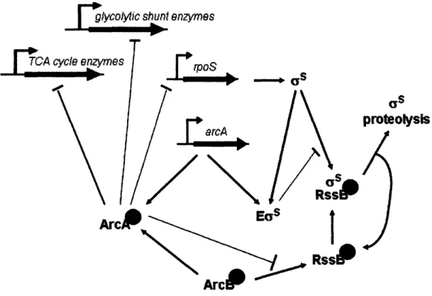
Figure 1.10. Three-component system involving the response regulators RssB and ArcA, and their cognate sensor kinase, ArcB. Competition between ArcA and RssB for phosphorylation by ArcB coordinates the transcriptional and proteolytic control of as (Mika and Hengge, 2005). Under certain stress conditions, ArcA acts as a transcriptional repressor of as (Sevcik et al., 2001). In contrast, as increases transcription of arcA.
Starvation-induced dephosphorylation of ArcB results in residual ArcA, but not RssB, phosphorylation; this leads to both de-repressed as transcription as well as as stabilization. Besides activating the starvation response program, increased levels of as also result in increased expression of arcA, and thus synthesis of more ArcA to compete for phosphorylation with RssB (Mika and Hengge, 2005).
A
GENERAL MODEL OF ADAPTOR FUNCTIONAs mentioned above, there are three known adaptors for ClpXP: SspB, which delivers ssrA-tagged proteins and N-RseA (Flynn et al., 2004; Levchenko et al., 2000); UmuD, which delivers a proteolytically processed form of itself, UmuD' (Gonzalez et al., 2000); and RssB, which delivers the master response regulator as (Zhou et al., 2001). RssB was the first of the three adaptor protein to be discovered (Bearson et al., 1996; Muffler et al., 1996a; Pratt and Silhavy, 1996). Yet more than a decade later, many details about its activity remain a mystery. We know a great deal more about SspB; biochemical and bioengineering experiments have illuminated its mode of interaction with both substrates and protease, independently and as part of a delivery complex. The mechanism of SspB-mediated substrate delivery to ClpXP has thus become the model for understanding adaptor function in general. In this section, we describe what we know about how SspB delivers its cargo to ClpXP. After using insights from studies of UmuD to extract common features, we then present the currently accepted model of adaptor
function. Finally, we discuss how the mechanism of RssB-mediated substrate delivery (see Chapter Two) fits the current SspB-based model in some ways while expanding it in others.
The SspB protein exists as a dimer (Wah et al., 2002). Each monomer is composed of three distinct regions: an N-terminal substrate-binding domain, a short C-terminal peptide that mediates interactions with ClpX; and a flexible linker region that joins the two (Levchenko et al., 2003; Wah et al., 2003). SspB is known to delivery at least two substrates: ssrA-tagged proteins and N-RseA (Flynn et al., 2004; Levchenko et al., 2000). Both are recognized by short peptide sequences, which fit neatly, albeit in different orientations, into SspB's substrate binding groove (Flynn et al., 2001; Levchenko et al., 2005; Levchenko et al., 2003). Bivalent
interactions of SspB's C-termini with the N-domain of ClpX tightly tether the SspB-substrate complex to ClpX (Fig 1.1 la; Bolon et al, 2004). The result is an increase in the effective concentration of substrate around ClpXP. This in turn increases the apparent affinity of ClpXP for the substrate. Following direct hand-off of its cargo to ClpXP, SspB disengages from the complex. (Bolon, 2004). It is now free for more rounds of substrate binding and delivery.
B C
/
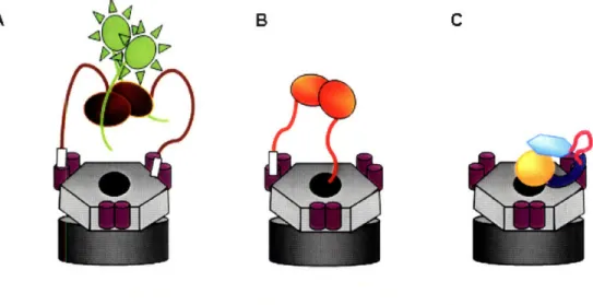
U R~ N i I I B iFigure 1.11. Adaptor-mediated substrate delivery to CIpXP. By making contacts with the N-terminal domains of CIpX, adaptors tether their cargo to CIpXP. a) The SspB dimer is shown carrying GFP-ssrA cargo. With its C-terminal tails, SspB makes bivalent contacts with the N-terminal domains of ClpX. b) UmuD delivers a processed version of itself UmuD'. With it's N-terminal extension, UmuD makes a monovalent contact with an
N-domain homodimer. c) RssB facilitates proteolysis of the master stress regulator ss. As both its N- and C-termini are involved in substrate binding, RssB contacts an N-domain dimer with its inter-domain linker region.
Studies with the adaptor protein UmuD indicated that only certain aspects of SspB function may be generalizable. Unlike SspB, UmuD functions as a monomer, forming a simple heterodimeric complex with the substrate to be delivered, UmuD' (Gonzalez et al, 2000). UmuD also does not clasp its substrate UmuD' in a substrate binding groove. As inferred from
structures of the UmuD and UmuD' homodimers, the UmuD-UmuD' complex is stabilized by association of the two C-termini, including the formation of an intermolecular anti-parallel structure from their individual f3-strands (Ferentz et al., 1997). Despite such close interaction between substrate and adaptor, UmuD is not degraded along with UmuD' (Gonzalez et al., 2000). UmuD interacts with the N-domain of ClpX via a ClpX-binding motif similar to that of
SspB (Neher et al., 2003b; Wah et al., 2003). This 'XB' peptide is located close to the N-terminus of UmuD (Gonzalez et al., 2000; Neher et al., 2003b). Despite the difference in location, the two XB motifs provide the same tethering function (Fig 1.1 lb). Replacement of
the ClpX-binding peptide of UmuD with that of SspB in fact increases the efficiency of UmuD' degradation by ClpXP. That UmuD function can also be competitively inhibited by the isolated ClpX-binding peptide of SspB suggests that the two adaptors bind to a common site on the N-domain of ClpX (Neher et al., 2003b).
Comparison of SspB and UmuD function facilitated the construction of a general model of adaptor-mediated delivery. According to the accepted model, adaptors carry ClpX-binding sequences at their flexible termini. They use these short peptides with similar amino acid sequences to tether their binding partners to the N-domain of ClpX. The combination of several weaker interactions, i.e. adaptor-ClpX, adaptor-substrate, and substrate-ClpX, promotes the formation of an efficient delivery complex. Thus ClpXP substrates are more efficiently turned over in the presence of their adaptor. Following substrate handoff to ClpXP, the adaptor protein is released back into the cytoplasm.
RssB - the maverick adaptor protein
Several features of the general model of adaptor function are conserved in the case of RssB-as (Fig 1.11 c). RssB uses a similar ClpX-binding sequence as SspB and UmuD to dock onto the N-domain of ClpX (Chapter Two). (Interestingly, this ClpX-binding sequence is more
similar to that of UmuD than SspB.) As in the case of SspB and UmuD, RssB facilitates degradation of its cargo by ClpXP by physically tethering it close to the ClpX processing pore.
The resulting increase in effective concentration promotes degradation of rs. Both SspB and UmuD combine fairly weak tethering interactions with other weak substrate-protease
interactions to create a robust delivery complex. Consistent with this model, RssB-ClpXP binary complexes have proved difficult to isolate, probably due to the weakness of the
interaction. However, the interaction between RssB and the N-terminal domain of ClpX, in combination with the interaction of the intrinsic degradation tag of as with the ClpX processing pore, stabilizes the quaternary RssB-as-ClpX-ClpP degradative complex enough to allow its
isolation by size chromatography (Zhou et al., 2001). As with SspB and UmuD, RssB is also
released from the complex as as is degraded (Klauck et al., 2001; Zhou et al., 2001).
There are also several important differences between RssB and the two other ClpXP
adaptor proteins. Firstly the regulation of RssB activity is more complex than for SspB or
UmuD. The affinity of RssB for its substrate is modulated by a post-translational modification. The modification itself is theoretically dependent on environmental conditions. Secondly, in contrast to SspB and UmuD, RssB is thought to use both its N- and C-termini to bind its
substrate. Thus, formation of the RssB-as complex may induce a conformational change in both partners (Studemann et al., 2003). We now also realize that assumptions based on SspB and UmuD regarding the location and specific nature of ClpX-binding motifs in ClpXP adaptor
proteins were overly simplistic. Unlike the other two adaptors, RssB does not use an N- or C-terminus to mediate interactions with ClpXP during substrate delivery. Instead it uses a ClpX-binding peptide (RssBxB) uniquely positioned within an inter-domain linker (Fig 1.11 c). Although RssBxB seems to interact with a common adaptor binding site on the surface of the ClpX N-domain, mutational analysis reveals that the ClpX-binding peptides of RssB and SspB make very different molecular contacts with this site (Chapter Two).
Implications of the Model - Competition for ClpXP
In times of stress, or in changing conditions, competition for protein partners is a common phenomenon within a cell. A well-known example is the titration of DnaK by
unfolded proteins during heat stress. This results in the reduced ability of DnaK to sequester oH and/or deliver it to FtsH for degradation (Muffler et al., 1997). As a consequence, oH levels rise, allowing effective competition for RNA core polymerase and induction of a heat shock response program. The fundamental mechanism of stress responsiveness in bacteria, i.e. formation of a condition-specific RNA polymerase holoenzyme by exchange of one a factor for another, is in fact predicated on competition for a limiting reagent (core polymerase) by competing binding
partners (alternate a factors). Thus, we see that intracellular competition for reagents is a
general mechanism by which cells sense a need for change and initiate new responses.
Competition requires that the limiting reagent is titrated by specific interactions with one factor versus another. There is no evidence that levels of ClpXP change dramatically under stress conditions. On the other hand, stress conditions can dramatically increase the pool of ClpXP substrates. Thus competition among substrates and substrate-complexes for ClpXP could function as an important means of regulating substrate priority. We and others have observed competition between substrates for ClpXP in vitro (Chien et al., 2007; Wojtyra et al., 2003). A recent paper provides evidence of competition for ClpXP in vivo. Fredriksson et al demonstrate that a reduction of translational fidelity upon carbon starvation results in an increase in aberrant and oxidatively damaged proteins. Occupation with these damaged substrates, and possibly with SspB-ssrA-protein complexes, titrates ClpXP away from RssBacs. Predictably,
stabilization of aS results in the induction of as-dependent gene expression, thus theoretically providing a response to the initiating oxidative stress as well as other potential stresses (Fredriksson et al., 2007).
The use of adaptor proteins by ClpXP adds complexity to the problem of substrate priorization. Conserved 'tether-and-deliver' mechanisms, which rely on conserved ClpX-binding motifs that bind a common adaptor ClpX-binding site, suggest that ClpXP adaptor proteins
have in fact evolved to facilitate competition amongst themselves. As suggested by the results above and our own work (Chapter Two), competition among adaptor proteins likely constitutes an additional means of regulating ClpXP activity, especially during the onset of stress. The relative abundance of adaptor proteins and protease components within the cell makes this
notion feasible (Farrell et al., 2005). The potential outcomes of competition among SspB, UmuD and RssB also make biological sense: Following extracytoplasmic stress, the newly created ClpXP substrate N-RseA needs priority over constitutive cellular substrates that may also be increased under the same conditions. SspB-mediated delivery of N-RseA to ClpXP