Publisher’s version / Version de l'éditeur:
Nanotechnology, 20, 6, pp. 065304-1-065304-6, 2009-02-01
READ THESE TERMS AND CONDITIONS CAREFULLY BEFORE USING THIS WEBSITE. https://nrc-publications.canada.ca/eng/copyright
Vous avez des questions? Nous pouvons vous aider. Pour communiquer directement avec un auteur, consultez la première page de la revue dans laquelle son article a été publié afin de trouver ses coordonnées. Si vous n’arrivez pas à les repérer, communiquez avec nous à PublicationsArchive-ArchivesPublications@nrc-cnrc.gc.ca.
Questions? Contact the NRC Publications Archive team at
PublicationsArchive-ArchivesPublications@nrc-cnrc.gc.ca. If you wish to email the authors directly, please see the first page of the publication for their contact information.
NRC Publications Archive
Archives des publications du CNRC
This publication could be one of several versions: author’s original, accepted manuscript or the publisher’s version. / La version de cette publication peut être l’une des suivantes : la version prépublication de l’auteur, la version acceptée du manuscrit ou la version de l’éditeur.
For the publisher’s version, please access the DOI link below./ Pour consulter la version de l’éditeur, utilisez le lien DOI ci-dessous.
https://doi.org/10.1088/0957-4484/20/6/065304
Access and use of this website and the material on it are subject to the Terms and Conditions set forth at
Fabrication of flexible superhydrophobic films by lift-up
soft-lithography and decoration with Ag nanoparticles
Yao, Tongjie; Wang, Chuanxi; Lin, Quan; Li, Xiao; Chen, Xiaolu; Wu, Jie;
Zhang, Junhu; Yu, Kui; Yang, Bai
https://publications-cnrc.canada.ca/fra/droits
L’accès à ce site Web et l’utilisation de son contenu sont assujettis aux conditions présentées dans le site LISEZ CES CONDITIONS ATTENTIVEMENT AVANT D’UTILISER CE SITE WEB.
NRC Publications Record / Notice d'Archives des publications de CNRC:
https://nrc-publications.canada.ca/eng/view/object/?id=1bc08fc6-49da-44fe-9623-e0efb490f7f3 https://publications-cnrc.canada.ca/fra/voir/objet/?id=1bc08fc6-49da-44fe-9623-e0efb490f7f3Nanotechnology 20 (2009) 065304 (6pp) doi:10.1088/0957-4484/20/6/065304
Fabrication of flexible superhydrophobic
films by lift-up soft-lithography and
decoration with Ag nanoparticles
Tongjie Yao
1, Chuanxi Wang
1, Quan Lin
1, Xiao Li
1,
Xiaolu Chen
1, Jie Wu
1, Junhu Zhang
1, Kui Yu
2and Bai Yang
1,31State Key Laboratory of Supramolecular Structure and Materials, College of Chemistry,
Jilin University, Changchun 130012, People’s Republic of China
2Steacie Institute for Molecular Sciences, National Research Council Canada, Ottawa,
K1AOR6, Canada
E-mail:byangchem@jlu.edu.cn
Received 17 August 2008, in final form 7 December 2008 Published 14 January 2009
Online atstacks.iop.org/Nano/20/065304
Abstract
Superhydrophobic films with excellent flexibility have been fabricated by combining the lift-up soft-lithography technique and chemical reduction of [Ag(NH3)2]+ions to Ag nanoparticles
(NPs) on the surface of silica spheres which are patterned on the polydimethylsiloxane (PDMS) films. Scanning electron microscopy (SEM) images reveal the presence of raspberry-like hierarchical structures on the PDMS films. The influence of the amount of Ag NPs and the size of the silica spheres on the wettability of the soft films is investigated carefully. Because PDMS films are elastomeric materials, our superhydrophobic films offer great flexibility. The resulting films can be easily transferred from one substrate surface to another without destroying their superhydrophobicity. These flexible and superhydrophobic films can be used repeatedly to satisfy a wide range of applications.
S Supplementary data are available fromstacks.iop.org/Nano/20/065304
1. Introduction
The wettability of solid surfaces is an important property, studied and widely used, from fundamental material research to many practical applications [1, 2]. The slippery superhydrophobic surface refers to that on which the water contact angle (CA) is larger than 150◦ and the sliding
angle (SA) is less than 10◦ [3]. In recent years, such
superhydrophobic surfaces have been widely used in many fields, for instance, in the areas of surface self-cleaning [4], marine coating [5], preventing the adhesion of snow, fog and raindrops to window surfaces and reducing flow resistance in microfluidic channels [6,7]. The superhydrophobic surface originated from the famous lotus effect of some natural leaves, which was first demonstrated by Barthlott and Neinhuis [8]. Further study revealed that the wettability of a surface was governed mainly by its chemical composition and geometrical structure [9]. Accordingly, superhydrophobic surfaces have
3 Author to whom any correspondence should be addressed.
been constructed usually based on two essential concerns: (i) micro- and nanoscale hierarchical structures to increase the roughness of a surface [10] and (ii) chemical modification to reduce the surface energy of a rough surface [11]. Based on these two principles, various techniques have been established to engineer superhydrophobic surfaces, with notable examples including lithographic patterning [12], layer-by-layer techniques [13,14], electrochemical deposition [15], sol–gel approach [16], electrospinning and plasma etch-ing/deposition [17–21]. Through these approaches, lots of superhydrophobic materials have been fabricated. For example, Jiang and co-workers have fabricated a series of superhydrophobic structures inspired by the superhydrophobic living organisms in nature, such as the eyes of mosquitoes, the legs of water striders, and rambutans [22–24]. Seeger and co-workers have reported a general method of growing silicone nanofilaments onto various materials to obtain superhydrophobic surfaces [25]. The recent development on superhydrophobic surfaces has been reported in several reviews [3,26–28].
Nanotechnology 20 (2009) 065304 T Yao et al
Figure 1.Schematic illustration of the fabrication procedure of a flexible superhydrophobic film.
To date, the efforts on fabrication of superhydrophobic surfaces focus mainly on the formation of hierarchical structures on rigid substrate surfaces [29, 30]. For many applications, there is an outstanding requirement for superhydrophobic films with flexibility that can be folded and transferred among a number of surfaces with different curvatures [31,32].
Here, we report a straightforward procedure to obtain hierarchical structures on soft poly(dimethylsiloxane) (PDMS) films by lift-up soft-lithography and post-decoration with Ag nanoparticles (NPs) to render the superhydrophobic surfaces. During the past decade, soft-lithography has been well developed as a new micro-fabrication method to prepare various patterned surfaces by using elastomeric stamps, molds and masks. Compared with other approaches, soft-lithography is relatively inexpensive with simple procedures and does not demand special instruments [33]. Obviously, the reported approach is low-cost and easy to handle. The advantages of our soft superhydrophobic films are appreciable: (i) they can be reused, and hence minimize material waste; (ii) various smooth surfaces can become superhydrophobic via ready coating with these films, especially for those hydrophilic materials which can hardly exhibit superhydrophobicity.
2. Experimental details
2.1. MaterialsPDMS elastomer kits (Sylgard 184) were purchased from Dow Corning (Midland, MI). 1H, 1H, 2H, 2H-perfluorodecanethiol (PDT) was obtained from Aldrich. (heptadecafluoro-1,1,2,2-tetrahydrodecyl)triethoxysilane (HTTS) was obtained from ABCR GmbH&Co.KG. Silver nitrate (AgNO3), ammonium hydroxide (NH3·H2O, 25 wt% in water), tetraethyl
orthosili-cate (TEOS), isopropanol and glucose were analytical grade and used as received. In all preparations, absolute ethanol and deionized water were used.
2.2. Preparation of superhydrophobic film
Monodisperse silica spheres were synthesized according to the well-known St¨ober method [34]. Afterwards, the resulting silica spheres were centrifuged and redispersed in ethanol.
Figure 1 outlines our procedures for the preparation of a superhydrophobic film, which begins with the assembly of monodisperse silica spheres on the surface of a silicon wafer by the evaporation of a suspension of silica spheres in ethanol (figure 1(a)) [35]. With soft-lithography, a single layer of close-packed spheres is transferred to the surface of the PDMS film (figure1(b)) [36]. Next, Ag NPs are deposited onto the silica spheres (figure 1(c)) [37]. Generally, the PDMS film containing the transferred silica spheres was immersed in a 0.5 M [Ag(NH3)2]+ion solution for four hours and dried under
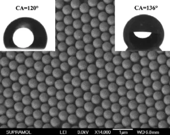
Figure 2.SEM image of a hexagonal close-packed silica array on the surface of a PDMS film; the diameter of the silica spheres is about 520 nm. The insets on the left and right top are the water droplets before and after the HTTS modification. The water CA before and after the surface modification is 120◦and 136◦, respectively.
the flow of nitrogen. The substrate was then immersed in a 0.5 M glucose solution for six hours at room temperature to reduce the [Ag(NH3)2]+ions to Ag NPs on the surface of the
silica spheres arranged in an ordered array. Finally, the surface of the as-prepared sample is chemically modified with 2 mM PDT solution in ethanol for one hour to reduce the surface energy (figure1(d)).
2.3. Characterization
The morphologies of the silica spheres on the surface of PDMS films and Ag NPs coated on the periodic silica spheres were investigated with a JEOL JEM-6700F field emission scanning electron microscope (SEM) with primary electron energy of 3 kV, and the samples were sputtered with a layer of Pt (ca. 2 nm thick) prior to imaging to improve conductivity. Water contact angle measurements were performed at room temperature using a drop-shape analysis system (DSA 10 MK2, KRUSS). A 2.0 µl droplet of deionized water was dropped onto the samples and the average of at least five measurements taken at different positions on each sample was adopted as static CAs. X-ray diffraction (XRD) data were collected on a Siemens D-5005 x-ray diffractometer with Cu Kα radiation (λ = 1.5418 ˚A).
3. Results and discussion
Figure2displays an SEM image of the ordered silica spheres which are pressed and sunk on the surface of the PDMS film. 2
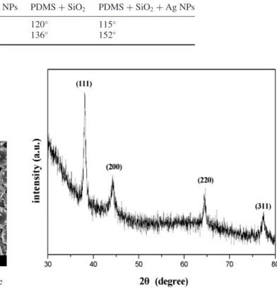
Table 1. Water CAs before and after hydrophobic modification of PDMS surfaces at different steps of the fabrication process. Fabrication step PDMS + Ag NPs PDMS + SiO2 PDMS + SiO2+Ag NPs
CA (before modification) 116◦ 120◦ 115◦
CA (after modification) 140◦ 136◦ 152◦
Figure 3.SEM image of silica spheres coated with Ag NPs whose diameters range from 30 to 40 nm; the silica spheres are pressed to sink partially into a PDMS film with a hexagonal array. The insets on the left and right top are the water droplets before and after the PDT modification. The water CA before and after the surface modification is 115◦and 152◦, respectively.
Table1lists the water CAs before and after modification by a grafting agent with low surface energy at different fabrication steps. The ordered silica spheres with a hexagonal close-packed array were transferred by lift-up soft-lithography onto the hydrophobic PDMS film (the CA of the flat hydrophobic PDMS film was about 114◦). The average diameter of the silica
spheres is about 520 nm and the PDMS film containing silica spheres shows hydrophobicity with a CA of 120◦(as shown in
the left inset of figure2, table1). After modification by HTTS, the CA of the silica pattern increases to 136◦(as shown in the
right inset of figure2, table1) [13].
Figure 3is the SEM image of the ordered silica spheres on the PDMS film but coated with Ag NPs. These silica spheres are uniformly coated with Ag NPs and also adopt an ordered hexagonal array. Therefore, the silica spheres on the PDMS film are robust, as their ordered structure is perfectly preserved during the entire chemical reduction procedure. It is easy to understand that there is a small portion of each sphere embedded in the PDMS film via the soft-lithography procedure, and the [Ag(NH3)2]+ ions are likely to locate
on the surface of the spheres left outside, resulting in the occurrence of Ag NPs on the silica spheres during the chemical reduction. Figure 3 clearly illustrates the existence of Ag NPs on the silica spheres leading to a raspberry-like structure, which is hierarchical. The diameter of the Ag NPs ranges from 30 to 40 nm. The water CA on this surface is about 115◦ (as shown in the left inset of figure3, table1), which
is a little smaller than that of the corresponding silica array
Figure 4.XRD pattern of the Ag NPs on the surface of silica spheres sunk partially into a PDMS film.
(as shown in the left inset of figure 2). This hierarchical surface is still hydrophobic. Moreover, the wettability of the structure changes from hydrophobicity to superhydrophobicity after surface modification with PDT, which is a chemical agent with low surface energy. The water CA increases significantly to 152◦ (as shown in the right inset of figure3, table 1). In
addition, the modified surface exhibits a small SA of about 4◦,
suggesting that the water droplet on the surface is not stable and easily rolls off (see figure S1 in supplementary information, available atstacks.iop.org/Nano/20/065304).
The XRD pattern of the sample in figure 4 shows four diffraction peaks corresponding to (111), (200), (220) and (311) Bragg reflection of silver, which is in good agreement with those reported for Ag NPs [38].
Let us turn our attention to surface roughness. For the PDMS film shown in figure 2 with silica spheres arranged in an ordered array, its surface roughness only comes from the presence of silica spheres. For the PDMS film shown in figure 3 with a raspberry-like surface, its surface roughness comes from the presence of both silica spheres and Ag NPs. Such a two-level structure is worthy of notice: the large silica spheres represent a micrometer-level structure and the Ag NPs symbolize a nanometer-level structure. Obviously, the raspberry-like structure was completely preserved during the fabrication of the superhydrophobic film. With the dual-size hierarchical structure, this superhydrophobic film resembled the surface of a lotus leaf.
The superhydrophobicity of the hierarchical struc-ture could be explained with the modified Cassie–Baxter
Nanotechnology 20 (2009) 065304 T Yao et al
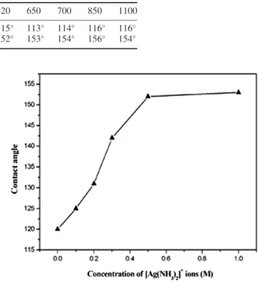
Table 2. Water CAs before and after hydrophobic modification of PDMS surfaces coated with silica spheres of various sizes. Diameter of silica spheres (nm) 520 650 700 850 1100
CA (before modification by PDT) 115◦ 113◦ 114◦ 116◦ 116◦
CA (after modification by PDT) 152◦ 153◦ 154◦ 156◦ 154◦
equation [39–42]:
cos θn+1=(1 − ωn)cos θn−ωn, (1) where n is the generation number of the hierarchical structure and θn is the contact angle on a surface of the nth generation. In our case, the first generation structure corresponded to the PDMS surface decorated with the Ag NPs. After hydrophobic modification, on such a surface, the CA was about 140◦ (see figure S2 in supplementary information
(available at stacks.iop.org/Nano/20/065304), table 1). The second generation structure consisted of the PDMS surface coated with silica spheres and decorated with Ag NPs. After hydrophobic modification on such a surface, the water CA was about 152◦(as shown in the right inset of figure3). Using the
measured values of the water CA, we can estimate a value for ωin equation (1), the fraction of the surface in contact with the droplet, equal to 0.5. This value is indicative of water droplets in contact with a part of the silica sphere surface corresponding to an angle of 45◦. This is reasonable for droplet sizes much
bigger than the silica spheres, which further indicates that our fabricated surfaces are in agreement with the modified Cassie– Baxter equation on a structure of dual-scale topography.
For the present methodology, the number of Ag NPs on the silica spheres could be controlled by the concentration of [Ag(NH3)2]+ ions. The lower the concentration of
[Ag(NH3)2]+ ions, the less the number of Ag NPs on the
silica spheres. Such a relationship is due to the fact that lower [Ag(NH3)2]+ion concentration led to less [Ag(NH3)2]+ions
on the silica spheres. Figure5shows the relationship between the water CA and the concentration of [Ag(NH3)2]+ions. The
water CA increases with the increase of the concentration of [Ag(NH3)2]+ions. When the [Ag(NH3)2]+ion concentration
is low, only small parts of the surface of silica spheres were decorated with Ag NPs, leading to small surface roughness and low water CA. As shown in figure 5, the concentration of the glucose is fixed at 0.5 M. When the concentration of [Ag(NH3)2]+ions is 0.1 M, the water CA of the film is about
125◦ after the PDT modification. This film does not achieve
superhydrophobicity. When the concentration of [Ag(NH3)2]+
ions reaches 0.5 M, the surface of the silica spheres is nearly completely coated by Ag NPs. Therefore, even if the concentration of [Ag(NH3)2]+ions further increases, the water
CA changes little.
The relationship between the size of the silica spheres and the wettability was also investigated. The concentrations of [Ag(NH3)2]+ ions and glucose were fixed at 0.5 M, and
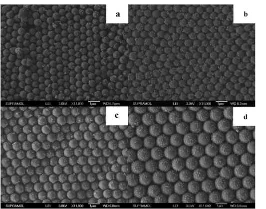
we investigated the wettability of the raspberry-like surfaces. Figure 6 shows the SEM images of our four raspberry-like surfaces with different silica spheres, whose average diameters are 650 (figure 6(a)), 700 (figure 6(b)), 850 (figure 6(c)) and 1100 nm (figure 6(d)), respectively. The hexagonal
Figure 5.The effect of the concentration of [Ag(NH3)2]+ions,
ranging from 0.0 to 1.0 M, on the water CA. The concentration of glucose is 0.5 M and the diameter of the silica spheres is 520 nm.
arrangement of the silica spheres and the diameters of the silica spheres can be distinguished clearly. Moreover, Ag NPs with diameters ranging from 30 to 40 nm distribute randomly on the surface of the silica spheres. Table2shows the relationship between the silica diameter and water CA of the film before and after the PDT modification. It demonstrates that the size of the silica spheres has little effect on the wettability of the resulting film. Namely, the dimensional change of the micrometer-level structure, ranging from 500 to 1100 nm, has little effect on the wettability of the hierarchical structure, whereas the amount of nanometer-level objects on the microstructures dominates the wettability of the hierarchical structure.
In our study, the silica spheres on the PDMS film were mechanically stable, because they sank into the polymeric film during the lift-up soft-lithography process (see figure S3 in supplementary information, available at stacks.iop.org/Nano/20/065304) [43,44]. The PDMS film is a soft material that could be easily rolled, stretched and folded. Accordingly, the superhydrophobic film could be easily peeled away from one substrate and then transferred to another one. During the transfer process, silica spheres with Ag NPs coating remained intact on the surface of the PDMS film. Thus, the superhydrophobicity of the film was preserved.
Figure7(a) shows the optical image of a superhydrophobic film. The array of the silica spheres decorated by the Ag NPs is presented in the center of the transparent PDMS film. The color of the pattern is brown, which is the feature color of Ag NPs. The dimensional size of the trapezium superhydrophobic film presented in figure7(a) is (2.0 + 3.5) × 2.5 cm2. The film can firmly stick on the flat glass without any glue, and it does not 4
Figure 6.SEM images of the silica arrays on PDMS films; the diameter of the silica spheres is (a) 650 nm; (b) 700 nm; (c) 850 nm and (d) 1100 nm. The surface of the silica spheres is decorated with Ag NPs.
Figure 7.Optical images of (a) a superhydrophobic film; the transparent margin is the PDMS film, and the silica spheres coated with Ag NPs are located in the center. (b) The water droplets, with a volume of 50 µl each, on a superhydrophobic film which is stuck on a glass substrate; the shape of the droplet is nearly spherical. (c) The film peels away from the glass and transfers onto the curved hydrophilic metal surface. fall off from the substrate even inverted 180◦. Figure7(b) is a
direct optical image of water droplets on the superhydrophobic film. The volume of every droplet is 50 µl. From the image we can see that the shape of the droplets is nearly spherical, indicating the film exhibits a good superhydrophobic property. When superhydrophobicity is demanded on a curved surface, the substrate can be dipped into ethanol where the PDMS film can be peeled away from the glass substrate; then the film is transferred carefully to the smooth, curved surface. As shown in figure 7(c), the film is coated on a curved and hydrophilic metal surface. A water droplet rolls down easily due to the small sliding angle of the superhydrophobic film. Consequently, a photo of the droplet on a curved surface could not be taken.
4. Conclusions
In summary, we have successfully fabricated a flexible superhydrophobic surface with a micrometer-level and
nanometer-level hierarchical structure on PDMS films. Lift-up soft-lithography and chemical reduction led to the presence of a raspberry-like structure on the PDMS films; the hierarchical structure originated from coating ordered silica spheres with Ag NPs. SEM images have been used to evaluate the roughness of the PDMS films patterned with silica spheres which were decorated with Ag NPs. The effects of the amount of Ag NPs and the size of silica spheres on the wettability of the PDMS films have been investigated: the former played a major role on the wettability. Our superhydrophobic films could be easily transferred to a number of smooth substrates with different curvatures. During the transfer process, the superhydrophobicity remained intact. Thus, our flexible superhydrophobic films could be used many times to satisfy a wide range of applications.
Acknowledgments
This work was supported by the National Nature Science Foundation of China (grant nos. 20534040 and 20674026), the
Nanotechnology 20 (2009) 065304 T Yao et al
‘111’ project (no. 1306009) and the National Basic Research Program (nos. 2007CB936402 and 2007CB936403).
References
[1] Gau H, Herminghaus S, Lenz P and Lipowsky R 1999 Science 28346
[2] Young T 1805 Trans. R. Soc. Lond.9565
[3] Sun T L, Feng L, Gao X F and Jiang L 2005 Acc. Chem. Res. 38644
[4] Gould P 2003 Mater. Today 6 44
[5] Honeychuck R V, Ho T, Wynne K J and Nissan R A 1993 Chem. Mater.51299
[6] Coulson S, Woodward I, Badyal J, Brewer S and Wills C 2000 J. Phys. Chem.B1048836
[7] Patankar N A 2003 Langmuir191249 [8] Barthlott W and Neinhuis C 1997 Planta2021 [9] Shull K R and Karis T E 1994 Langmuir10334
[10] Ming W, Wu D, Benthem R V and With G D 2005 Nano Lett. 52298
[11] Feng L, Shi S H, Li Y S, Li H J, Zhang L J, Zhai J, Song Y L, Liu B Q, Jiang L and Zhu D B 2002 Adv. Mater.141857 [12] F¨ursmer R, Barthlott W, Neinhuis C and Walzel P 2005
Langmuir21956
[13] Zhang L B, Chen H, Sun J Q and Shen J C 2007 Chem. Mater. 19948
[14] Zhang X, Shi F, Yu X, Liu H, Fu Y, Wang Z Q, Jiang L and Li X Y 2004 J. Am. Chem. Soc.1263064
[15] Tian Y, Liu H Q and Zheng Z F 2006 Chem. Mater.185820 [16] Ebril H Y, Demirel A L, Avci Y and Mert O 2003 Science
2991377
[17] Lim J M, Yi G R, Moon J H, Heo C J and Yang S M 2007 Langmuir237981
[18] Tsougeni K, Tserepi A, Boulousis G, Constantoudis V and Gogolides E 2007 Plasma Process. Polym.4398 [19] Satyaprasad A, Jain V and Nema S K 2007 Appl. Surf. Sci.
2535462
[20] Cortese B, D’Amone S, Manca M, Viola I, Cingolani R and Gigli G 2008 Langmuir242712
[21] Manca M, Cortese B, Viola I, Arico A S, Cingolani R and Gigli G 2008 Langmuir241833
[22] Gao X F, Yan X, Yao X, Xu L, Zhang K, Zhang J H, Yang B and Jiang L 2007 Adv. Mater.192213 [23] Gao X F and Jiang L 2004 Nature43236
[24] Zhu Y, Hu D, Wan M X, Jiang L and Wei Y 2007 Adv. Mater. 192092
[25] Artus G R J, Jung S, Zimmermann J, Gautschi H P, Marquardt K and Seeger S 2006 Adv. Mater.182758 [26] Feng X J and Jiang L 2006 Adv. Mater.183063
[27] Li X M, Calama M C and Reinhoudt D N 2007 Chem. Soc. Rev. 361350
[28] Zhang X, Shi F, Niu J, Jiang Y G and Wang Z Q 2008 J. Mater. Chem.18621
[29] Li Y, Lee E J and Cho S O 2007 J. Phys. Chem. C11114813 [30] Tsai H J and Lee Y L 2007 Langmuir2312687
[31] Sun M H, Luo C X, Xu L P, Ji H, Ouyang Q, Yu D P and Chen Y 2006 Langmuir218978
[32] Tserepi A D, Vlachopoulou M E and Gogolides E 2005 Nanotechnology173977
[33] Xia Y N and Whitesides G M 1998 Angew. Chem. Int. Edn 37550
[34] St¨ober W, Fink A and Bohn E 1968 J. Colloid Interface Sci. 2662
[35] Denkov N D, Velev O D, Kralchevsky P A, Ivanov I B and Nagayama H K 1993 Nature36126
[36] Yan X, Yao J M, Lu G, Chen X, Zhang K and Yang B 2004 J. Am. Chem. Soc.12610510
[37] Chen Z M, Chen X, Zheng L L, Gang T, Cui T Y, Zhang K and Yang B 2005 J. Colloid Interface Sci.285146
[38] Zhang Z P, Zhang L D and Wang S X 2001 Polymer428315 [39] Cassie A and Baxter S 1944 Trans. Faraday Soc.40546 [40] Herminghaus S 2000 Europhys. Lett.52165
[41] Bormashenko E, Stein T, Whyman G, Bormashenko Y and Pogreb R 2006 Langmuir229982
[42] Patankar N A 2004 Langmuir208209
[43] Yan X, Yao J M, Lu G, Li X, Zhang J H, Han K and Yang B 2005 J. Am. Chem. Soc.1277688
[44] Yao J M, Yan X, Lu G, Zhang K, Chen X, Jiang L and Yang B 2004 Adv. Mater.1681