HAL Id: hal-03030782
https://hal.archives-ouvertes.fr/hal-03030782
Submitted on 14 Dec 2020
HAL is a multi-disciplinary open access
archive for the deposit and dissemination of
sci-entific research documents, whether they are
pub-lished or not. The documents may come from
teaching and research institutions in France or
abroad, or from public or private research centers.
L’archive ouverte pluridisciplinaire HAL, est
destinée au dépôt et à la diffusion de documents
scientifiques de niveau recherche, publiés ou non,
émanant des établissements d’enseignement et de
recherche français ou étrangers, des laboratoires
publics ou privés.
Mediates Temporal Control of an Ecdysteroid Target
Gene at the Onset of Drosophila Metamorphosis
Bruno Mugat, Véronique Brodu, Jana Kejzlarova-Lepesant, Christophe
Antoniewski, Cynthia Bayer, James Fristrom, Jean-Antoine Lepesant
To cite this version:
Bruno Mugat, Véronique Brodu, Jana Kejzlarova-Lepesant, Christophe Antoniewski, Cynthia Bayer,
et al.. Dynamic Expression of Broad-Complex Isoforms Mediates Temporal Control of an Ecdysteroid
Target Gene at the Onset of Drosophila Metamorphosis. Developmental Biology, Elsevier, 2000, 227,
pp.104 - 117. �10.1006/dbio.2000.9879�. �hal-03030782�
Dynamic Expression of Broad-Complex Isoforms
Mediates Temporal Control of an Ecdysteroid
Target Gene at the Onset of Drosophila
Metamorphosis
Bruno Mugat,* Ve´ronique Brodu,* Jana Kejzlarova-Lepesant,*
Christophe Antoniewski,* Cynthia A. Bayer,† James W. Fristrom,‡
and Jean-Antoine Lepesant*
,1*Institut Jacques-Monod, CNRS et Universite´s Paris 6 –P. et M. Curie et Paris 7–Denis-Diderot, 2, place Jussieu, F-75251 Paris Cedex 05, France;†Department of Biology, University of
Central Florida, 4000 Central Florida Boulevard, Orlando, Florida 32816-2368; and ‡Department of Molecular and Cellular Biology, University of California
at Berkeley, Berkeley, California 94720-3200
Metamorphosis in Drosophila melanogaster is orchestrated by the steroid hormone ecdysone, which triggers a cascade of primary-response transcriptional regulators and secondary effector genes during the third larval instar and prepupal periods of development. The early ecdysone-response Broad-Complex (BR-C) gene, a key regulator of this cascade, is defined by three complementing functions (rbp, br, and 2Bc) and encodes several distinct zinc-finger-containing isoforms (Z1 to Z4). Using isoform-specific polyclonal antibodies we observe in the fat body a switch in BR-C isoform expression from the Z2 to the other three isoforms during the third instar. We show that the 2Bcⴙfunction that corresponds presumably to the Z3
isoform is required for the larval fat body-specific expression of a transgenic construct (AE) in which the lacZ gene is under the control of the ecdysone-regulated enhancer and minimal promoter of the fat body protein 1 (Fbp1) gene. Using hs(BR-C) transgenes, we demonstrate that overexpression of Z1, Z3, or Z4, but not Z2, is able to rescue AE activity with faithful tissue specificity in a BR-C null (npr1) genetic context, demonstrating a partial functional redundancy between Z1, Z3, and Z4 isoforms. We also show that continuous overexpression of Z2 during the third instar represses AE, while conversely, expression of Z3 earlier than its normal onset induces precocious expression of the construct. This finding establishes a tight correlation between the dynamic pattern of expression of the BR-C isoforms and their individual repressive or inductive roles in AE regulation. Altogether our results demonstrate that the balance between BR-C protein isoforms in the fat body mediates, in part, the precise timing of the ecdysone activation of the AE construct but does not modulate its tissue specificity. © 2000 Academic Press
Key Words:larval fat body; ecdysone; fat body protein 1 gene; temporal regulation; zinc fingers.
INTRODUCTION
Metamorphosis in holometabolous insects is a remark-able developmental process leading to the remodeling of the larva into the adult. During this period, initiation of new
programs of gene expression lead to important morphologi-cal and physiologimorphologi-cal modifications within larval and imaginal tissues. Larval tissues are histolyzed or reorga-nized while imaginal tissues develop into adult structures. In Drosophila, this metamorphic transformation is orches-trated by successive waves of the steroid hormone 20-hydroxyecdysone (referred to here as ecdysone) (Riddiford, 1993). How the hormonal signal is correctly interpreted in each target tissue remains an open question. Cytological studies of ecdysone-induced puffing of polytene chromo-1To whom correspondence should be addressed at the Institut
Jacques Monod, CNRS et Universite´s Paris 6 –P. et M. Curie et Paris 7–Denis-Diderot, 2, place Jussieu, F-75251 Paris Cedex 05, France. Fax:⫹33 1 44 27 52 65. E-mail: [email protected].
somes led Ashburner and his colleagues (1974) to propose a model of action of ecdysone in larval salivary glands that was further refined and extended to include a variety of ecdysone target tissues (Ashburner et al., 1974; Russell et
al.,1996; Thummel, 1990, 1996). According to this model,
the release of the hormonal signal during the third larval instar represses intermolt genes and induces a small set of early regulatory genes simultaneously. The products of the early genes subsequently repress their own expression and activate a large set of late genes allowing tissues to ensure the correct execution of their metamorphic program.
To date four early genes have been characterized at the molecular level, Broad-Complex (BR-C), E74, E75, and E63-1 (Andres and Thummel, 1995; Burtis et al., 1990; DiBello et al., 1991; Segraves and Hogness, 1990). Consis-tent with their regulatory function, the first three of these genes direct the synthesis of putative transcription factors. The BR-C, located in the 2B5 region of the X chromosome, plays a key role in the genetic control of metamorphic responses to ecdysone because it is implicated in the regulation of intermolt, early, and late gene activities (Belyaeva et al., 1981; Guay and Guild, 1991; Karim et al., 1993; Zhimulev et al., 1982).
The BR-C is organized in overlapping transcription units (Bayer et al., 1996; DiBello et al., 1991) that give rise to an extensive family of RNA transcripts (Chao and Guild, 1986; Karim et al., 1993; Karim and Thummel, 1992) encoding several related protein isoforms. These isoforms share a common amino-terminal “core” region containing a BTB/ POZ domain likely involved in protein/protein interactions (Bardwell and Treisman, 1994; Zollman et al., 1994). The core exons are alternatively spliced to one of four isoform-specific exons encoding pairs of zinc-finger motifs (Z1 to Z4) (Bayer et al., 1996; DiBello et al., 1991).
The BR-C locus encodes three genetic subfunctions each represented by a complementation group: broad (br), re-duced bristles on the palpus(rbp), and 2Bc. All mutations affecting these functions fail to complement the nonpupari-atingmutations (npr1 alleles) that are phenotypically indis-tinguishable from deletions of the locus (Belyaeva et al., 1980; Kiss et al., 1988). Although several studies have found converging results supporting a plausible correlation be-tween BR-C isoforms and complementing genetic func-tions, there is not a simple one-to-one correspondence
between BR-C proteins and BR-C⫹genetic functions (Bayer
et al.,1997; Emery et al., 1994). Using heat-inducible BR-C transgenes (hs(BR-C)) to rescue the lethality associated with each BR-C subfunction and restore transcriptional
activity of tissue-specific BR-C⫹-dependent target genes,
Bayer et al. (1997) demonstrated that the br⫹ function is
provided by the Z2 isoform only. In contrast, although the
rbp⫹function was fully supplied by the Z1 isoform, it was
also partially provided by Z4, indicating functional redun-dancy between these two isoforms (Bayer et al., 1996;
Sandstrom et al., 1997). Finally, the 2Bc⫹ function was
provided by the Z3 isoform. However, the other BR-C isoforms were able to at least partially compensate for the
absence of this function. This last observation also indi-cated a functional redundancy between Z3 and the other
BR-C isoforms, but further evidence to confirm this
hy-pothesis is lacking. Alternatively, these results could sug-gest a role of Z3 in the autoregulation of BR-C expression, either directly or indirectly through the modulation of the activity of other genes regulating this locus.
The npr1 mutations lead to lethality at the onset of metamorphosis without puparium formation. Alleles be-longing to the br, rbp, and 2Bc complementing functions result in lethality somewhat later in development, with severe morphological defects (Belyaeva et al., 1980; Fris-trom et al., 1981; Kiss et al., 1988; Restifo and Merrill, 1994; Restifo and White, 1991, 1992). Several lines of evidence demonstrate that these developmental failures are not due to an ecdysteroid deficiency, but rather to defects in the response to the hormonal signal (Fristrom et al., 1981; Kiss et al.,1976, 1978; Murphy et al., 1977; Stewart et al., 1972). Together these data point to the BR-C as essential for the proper response of tissues to the ecdysone signal during metamorphosis.
The regulatory role and mode of action of the proteins encoded by the BR-C remain to be thoroughly determined.
BR-C isoforms and BR-C mRNAs are expressed
differen-tially in a number of tissues around the time of metamor-phosis (Emery et al., 1994; Huet et al., 1993). This supports the postulate of the tissue coordination model (Burtis et al., 1990; Thummel, 1990) that the relative levels of BR-C isoforms and other early ecdysone-induced gene products, in hormone target tissues, may contribute to the tissue specificity of the ecdysone response. However, several stud-ies support the idea that the BR-C is an important stage regulator of the transcriptional response to ecdysone. The study of the effect of BR-C mutations on a series of ecdysone-inducible genes in salivary glands led Karim et al. (1993) to conclude that the BR-C is implicated in both the timing and the level of transcription of these genes. Cross-grove et al. (1996) have suggested that the sequential expression and antagonistic activities of BR-C protein iso-forms may be responsible for the timing of L71 late gene expression in salivary glands.
With the aim of further investigating this role of the
BR-Cprotein isoforms as temporal regulators, we examined
the requirement for and function of individual BR-C iso-forms in the expression of the fat body protein 1 gene (Fbp1) as a model target gene. Fbp1 is a primary ecdysone-responsive gene which is expressed exclusively in the fat body during the second half of the third larval instar (Andres et al., 1993; Lepesant et al., 1982, 1986; Maschat et
al.,1990). Analysis of its cis-regulatory sequences led to the
characterization of a 70-bp enhancer (⫺69 to ⫺138) (referred
to as element E) (Laval et al., 1993) and a 32-bp amplifier
(⫺194 to ⫺162) (referred to as element A) (Lapie et al., 1993)
that are sufficient to confer the correct temporal and spatial patterns of ecdysone-induced expression of the Fbp1 gene
pro-moter driving the lacZ reporter gene in the AE construct (Antoniewski et al., 1996; Brodu et al., 1999).
We demonstrate here a strong requirement for BR-C⫹
genetic functions for the normal pattern of expression of the
AEconstruct in the larval fat body. We identify which BR-C
genetic functions are implicated by testing the effects of mutations of each complementation group and the overex-pression of individual BR-C isoforms. The potential activat-ing or repressactivat-ing function attributed to individual BR-C isoforms from these experiments is supported by the de-tailed analysis of the expression profile of the four BR-C isoforms in the fat body throughout the third larval instar. Finally, the observation of a repression or a temporal deregulation of AE expression following heat-shock induc-tion of specific BR-C isoforms allows us to assign a role to these isoforms in the timing of the expression of this target construct.
MATERIALS AND METHODS
Drosophila Strains and Crosses
Stocks are listed in Table 1. The br28
, 2Bc2
, rbp5
, and npr16
mutations and the Df(1)S39 and D(1; Y)Sz280 deficiencies and translocations are described in Lindsley and Zimm (1992), except
br28
, which is described in DiBello et al. (1991). The AE stock harbors the transgenic AE reporter construct described in Brodu et
al. (1999). The AEhsZ1– 4 stocks, homozygous for the hsZ1– 4 transgenic constructs (Crossgrove et al., 1996) and the AE reporter construct were obtained by appropriate crosses. Flies and larvae were reared at 25°C on a standard Drosophila medium supple-mented with dry yeast. w1118
was used as the reference wild-type strain.
To test the effect of BR-C mutations on the AE construct (Fig. 5), AE males were crossed with heterozygous mutant females carrying
BR-Calleles depicted in Table 1 to generate male progeny
hemi-zygous for the BR-C mutation and heterohemi-zygous for the AE trans-gene. These animals were staged as described below and harvested at 114 –119 h after egg-laying (AEL).
For the rescue assay (Fig. 6), homozygous males of the AEhsZ1– 4 stocks (see Table 1) were crossed to y npr16/FM6 l(1)69jfemales to
generate male progeny hemizygous for y npr16and heterozygous for
both the hs(BR-C) and the AE transgenes. As a control, male progeny from y npr16/FM6 l(1)69jfemales crossed to AE males were
generated.
Developmental Staging of Larvae
To simplify presentation and comparison of developmental data, all times of development are indicated as hours of development AEL. Under our culture conditions at 25°C, transition from the second to the third instar occurred about 72 h AEL and pupariation at 120 h AEL, and the white prepupal stage was reached about 120 –124 h AEL (Bainbridge and Bownes, 1981). It should be noted that the onset of expression of the AE transgene takes place after 106 h AEL (Brodu et al., 1999). Two staging methods, as follows, that proved to be reproducible were used.
Test of BR-C mutations and rescue of AE expression assay.
w1118
and mutant larvae were reared at 25°C in uncrowded vials on the standard medium supplemented with 0.05% (w/v) bromophe-nol blue. This dye is clearly visible inside the gut of live animals and its clearance is a reliable indicator of the developmental stage of late third-instar larvae. Larvae with lightly stained gut were approximately 5–12 h away from pupariation, i.e., 108 –115 h AEL, and those with an almost completely clear gut were approximately 1– 6 h away from pupariation, i.e., 114 –119 h AEL (Andres and Thummel, 1994). We verified that larvae hemizygous for the br,
rbp,and 2Bc mutations did not develop any slower than wild-type larvae according to the time of hindgut clearing. Larvae hemizy-gous for the npr16
mutation never pupariated, but complete gut clearing occurred normally and we relied on this criterion for selecting larvae for assays.
Immunostaining and temporal deregulation experiments with heat-inducible hs(BR-C) transgenes. First-instar larvae from 1-h egg lays were collected within 30 min of hatching, transferred to TABLE 1
DrosophilaStocks
Chromosome/balancer Nature of BR-C lesion Reference
Mutant stocks
br28 br28/FM6, l(1)69j* P-element insertion in Z2 zinc-finger exon (a)
2Bc2 y 2Bc2/Binsn Unknown (b)
rbp5 y rbp5/Binsn Stop codon in Z1 linker region (b, c)
npr16 y npr16/FM6, l(1)69j Unknown (b)
Df(1)S39 Df(1)S39/FM6, l(1)69j Df(1)1E1-2;2B5-6 (b)
Dp (1; Y)Sz280 y l(1)/FM6, l(1)69j/Dp(1;Y)Sz280 Dp(1;Y)1A;2C1-2 with internal deficiency Df(1)2B3-4;2B7-8. (b)
Transgenic stocks
AE w1118;⫹; AE (d)
AEhsZ1 w1118; hsp70-dm708; AE This study
AEhsZ2 w1118; hsp70-cD5; AE This study
AEhsZ3 w1118; hsp70-dm797; AE This study
AEhsZ4 w1118; hsp70-28.I; AE This study
Note.(a) DiBello et al. (1991), (b) Lindsley and Zimm (1992), (c) Bayer et al. (1997), (d) Antoniewski et al. (1996); Brodu et al. (1999). * FM6, l(1)69jwas described in Belyaeva et al. (1980).
standard medium in batches of about 50 larvae, and reared at 25°C. Samples of larvae for immunostaining were taken at 72 (early L3), 96 (middle L3), 118 (Late L3), and 120 –124 h AEL (white prepupae) (Fig. 4). For experiments on AE deregulation by overexpression of Z3, larvae were taken prior to any detectable expression of the AE transgene at 96 h AEL (Fig. 8).
Heat-Shock Protocols
To induce expression of the heat-inducible hs(BR-C) transgenes, staged larvae were placed in hermetically closed plastic vials and submerged for 35 min in a 37°C water bath. After heat shocks the vials were placed at 25°C.
Rescue of AE expression. y npr16/Y; hs(BR-C)/⫹; AE/⫹ males
were heat shocked at the “light blue gut” stage and then allowed to develop at 25°C for 8 h. “Clear gut” larvae were then collected in batches of three larvae in Eppendorf tubes and frozen on dry ice before storage at⫺80°C.
Temporal deregulation of AE expression. Heat shocks were performed on AEhsZ2 larvae at 96 h AEL and again at 100 h AEL while AEhsZ1, AEhsZ3, and AEhsZ4 larvae were heat shocked at 73 h AEL and again at 90 h AEL. Larvae were allowed to develop at 25°C until they were collected for-galactosidase assay.
Generation of Antibodies
Fusion proteins were used as immunogens to produce polyclonal antibodies in New Zealand female rabbits.
Except for Z2, BR-C protein-specific regions were chosen outside of the zinc-finger motif. Isoform-specific regions of BR-C cDNAs [dm527(Q-Z1), cD5(Z2), dm797(Z3), and 28.I(Z4)] (Bayer et al., 1996; DiBello et al., 1991) were amplified by PCR with primers containing restriction enzyme sites at their ends for subsequent cloning in frame into both pGEX (Pharmacia Biotech) and pMALc2 (Biolabs) bacterial expression vectors in Escherichia coli BL21 strain. The amplified regions correspond to nucleotide positions 1899 –2107 for the Z1 isoform, 1680 –1931 for the Z2 isoform, 2307–2501 for the Z3 isoform, and 1966 –2163 for the Z4 isoform. Constructs were verified by sequence determination of the cloned DNA fragments in the resulting pGEXz1 to pGEXz4 and pMALz1 to pMALz4 plasmids.
Bacterial cultures were grown until OD600nm reached 0.5 and induced with 2 mM IPTG for 3 h. GST fusion proteins were purified on glutathione Sepharose 4B (Pharmacia Biotech) while MBP fusion proteins were purified on an amylose resin (Biolabs) in accordance with the manufacturer’s specifications.
For the production of anti-Z1, anti-Z2, anti-Z3, and anti-Z4 polyclonal antibodies, 100- to 500-g aliquots of purified GSTz1 (from pGEXz1), MBPz2 (from pMALz2), GSTz3 (from pGEXz3), and GSTz4 (from pGEXz4) proteins were used to immunize and boost via subcutaneous route two New Zealand female rabbits for each fusion protein. The first injection was in Freund’s complete adju-vant and the boost, spaced for at least 3 weeks, was in Freund’s incomplete adjuvant. A total of four to five boosts were performed on each rabbit, and sera taken 2 weeks after the last boost were tested for their immunoreactivity against the injected proteins.
Immunoaffinity Purification of Antibodies
About 10 mg of GST, MBP, GSTz1, MBPz2, MBPz3, and MBPz4 proteins were independently immobilized on CNBr-activated Sepharose 4 Fast Flow (Pharmacia Biotech), according to the manufacturer’s recommendations.
In the case of the anti-Z1 and anti-Z2 sera, polyreactive antibod-ies were retained on a GST- or a MBP-coupled Sepharose column and the flowthrough was applied at a flow rate of about 10 ml䡠 cm⫺2 䡠 h⫺1 to a GSTz1- or a MBPz2-coupled Sepharose column, respectively. In the case of the anti-Z3 and anti-Z4 sera, about 20 ml of serum was applied directly to a MBPz3- or a MBPz4-coupled Sepharose column, respectively. Each of the four affinity columns was washed with 1⫻ PBS, 500 mM NaCl until the OD280 nmreached a null value. The specific antibodies retained were eluted with 100 mM glycine–HCl buffer (pH 2.5) and diluted in 100 mM Tris–HCl (pH 8) in a 1:3 ratio (v/v). About 6 gel volumes of elution buffer were used and the protein content of each collected fraction was determined by OD280 nm measurement. Fractions with significant amounts of protein were pooled and concentrated on an Amicon concentrator. Purified antibodies were stored in 50% glycerol at ⫺20°C.
In Vitro Translation of BR-C Protein Isoforms
In vitrotranslation of dm527, cD5, dm797, and 28.I cDNAs was performed using the TNT coupled reticulocyte lysate system from Promega.
Protein Extracts and Western Blotting
Ten to twenty late third-instar w1118 or BR-C null larvae were
homogenized in 40 – 80l cracking buffer [125 mM Tris (pH 6.8), 5%-mercaptoethanol, 2% SDS, 4 M urea] by vortexing. Samples were boiled (2 min) and centrifuged prior to electrophoresis on a 7% SDS–polyacrylamide gel. Four microliters of extract or 5l of in
vitro-translated BR-C protein was loaded in the corresponding lane. Transfers were performed onto nitrocellulose membranes (Schleicher and Schuell) using a Novablot Electrophoretic Transfer Kit (LKB). All subsequent steps were carried out at room tempera-ture. The membranes were blocked in TNT buffer [50 mM Tris (pH 7.5), 150 mM NaCl, 0.25% Triton X-100] containing 5% nonfat dried milk for 1 h. All antibodies were diluted 1:104in this blocking buffer. Incubations with these antibodies were performed for 1–2 h. Blots were rinsed three times for 10 min in TNT and incubated for 1 h with horseradish peroxidase-conjugated goat rabbit anti-bodies (Vector Laboratories) diluted 1:5000. Blots were rinsed as before, followed by three washes with sterile distilled water. Detection of the enzyme-conjugated antibodies was achieved using the ECL kit (Amersham) and X-OMAT AR film (Kodak).
Whole-Mount Immunostaining
Immunostaining of dissected tissues was performed as described in Brodu et al. (1999) with the following modifications. Normal goat serum (NGS) (Jackson Immunoresearch) was used instead of BSA. Ten percent NGS was used in the blocking step and 5% NGS was added in PBT to dilute antibodies. The anti-Z1, anti-Z2, and anti-Z3 were diluted 1:3000 and the anti-Z4, 1:4000, and used without preadsorption on embryos.
Histochemical and Spectrophotometric Assays
of
-Galactosidase Activity
Histochemical staining and CPRG spectrophotometric assays of -galactosidase activity were performed as described in Brodu et al. (1999).
RESULTS
Isoform-Specific Anti-BR-C Antibodies
Each BR-C isoform is characterized by unique C-terminal zinc-finger and linker domains fused to a common N-terminal core region (Bayer et al., 1996; DiBello et al., 1991). Small portions of these isoform-specific protein re-gions were expressed in E. coli and used as immunogens to generate polyclonal antibodies directed against each BR-C protein isoform (Fig. 1). In order to test the specificity of these antibodies, Western blots were performed using in vitro-translated BR-C proteins and protein extracts from wild-type larvae and larvae carrying a deletion of the entire
BR-Clocus. In vitro translation of each of the four BR-C
cDNAs gave rise to two 35S-labeled bands in SDS–
polyacrylamide gel (Fig. 2E). In all four cases, the lower molecular weight band (arrowheads in Fig. 2E) corresponded to the expected size deduced from the cDNA sequence (Bayer et al., 1996; DiBello et al., 1991). We presume that the upper band corresponds to a translational readthrough product. Each anti-BR-C antibody recognized specifically the two products of the corresponding in vitro-translated
BR-CcDNA, and no cross-reactivity was observed between
the various antibodies (Figs. 2A to 2D, lanes Z1, Z2, Z3, Z4). In each blot, the signal detected in the extracts from wild-type larvae (lanes WT) revealed a group of several bands migrating slower than the lower migrating in vitro-translated product (arrowheads in Fig. 2). Because no equivalent signal was detected in the BR-C deletion extract (null lanes) we concluded that the antibodies recognized
specifically the endogenous BR-C isoforms. The multiple specific products detected with each antibody may corre-spond either to various posttranslational modifications (Bayer et al., 1996; Emery et al., 1994) or to translational products from transcripts resulting from a differential splic-ing of isoform-specific exons. The isolation of a family of Z1 cDNAs encoding several Z1 protein isoforms differing by the length of the linker between the core region and the zinc-finger domain (DiBello et al., 1991) supports this hypothesis. Similarly, Bayer et al. (1996) have identified a family of partially spliced Z4 mRNAs.
The specificity of these antibodies was further tested when we examined the protein defects induced by muta-tions representative of each BR-C complementation group
listed in Table 1. The BR-C null mutation npr16
results in
a total loss of BR-C⫹genetic functions and accordingly no
BR-Cprotein isoforms were detected in mutant larvae (Fig.
3). The br28
mutation results in a loss of br⫹ function
associated with a P-element insertion within the Z2 zinc-finger pair (DiBello et al., 1991). We confirmed the previous finding of Emery et al. (1994) that this mutation gives rise to the accumulation of truncated Z2 protein products (Fig. 3B) while no other isoform was affected (Fig. 3D and data not shown; see also Bayer et al., 1997). In larvae carrying the 2Bc2
mutation the multiple Z3-specific bands detected in wild-type larvae were replaced by a single band which migrated slightly faster. This may reflect an alteration of the posttranslational modifications of the Z3 protein (Fig.
3C). A similar effect appeared to take place in the 2Bc1
mutant larvae (Emery et al., 1994, and our unpublished
data). The other BR-C isoforms in the 2Bc2
mutant were detected at the expected molecular weight but appeared to
be underrepresented (Fig. 3D and data not shown). The rbp5
mutation is associated with a stop codon in the Z1 linker region (Bayer et al., 1997) and as expected no Z1 isoforms were detected with our antibodies (Fig. 3A). The other BR-C isoforms were detected at their respective expected molecu-lar weights (Fig. 3D and data not shown).
The Developmental Expression Profile of BR-C
Protein Isoforms in the Larval Fat Body
Is Dynamic during the Third Instar
Anti-BR-C polyclonal antibodies were used to establish a detailed spatial and temporal distribution of BR-C proteins in larval and imaginal tissues during the third larval instar (Mugat et al., in preparation). Here, we focused on the characterization of the temporal distribution of BR-C pro-teins in the larval fat body where the BR-C target AE construct is specifically expressed.
Representative immunohistochemical stainings of dis-sected fat bodies are presented in Fig. 4. Immunostaining on
BR-Cnull larvae carried out in parallel as a control showed
no significant specific signal over background (data not shown). A nuclear signal was detected with each antibody in agreement with the role of transcriptional regulator proposed for the BR-C proteins. These experiments revealed
FIG. 1. Schematic representation of the Q1-Z1, Z2, Z3, and Z4 protein isoforms encoded by the BR-C locus. The first 431 amino acids defining the core region, common to all BR-C proteins, are linked to isoform-specific domains as described in DiBello et al. (1991) and Bayer et al. (1996). The isoform-specific domains used to produce polyclonal antibodies are each delimited by a line with amino acid coordinates of the peptide sequence indicated. Acces-sion numbers for sequences in GenBank are QZ1, X54663 S79435; Z2, X54665; Z3, X54664 S79445; and Z4, U51585.
that Z2 is the only isoform present in the fat body at the beginning of the third instar (ca. 72 h AEL) (Fig. 4B). Z2 staining decreased to a very low level in mid-third-instar (ca. 96 h AEL) fat body while Z3 expression became detect-able (Figs. 4F and 4G). At this time of development, Z2 and Z3 were the only two isoforms detectable in the fat body. In late third-instar fat body (ca. 118 h AEL), Z2 expression was undetectable while Z1, Z3, and Z4 were strongly expressed (Figs. 4I, 4J, 4K, and 4L). The same expression profile was maintained in white prepupae (120 h AEL) (Figs. 4M– 4P). Interestingly we observed that the Z2 truncated products
detected with our antibody in a br28
mutant persisted in the
late third-instar fat body (Fig. 4J⬘).
BR-C Loss-of-Function Mutations Affect
the Expression of the AE Construct
We used the AE construct, which is expressed in an ecdysone-dependent manner in the fat body of late third-instar larvae (Antoniewski et al., 1996), to test whether this
sequential appearance of BR-C isoforms was involved in the regulation of BR-C downstream target genes in the fat body. To this end, we investigated the effect of representative
BR-C mutations of the four mutational classes described
above (see also Table 1) on the fat body-specific expression of the construct.
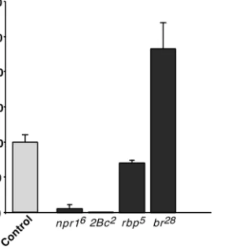
All BR-C mutations tested but rbp5
had a strong effect on the level of AE expression in late third-instar larvae (114 –
120 h AEL) (Fig. 5). The npr16
and 2Bc2
genetic context led to a strong decrease of the expression of the construct to only 5 and 0.4%, respectively, of that measured in the
wild-type control larvae. In contrast, in an rbp5
mutant, the
level of AE expression reached 71% of the level in BR-C⫹
control larvae, indicating only a minor effect of the rbp5
mutation on the construct. Finally, in a br28
mutant, the AE expression level reached 230% of the corresponding control (Fig. 5) but remained limited to the fat body (data not shown).
Together, these experiments revealed that multiple BR-C genetic functions were involved in the activation of the
FIG. 2. Test of the specificity of anti-BR-C isoforms antibodies. Polyclonal antibodies directed against the Z1 (A), Z2 (B), Z3 (C), and Z4 (D) specific domains were used to probe blots containing protein extract from male third-instar larvae [Df(1)S39/Dp(1;Y)Sz280 transheterozygotes] carrying a deletion of the BR-C locus (null) and from wild-type (WT) late third-instar larvae and each of the in
vitro-translated BR-C isoforms. About one animal equivalent of total extract was loaded in the null and WT lanes. The mobilities of the molecular weight markers are indicated on the right in kDa. Arrowheads indicate the expected molecular weight of the in vitro-translated proteins detected with antibodies (A, B, C, D) or radiolabeled (E). (A) A nonspecific high-molecular-weight product (asterisk) was detected with the anti-Z1 in both BR-C null and wild-type extracts. (C) A protein from the rabbit reticulocyte lysate was detected with the anti-Z3 (asterisk). (E) Autoradiogram of the35
Fbp1enhancer and promoter included in the AE construct
and suggested that the br⫹ genetic function, and
corre-spondingly, the Z2 isoform, has a repressive function. These conclusions were further tested by overexpression of individual BR-C isoforms in a BR-C null genetic context.
Isoform-Specific Rescue of the Expression
of the AE Construct
hsZ1, hsZ2, hsZ3,and hsZ4 constructs in which cDNAs encoding the Z1, Z2, Z3, and Z4 isoforms are expressed under the control of the hsp70 promoter (Crossgrove et al., 1996) were tested for their ability to restore AE expression
in a npr16
mutant background. npr16
males carrying the AE construct and one of the four hs(BR-C) transgenes were heat-shocked at late third instar (108 –115 h AEL). After an
8-h recovery period,-galactosidase activity was assayed on
extracts from whole larvae. Ubiquitous overexpression of each BR-C isoform following heat shock was verified by Western blotting and immunostaining of dissected tissues (data not shown). Results are presented in Fig. 6 in which -galactosidase activity is expressed as a percentage of the
corresponding heat-shocked BR-C⫹ control male larvae.
Control animals that did not carry a hs(BR-C) transgene failed to induce AE expression (Fig. 6).
We found that induction of the hsZ1, hsZ3, or hsZ4
transgenes in npr16
mutant larvae was able to restore 60, 33, and 68% of AE expression, respectively, compared to
matching control heat-shocked [BR-C⫹; AE; hsZN] male
larvae. Rescued AE expression remained limited to the fat body as determined by histochemical staining (not shown). In contrast, hsZ2 induction was unable to rescue AE
trans-gene expression in npr16
mutant larvae (Fig. 6).
A Switch in BR-C Isoforms Is Responsible
for the Proper Temporal Expression of ARE
We find that the Z2 isoform is expressed at the beginning of the third larval instar, before the AE construct becomes active at approximately 108 h of development and before the Z3 isoform starts to accumulate. We also find that in
the absence of a functional Z2 isoform, i.e., in a br28
genetic context, the AE construct is significantly derepressed. These observations prompted us to ask whether the sequen-tial appearance of the four BR-C isoforms is required for the proper timing of the expression of the AE construct during the third instar. To approach this question, we analyzed how AE expression was affected if a high level of Z2 isoform were maintained past its normal time of expression, i.e., after 96 h AEL. Conversely, we attempted to deregulate AE expression by overexpressing Z1, Z3, or Z4 at the beginning of the third instar (ca. 72 h AEL), i.e., at least 24 h earlier than their normal onset of appearance in the fat body. We took care to determine if the animals were alive after the heat-shock recovery period because previous studies had shown that overexpression of BR-C isoforms at a time in development when they are not normally present at high levels leads to a strong lethality (Bayer et al., 1997).
AEhsZ2 larvae (see Table 1) were heat-shocked at 96 and 100 h after egg-laying and animals were collected at 118 h.
Under these conditions, the level of-galactosidase reporter
activity was only 25% of that measured in control larvae that were not heat-shocked (Figs. 7A and 7B). We verified that this down-regulation of AE expression was due to the overexpression of the hsZ2 construct and not to a nonspe-cific effect of the heat shocks by assaying larvae which carry only the AE construct. No significant difference in the level
of -galactosidase activity was observed between animals
that were heat-shocked and those that were not (Fig. 7B). These results showed that the Z2 isoform, when overex-pressed during the second half of the third instar, prevented
AE from reaching its wild-type level of expression and
support the hypothesis that, under physiological condi-tions, the Z2 isoform maintains AE in an inactive state until the middle of the third instar.
In contrast to hsZ2, induction of the hsZ1, hsZ3, or hsZ4 transgene at the beginning of the third instar led to different effects on AE expression.
The strong lethality of larvae following Z1 overexpres-sion under our heat-shock conditions precluded any
conclu-FIG. 3. Western blotting analysis of BR-C mutants. Polyclonal anti-BR-C isoform-specific antibodies were used to probe blots containing protein extracts from BR-C mutants and wild-type animals (wt). The amount of total extract loaded in each lane corresponded to about one animal equivalent. Specific signals are indicated by brackets. The mobilities of the molecular weight markers are indicated on the right in kDa. (A) A nonspecific high-molecular-weight product (asterisk) was detected with the anti-Z1 in rbp5, npr16,and wild-type extracts; no Z1 protein was
detected in the rbp5 and npr16 lanes. Truncated products were
detected with the anti-Z2 antibody in the br28mutant and no Z2
isoform was detected in npr16 mutant larvae (B). A single band
migrating slightly faster than the wild-type bands was observed in a 2Bc2mutant with the anti-Z3 antibody while no Z3 isoform was
detected in the npr16mutant (C). (D) Z4 was undetectable in the
npr16mutant extract; similar background bands were detected in
sions from being drawn concerning this isoform. No effect on AE expression was observed after hsZ4 activation. In contrast, induction of hsZ3 at the beginning of third instar led to the premature expression of AE in the middle of the third instar (96 h AEL) exclusively in the fat body (Fig. 8A). In the course of these experiments, we observed that larvae carrying a single copy of the hsZ3 and AE constructs did not show any significant staining in the fat bodies of 96-h larvae after heat shock (Fig. 8B). We asked whether this absence of response was due to a reduction of the dose of Z3 or to a lack of sensitivity of the histochemical assay when only one copy of the AE construct was present. Males homozy-gous for the two transgenes were crossed with females homozygous for hsZ3 in order to obtain larvae with two
copies of hsZ3 and one copy of AE (hsZ3/hsZ3; AE/⫹).
When submitted to the heat-shock regimen these animals showed AE expression in the fat body at 96 h of develop-ment (Fig. 8C). This indicated that two copies of hsZ3 were
required for premature induction of AE. In striking contrast,
in a br28
genetic context, i.e., in the absence of functional Z2 protein, a single copy of the hsZ3 transgene was sufficient to induce precocious expression of AE in the fat bodies of 96-h heat-shocked larvae (Fig. 8D). This result lends further support to our hypothesis that the Z2 isoform represses AE in the first stages of the third larval instar.
DISCUSSION
Our study based on the use of specific anti-BR-C poly-clonal antibodies provides the first evidence for a dynamic expression profile of BR-C protein isoforms in the fat body throughout the third larval instar. Although we tested four stages of larval development only, we can establish a sequential order of appearance of these isoforms: Z2 is the first and only isoform detected at the early third-instar
FIG. 4. Dynamic expression profile of BR-C protein isoforms in the fat body during the third larval instar. Polyclonal antibodies directed against the Z1, Z2, Z3, or Z4 specific domains were used to immunostain dissected fat bodies, including gonads, from early L3 (72 h AEL) (A, B, C, D), middle L3 (96 h AEL) (E, F, G, H), or late L3 (118 h AEL) (I, J, K, L) larvae or white prepupae (120 h AEL) (M, N, O, P) of the
w1118stock. (J⬘) Late third-instar br28mutant fat body immunostained with anti-Z2; the truncated Z2 isoform persists in the mutant larval
tissue while the wild-type Z2 isoform has become undetectable in the wild-type fat body (J). Detection was achieved using horseradish peroxidase-conjugated secondary antibody and diaminobenzidine.
stage, Z3 appears at mid-third instar, while Z1 and Z4 do not appear until the end of larval development. The AE construct’s requirement for different BR-C functions at different times in third instar development, as tested with
BR-Closs-of-function mutations and overexpression of
in-dividual isoforms, demonstrates that this temporal pattern can be correlated with the regulation of an ecdysone target gene specifically expressed in the fat body tissue.
Activator Role for Z1, Z3, and Z4
The strong negative effect of the 2Bc2
mutation on AE expression and the 33% rescue by overexpression of the Z3 isoform in a BR-C null genetic context are strong indica-tions that the Z3 isoform is required for activation of the
reporter construct. The moderate effect of the rbp5
muta-tion suggests that the Z1 isoform is not essential for AE expression. This result is in contrast with the strong rescue observed by overexpression of Z1 in the BR-C null context. However, the partial functional redundancy between Z1 and Z4 reported previously (Bayer et al., 1997; Sandstrom et
al.,1997) and correlated with the high sequence similarity
between the Z4 and the Z1 zinc-finger regions could mask
the actual effect of the loss of Z1 in the rbp5
mutant context. The possibility that Z4 may compensate partially for the lack of Z1 is supported by the strong rescue of AE expression upon overexpression of Z4 in the BR-C null context. Together these results provide further evidence
that the Z4 isoform provides a BR-C function and suggest that it may have a specific role in the expression of the AE target transgene in the fat body. It is thus possible that both Z1 and Z4 are required for correct expression of the AE construct. Because Z4 loss-of-function mutations are not available, it is not yet possible to assess further the specific individual roles of Z1 and Z4 in this process.
The level of rescued AE expression following induction of Z1, Z3, or Z4 isoforms in a BR-C null context is partial in each case and does not exceed 68% of control larvae. A simple explanation is that longer recovery times would be actually necessary to achieve 100%. It has been reported that the increase in Hsp70 protein levels following a heat shock is delayed by several hours in the fat body, in contrast to other tissues (Krebs and Feder, 1997). However, extend-ing the recovery period to 18 h in our experiments gave similar results (data not shown). We are thus led to propose that the simultaneous contribution of more than a single
BR-Cisoform is required to ensure full expression of the
target AE transgene. The fact that more than one BR-C genetic function was required for its correct expression supports this hypothesis.
We also noticed that the degree of rescue was different for each isoform: Z1 and Z4 were able to rescue AE activity to higher levels than Z3 could. It is unlikely that this is due to a limiting availability of the Z3 active isoform after heat shock because Western blot analysis (data not shown) showed that overexpression led to the accumulation of a
FIG. 6. Rescue of AE construct expression in late third-instar larvae by overexpression of BR-C isoforms. Light blue gut (i.e., 108 –115 h AEL) y npr16
male larvae carrying one copy of AE and either one copy of an hs(BR-C) transgene (hsZ1, hsZ2, hsZ3, or
hsZ4) or no BR-C transgene (no hs(BR-C)) were heat-shocked at 37°C for 35 min and then allowed to recover at 25°C prior to harvesting 8 h later as clear gut animals. In each case, -galactosidase activity was assayed on crude extracts of at least five pools of three heat-shocked (⫹) or non-heat-shocked (⫺) larvae and the mean was expressed as a percentage of the-galactosidase activity measured in the corresponding BR-C⫹control larvae. Error bars represent the standard error of the mean.
FIG. 5. Effect of BR-C mutations on the expression of the AE transgene in late third-instar larvae. AE males were crossed to females carrying a balanced BR-C mutation (Table 1) to generate male progeny hemizygous for the BR-C mutation and heterozygous for the AE construct. In each case -galactosidase activity was assayed on at least five independent batches of three late third-instar larvae at the clear gut stage (i.e., 114 –119 h AEL) and the mean was expressed as a percentage of -galactosidase activity measured in the corresponding heterozygous females (⫹/BR-C⫺; ⫹/AE) (Control). Error bars represent the standard error of the mean.
high level of the corresponding protein for each hs(BR-C) transgene. The fact that Z3 is normally expressed earlier than Z1 and Z4 in the larval fat body leads us to propose the following hypothesis to account for these differences in rescuing activity. The early expression of the Z3 isoform may prime AE induction to an initial plateau level which is further raised to full activation by the later expression of Z1 and/or Z4. This would explain why overexpression of Z3 alone in the BR-C null context, i.e., in the absence of endogenous Z1 and Z4, leads to a limited rescue. The
observation that overexpression of Z3 in a 2Bc1
genetic context, i.e., in the absence of functional Z3 and presence of endogenous Z1 and Z4 (unpublished data), leads to a higher level of rescue (63%) than in the BR-C null context supports this hypothesis (data not shown). Overexpression of either Z1 or Z4 alone, in the BR-C null genetic context, leads to a 60 – 68% rescue, indicating that these isoforms can act without previous Z3 expression and that their independent overexpression bypasses its absence. The observation that
overexpression of either Z1 or Z4 in the 2Bc1
mutant context leads to 100% rescue confirms this point and indicates that Z1 and Z4 act synergistically to achieve the full expression of the AE construct (data not shown).
A Repressor Role for Z2
The finding that overexpressed Z2 is unable to rescue AE
activity in a npr16
genetic context indicates that either Z2 is not normally required for induction of AE or it is not active on this construct. However, the significant elevation
of AE expression in the absence of br⫹ function indicates
that br⫹, i.e., Z2, has a repressor effect on the expression of
this construct. The strong decrease observed in the level of
AE expression resulting from the continuous
overexpres-sion of the Z2 isoform during the second half of the third
instar (Fig. 7) and the facilitation by the br28
genetic context of the precocious expression of the AE construct by prema-ture induction of hsZ3 (Fig. 8D) strongly support a repressor function for Z2. However, we observed no precocious expression of the AE construct in the absence of functional
Z2 in the br28
context (data not shown) or in the br28
/Y; hsZ3/⫹; AE/⫹ context prior to heat shock (Fig. 8D). These results indicate that the absence of the Z2 repressor is not sufficient by itself to allow the expression of the AE construct and support the essential requirement for Z3, Z1, and Z4 as activators in this process.
Interestingly, repression of target genes by BR-C has been observed previously as well. Karim et al. (1993) have shown
that the 2Bc⫹function is required for the repression of the
71E gene VII and Sgs-3, Sgs-4, and Sgs-5 salivary gland-specific genes at the prepupal stage. The Sgs-4 gene is repressed by overexpression of Z3 (Bayer et al., 1996) and
L71 genes by Z3 and Z4 (Crossgrove et al., 1996). The
component of the Ddc gene expressed in the epidermis in late third-instar larvae and white prepupae (Andres et al., 1993; Scholnick et al., 1983) is repressed by induction of
hsZ1 and hsZ4 in wild-type animals (Bayer et al., 1996).
Expression of the Brg-P9 gene in imaginal discs at puparium
formation is repressed by br⫹/Z2 function (Bayer et al.,
1996). The observation that Z2 is expressed in larval tissues that survive metamorphosis (e.g., the ring of diploid imagi-nal cells at the base of the salivary glands and the small islands of diploid imaginal cells in the larval midgut) (Mugat et al., in preparation) suggests that this isoform could also be involved in the repression of the genes that direct the death response.
BR-C Isoforms Are Involved in the Temporal but
Not in the Spatial Control of AE Expression
Together with the sequential appearance of the Z2, Z3, Z1, and Z4 isoforms, our finding that overexpression of the
Z3 isoform at the beginning of the third instar in a BR-C⫹
context results in a premature expression of AE with tissue-specific fidelity provides evidence that the BR-C is implicated in the temporal regulation of the activation of the Fbp1 enhancer in the AE construct. This leads us to integrate our data into a tentative model of BR-C protein action in the regulation of the AE construct in the fat body during the third instar (Fig. 9). We propose that at the beginning of the third instar, the predominant Z2 isoform acts as a repressor of AE expression. At mid-third instar Z3 may both antagonize Z2 repression and initiate activation of the construct that is then up-regulated to full levels in late third instar by the Z4 and/or Z1 isoforms. It should be noted, however, that overexpression of Z3 did not induce any precocious AE activation earlier than 12 h before the normal onset of expression of the construct (data not shown) and that this premature level of expression re-mained low (Fig. 8A). This strongly suggests that the correct timing of AE expression involves additional temporally regulated transactivating factors. One such factor could be the ecdysone receptor itself. We had demonstrated earlier that the EcR/USP ecdysone receptor acts as a hormone-dependent timer for the activation of the Fbp1 enhancer (Brodu et al., 1999). Along the same line, Schubiger and Truman (2000) have presented recently a model for silenc-ing of hormone-responsive genes at mid-third instar by the ligand-free ecdysone receptor that is released by the rise of the hormone titer. Thus, we propose that the low titer in ecdysone in mid-third instar limits the response of AE to Z3 both under normal physiological conditions and in the context of Z3 overexpression (Fig. 9).
It is interesting to note, in this context, that a cooperation of the BR-C proteins with stage-specific regulators has been proposed previously by Jiang et al. (1997) for the appropriate induction of reaper (rpr) and head involution defective (hid) cell death genes in larval tissues that are fated to die.
No ectopic expression of the AE construct was detected following ubiquitous overexpression of individual BR-C
isoform in npr16
or BR-C⫹ animals after heat shock. The
lack of induction of the construct in tissues other than the fat body suggests that one or several fat body-specific factors are required for AE expression in addition to the
FIG. 7. Effect of Z2 overexpression on AE. AEhsZ2 larvae were heat-shocked (⫹ HS) at 37°C for 35 min at 96 h and again at 100 h o f development after egg-laying and allowed to recover for 18 h at 25°C before assaying for  -galactosidase activity by histochemical staining (A) or CPRG spectrophotometric assay (B). As a control, the AE line was treated identically in parallel. (B) Results are expressed in % o f -galactosidase activity measured in non-heat-shocked larvae. FIG. 8. Temporal deregulation of AE. Larvae of the indicated genotypes were staged and separated into two batches. One group was heat-shocked (⫹ HS) at 73 h and again at 90 h o f development AEL for 35 min at 37°C and returned to 25°C, the other was not subjected to heat shock (⫺ HS) as a control and maintained at 25°C. Larvae from the two groups were dissected at 96 h AEL and tissues stained for  -galactosidase activity.
BR-Cproteins. This is in agreement with our demonstra-tion that the dGATAb/serpent factor plays an indispensable role in the fat body-specific activation of the Fbp1 enhancer (Brodu et al., 1999; Brodu et al., in preparation).
Mode of Action of the BR-C Proteins
To date the molecular mechanisms of action of the BR-C proteins remain unclear. The presence of the zinc-finger motifs suggests that their action is mediated by direct sequence-specific interactions with DNA. Several studies based on the use of recombinant BR-C isoforms produced in E. colifor in vitro footprinting experiments reported a direct in vitro binding of these proteins to target sequences in promoter regions of the BR-C-regulated Ddc (Hodgetts et
al.,1995), Sgs-4 (von Kalm et al., 1994), and L71 (Crossgrove
et al.,1996) genes. Consensus binding sequences for BR-C proteins were deduced from these studies but their func-tional significance has not been tested thoroughly in a transgenic assay. In contrast, comparative mapping of DNase I hypersensitive sites in the vicinity of the small heat shock proteingenes in salivary glands of wild-type and
BR-Cmutant third-instar larvae led Dubrovsky et al. (1994)
to suggest that BR-C proteins may act more indirectly by opening chromatin to allow efficient binding of DNA by other trans-acting factors. In addition, the possibility that
the BR-C proteins may require the interaction with other proteins for binding to their target sites, in vivo, was raised by Lehmann and Korge (1996).
We were unable to observe direct binding of BR-C iso-forms to Fbp1 regulatory sequences using larval fat body nuclear extracts (unpublished data). Yet, two observations strongly suggest that the zinc-finger motifs of the BR-C proteins are essential for their function. We report here that a truncated Z2 protein product is present throughout the
third larval instar in the fat body of br28
mutant larvae (Fig.
4J⬘). The level of AE expression is higher in these mutant
larvae than in wild-type larvae, which shows that this mutant protein devoid of zinc fingers has lost the repressor activity of the intact Z2 isoform. In addition, a transgenic line containing a Z4 cDNA lacking the zinc-finger region and placed under the control of the hsp70 promoter is
unable to rescue AE expression in a npr16
genetic context after heat shock (unpublished data).
Regardless of the nature of the molecular interactions of the BR-C proteins with chromatin and/or DNA, it is very likely that their activity is modulated by interactions with other trans-acting factors as suggested by Lehmann and Korge (1996). The presence of the BTB/POZ domain of protein–protein interaction in the N-terminal region of the core domain common to all isoforms supports this assump-tion. Additional evidence is provided by the fact that the
FIG. 9. Tentative model for repression and activation of the Fbp1 enhancer in the AE construct by the BR-C isoforms during the third larval instar. In early third instar, Z2 maintains an inactivated state of the enhancer. In mid-third instar, Z3 antagonizes Z2 repression and primes enhancer activation. However, because of the presence of the ligand-free EcR/USP ecdysone receptor bound to the ecdysone response element (EcRE), the expression of the AE construct remains suppressed. As the hormone titer rises in late third instar, the ligand-bound receptor no longer silences and together with the Z1, Z3, and Z4 BR-C isoforms activates transcription.
individual isoforms can exert opposite effects on different target genes; e.g., Z3 and Z4 are able to activate AE in the larval fat body while they can mediate the repression of the
L71genes in larval salivary glands as shown by Crossgrove
et al.(1996). Similarly Z2 is unable to activate AE in the fat body in our rescue assay while Bayer et al. (1997) have shown that it can partially rescue the expression of the
Fbp2gene in the same tissue in a 2Bc1
genetic background. How much of these interactions are target-, stage-, and tissue-specific remains to be more thoroughly determined. Identification of protein partners of the BR-C proteins would be a major step toward the understanding of the mode of action of these ubiquitous trans-acting regulators of the ecdysone response and their integration in the molecular complexes that recruit the stage- and tissue-specific transcription factors that direct the appropriate expression of hormone-regulated genes.
ACKNOWLEDGMENTS
We are indebted to Laurie von Kalm for helpful discussions and the generous gift of material and to Anne Rascle, Genevie`ve Gonzy-Treboul, and Jean Deutsch for helpful suggestions and for critically reading the manuscript. This work was supported by grants from the USPHS (GM-50264) to James W. Fristrom and from the Centre National de la Recherche Scientifique and the Associa-tion pour la Recherche contre le Cancer (Grant 6294) to Jean-Antoine Lepesant.
REFERENCES
Andres, A. J., Fletcher, J. C., Karim, F. D., and Thummel, C. S. (1993). Molecular analysis of the initiation of insect metamor-phosis: A comparative study of Drosophila ecdysteroid-regulated transcription. Dev. Biol. 160, 388 – 404.
Andres, A. J., and Thummel, C. S. (1994). Methods for quantitative analysis of transcription in larvae and prepupae. Methods Cell
Biol.44, 565–573.
Andres, A. J., and Thummel, C. S. (1995). The Drosophila 63E early puff contains E63-1, an ecdysone-inducible gene that encodes a novel Ca2⫹binding protein. Development 121, 2667–2679. Antoniewski, C., Mugat, B., Delbac, F., and Lepesant, J. A. (1996).
Direct repeats bind the EcR/USP receptor and mediate ecdy-steroid responses in Drosophila melanogaster. Mol. Cell. Biol. 16, 2977–2986.
Ashburner, M., Chihara, C., Meltzer, P., and Richards, G. (1974). Temporal control of puffing activity in polytene chromosomes.
Cold Spring Harbor Symp. Quant. Biol.38, 655– 662.
Bainbridge, S. P., and Bownes, M. (1981). Staging the metamorpho-sis of Drosophila melanogaster. J. Embryol. Exp. Morphol. 66, 57– 80.
Bardwell, V. J., and Treisman, R. (1994). The POZ domain: A conserved protein–protein interaction motif. Genes Dev. 8, 1664 –1677.
Bayer, C. A., Holley, B., and Fristrom, J. W. (1996). A switch in
Broad-Complexzinc-finger isoform expression is regulated post-transcriptionally during the metamorphosis of Drosophila imagi-nal discs. Dev. Biol. 177, 1–14.
Bayer, C. A., von Kalm, L., and Fristrom, J. W. (1997). Relationships between protein isoforms and genetic functions demonstrate functional redundancy at the Broad-Complex during Drosophila metamorphosis. Dev. Biol. 187, 267–282. [Published erratum appears in Dev. Biol., 1997, 191, 311–312]
Belyaeva, E. S., Aizenzon, M. G., Semeshin, V. F., Kiss, I. I., Koczka, K., Baritcheva, E. M., Gorelova, T. D., and Zhimulev, I. F. (1980). Cytogenetic analysis of the 2B3-4 –2B11 region of the X-chromosome of Drosophila melanogaster. I. Cytology of the region and mutant complementation groups. Chromosoma 81, 281–306.
Belyaeva, E. S., Vlassova, I. E., Biyasheva, Z. M., Kakpakov, V. T., Richards, G., and Zhimulev, I. F. (1981). Cytogenetic analysis of the 2B3-4 –2B11 region of the X chromosome of Drosophila
melanogaster.II. Changes in 20-OH ecdysone puffing caused by genetic defects of puff 2B5. Chromosoma 84, 207–219.
Brodu, V., Mugat, B., Roignant, J. Y., Lepesant, J. A., and An-toniewski, C. (1999). Dual requirement for the EcR/USP nuclear receptor and the dGATAb factor in an ecdysone response in
Drosophila melanogaster. Mol. Cell. Biol.19, 5732–5742. Burtis, K. C., Thummel, C. S., Jones, C. W., Karim, F. D., and
Hogness, D. S. (1990). The Drosophila 74EF early puff contains E74, a complex ecdysone-inducible gene that encodes two ets-related proteins. Cell 61, 85–99.
Chao, A. T., and Guild, G. M. (1986). Molecular analysis of the ecdysterone-inducible 2B5 “early” puff in Drosophila
melano-gaster. EMBO J.5, 143–150.
Crossgrove, K., Bayer, C. A., Fristrom, J. W., and Guild, G. M. (1996). The Drosophila Broad-Complex early gene directly regu-lates late gene transcription during the ecdysone-induced puffing cascade. Dev. Biol. 180, 745–758.
DiBello, P. R., Withers, D. A., Bayer, C. A., Fristrom, J. W., and Guild, G. M. (1991). The Drosophila Broad-Complex encodes a family of related proteins containing zinc fingers. Genetics 129, 385–397.
Dubrovsky, E. B., Dretzen, G., and Bellard, M. (1994). The
Drosoph-ila Broad-Complex regulates developmental changes in tran-scription and chromatin structure of the 67B heat-shock gene cluster. J. Mol. Biol. 241, 353–362.
Emery, I. F., Bedian, V., and Guild, G. M. (1994). Differential expression of Broad-Complex transcription factors may forecast tissue-specific developmental fates during Drosophila metamor-phosis. Development 120, 3275–3287.
Fristrom, D. K., Fekete, E., and Fristrom, J. W. (1981). Imaginal disc development in a non-pupariating lethal mutant in Drosophila
melanogaster. Wilhelm Roux’s Arch. Dev. Biol.190, 11–21. Guay, P. S., and Guild, G. M. (1991). The ecdysone-induced puffing
cascade in Drosophila salivary glands: A Broad-Complex early gene regulates intermolt and late gene transcription. Genetics 129, 169 –175.
Hodgetts, R. B., Clark, W. C., O’Keefe, S. L., Schouls, M., Cross-grove, K., Guild, G. M., and von Kalm, L. (1995). Hormonal induction of Dopa decarboxylase in the epidermis of Drosophila is mediated by the Broad-Complex. Development 121, 3913– 3922.
Huet, F., Ruiz, C., and Richards, G. (1993). Puffs and PCR: The in
vivo dynamics of early gene expression during ecdysone re-sponses in Drosophila. Development 118, 613– 627.
Jiang, C., Baehrecke, E. H., and Thummel, C. S. (1997). Steroid regulated programmed cell death during Drosophila metamor-phosis. Development 124, 4673– 4683.
Karim, F. D., Guild, G. M., and Thummel, C. S. (1993). The
Drosophila Broad-Complex plays a key role in controlling ecdysone-regulated gene expression at the onset of metamorpho-sis. Development 118, 977–988.
Karim, F. D., and Thummel, C. S. (1992). Temporal coordination of regulatory gene expression by the steroid hormone ecdysone.
EMBO J.11, 4083– 4093.
Kiss, I., Beaton, A. H., Tardiff, J., Fristrom, D., and Fristrom, J. W. (1988). Interactions and developmental effects of mutations in the Broad-Complex of Drosophila melanogaster. Genetics 118, 247–259.
Kiss, I., Bencze, G., Fodor, G., Szabad, J., and Fristrom, J. W. (1976). Prepupal larval mosaics in Drosophila melanogaster. Nature 262, 136 –138.
Kiss, I., Szabad, J., and Major, J. (1978). Genetic and developmental analysis of puparium formation in Drosophila melanogaster.
Mol. Gen. Genet.164, 77– 83.
Krebs, R. A., and Feder, M. E. (1997). Tissue-specific variation in Hsp70 expression and thermal damage in Drosophila
melano-gasterlarvae. J. Exp. Biol. 200, 2007–2015.
Lapie, P., Nasr, F., Lepesant, J. A., and Deutsch, J. (1993). Deletion scanning of the regulatory sequences of the Fbp1 gene of
Dro-sophila melanogasterusing P transposase-induced deficiencies.
Genetics135, 801– 816.
Laval, M., Pourrain, F., Deutsch, J., and Lepesant, J. A. (1993). In
vivo functional characterization of an ecdysone response en-hancer in the proximal upstream region of the Fbp1 gene of D.
melanogaster. Mech. Dev.44, 123–138.
Lehmann, M., and Korge, G. (1996). The fork head product directly specifies the tissue-specific hormone responsiveness of the
Dro-sophila Sgs-4gene. EMBO J. 15, 4825– 4834.
Lepesant, J. A., Levine, M., Garen, A., Lepesant-Kejzlarvoa, J., Rat, L., and Somme-Martin, G. (1982). Developmentally regulated gene expression in Drosophila larval fat bodies. J. Mol. Appl.
Genet.1, 371–383.
Lepesant, J.-A., Maschat, F., Kejzlarova-Lepesant, J., Benes, H., and Yanicostas, C. (1986). Developmental and ecdysteroid regulation of gene expression in the larval fat body of Drosophila
melano-gaster. Arch. Insect Biochem. Physiol.Suppl. 1, 133–141. Lindsley, D. L., and Zimm, G. G. (1992). “The Genome of
Drosoph-ila melanogaster.” Academic Press, San Diego.
Maschat, F., Dubertret, M. L., Therond, P., Claverie, J. M., and Lepesant, J. A. (1990). Structure of the ecdysone-induc-ible P1 gene of Drosophila melanogaster. J. Mol. Biol. 214, 359 –372.
Murphy, C., Fristrom, J. W., and Shearn, A. (1977). Aspects of development of imaginal discs in a non-pupariating lethal mu-tant in Drosophila melanogaster. Cell Differ. 6, 319 –330. Restifo, L. L., and Merrill, V. K. (1994). Two Drosophila regulatory
genes, deformed and the Broad-Complex, share common func-tions in development of adult CNS, head, and salivary glands.
Dev. Biol.162, 465– 485.
Restifo, L. L., and White, K. (1991). Mutations in a steroid hormone-regulated gene disrupt the metamorphosis of the cen-tral nervous system in Drosophila. Dev. Biol. 148, 174 –194.
Restifo, L. L., and White, K. (1992). Mutations in a steroid hormone-regulated gene disrupt the metamorphosis of internal tissues in Drosophila: Salivary glands, muscle, and gut. Wilhelm
Roux’s Arch. Dev. Biol.201, 221–234.
Riddiford, L. M. (1993). Hormones and Drosophila development. In (M. A. Bate, Ed.), pp. 899 –939.
Russell, S. R., Heimbeck, G., Goddard, C. M., Carpenter, A. T., and Ashburner, M. (1996). The Drosophila Eip78C gene is not vital but has a role in regulating chromosome puffs. Genetics 144, 159 –170.
Sandstrom, D. J., Bayer, C. A., Fristrom, J. W., and Restifo, L. L. (1997). Broad-Complex transcription factors regulate thoracic muscle attachment in Drosophila. Dev. Biol. 181, 168 –185. Scholnick, S. B., Morgan, B. A., and Hirsh, J. (1983). The cloned
dopa decarboxylase gene is developmentally regulated when reintegrated into the Drosophila genome. Cell 34, 37– 45. Schubiger, M., and Truman, J. W. (2000). The RXR ortholog USP
suppresses early metamorphic processes in Drosophila in the absence of ecdysteroids. Development 127, 1151–1159. Segraves, W. A., and Hogness, D. S. (1990). The E75
ecdysone-inducible gene responsible for the 75B early puff in Drosophila encodes two new members of the steroid receptor superfamily.
Genes Dev.4, 204 –219.
Stewart, M., Murphy, C., and Fristrom, J. W. (1972). The recovery and preliminary characterization of X chromosome mutants affecting imaginal discs of Drosophila melanogaster. Dev. Biol. 27, 71– 83.
Thummel, C. S. (1990). Puffs and gene regulation—Molecular insights into the Drosophila ecdysone regulatory hierarchy.
BioEssays12, 561–568.
Thummel, C. S. (1996). Flies on steroids—Drosophila metamor-phosis and the mechanisms of steroid hormone action. Trends
Genet.12, 306 –310.
von Kalm, L., Crossgrove, K., Von Seggern, D., Guild, G. M., and Beckendorf, S. K. (1994). The Broad-Complex directly controls a tissue-specific response to the steroid hormone ecdysone at the onset of Drosophila metamorphosis. EMBO J. 13, 3505– 3516.
Zhimulev, I. F., Vlassova, I. E., and Belyaeva, E. S. (1982). Cytoge-netic analysis of the 2B3-4 –2B11 region of the X chromosome of
Drosophila melanogaster. III. Puffing disturbance in salivary gland chromosomes of homozygotes for mutation l(1)pp1t10.
Chromosoma85, 659 – 672.
Zollman, S., Godt, D., Prive, G. G., Couderc, J. L., and Laski, F. A. (1994). The BTB domain, found primarily in zinc finger proteins, defines an evolutionarily conserved family that includes several developmentally regulated genes in Drosophila. Proc. Natl.
Acad. Sci. USA91, 10717–10721.
Received for publication April 7, 2000 Revised July 31, 2000 Accepted July 31, 2000 Published online September 28, 2000