HAL Id: cea-02134696
https://hal-cea.archives-ouvertes.fr/cea-02134696
Submitted on 20 May 2019
HAL is a multi-disciplinary open access
archive for the deposit and dissemination of
sci-entific research documents, whether they are
pub-lished or not. The documents may come from
teaching and research institutions in France or
abroad, or from public or private research centers.
L’archive ouverte pluridisciplinaire HAL, est
destinée au dépôt et à la diffusion de documents
scientifiques de niveau recherche, publiés ou non,
émanant des établissements d’enseignement et de
recherche français ou étrangers, des laboratoires
publics ou privés.
Expression of clusterin and C4 mRNA during rat
peripheral nerve regeneration
Anne-Sophie Bonnard, Philippe Chan, Marc Fontaine
To cite this version:
Anne-Sophie Bonnard, Philippe Chan, Marc Fontaine. Expression of clusterin and C4 mRNA during
rat peripheral nerve regeneration. International Immunopharmacology, Elsevier, 1997, 38 (1-2),
pp.81-86. �10.1016/S0162-3109(97)00073-8�. �cea-02134696�
ELSEVIER
Immunopharmacology 38 (1997) 81-86Immunophannacology
Expression of clusterin and C4 mRNA during rat peripheral nerve
regeneration
Anne-Sophie Bonnard *, Philippe Chan, Marc Fontaine
IFRMP No. 23--1NSERM U78, 543, Chemin de la Bretbque, BP73, 76233 Bois-Guillaume Cedex, France Received 30 May 1997; accepted 22 July 1997
Abstract
The complement system (C) is a major piece of the inflammatory processes triggered after tissue injury. Since implication of the C has been demonstrated during neurodegeneration and Wallerian degeneration, without being clearly explained, we investigated the expression of C4 and clusterin mRNA, at the lesion site, after rat sciatic nerve crush injury. This pilot study was then realized over 28 days during peripheral nerve regeneration. We determined mRNA expression levels in naive control animals (N) and 2, 7, 14 and 28 days (D) after crush experiment, by using semi-quantitative RT-PCR. We observed a basic constitutive expression of both mRNAs in group N. Clusterin mRNA level increased between D2 and D7 to reach 2.5-fold the basic level of expression (N) at D7 and D14, and slightly decreased until D28. C4 mRNA underwent a rapid and marked increase and represented 2-3-fold the N level from D2 to D14, then it decreased until D28 to return to the basic level of expression (N). These preliminary data exhibit very interesting individual variations in mRNA expression and show that a peripheral nerve trauma can stimulate the expression of C4 and clusterin mRNA at the lesion site. © 1997 Elsevier Science B.V.
Keywords: Inflammation; C4; Clusterin; Sciatic nerve; Rat
1. Introduction
Promoting peripheral nerve regeneration after traumatic injury is yet the major challenge encoun- tered by surgeons and neuroscientists. Many ways
Abbreviations: C, complement system; cDNA, complementary deoxyribonucleic acid; CNS, central nervous system; D, postoper- ative day; MAC, membrane attack complex; mRNA, messenger ribonacleic acid; PNS, peripheral nervous system; RT-PCR, re- verse transcriptase-polymemse chain reaction
Corresponding author. Present address: Laboratory of Experi- mental Neurosurgery, Neurosurgery Department, Bic~tre Hospital, 78, Rue du G~n6ral Leclerc, 94275 Le Kremlin-Bic~tre Cedex, France. Tel.: + 33-1-45212649; fax: + 33-1-45212422.
have been explored to enhance axonal regrowth fol- lowing tissue insult, 'mechanical' ones, like graft or tubular prosthesis, and 'biochemical' ones, consist- ing in exogenous factor (cellular or molecular) appli- cation at the lesion site (Seckel, 1990).
Tissue modifications occurring after peripheral nerve injury have been well documented (Danielsen, 1990), but the role of inflammatory reaction and mainly of its mediators, is still badly understood (Hirschberg et al., 1994; Lu and Richardson, 1991). A m o n g these mediators, produced by inflammatory cells (leukocytes and macrophages), the complement system (C) has an essential role. Indeed, C activates phagocytosis via macrophage recruitment and op- sonization and ends up to cytolysis via the mem- 0162-3109/97/$17.00 © 1997 Elsevier Science B.V. All rights reserved.
82 A.-S. Bonnard et al. / lmmunopharmacology 38 (1997) 81-86
brane attack complex (MAC). Moreover, the C has been demonstrated to act as an effector of demyeli- nating pathologies (Gay and Esiri, 1991) and one of its regulators, clusterin, is known to be involved in neurodegenerative diseases (McGeer and McGeer, 1992; Rosenberg and Silkensen, 1995). It has already been established that the classical pathway of the C cascade is activated and clusterin up-regulated, fol- lowing sensory and motor axon injury (Liu et al., 1995). As regards the C classical pathway activation, Liu et al. (1995) have only inquired about CI and C l q expression, but this pathway consists in C1, C2 and C4, this latter being one of the most regulated C factors (Liszewski et al., 1996) and one of the com- ponents of C3 and C5 convertases. Thus, the dysreg- ulation of C4 expression could induce non-negligible side effects. The role of clusterin in tissue injury must also be emphasized as it consists in tissue remodelling and setting of injury, including inhibi- tion of the C, lipid recycling, membrane protection and maintenance of cell interactions (Silkensen et al., 1994).
Then, C and regulators must be considered not only as exerting a degenerative activity but also as triggering repair mechanisms. To go further into the understanding of C involvement in peripheral nerve regeneration after a trauma, it appears indispensable to know how expression of C and regulators evolves at the lesion site. The aim of the present study was to investigate, primarily in a simple model of rat sciatic nerve crush, the time course of C4 and clusterin mRNA expression, using the accurate semi-quantita- tive RT-PCR method.
2. Materials and methods
2.1. Animals and surgery
(except non-operated naive animals). Under a surgi- cal microscope and, as far as possible, under aseptic conditions, skin was cut and muscle was opened. Then, the right sciatic nerve was freed from sur- rounding tissue and crushed, at a midway point between the sciatic notch and the popliteal space, with a hemostatic clamp for 5 min. Superficial wounds were closed in layers (muscle and skin) by standard sutures and animals were allowed to re- cover in separate cages. Upon regaining conscious- ness, the animals were returned to their cage and allowed free access to food and water until sacrifice. This procedure was in agreement with French laws concerning animal experimentation.
2.2, Total RNAs preparation
Expression of C4 and clusterin genes was exam- ined in sciatic nerve samples taken from naive con- trol animals (N) and 2, 7, 14, 28 days (D) following surgery. At times of sacrifice, rats were anaes- thetized with the same ketamine/xylazine cocktail as for surgery, both ipsilateral and contralateral sci- atic nerves were exposed and quickly removed, placed in a sterile microcentrifuge tube and frozen in liquid nitrogen before storage at - 7 0 ° C until use. Total RNAs were extracted from the sciatic nerves with the RNA QUICK TM II kit (Bioprobe Systems, Montreuil, France) according to the manufacturer instructions. RNA concentrations were determined from the absorbance at 260 nm. RNA integrity was controlled by visualization of the 18 S and 28 S ribosomal bands after ethidium bromide staining in 1% agarose gels. The RNA were stored at - 7 0 ° C until use.
2.3. Semi-quantitative reverse transcriptase-poly- merase chain reaction (RT-PCR)
Adult male Sprague-Dawley rats (Charles River, Saint-Aubin l~s Elbeuf, France), weighing 350-400 g, were used. Housed two or three to a cage, at a constant temperature of 21 + I°C, with a photope- riod of 12 h, they had free access to food and water. The rats were anaesthetized by intramuscular injec- tion with a 0.3 ml of ketamine (Imalg~ne 1000, Rhrne M~rieux, Lyon, France) and 0.3 ml xylazine (Rompum 2%, Bayer Pharma, Sens, France) cocktail
2.3.1. RT
cDNA synthesis was performed with constant amounts of total RNAs (1.5 /xg), containing /3-actin (endogenous internal standard). Prior to PCR steps, the RT was carried out for 60 min at 37°C in 30 /xl (final volume) with 1.5 /xg of total RNAs, 30 U RNAsin (Promega, Charbonnibre, France), 250 pmol random hexamer primers (pdN6 from Pharmacia Biotech, Orsay, France), 5 mM DTT (Life Technolo-
A.-S. Bonnard et al./ Immunopharmacology 38 (1997) 81-86 83 gies, Cergy-Pontoise, France) and 200 U murine
Moloney leukaemia virus (M-MLV) RT (Life Tech- nologies) in the reaction buffer (50 mM Tris-HC1, 75 mM KC1 and 3 mM MgC12). The cDNA obtained were stored at -20°C.
2.3.2. PCR
PCR was carried out with 7/zl of cDNA mixture, in 100 /zl final reaction with 2.5 mM MgC12 (Pro- mega), 200 /zM dNTPs (Pharmacia), 50 pmol (/3- actin primers) to 250 pmol (clusterin and C4 primers) of specific primers (Bioprobe Systems, see Section 2.4) and 2.5 U Taq DNA polymerase (Promega) in the reaction buffer (50 mM KC1, 10 mM Tris-HC1, pH = 9, and 0.1% Triton X-100). PCR parameters were: denaturation step for 3 rain at 94°C; 5 cycles (94°C, 40 s; 57°C (clusterin) or 59°C (C4), 50 s; 72°C, 1 rain) with ramping (6 s/°C, from annealing temperature to 72°C); 21 cycles without ramping; last elongation step (72°C, 10 min). PCR was per- formed in a Hybaid Omnigene thermocycler (Schleicher and Schuell, Crra-Labo, Ecquevilly, France). PCR products were stored at +4°C. The absence of contaminant was routinely checked by PCR on negative control samples in which either RNA were replaced with sterile water before PCR, or M-MLV RT was omitted before RT. As expected, no non-specific amplification products were ob- served at the size of studied factors. Amplicon sizes were 535 bases pairs (bp), 252 bp and 258 bp respectively for/3-actin, clusterin and C4.
2.4. Probes
Specific probes were cloned from rat monocytes (/3-actin), rat testis (clusterin) and rat liver (C4) total RNAs, by RT-PCR with the following specific primers (5'-3' orientation): /3-actin (EMBL V01217) 5'-CAGAGCAAGAGAATCCT, 3'-GGCAGCTCR- TAGCTCTrCTC; clusterin (EMBL M16975) 5'- TCTCWGACAATGAGCTCCA, 3'-CAGAGGGC- CATCATGGCTT; C4 (EMBL K02403 and EMBL M11729) 5'-CGGGTCTTrGCWCTGGATCA, 3'- CTTCACCTCRAAGTTGGGAA. cDNAs were puri- fied on agarose gel using the Jet Sorb DNA Extrac- tion kit (Genomed, Bioprobe Systems) and sub- cloned in the pGEM-T TM Vector (Promega). Plas-
raids were used to transfect E. coli DH5a, by electroporation (Easyject + , Eurogentec, Belgium)
and recombinant clones were identified by blue/white screening. Identity and orientation of inserts were checked by dideoxy sequencing at both ends using primers for pUC-based plasmids (Boeh- ringer Mannheim, Meylan, France), [aS3p]-dATP (3000 Ci/mmol, Isotopchim, Ganagobie-Peyruis, France) and a T7 polymerase sequencing kit (Phar- macia). The sequencing reactions were separated onto buffer-gradient gel, sequences were compared to rat sequences in DNASIS Software (Pharmacia), and found to be analogous to these latters.
Specific cDNA probes were prepared by PCR from positive clones and purified on agarose gel with the same procedure described above. For Southern blotting, probes were labelled by the random oligo procedure with [a32p]-dCTP (3000 Ci/mmol, Amersham, Les Ulis, France), and had a specific activity of 0.5 to 2 × 10 9 cpm/mg.
2.5. Southern blots
Southern blotting was performed by way of mi- gration of 7 /xl of RT-PCR products. After slight staining with ethidium bromide, the gel was treated 30 min in 0.4 NaOH and Southem blotting was realized in 0.4 NaOH with a vacuum apparatus (Vacugene TM XL, Pharmacia) onto Nylon membranes
(Hybond N + , Amersham). Pre-hybridization (3 to 4 h, 42°C) and hybridization (16 to 20 h, 42°C) were done in a hybridization oven at high stringency: 50% deionized formamide; 5 x SSPE (150 mM NaC1, 10 mM NaHzPO 4, 1 mM EDTA, pH = 7.4); 5 X Den- hardt's reagent; 1% SDS; 5% dextran sulphate; and 1 0 0 / z g / m l denatured and fragmented herring sperm DNA. Post-hybridization washings were done in: 2 X SSPE, 0.1% SDS, four times at room tempera- ture; 2 X SSPE, 0.1% SDS, for 1 h at 68°C; 1 x SSPE, 0.1% SDS, for 1 h at 68°C. Autoradiographic exposures were performed onto Cronex 4 films (Dupont-NEN, Les Ulis, France) for a time ranging from 30 rain to 1 h.
2.6. Semi-quantitative analysis of amplification products
Semi-quantitative analysis was performed using Lecphor 2.0 software (Biocom Image Station, Bio- corn, Les Ulis, France) to estimate intensity of sig- nals featuring either standard (fl-actin) or target
84 A.-S. Bonnard et al./ lmmunopharmacology 38 (1997) 81-86
(clusterin or C4) mRNA (Fig. 1). Values of target mRNA expression were calculated as follows: for each group, image analysis gave two series of val- ues, one for standard and the other one for target mRNA (3 or 4 values per group); each target value was corrected for the difference of percentage be- tween its corresponding standard value and the mean standard value (standard values are considered not to vary during all the experiment). Mean + SD was calculated for each group (N, D2, D7, D14, D28), and reported on graphics, as relative units, as func- tion of surgical groups.
2. 7. Statistical analysis
Statistical analysis of the data was performed by ANOVA and Fisher's PLSD F test using StatView 512 + TM 1.0 software for Macintosh.
3. Results
3.1. Semi-quantitative RT-PCR optimization
During RT and PCR, manipulation variations can be corrected by performing a coamplification of the factor of interest with an endogenous internal stan- dard (which expression is considered as constant and constitutive) or an exogenous internal standard (which expression is fixed up by introducing the same quantity of its RNA in all samples before performing RT) (Dukas et al., 1993). Competition between standard and target templates during PCR implies many trials to determine optimal primer ratio
(1/5), amplification cycle number (16) and anneal- ing temperature (57°C and 59°C), so as to obtain uniform coamplification (data not shown). The opti- mal number of PCR cycles allows the quantification of amplification products while remaining in the exponential amplification zone, beneath the 'plateau phase' (Morrison and Gannon, 1994). PCR primers were chosen, whenever it was possible, at splice junction on the RNA or DNA sequence or in two different exons, respectively to avoid or recognize genomic amplification products. Each couple of primers was specific for each studied factor, A / T and G / C contents were approximately equivalent to have similar Tm for 5' and 3' primers, and finally complementary sequences in primers were avoided to impede dimer formation and artefactual amplifica- tions.
3.2. Clusterin mRNA (Fig. 2A) and C4 mRNA (Fig. 2B) expression
For both factors, we observed a constitutive mRNA expression at a low but notable level, since we detected signals in both N groups.
Clusterin mRNA expression began to increase after D2. It reached a maximum of expression at D7-D14, that represented 2.5-fold the N level. Then, although the D28 mean ___ S.D. was not significantly different from the N one, it seamed that clusterin mRNA expression decreased from D14 to D28, to a level still higher than the N one.
Compared to clusterin, C4 mRNA expression un- derwent an earlier increase, that occurred before D2.
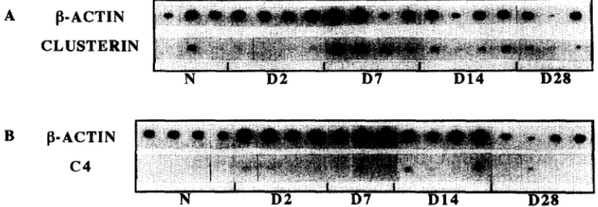
A [ ~ - A C T I N C L U S T E R I N N D 2 D7 D 1 4 D 2 8 B ~ - A C T I N C 4 N D 2 D7 D I 4 D 2 8
Fig. 1. Southern blots representing the co-expression of fl-actin/clusterin (A) and fl-actin/C4 (B) mRNA in sciatic nerves of adult rats: naive control animals (N) and 2 (D2), 7 (D7), 14 (D14), 28 (D28) days after crush injury. /3-actin mRNA was used as an endogenous internal standard for both semi-quantitative RT-PCR experiments, n = 3 or 4 animals per group.
A.-S. Bonnard et al. / lmmunopharmacology 38 (1997) 81-86 85 70000 60000 => soo00 .~_ 40000 ~ 3 0 0 0 0 "6 200o0 z < 10000 rw E 0 ¢r I I I I I N D2 D7 D14 D28 GROUPS 20000 150OO 10000 5000 E 0 I I I I N D2 D7 D14 D28 GROUPS
Fig. 2. Graphic representation of clusterin (A) and C4 (B) m R N A expression in sciatic nerves of adult rats: naive control animals (N) and 2 (D2), 7 (D7), 14 (D14), 28 (D28) days after crush injury. Quantification of values was done by scanning autoradio- graphies of Southern blots (presented in Fig. 1). Each value of clusterin or C4 m R N A expression was corrected for the difference of percentage between its corresponding /3-actin value and the mean/3-actin value (for details, see Section 2). Means + S.D. were calculated at each time, and are represented on two distinct scales because of the differences between clusterin and C4 rnRNA levels of expression. ANOVA analysis of variance ( F clusterin = 4.3, F C4 = 21.8) and Fisher's F test ( p < 0 . 0 5 ) were performed. (A) ( * ) versus N and D2 groups. (B) ( * ) versus N group.
At D2, D7 and DI4, C4 mRNA expression remained at a high level, representing respectively 2.5-, 2- and 3-fold the basic level of expression (N). Then it decreased progressively to reach back the N level of expression between D14 and D28.
4. Discussion
In this study, we demonstrate that both clusterin and C4 mRNAs are constitutively expressed and
increased at the lesion site following rat sciatic nerve crush. Indeed, clusterin mRNA expression reaches a maximum at D7-D14, and C4 mRNA expression at D 2 - D I 4 .
Liu et al. (1995) provided evidence for C classical pathway and regulator activation after sciatic nerve transection. They observed an increase of C1 and C l q immunoreactivity as soon as D2, which re- mained high until D28. These authors also demon- strated the up-regulation of clusterin mRNA from D2 to D7, and of clusterin immunoreactivity from D2 to D28. The slight discrepancies observed at D2 and D28 could be explained by the fact that their experi- ments were carded out in the spinal cord, after sciatic nerve transection, and by other technical ap- proaches. Our results confirm and extend this study, since we demonstrate a markedly enhanced C4 mRNA expression at the crush site, which strength- ens the hypothesis of C classical pathway activation. Following a peripheral nerve injury, regeneration cannot occur properly if Wallerian degeneration is not achieved (Beuche and Friede, 1984; Dahlin, 1995; Perry et al., 1987). Macrophage recruitment and myelin debris phagocytosis are critical pro- cesses, as they clear the way for proliferating Schwann cells, which are the main support for spon- taneously regenerating peripheral axons. Previous studies, realized in our laboratory, have demon- strated that microglial cells and astrocytes are both able to synthesize all the C components as well as clusterin (Gasque et al., 1992, 1993, 1995). By anal- ogy with CNS cells, we could postulate that Schwann cells, participating to the PNS scaffold tissue like astrocytes in the CNS, and macrophages recruited at the lesion site represent the best candidates to as- sume the production of C components and clusterin. We observed that the stimulation of C4 mRNA expression started earlier than the clusterin one. That could support the idea that the activation of C cas- cade plays a role in Wallerian degeneration, mainly by stimulating macrophage recruitment (anaphy- latoxins) and myelin phagocytosis (opsonization). Schwann cells could also participate to this step, since they are able to exert a phagocytic activity (Fernandez-Valle et al., 1995; Reichert et al., 1994). Moreover, the delayed expression of clusterin mRNA could be emphasized by its functions in the regulation of the C terminal pathway and in tissue
86 A.-S. Bonnard et al. / lmmunopharmacology 38 (1997) 81-86
remodelling (Rosenberg and Silkensen, 1995; Silkensen et al., 1994). On the one hand, clusterin is considered as an inhibitor of the MAC formation and subsequently of the C-mediated cytolysis. On the other hand, clusterin is involved in lipid recycling, membrane protection and maintenance of cell-cell or cell-substratum interactions. The combination of these effects leads us to think that clusterin produc- tion at the lesion site could provide a favorable environment to Schwann cell proliferation and ax- onal regrowth.
This pilot work on C4 and clusterin expression at the site of the crush injury needs obviously to be further investigated. Determination of the time course of other C components and regulators expression has to be achieved as well as the identification of their cellular sources. Furthermore, it would be very inter- esting to inquire about the in situ effects of the administration of C components and regulators, at the lesion site. For instance, using a model of periph- eral nerve regeneration through a tubular prosthesis (Seckel, 1990) would be greatly valuable to point out neuroprotective or neurodestructive effects of in- flammatory mediators mainly recognized yet for their immune properties.
Acknowledgements
The authors would like to address very special thanks to Dr. N. Bodjarian for precious help in the editing of the manuscript.
References
Beuche, W., Friede, R.L., 1984. The role of non-resident cells in Wallerian degeneration. J, Neurocytnl. 13, 767-796. Dahlin, L.B., 1995. Prevention of macrophage invasion impairs
regeneration in nerve grafts. Brain Res. 679, 274-280. Danielsen, N., 1990. Regeneration of the rat sciatic nerve in the
silicone chamber model. Restor. Neurol. Neurosci. 1,253-259. Dukas, K., Sarfati, P., Vaysse, N., Pradayrol, L., 1993. Quantifica- tion of changes in the expression of multiple genes by simulta- neous polymerase chain reaction. Anal. Biochem. 215, 66-72. Fernandez-Valle, C., Bunge, R.P., Bunge, M.B., 1995. Schwann cells degrade myelin and proliferate in the absence of
macrophages: evidence from in vitro studies of Wallerian degeneration. J. Neurocytol. 24, 667-679.
Gasque, P., Fontaine, M., Morgan, B.P., 1995. Complement ex- pression in human brain. Biosynthesis of terminal pathway components and regulators in human glial cells and cell lines. J. Immunol. 154, 4726-4733.
Gasque, P., Julen, N., Ischenko, A., Picot, C., Mauger, C., Chauzy, C., Ripoche, J., Fontaine, M., 1992. Expression of comple- ment components of the alternative pathway by glioma cell lines. J. Immunol. 149, 1381-1387.
Gasque, P., Ischenko, A., Legoedec, J., Mauger, C., Schouft, M.T., Fontaine, M., 1993. Expression of complement classical pathway by human glioma in culture. A model for comple- ment expression by nerve cells. J. Biol. Chem. 268, 25068- 25074.
Gay, D., Esiri, M., 1991. Blood-brain barrier damage in acute multiple sclerosis plaques. An immunocytological study. Brain 114, 557-572.
Hirschberg, D.L., Yoles, E., Belkin, M., Schwartz, M., 1994. Inflammation after axonal injury has conflicting consequences for recovery of function: rescue of spared axons is impaired but regeneration is supported. J. Neuroimmunol. 50, 9-16. Liszewski, M.K., Farries, T.C., Lublin, D.M., Rooney, I.A.,
Atkinson, J.P., 1996. Control of the complement system. Adv. lmmunol. 61, 201-283.
Liu, L., Trrnqvist, E., Mattsson, P., Eriksson, N.P., Persson, J.K.E., Morgan, B.P., Aldskogius, H., Svensson, M., 1995. Complement and clusterin in the spinal cord dorsal horn and gracile nucleus following sciatic nerve injury in the adult rat. Neuroscience 68, 167-179.
Lu, X., Richardson, P.M., 1991. Inflammation near the nerve cell body enhances axonal regeneration. J. Neurosci. 11,972-978. McGeer, P.L., McGeer, E.G., 1992. Complement proteins and complement inhibitors in Alzheimer's disease. Res. lmmunol.
143, 621-623.
Morrison, C., Gannon, F., 1994. The impact of the PCR plateau phase on quantitative PCR. Biochim. Biophys. Acta 1219, 493-498.
Perry, V.H., Brown, M.C., Gordon, S,, 1987. The macrophage response to central and peripheral nerve injury: a possible role for macrophages in regeneration. J. Exp. Med. 165, 1218-
1223.
Reichert, F., Saada, A., Rotshenker, S., 1994. Peripheral nerve injury induces Schwann cells to express two macrophage phenotypes: phagocytosis and the galactose-specific lectin MAC-2. J. Neurosci. 14, 3231-3245.
Rosenberg, M.E., Silkensen, J., 1995. Clusterin: physiologic and pathophysiologic considerations. Int. J. Biochem. Cell Biol. 27, 633-645.
Seckel, B.R., 1990. Enhancement of peripheral nerve regenera- tion. Muscle Nerve 13, 785-800.
Silkensen, J.R., Schwochau, G.B., Rosenberg, M.E., 1994. The role of clusterin in tissue injury. Biochem. Cell Biol. 72, 483-488.