HAL Id: hal-02011063
https://hal.univ-grenoble-alpes.fr/hal-02011063
Submitted on 7 Feb 2019
HAL is a multi-disciplinary open access
archive for the deposit and dissemination of
sci-entific research documents, whether they are
pub-lished or not. The documents may come from
teaching and research institutions in France or
abroad, or from public or private research centers.
L’archive ouverte pluridisciplinaire HAL, est
destinée au dépôt et à la diffusion de documents
scientifiques de niveau recherche, publiés ou non,
émanant des établissements d’enseignement et de
recherche français ou étrangers, des laboratoires
publics ou privés.
The cAMP pathway regulates mRNA decay through
phosphorylation of the RNA-binding protein
TIS11b/BRF1
Felicitas Rataj, Séverine Planel, Agnès Desroches-Castan, Juliette Le Douce,
Khadija Lamribet, Josiane Denis, Jean-Jacques Feige, Nadia Cherradi
To cite this version:
Felicitas Rataj, Séverine Planel, Agnès Desroches-Castan, Juliette Le Douce, Khadija Lamribet, et
al.. The cAMP pathway regulates mRNA decay through phosphorylation of the RNA-binding protein
TIS11b/BRF1. Molecular Biology of the Cell, American Society for Cell Biology, 2016, 27 (24),
pp.3841-3854. �10.1091/mbc.E16-06-0379�. �hal-02011063�
MBoC |
ARTICLE
The cAMP pathway regulates mRNA decay
through phosphorylation of the RNA-binding
protein TIS11b/BRF1
ABSTRACT TPA-inducible sequence 11b/butyrate response factor 1 (TIS11b/BRF1) belongs to the tristetraprolin (TTP) family of zinc-finger proteins, which bind to mRNAs containing AU-rich elements in their 3′-untranslated region and target them for degradation. Regulation of TTP family function through phosphorylation by p38 MAP kinase and Akt/protein kinase B signaling pathways has been extensively studied. In contrast, the role of cAMP-dependent protein kinase (PKA) in the control of TTP family activity in mRNA decay remains largely un-known. Here we show that PKA activation induces TIS11b gene expression and protein phos-phorylation. Site-directed mutagenesis combined with kinase assays and specific phosphosite immunodetection identified Ser-54 (S54) and Ser-334 (S334) as PKA target amino acids in vitro and in vivo. Phosphomimetic mutation of the C-terminal S334 markedly increased TIS11b half-life and, unexpectedly, enhanced TIS11b activity on mRNA decay. Examination of protein–protein interactions between TIS11b and components of the mRNA decay machinery revealed that mimicking phosphorylation at S334 enhances TIS11b interaction with the decapping coactivator Dcp1a, while preventing phosphorylation at S334 potentiates its in-teraction with the Ccr4-Not deadenylase complex subunit Cnot1. Collectively our findings establish for the first time that cAMP-elicited phosphorylation of TIS11b plays a key regula-tory role in its mRNA decay-promoting function.
INTRODUCTION
Besides transcription, posttranscriptional mechanisms play a major role in the regulation of gene expression. In particular, mRNA stabil-ity is a key step that progressively appears to be a highly regulated step. Importantly, this mechanism is responsive to modifications of the cellular environment (hormonal variations, hypoxia, etc.) and regulates the expression of subsets of proteins whose levels need to be rapidly adjusted. The regulation of mRNA stability involves cis sequences located mainly in the 3′ untranslated region (UTR) of the target mRNA that are bound by trans-acting factors. The most stud-ied cis element is the AU-rich element (ARE) located in the 3′ UTR of short-lived mRNAs encoding proteins such as cytokines, growth fac-tors, or metabolic regulators.
A great effort has been devoted over the past two decades to the identification of ARE-binding proteins and analysis of their contribution to the control of mRNA stability (Garneau et al., 2007). mRNA-stabilizing proteins include the members of the Hu
Monitoring Editor Marvin P. Wickens University of Wisconsin Received: Jun 7, 2016 Revised: Sep 29, 2016 Accepted: Sep 30, 2016
This article was published online ahead of print in MBoC in Press (http://www .molbiolcell.org/cgi/doi/10.1091/mbc.E16-06-0379) on October 5, 2016. †These authors contributed equally to this work.
*Address correspondence to: Nadia Cherradi (nadia.cherradi@cea.fr).
© 2016 Rataj, Planel, et al. This article is distributed by The American Society for Cell Biology under license from the author(s). Two months after publication it is available to the public under an Attribution–Noncommercial–Share Alike 3.0 Un-ported Creative Commons License (http://creativecommons.org/licenses/by-nc -sa/3.0).
“ASCB®,” “The American Society for Cell Biology®,” and “Molecular Biology of the Cell®” are registered trademarks of The American Society for Cell Biology. Abbreviations used: ACTH, adrenocorticotropin; ARE, AU-rich element; BAC, bovine adrenocortical; BRF1, butyrate response factor 1; CHIP, chromatin immu-noprecipitation; CI, confidence interval; CRE, cAMP response element; CTD, C-terminal; DRB, 5,6-dichloro-1-β-d-ribofuranosylbenzimidazole; IgG,
immunoglob-ulin G; MAPK, MAP kinase; MK2, MAPK-activated protein kinase 2; NTD, N-terminal domain; OA, okadaic acid; PBS, phosphate-buffered saline; PKA, cAMP-dependent protein kinase; PKB, protein kinase B; PP2A, protein phospha-tase 2A; RT-qPCR, quantitative reverse transcription-PCR; TIS11b, TPA-inducible sequence 11b; TSS, transcription start site; TTP, tristetraprolin; CIM, TTP-Ccr4-Not interaction motif; VEGF, vascular endothelial growth factor; WT, wild type.
Felicitas Rataj†, Séverine Planel†, Agnès Desroches-Castan, Juliette Le Douce, Khadija Lamribet, Josiane Denis, Jean-Jacques Feige, and Nadia Cherradi*
Institut National de la Santé et de la Recherche Médicale, INSERM U1036, Commissariat à l’Energie Atomique et aux Energies Alternatives, Institut de Biosciences et Biotechnologies de Grenoble, Laboratoire Biologie du Cancer et de l’Infection, and Université Grenoble Alpes, Unité Mixte de Recherche-S1036, F-38000 Grenoble, France
TIS11b exogenously delivered to preestab-lished tumors in mice exerts antiangiogenic and antitumoral effects through multitarget destabilization of tumor mRNAs (Planel
et al., 2010).
TIS11 family proteins are distal targets for signaling pathways, allowing transfer of external stimuli to the mRNA decay ma-chinery (Venigalla and Turner, 2012). The p38 MAP kinase (MAPK) and its down-stream kinase MAPK-activated protein ki-nase 2 (MK2) play a pivotal role in ARE-mediated mRNA decay. Activation of p38 MAPK has been shown to impair deadenyl-ation of ARE-containing mRNAs, leading to mRNA stabilization (Marchese et al., 2010). Macrophages from MK2−/− mice
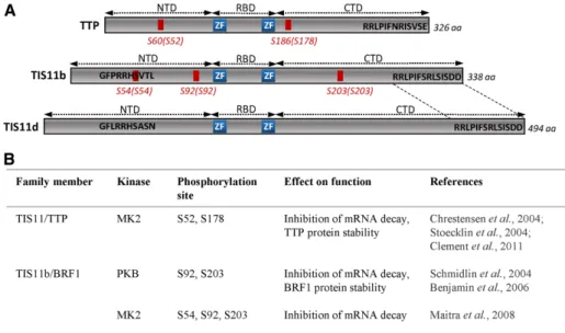
show severely reduced levels of TNFα, IL-1, IL-6, and IFNγ due to decreased cyto-kine mRNA stability (Kotlyarov et al., 1999; Neininger et al., 2002). A major target of MK2 is TTP, which is directly phosphory-lated at Ser-52 (S52) and Ser-178 (S178) (Figure 1), allowing binding of 14-3-3 adaptor proteins (Chrestensen et al., 2004). This interaction impairs the recruit-ment of the deadenylation machinery to the target mRNA (Stoecklin et al., 2004; Clement et al., 2011). On the other hand, phosphorylation of TTP by MK2 on S52 and S178 stabilizes TTP protein by preventing TTP degradation by the proteasome (Brook et al., 2006). It has been suggested that MK2 is counterbalanced by protein phosphatase 2A (PP2A), which directly competes with 14-3-3 protein for binding to TTP. PP2A dephosphorylates TTP at Ser-178 (and possibly at other serine residues) and thereby activates mRNA decay (Sun et al., 2007). More recently, phosphopeptide mapping and mass spectrometry analyses allowed the identification of a major tryptic phosphopep-tide containing S90 and S93 in human TTP (Cao et al., 2014).
In contrast with the accumulating data on the impact of TTP phosphorylation on its function, few studies address the role of TIS11b phosphorylation in ARE-mediated mRNA decay (Figure 1). Phosphorylation of TIS11b by the Akt/protein kinase B (PKB) at S92 and S203 abrogates mRNA decay of an IL-3 ARE-containing transcript and leads to TIS11b binding to 14-3-3 proteins and to TIS11b protein stabilization (Schmidlin et al., 2004; Benjamin et al., 2006). Phosphorylation of TIS11b by MK2 at S54, S92, and S203 inhibits its ability to destabilize ARE-containing mRNAs (Maitra
et al., 2008). The striking number of potential phosphorylation
sites in the TIS11b sequence suggests that the protein has to inte-grate multiple signals to provide the appropriate cellular response. In this study, we show that the cAMP signaling pathway orches-trates all the steps controlling TIS11b expression and function, from TIS11b promoter activation to TIS11b protein phosphoryla-tion. We identified two cAMP-dependent protein kinase (PKA) phosphorylation sites in living cells, S54 and S334, and investi-gated the role of these phosphosites in the control of TIS11b pro-tein half-life and function in mRNA decay. Our data indicate that phosphorylation of S334 regulates the stability of TIS11b and its ability to interact with binding partners and to trigger mRNA degradation.
family (HuR, HuC, and HuD; Meisner and Filipowicz, 2010), whereas major mRNA-destabilizing proteins comprise members of the TIS11 family of double zinc-finger proteins, KSRP and AUF1 (Briata et al., 2013; Brooks and Blackshear, 2013; White et al., 2013). The TIS11 protein family is composed of four known mem-bers: TTP (Tristetraprolin/TIS11/ZFP36), TIS11b (ZFP36L1/BRF1), TIS11d (ZFP36L2/BRF2), and ZFP36L3, which is expressed exclu-sively in mouse placental tissue (Blackshear et al., 2005). In vitro, the three main members of the TIS11 family (TTP, TIS11b, and TIS11d) have been shown to interact with several ARE-containing mRNAs and to trigger deadenylation and degradation of the tar-get mRNAs (Baou et al., 2009). However, genetic knockout of each member in mice leads to different phenotypes, thus suggesting that TIS11 proteins might have specific target mRNAs in vivo (Taylor et al., 1996; Bell et al., 2006; Stumpo et al., 2009). TTP, the most-studied member of the TIS11 family, has been shown to trig-ger degradation of several transcripts, including TNF-α, GM-CSF, IL-2, and IL-10 mRNAs (Carballo et al., 1998, 2000; Ogilvie et al., 2005; Stoecklin et al., 2008). TTP and TIS11b are able to recruit the components of the mRNA decay machinery (Fenger-Gron et al., 2005; Lykke-Andersen and Wagner, 2005; Clement et al., 2011). We have previously shown that TIS11b destabilizes the mRNA of the angiogenic cytokine vascular endothelial growth factor (VEGF; Ciais et al., 2004) and the mRNA of the steroidogenic acute regu-latory protein (Duan et al., 2009) through interaction with ARE mo-tifs in their 3′ UTRs. In endocrine cells, transient hormone-induced expression of VEGF mRNA is regulated through antagonistic ac-tions of HuR and TIS11b (Cherradi et al., 2006). More recently, we demonstrated that TIS11b controls mineralocorticoid receptor mRNA stability in renal cells exposed to hypertonicity in vitro and in vivo (Viengchareun et al., 2014). Importantly, a cell-penetrating
FIGURE 1: Schematic representation of known phosphorylation sites in human TTP family members. (A) NTD and CTD represent the N-terminal and C-terminal activation domains on either sides of the zinc-finger (ZF) RNA-binding domain (RBD). The serines that have been shown previously to regulate TTP family function upon phosphorylation are indicated in red.
Corresponding serines in mouse proteins are indicated with brackets. The conserved C-terminal sequence between TIS11b and TIS11d is indicated between dotted lines. Other phosphoserines have been identified in vitro and in phosphoproteomic studies of ectopically expressed TTP (reviewed in Venigalla and Turner, 2012). However, the relevance of these phosphosites remains to be validated experimentally. No phosphorylated serines in TIS11d/BRF2 have been reported so far. (B) Protein kinases regulating TTP family fate and function. aa, amino acid.
found at −30 to −27 and at −402 to −394 relative to the TSS, respec-tively (Figure 3A). This sequence was highly conserved among spe-cies, suggesting an important role in cAMP-regulated TIS11b ex-pression (Figure 3B). No classical CRE was identified within the −2000 base pairs upstream of the TSS of the two other family mem-bers TTP and TIS11d (unpublished data). A promoter sequence comprising the region −1038 to +52 relative to the TSS of human TIS11b was cloned and inserted in pGL3-luciferase reporter plas-mid to study the activity of the canonical CRE (pWT construct; Figure 3C). Transfection of COS7 cells with pWT and subsequent treatment with forskolin to increase cAMP levels significantly stimu-lated the basal activity of the reporter gene about threefold, thus indicating that TIS11b promoter is regulated by the cAMP signaling pathway (Figure 3D). Cotreatment of COS7 cells with the cAMP-dependent protein kinase (PKA) inhibitor RpcAMP strongly inhib-ited the cAMP-induced activation of the reporter gene. To investi-gate the role of the putative CRE in mediating the response to cAMP, we mutated the CRE sequence TGACGTCA into TCTC-GAGA (pmutCRE construct). Transfection of COS7 cells with pmut-CRE completely abrogated forskolin-mediated stimulation of lucif-erase activity, thus indicating a key role of the CRE in cAMP-mediated transactivation of TIS11b promoter (Figure 3D). As the CRE-binding protein CREB functions as a trans-acting regulator of genes contain-ing a CRE sequence in their promoter, we examined whether CREB was involved in transactivation of TIS11b promoter by cAMP using anti–phospho-CREB antibodies. Forskolin or ACTH triggered a time-dependent phosphorylation of CREB peaking at 30 min in COS7 and BAC cells, respectively (Supplemental Figure S1, A–D). To provide further evidence that the CRE is implicated in CREB binding to DNA upon cAMP stimulation, we performed chromatin immunoprecipitation (CHIP) experiments in COS7 cells transfected with pWT promoter construct. A specific interaction occurred in vivo between CREB and TIS11b promoter at the basal level, and this interaction was increased at 30 min posttreatment with forskolin (Supplemental Figure S1E). Similarly, CHIP experiments in BAC cells challenged with ACTH revealed that CREB was bound to endoge-nous TIS11b promoter at 30 min poststimulation (Supplemental Figure S1F). Collectively these data indicate that the PKA signaling pathway regulates TIS11b gene transcription through CREB activation.
The cAMP-dependent protein kinase regulates TIS11b expression and phosphorylation
We have previously shown that ACTH increases TIS11b protein ex-pression and that silencing of TIS11b compromises VEGF mRNA decay in endocrine cells (Chinn et al., 2002; Cherradi et al., 2006). In addition, activation of the cAMP signaling pathway induced a broad series of 38–50 kDa TIS11b bands that collapsed into a single band after λ-phosphatase treatment, indicating that the slower-mobility species arose from phosphorylation events (Duan et al., 2009). For investigation of the role of PKA in the hormonal induction of TIS11b and its target mRNA, BAC cells were stimulated by ACTH for various periods of time in the absence or presence of H89, a PKA-specific inhibitor. Western blot analysis of cell lysates showed that the hor-mone induced a marked time-dependent increase in TIS11b protein levels (Figure 4, A and B) that was accompanied by a shift toward high-molecular-weight species (Figure 4A; see dotted lane as a ref-erence for migration). In contrast, the levels of the three major VEGF isoforms (VEGF121, VEGF165, and VEGF189; Planel et al., 2010) peaked at 2 h, then decreased toward basal levels (Figure 4, A and C). Quantitative PCR analysis of total RNA revealed a transient ACTH-induced increase in VEGF mRNA levels, consistent with
RESULTS
The cAMP signaling pathway regulates TIS11b gene transcription through the binding of the cAMP-response element (CRE)-binding protein CREB to TIS11b promoter
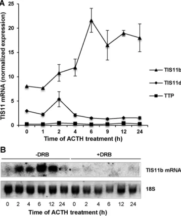
In adrenocortical cells in primary culture, the TIS11b transcript is expressed at very low levels and is rapidly induced by the cAMP-mobilizing hormone adrenocorticotropin (ACTH; Chinn et al., 2002; Cherradi et al., 2006). For defining the mechanisms involved in TIS11b mRNA induction, bovine adrenocortical (BAC) cells were stimulated by ACTH in the presence of the transcription inhibitor 5,6-dichloro-1-β-d-ribofuranosylbenzimidazole (DRB). The hormone
induced a marked increase in TIS11b mRNA levels, which was com-pletely abrogated in the presence of DRB (Figure 2, A and B), thus suggesting a transcription-dependent mechanism. Interestingly, TTP or TIS11d mRNA levels were not significantly changed upon ACTH challenge, pointing at a specific induction of TIS11b by the hormone (Figure 2A). For testing potential promoter elements, up to 2000 nucleotides upstream of the transcription start site (TSS) of the TIS11b gene (zfp36l1) were inspected using Ensembl Genome Browser (www.ensembl.org) and the previously published partial promoter sequence of the rat TIS11b gene (cMG1) (Corps et al., 1995). A TATAAA box and a cAMP response element (CRE) were
FIGURE 2: ACTH-induced expression of TIS11b mRNA in adrenocortical cells is transcription dependent. (A) Time-course analysis of TIS11 family member expression in BAC cells challenged with ACTH. RT-qPCR was performed as described in the Supplemental Material. Expression levels of TTP, TIS11b, and TIS11d mRNAs were normalized to RPL27 mRNA. Values are means ± SD of four independent experiments. (B) Northern blot analysis of TIS11b transcript levels in BAC cells challenged with ACTH in the absence or presence of the transcription inhibitor DRB. The membrane was hybridized to a radiolabeled TIS11b cDNA probe and rehybridized to 18S RNA probe for loading control. Shown is a representative Northern blot of six independent experiments.
VEGF protein expression (Figure 4D). Re-markably, the high induction of TIS11b at 6 h was correlated with low VEGF mRNA and protein levels. Stimulation of TIS11b and VEGF expression by ACTH were dra-matically reduced in the presence of H89 (Figure 4, A–D) with disappearance of high-molecular-weight forms of TIS11b.
To further establish the effect of ACTH on TIS11b phosphorylation, we performed immunoprecipitation experiments in [32P]
orthophosphate-labeled BAC cells. A basal phosphorylation level of TIS11b was de-tected in control cells, while ACTH induced a robust and time-dependent increase of
32P incorporation into TIS11b, which was
markedly impaired in the presence of H89 (Figure 4E). Quantification of phospho-TIS11b/total TIS11b ratio in independent experiments revealed that ACTH increased TIS11b phosphorylation by 2.4 ± 0.4-fold at 6 h poststimulation (Figure 4F). We next performed in vitro phosphorylation experi-ments to determine whether TIS11b is a di-rect substrate of PKA. Purified recombinant glutathione S-transferase (GST)-TIS11b was incubated in the presence of the catalytic subunit of PKA and [γ-32P]-labeled ATP. As
shown in Figure 4G, TIS11b was efficiently phosphorylated by PKA in vitro. Taken to-gether, these results suggest that activation of PKA signaling in endocrine cells is a piv-otal mechanism whereby concomitant ex-pression and phosphorylation of TIS11b are associated to a transient induction of its tar-get mRNA VEGF.
Identification of PKA target sites within the TIS11b sequence: conserved S54 and S334 are phosphorylated in vitro and in vivo
TTP, TIS11b, and TIS11d each consist of an RNA-binding zinc-finger domain flanked by N-terminal (NTD) and C-terminal (CTD) do-mains that can activate mRNA decay (Figure 1; Lykke-Andersen and Wagner, 2005). In-spection of the coding sequence of TIS11b (SwissProt, accession number Q07352) us-ing the Phosphorylation Site Predictor soft-ware DISPHOS 1.3 (www.dabi.temple.edu/ disphos) revealed that the CTD of TIS11b harbors a majority of phosphorylatable ser-ine residues as compared with the NTD (Supplemental Figure S2). By contrast, anal-ysis of the TTP sequence showed that phos-phorylatable serines were almost equally distributed between the NTD and the CTD of TTP. Interestingly, TIS11d displayed a similar profile of phosphorylatable serines in its distal CTD to that of TIS11b, while an
FIGURE 3: Cloning and analysis of TIS11b promoter. (A) The sequence of the human TIS11b promoter region from −1670 nucleotides upstream of the transcription start site (+1) to the translation initiation ATG was determined using Ensembl Genome Browser (www.ensembl.org). The CRE is shown in red, and the TATAA box (bold) is underlined. (B) The CRE in TIS11b promoter is conserved among species. (C) Schematic representations of TIS11b promoter region constructs. The WT construct contains 1088 base pairs of the promoter region. The mutCRE construct results from the substitution of four nucleotides within the CREB consensus sequence as described in the Supplemental Material. (D) WT and mutCRE constructs were inserted in pGL3-luciferase reporter plasmid and transfected in COS7 cells as described in the
Supplemental Material. Promoter-driven luciferase activity was measured after cell stimulation with 25 μM forskolin in the presence or absence of RpcAMP (10 nM) for 24 h. Results are represented as a percentage of luciferase activity in control nonstimulated cells. Transfections were performed in triplicate, and values are means ± SD from three independent experiments. *, significantly different from pWT luciferase activity in control nonstimulated cells, with p < 0.05; #, significantly different from pWT luciferase activity in forskolin-treated cells, with p < 0.05.
served not only between TIS11b and TIS11d but also between species (Figure 5A), we focused on both of these serines.
We first tested whether TIS11b peptides spanning either S54 (amino acids [aa] 50–60) or S334 (aa 330–338) were phosphorylated by PKA in vitro. SDS–PAGE analysis and au-toradiography revealed a strong dose-de-pendent phosphorylation of the S54-bear-ing peptide, while PKA also phosphorylated the S334-bearing peptide, but to a lesser extent (Figure 5B). To determine whether S54 and S334 were directly targeted by PKA, we generated Flag-tagged wild-type (WT) and mutant TIS11b in which S54 or S334 was replaced by an alanine to prevent phosphorylation (S54A or S334A). Purified WT or mutant proteins were incubated in vitro with PKA. As shown in Figure 5, C and D, phosphorylation of TIS11b S54A or S334A mutants was significantly decreased as compared with the WT protein. These observations led us to investigate whether S54 and S334 were phosphorylated in vivo. We therefore generated phospho-S54–spe-cific and phospho-S334–spephospho-S54–spe-cific antibodies in rabbits immunized with phosphopeptides spanning S54 (CAGGGFPRRH(Sp)VTL) or S334 (RRLPIFSRL(Sp)ISD). The specificity of the sera was first evaluated by Western blot analysis of the phosphorylated and unphos-phorylated control peptides (Figure 5E and Supplemental Figure S3). A site-specific and dose-dependent increase in phosphopep-tide detection was observed, while the controls were not recognized by the phos-phospecific antibodies. For further charac-terization of the phospho-S54–specific and phospho-S334–specific antibodies, phos-phorylation of WT TIS11b or of the non-phosphorylatable TIS11b mutants (S54A or S334A) was monitored in COS7 cells trans-fected with pTarget-TIS11b constructs, then stimulated with forskolin in the absence or presence of H89 (Figure 5F). Endogenous TIS11b expression was barely detectable in COS7 cells (Figure 5F, empty pTarget plas-mid or Vect). In basal conditions, a weak phosphorylation of endogenous TIS11b at S54 was detected, while the protein dis-played three distinct forms phosphorylated at S334. These findings suggest that basal PKA activity in COS7 cells is sufficient for endogenous TIS11b phosphorylation. For-skolin-induced PKA activation led to a ro-bust phosphorylation at both S54 and S334, which was prevented in the presence of H89. Importantly, the overexpressed TIS11b mu-tants S54A and S334A were not recognized by the phosphospecific antibodies (Figure 5F). These results indicate that the newly gener-ated antibodies are phosphospecific and that the PKA-induced phosphorylation event is efficient on WT TIS11b but is not on the alanine substitution mutants.
additional phosphorylation hotspot was predicted close to TIS11d zinc-finger domain (Supplemental Figure S2). To identify putative PKA phosphorylation sites within TIS11b, we used the NetPhosK (www.cbs.dtu.dk/services/NetPhos) software. Four motifs corre-sponding to PKA consensus phosphorylation sites (RXS or RRXS), including S54, S92, S192, and S334 were found. As S54 and S334 presented the highest predictive score and as they were highly
con-FIGURE 4: ACTH induces a cAMP-dependent expression and phosphorylation of TIS11b. (A) BAC cells were preincubated in the absence or presence of H89 (5 μM) for 30 min before addition of 10 nM of ACTH for the indicated periods of time. TIS11b and VEGF protein levels of whole-cell extracts (20 μg) were analyzed by Western blot. The blot was subsequently probed with an anti–β-actin to assess equal loading of samples. (B–D) Quantification of TIS11b, VEGF mRNA, and protein levels from independent experiments (n = 5, means ± SEM). Protein-level values were normalized to actin and are expressed as percentage of control values at time 0 (unstimulated cells). VEGF mRNA levels were measured by quantitative PCR and normalized to hypoxanthine-guanine phosphoribosyltransferase (HPRT). (E) Time-course of TIS11b phosphorylation in BAC cells stimulated with 10 nM of ACTH in the presence of [32P]orthophosphate and in the presence
or absence of H89. TIS11b was immunoprecipitated (IP) from cell extracts, resolved by SDS– PAGE, and then visualized by autoradiography. One representative experiment of four is shown. (F) Quantification of phospho-TIS11b/total TIS11b ratio in ACTH-stimulated BAC cells (n = 4, means ± SEM). (G) Phosphorylation of recombinant TIS11b by the catalytic subunit of PKA. Purified GST-TIS11b fusion protein was produced as described previously (Ciais et al., 2004). Increasing doses of GST-TIS11b were subjected to in vitro phosphorylation as described in
Materials and Methods. Protein extract from Escherichia coli (30 μg) transformed with empty
TIS11b is phosphorylated at S54 and S334 in both endocrine and cancer cells
To assess whether TIS11b is phosphorylated at S54 and S334 in different cell types, we used the highly differentiated BAC cells and the human lung carcinoma cell line A549. BAC cells were challenged with ACTH for various periods of time (Figure 6A). Cell ly-sates were probed either with specific anti– total TIS11b/TIS11d (Chamboredon et al., 2011) or with phosphospecific antibodies. It is worth mentioning that TIS11d expression was variable between primary cultures of BAC cells. TIS11d was detected at the basal level and slightly decreased at 6 h poststimu-lation, while TIS11b was nearly undetectable at the basal level and markedly up-regulated at 6 h by ACTH treatment. ACTH also in-creased the phospho-S54 signal, which par-alleled the increase in total TIS11b protein levels. Interestingly, TIS11d was heavily la-beled with the anti–phospho-S54 due to the conservation of the RRHS motif (S57 in TIS11d) between TIS11b and TIS11d. No phospho-S334 signal was detected for TIS11b before 6 h of stimulation by the hor-mone, suggesting a delayed ACTH-induced phosphorylation of this residue (Figure 6A). The single band detected belongs to the high-molecular-weight species of TIS11b (shown by an asterisk). As observed with the anti–phospho-S54, TIS11d was markedly la-beled with the anti–phospho-S334 antibod-ies (S490 in TIS11d). Again, this result is likely due to the perfect conservation of the C-ter-minal peptide between TIS11b and TIS11d. Importantly, cotreatment of BAC cells with ACTH and the PKA inhibitor H89 abrogated TIS11b induction and TIS11b phosphoryla-tion at S54 and S334 (Figure 6A).
We next evaluated whether phosphory-lation of S54 and S334 could occur in an-other cellular context. Besides hormones, hypoxia is another regulator of TTP protein family expression (Sinha et al., 2009; Kim
et al., 2010; Chamboredon et al., 2011). In
particular, TIS11b expression was shown to be increased in von Hippel–Lindau (VHL)-expressing renal cell carcinoma in response to hypoxia (Sinha et al., 2009). A549 lung carcinoma cells were exposed to normoxia or hypoxia (1.5% O2) for 2–8 h, then total
cell extracts were analyzed by Western blot. A transient but robust increase in TIS11b levels was observed under hypoxia, peaking between 2 and 4 h of exposure and declin-ing at 8 h (Figure 6B). This increase in total protein level was accompanied by an in-crease in the quantity of phosphorylated TIS11b at the S54 and S334 residues. Inter-estingly, as observed for TIS11b, TIS11d was
FIGURE 5: S54 and S334 are PKA target sites in vitro and in vivo. (A) Sequence alignment of conserved amino acid within the N-terminus and the C-terminus between TIS11b, TIS11d, and TTP showing PKA consensus motifs (highlighted in red, RRHS and RLS). These motifs are also conserved between species in TIS11b sequence and harbor S54 S334 (hs, Homo sapiens; Bt, Bos
taurus; Rt, Rattus norvegicus; mm, Mus musculus; xl, Xenopus laevis). (B) Dose-dependent in
vitro phosphorylation of synthetic N-terminal and C-terminal peptides of TIS11b by the catalytic subunit of PKA. Both peptides contain the PKA consensus motifs RRHS or RLS (S54 and S334 are shown in bold; aa, amino acids). Phosphorylated peptides were resolved by SDS–PAGE (15%) and visualized by autoradiography. (C) In vitro phosphorylation of recombinant Flag-WT TIS11b and Flag-TIS11b mutants S54A and S334A (1 μg purified proteins). Protein extracts from
E. coli transformed with empty vector (pET15b) served as control (Vect). (D) PKA-mediated
phosphorylation of recombinant TIS11b was significantly impaired when S54 and S334 were replaced by an alanine. Ratios of phosphorylated protein/total protein are reported (n = 4 independent experiments, mean ± SEM). Asterisks: significantly different from the WT with **p < 0.01 and ***p < 0.001. (E) Characterization of the phosphospecific antibodies in vitro. Unphosphorylated control peptides were run alongside phosphorylated peptides to determine whether the antibodies could detect the phospho-S34 (pS54) or phospho-S334 (pS334). (F) Characterization of the phosphospecific antibodies in forskolin-stimulated cells. COS7 cells were transfected with empty pTarget Vector (Vect), pTarget-WT TIS11b (WT), pTarget-TIS11b-S54A, or pTarget-TIS11b-S334A plasmids and then stimulated or not (basal) with 25 μM of forskolin for 60 min in the presence or absence of H89. Cell lysates were analyzed by Western blot to assess the specificity of the anti-pS54 or anti-pS334 antibodies. Blots were imaged simultaneously with the Chemidoc Imaging system for 5 s to accurately detect strong and weak bands for all conditions.
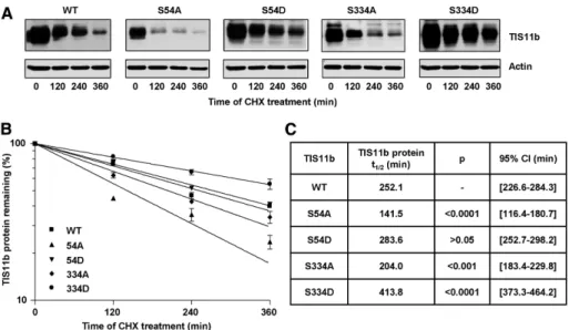
S54 and S334 are involved in the control of TIS11b protein half-life
TIS11b protein turnover was shown to be regulated by PKB through S92 and S203 phosphorylation sites (Benjamin et al., 2006). We sought to investigate whether S54 and S334 were also involved in the regulation of TIS11b protein half-life. COS7 cells were transfected with WT TIS11b, TIS11b-S54A, TIS11b-S54D, TIS11b-S334A, or TIS11b-S334D mutants, and then transla-tion was arrested by cycloheximide at the indicated time points. The steady-state lev-els of TIS11b alanine mutants (S54A and S334A) were lower than those of WT TIS11b (time point t = 0, Figure 7A), while the steady-state levels of TIS11b aspartate mu-tants (S54D and S334D) were higher than those of WT TIS11b. These results suggest that S54 and S334 substitutions impact TIS11b protein turnover. Nonlinear regres-sion to a first-order exponential decay model yielded a half-life for WT TIS11b of 252.1 min (Figure 7, B and C). The mutant TIS11b-S54A displayed a shorter half-life of 141.5 min, while the half-life of the TIS11b-S54D mutant was higher than that of the WT (t1/2= 273.6 min). The TIS11b-S334A mutant was less stable (t1/2 = 204.0 min)
than WT TIS11b. By contrast, TIS11b pro-tein stability was substantially increased when the S334 was replaced by the phos-phomimetic aspartate (TIS11b-S334D), with a half-life of 413.8 min. Comparison of the best-fit values showed that the half-lives of TIS11b-S54A and TIS11b-S334D were the most significantly different from the half-life of WT TIS11b (Figure 7C). These data indi-cate that S54 and S334 are critical residues in the control of TIS11b turnover.
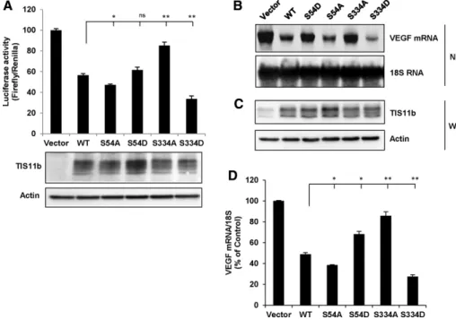
Mimicking phosphorylation at S334 potentiates TIS11b-mediated decrease in reporter mRNA activity and VEGF mRNA steady-state levels
TIS11b is a direct substrate of the kinases Akt/PKB (Schmidlin et al., 2004; Benjamin et al., 2006) and MK2 (Maitra et al., 2008). PKB- and MK2-induced phosphorylations of TIS11b at Ser-90/Ser-92/Ser-203 were reported to inhibit TIS11b-mediated ARE mRNA decay (Schmidlin et al., 2004; Stoecklin et al., 2004; Benjamin et al., 2006). For determining the role of S54 and S334 in TIS11b function, the mRNA-destabilizing activity of WT TIS11b, S54A, TIS11b-S54D, TIS11b-S334A, and TIS11b-S334D mutants was first assessed using a Luciferase-VEGF 3′ UTR fusion construct (Ciais et al., 2004) whose activity is driven by the ARE-containing 3′ UTR of VEGF. To ensure that differences in reporter activity were not due to differ-ences in TIS11b protein expression levels, we transfected variable doses (3–10 ng) of TIS11b constructs on the basis of half-life deter-minations (Figure 8) to achieve similar basal levels of TIS11b expres-sion. As shown in Figure 8A, overexpression of WT TIS11b de-creased luciferase activity by 43.4 ± 1.5%. Substitution of S54 by an alanine (S54A) modestly, although significantly, affected TIS11b-mediated decrease in luciferase activity (55.9 ± 1.0% inhibition). The heavily labeled with anti–phospho-S54-antibodies in response to
hypoxia. By contrast, TIS11d was labeled with anti–phospho-S334-antibodies in both normoxic and hypoxic conditions.
As the phosphorylation status of the TIS11b protein family is under the dual control of kinases and phosphatases (Benjamin et al., 2006; Brook et al., 2006; Sun et al., 2007), we sought to determine whether inhibition of serine/threonine phosphatases could impact the level of hypoxia-induced phosphorylation of S54 and S334 within TIS11b. Therefore A549 cells were exposed to normoxia or hypoxia for 8 h in the absence or presence of okadaic acid (OA), a potent inhibitor of protein phosphatases PP1 and PP2A. As shown in Figure 6C, treat-ment of A549 cells by OA markedly increased total TIS11b protein levels in normoxia, and this up-regulation was even more pronounced in response to hypoxia. In addition, OA induced accumulation of low-mobility bands of TIS11b under hypoxia (Figure 6C, top, asterisk). TIS11b protein was highly phosphorylated at S54 after OA treatment in normoxia and hypoxia (Figure 6C). Remarkably, OA-induced TIS11b was heavily phosphorylated at S334 under hypoxia with ap-pearance of low-mobility forms of the protein (Figure 6C, bottom, asterisk). Altogether these results indicate that the S54 and S334 resi-dues of TIS11b and their counterparts in TIS11d are phosphorylation target sites. Moreover, both residues were phosphorylated under dif-ferent physiological conditions and in difdif-ferent cell types, suggesting that they may play a wide biological role in TIS11b function.
FIGURE 6: TIS11b is phosphorylated at S54 and S334 in hormone- and hypoxia-stimulated cells. (A) BAC cells were stimulated with ACTH in the presence or absence of H89 for the indicated periods of time. Total-cell extracts were probed for total TIS11b/TIS11d, pS54-TIS11b, and pS334-TIS11b. Owing to the conserved PKA consensus motifs (RRHS and RHS; see Figure 2) within TIS11b and TIS11d sequences, both phosphoproteins were detected. Note the shift of TIS11b toward high-molecular-weight species. The asterisk indicates the phospho-(S334)-TIS11b species appearing at 6 h poststimulation by ACTH. (B) A549 lung carcinoma cells were exposed to normoxia (0 h) or hypoxia for 2, 4, and 8 h. Cells extracts were probed by Western blot as described in A. (C) A549 cells were exposed to normoxia or hypoxia for 8 h in the absence or presence of OA, an inhibitor of the phosphatases PP2A/PP1. Note the pS334-TIS11b species appearing under hypoxia in the presence of OA (indicated by an asterisk). Blots were subsequently probed with an anti–β-actin to assess equal loading of samples.
TIS11b-S334A mutant was significantly less active than WT TIS11b in promoting VEGF mRNA decay (t1/2 = 95.5 min). By contrast,
we observed that VEGF mRNA decay was markedly accelerated by the TIS11b-S334D mutant when compared with WT TIS11b (t1/2 = 40.6 min). Comparison of the best-fit
values showed that TIS11b-S334D–elicited mRNA decay was highly significantly differ-ent from WT TIS11b–elicited mRNA decay (Figure 9C). These results suggest that mim-icking phosphorylation at the residue S334 enhances the mRNA-destabilizing function of TIS11b.
S334 is a key residue in TIS11b interaction with the mRNA decay machinery
TTP has been reported to activate both 3′→5′ mRNA decay, by recruiting the multi-subunit Ccr4-Not deadenylase complex to the mRNA, and 5′→3′ mRNA decay, by recruiting the decapping enzymes Dcp2/ Dcp1a and the exonuclease Xrn1 (Fenger-Gron et al., 2005; Lykke-Andersen and Wagner, 2005; Marchese et al., 2010; Clement et al., 2011; Sandler et al., 2011; Fabian et al., 2013). The Ccr4-Not complex comprises several proteins named Ccr4-Not (Cnot) subunits. The Cnot1 subunit is a scaf-fold for the other Ccr4-Not components, including Ccr4 and Caf1 deadenylases (Goldstrohm and Wickens, 2008). While some studies have shown that the Ccr4-Not complex interacts with the NTD of TTP (Lykke-Andersen and Wagner, 2005), more recent data pointed at interactions of the complex with the CTD of TTP (Sandler et al., 2011; Fabian et al., 2013). A crystal struc-ture of Cnot1, a core subunit of Ccr4-Not, associated with a C-ter-minal peptide of TTP, led to the identification of a TTP-Ccr4-Not interaction motif (CIM; Figure 10A; Fabian et al., 2013). TTP-CIM is highly conserved between TTP family members and com-prises the S323 residue in TTP that corresponds to the S334 residue in TIS11b. To understand the functional effects of the phosphomi-metic TIS11b-S334D mutant, we examined protein–protein interac-tions between WT TIS11b or its S334 mutants and components of the mRNA decay machinery. As it was technically challenging to detect some of the mRNA decay enzymes in COS7 cells, coimmu-noprecipitation experiments were performed using HEK 293T cells transfected with WT TIS11b, TIS11b-S334A, or TIS11b-S334D mu-tant expression plasmids. TIS11b was subsequently immunoprecipi-tated from cell extracts, and the amounts of Cnot1, Dcp2, Dcp1a, or Xrn1 in the immunoprecipitates were detected by immunoblotting. Figure 10B shows that WT and mutant TIS11b were expressed at similar levels in transfected cells (input panel) and immunoprecipi-tated equally efficiently from cell extracts (IP panel). Protein com-plexes immunoprecipitated by anti-TIS11b antibodies contained Cnot1, Dcp2, Dcp1a, and Xrn1 but not HuR (negative control; Figure 10B), an ARE-binding protein that is not an activator of mRNA decay (Fan and Steitz, 1998). This result indicates that WT TIS11b exists in a complex with the major mRNA decay proteins. The nonphosphorylatable mutant TIS11b-S334A showed an in-creased interaction with Cnot1 as compared with that observed with the phosphomimetic mutant TIS11b-334D (Figure 10, B and C). activity of the mutant TIS11b-S54D was not statistically different
from that of the WT. When S334 was replaced by an alanine (S334A), the inhibitory effect of TIS11b was significantly impaired (15.3 ± 3.2% inhibition). Replacing S334 by an aspartate (S334D) signifi-cantly potentiated the inhibitory effect of TIS11b on luciferase activ-ity (65.2 ± 2.9% inhibition). Importantly, the observed differences in luciferase activities were not due to differences in expression levels of TIS11b (Figure 8A, bottom panel).
We next examined the effect of TIS11b mutant expression on endogenous VEGF mRNA steady-state levels. Overexpression of each mutant followed by Northern blot analysis revealed that the TIS11b-S54A mutant was slightly more active than the WT in de-creasing VEGF mRNA, while TIS11b-S54D was less efficient (Figure 8, B–D). Remarkably, the mutant TIS11b-S334D was significantly more active than the WT, while the activity of TIS11b-S334A mutant was significantly impaired. Altogether these results suggest that phosphorylation at S54 leads to a modest impairment of TIS11b-elicited decrease in VEGF transcript, whereas phosphorylation at S334 potentiates TIS11b function.
Mimicking phosphorylation at S334 enhances TIS11b-elicited VEGF mRNA decay
We next examined the effect of TIS11b mutant expression on en-dogenous VEGF mRNA turnover (Figure 9). COS7 cells were trans-fected either with WT or mutant TIS11b constructs and then treated with DRB for mRNA decay analysis (Figure 9A). Nonlinear regression to a first-order exponential decay model yielded a half-life of 177.5 min for VEGF mRNA in cells transfected with the empty vector (Figure 9, B and C). Overexpression of WT TIS11b significantly de-creased VEGF mRNA half-life to 71.5 min, while the TIS11b-S54A mutant modestly affected VEGF mRNA decay (t1/2 = 63.8 min). The
FIGURE 7: S54 and S334 regulate TIS11b protein stability. (A) COS7 cells were cotransfected with 10 ng of pTarget plasmids encoding WT TIS11b (WT), TIS11b S54A, TIS11b S54D, TIS11b S334A, or TIS11b S334D mutants and then treated with cycloheximide (CHX) as described in
Materials and Methods. The half-life of WT or mutant TIS11b was analyzed by Western blot. All
the membranes were exposed simultaneously for 15 s. Quantification of TIS11b steady-state levels (TIS11b/actin ratio) at t = 0 yielded the ratios 1, 0.61, 1.27, 0.85, and 1.58 for WT TIS11b, TIS11b-S54A, S54D, TIS11b-S334A, and TIS11b-S334D mutants, respectively. (B) TIS11b protein levels were normalized to actin levels and plotted as a percentage of the initial value against time using nonlinear regression to a first-order exponential decay model. (C) Calculated half-lives of TIS11b from four independent experiments. Half-lives of TIS11b mutants were compared with the half-life of WT TIS11b. p Values and 95% confidence intervals (CIs) were determined using an F-test.
way. We uncovered novel PKA-dependent phosphorylations of the NTD and CTD of TIS11b in vitro and in vivo and identified S54 and S334 as physiological phosphoryla-tion sites of PKA. Our major findings sug-gest that phosphorylation at S334 markedly enhances TIS11b protein stability and TIS11b association with the decapping co-activator Dcp1a. Both events might contrib-ute to the increased efficiency of phospho– S334-TIS11b to trigger mRNA turnover.
We have previously shown that ACTH induces a transient increase in VEGF mRNA levels and that TIS11b is involved in the de-cay phase of VEGF mRNA expression (Chinn
et al., 2002; Cherradi et al., 2006). The
pres-ent study indicates that the hormone also induces a rapid phosphorylation of TIS11b that is significantly blunted upon inhibition of PKA. Because the NTD and CTD of TIS11 proteins family have been suggested to play different roles in the recruitment of the mRNA decay machinery (Lykke-Andersen and Wagner, 2005; Sandler et al., 2011), we studied the most distal serines in the NTD and CTD that were predicted as target resi-dues for PKA, namely S54 and S334. Using peptide fragments containing S54 or S334 residues, we found that these sites are in-deed phosphorylated by PKA in vitro. Sub-stitution of S54 or S334 by an alanine in re-combinant proteins impaired PKA-mediated phosphorylation, thus confirming that they are PKA target sites. Interestingly, S54 has been reported previously as a target of MK2 in vitro in conjunction with S92 and S203 (Maitra et al., 2008). The similarity between MK2 and PKA consensus phosphorylation sites (RXXS and RRXS, respectively) could result in shared serine tar-gets. Nevertheless, the role of S54 or S334 alone in the regulation of TIS11b-dependent mRNA decay has not been investigated. Our finding that S54 and S334 are phosphorylated not only in a hormon-ally regulated cellular context but also during cancer cell response to hypoxic stress suggests that both serines are important regula-tory phosphosites in TIS11b-dependent mRNA decay. Remarkably, PKA-dependent phosphorylation at S334 occurs in the late phase of TIS11b induction by ACTH, which is associated with decreased lev-els of VEGF mRNA. In this context, we hypothesized that S334 phosphorylation contributes to the destabilization of VEGF mRNA by TIS11b. The kinase involved in the phosphorylation of S54 and S334 in response to hypoxia remains to be identified. A study re-porting the mammalian target of rapamycin–regulated phospho-proteome identified S334 as a target residue in TIS11b (Hsu et al., 2011). Our observation that inhibition of phosphatase PP2A by OA increases the phosphorylation of both serines is in line with previous studies showing that PP2A inhibition increases TTP phosphorylation in macrophages (Sun et al., 2007). The phosphorylation status of TIS11 proteins thus depends on a dynamic equilibrium between ki-nase and phosphatase activities.
Phosphorylation of TIS11b by Akt/PKB at S92 and S203 abro-gates mRNA decay of an IL-3 ARE-containing probe and leads to TIS11b binding to 14-3-3 proteins and to TIS11b protein stabilization These findings suggest that S334 is a regulatory residue in
TIS11b-mediated recruitment of the Ccr4-Not complex and that phosphor-ylation at this site might impair TIS11b and Ccr4-Not interaction. Therefore it is unlikely that the observed potentiated activity of TIS11b-S334D mutant on mRNA decay results from an enhanced recruitment of the deadenylase complex at the CTD of TIS11b. We thus investigated the impact of S334 substitutions on TIS11b inter-action with the 5′→3′ mRNA decay machinery proteins. Figure 10B shows that Xrn1, Dcp1a, and Dcp2 coprecipitated with TIS11b. No differences were observed in immunoprecipitated levels of Xrn1 or Dcp2 in the presence of WT TIS11b or its mutants S334A and S334D. By contrast, the S334D mutant coprecipitated with a greater amount of Dcp1a than did the S334A nonphosphorylatable mutant. Together these results suggest that mimicking phosphorylation at S334 in the CTD of TIS11b stimulates its interaction with the decap-ping cofactor Dcp1a and that the phosphorylation status of TIS11b at the S334 site could modulate decapping efficiency.
DISCUSSION
TIS11 proteins are targets of several protein kinases that modulate their mRNA-decay promoting activity and their protein stability (Baou et al., 2009; Brooks and Blackshear, 2013). In this report, we provide the first evidence that TIS11b transcription, expression, and phosphorylation are under the control of the cAMP signaling
path-FIGURE 8: The phosphomimetic TIS11b-S334D mutant is more potent than WT TIS11b in decreasing VEGF 3′ UTR–driven luciferase activity and endogenous VEGF mRNA steady-state levels. (A) COS7 cells were cotransfected with pLuc 3′ UTR and pTarget plasmids encoding WT TIS11b, TIS11b-S54A, TIS11b-S54D, TIS11b-S334A, or TIS11b-S334D mutants. Firefly/Renilla luciferase activities of cell lysates were measured as described in Materials and Methods. Results are expressed as relative light units of firefly luciferase activity over relative light units of Renilla luciferase activity and are represented as a percentage of the luciferase activity in control cells transfected with empty pTarget plasmid. Transfections were performed in triplicate, and values are means ± SEM from seven independent experiments. The lower panel is a representative Western blot analysis of overexpressed TIS11b proteins showing that equivalent amounts of overexpressed TIS11b were recovered. (B) Northern blot (NB) analysis of endogenous VEGF mRNA in COS7 cells transfected as in A. (C) Western blot (WB) analysis of TIS11b protein expression levels in the COS7 cells used for the Northern experiment shown in B.
(D) Quantification of VEGF mRNA steady-state levels in four independent experiments (means ± SEM). Asterisks: significantly different from WT with *p < 0.05 and **p < 0.01 (one-way ANOVA).
gated whether mimicking phosphorylation at S334 could impact TIS11b association with endogenous mRNA decay proteins. Recruitment of the Ccr4-Not deadenylase complex by TTP is emerging as a key mech-anism in the initiation of ARE-mediated mRNA decay (Clement et al., 2011; Sandler
et al., 2011; Fabian et al., 2013). Very
in-terestingly, Fabian et al. (2013) identified Ser-323 (homologous to S334 in TIS11b) in the CTD of TTP as a critical serine for its in-teraction with Cnot1. Structural analyses re-vealed that hydrogen bonding between Tyr-900 of Cnot1 and S323 of TTP contributes to a stable closed-loop conformation of TTP-CIM. Moreover, it was shown that a TTP-CIM peptide containing a phospho-S323 binds with a lower affinity to Cnot1 as compared with WT. Our study demonstrates for the first time that TIS11b also exists in a complex with endogenous Cnot1. Coim-munoprecipitation experiments show that replacing S334 by an alanine, thus pre-venting phosphorylation at this residue, potentiates the association of TIS11b with Cnot1. This observation thus supports the hypothesis of Fabian et al. (2013) suggest-ing that phosphorylation at S323 in TTP or at S334 in TIS11b would impair deadenyl-ase recruitment to TTP or TIS11b. Never-theless, the enhanced interaction that we found between the TIS11b-S334A mutant and Cnot1 was para-doxical with respect to the low activity of this mutant in reporter and mRNA decay experiments. These observations suggest that preventing phosphorylation of S334 alone is not sufficient to pro-mote deadenylation. An alternative explanation could be that substitution of S334 alone by an alanine induces conformational changes of the C-terminus of TIS11b, which is not compatible with optimal recruitment of the deadenylases Caf1 and Ccr4 to TIS11b/Cnot1.
In contrast to TIS11b-S334A, we found that the phosphomi-metic TIS11b-S334D was more active in decreasing VEGF mRNA half-life when compared with WT TIS11b, even though this mu-tant was not significantly different from WT TIS11b in its ability to associate with Cnot1. These results led us to evaluate whether recruitment to TIS11b of the 5′→3′ mRNA decay regulators, namely the Dcp2/Dcp1a decapping complex and the exonucle-ase Xrn1, was increexonucle-ased upon phosphorylation of S334. Yeast Dcp2, which has decapping activity, directly interacts with Dcp1a, which modulates its function. However, the human Dcp1a-Dcp2 interaction is believed to require additional cofactors (Lsm1-7, en-hancers of decapping Edc3 and Edc4, and the RNA helicase RCK; van Dijk et al., 2002; Fenger-Gron et al., 2005; She et al., 2008). Dcp1a not only accelerates the catalytic step of decapping, but it also links other decapping activators to Dcp2 (She et al., 2008). Ectopically expressed TTP and TIS11b were shown to coprecipi-tate with Dcp1a and Dcp2 (Lykke-Andersen and Wagner, 2005). In addition, the NTD of TTP, but not the CTD, was found to associate with overexpressed Dcp2 and Dcp1a (Fenger-Gron et al., 2005; Lykke-Andersen and Wagner, 2005). The interactions of the NTD and CTD of TIS11b with the decapping complex have not been tested in these studies. Nonetheless, the CTD of TIS11b was (Schmidlin et al., 2004; Benjamin et al., 2006). We therefore tested
the impact of S54 and S334 mutations on TIS11b protein turnover. We found a half-life of 4 h for WT TIS11b, whereas the mutant TIS11b-S54A was highly unstable compared with the WT (t1/2 ∼ 2 h).
Notably, the phosphomimetic mutant TIS11b-S334D displayed a markedly enhanced stability (t1/2 ∼ 7 h) when compared with WT
TIS11b, suggesting that the S334D mutation could favor a long-last-ing action of the protein in mRNA decay.
Using in vitro kinase assays, Maitra et al. (2008) reported that phosphorylation of TIS11b by MK2 at S54, S92, and S203 did not affect its ability to bind to ARE or to recruit mRNA degradation enzymes but did nonetheless inhibit its ability to destabilize ARE-containing mRNA. In this study, we found that substitution of S54 alone by an alanine has a modest effect on VEGF mRNA turnover. This result suggests that phosphorylation of additional PKA tar-get residues is needed to significantly alter TIS11b function. A potential candidate serine is S92, which was identified as a target of both PKA (our in silico prediction analysis) and MK2 (Maitra
et al., 2008). By contrast, we show for the first time that
substitu-tion of S334 by an alanine impairs TIS11b funcsubstitu-tion in VEGF 3′ UTR–driven reporter assays and mRNA decay. Strikingly, the mu-tant TIS11b-S334D even proved to be more active than WT TIS11b in triggering VEGF mRNA degradation. Given the huge number of phosphorylatable serines in TIS11b (49 out of 338 amino acids), in particular in the CTD of the protein (Supplemen-tal Figure S2), we hypothesized that TIS11b function in ARE-me-diated mRNA decay could be modulated by antagonistic phos-phorylation events that allow recruitment of different binding partners.
As TTP and TIS11b bind to ARE-containing mRNAs and function by recruiting mRNA decay enzymes onto the mRNAs, we
investi-FIGURE 9: VEGF mRNA is more efficiently destabilized by the phosphomimetic TIS11b S334D mutant. (A) COS7 cells were transfected in 12-well plates with pTarget empty plasmid (Vector) or plasmids encoding WT or mutant TIS11b. AT 48 h after transfection, the transcription inhibitor DRB (10 μg/ml) was added, and total RNA was extracted at the indicated time points and analyzed by Northern blot. The membrane was hybridized to a radiolabeled VEGF 3′ UTR probe and rehybridized to 18S RNA probe for loading control. (B) VEGF mRNA levels were normalized to 18s RNA levels and plotted as a percentage of the initial value against time using nonlinear regression to a first-order exponential decay model. Shown are the mRNA decay rates from three pooled independent experiments. (C) VEGF mRNA half-lives were calculated and compared with the half-life of the transcript in the presence of WT TIS11b. p Values and 95% CIs were determined using an F-test.
expression and phosphorylation of TIS11b is correlated with the lowest VEGF mRNA levels in endocrine cells. These observa-tions suggest that combinatorial phos-phorylations of TTP or TIS11b on specific residues, at least in some physiological sit-uations, do not abrogate their mRNA de-stabilizing capability but rather fine-tune their interactions with the mRNA decay machineries.
MATERIALS AND METHODS
Cell culture
BAC cells were prepared by enzymatic dis-persion of adrenal zona fasciculata-reticu-laris with trypsin, and primary cultures were established as previously described in detail elsewhere (Duperray and Cham-baz, 1980). On day 4, 3 × 10⁶ cells/10
cm-Petri dish were stimulated with 10 nM ACTH for the indicated periods of time in the presence or absence of the PKA inhibi-tor H89 (5 μM; Sigma-Aldrich, Saint-Quen-tin Fallavier, France). A549 cells were pur-chased from ATCC and cultured in DMEM GlutaMAX high glucose (Thermo Fisher, Saint Aubin, France) containing 10% fetal bovine serum (GE Healthcare, Velizy-Villa-coublay, France) and 100 U/ml penicillin, 100 μg/ml streptomycin, and 30 μg/ml gentamicin. A549 cells (7.5 × 10⁵) were
seeded in 35 mm Petri dishes. One day later, they were exposed to hypoxia (1.5% O2) in a hypoxia workstation (Don Whitley Scientific, H135, Shipley, UK) and har-vested at the indicated time points for Western blot analyses. Alternatively, A549 cells were treated with 100 nM OA or di-methyl sulfoxide (Sigma-Aldrich) and then incubated under hypoxia. COS7 cells and HEK 293T cells were cultured as previously described (Planel et al., 2010). All cell types were grown at 37°C in a 5% CO2–95% air atmosphere.
Cloning and analysis of TIS11b promoter regulation by cAMP
Details are provided in the Supplemental Material.
Quantitative reverse transcription-polymerase chain reaction (RT-qPCR)
Details are provided in the Supplemental Material.
Plasmids
The plasmids pTarget-TIS11b-S54A, pTarget-TIS11b-S54D, pTar-get-TIS11b-S334A, and pTarget-TIS11b-S334D were generated from pTarget-WT TIS11b (Ciais et al., 2004) by site-directed muta-genesis (QuikChange XL site-directed mutamuta-genesis kit; Agilent Technologies, Massy, France), using the primers indicated in Sup-plemental Table S1. For recombinant TIS11b, the constructs used were pET15b-Flag-TIS11b-S54A and pET15b-Flag-TIS11b-S334A, which were constructed as previously described for pET15b-Flag-TIS11b (Planel et al., 2010), using their respective pTarget plas-mids. Recombinant proteins were purified as previously described found to be more active than the NTD in “tethered” mRNA decay
(Lykke-Andersen and Wagner, 2005). In agreement with previous work, our results show that TIS11b is indeed associated with Dcp1a, Dcp2, and the 5′→3′exonuclease Xrn1 (Lykke-Andersen and Wagner, 2005). No difference was observed in TIS11b-S334A or TIS11b-S334D mutant interactions with Dcp2 and Xrn1. In con-trast, we found that mimicking phosphorylation at S334 signifi-cantly enhanced TIS11b association with Dcp1a. These data sug-gest that the CTD of TIS11b, through the phosphorylation of this distal serine, could modulate TIS11b/Dcp1a interaction. Whether phosphorylation of S334 in TIS11b enhances the assembly and the efficiency of the decapping complex Dcp1a/Dcp2 remains to be determined in future studies.
A key finding of this study is that phosphorylation of TIS11b/ BRF1 at a specific site is associated with activation of its function in mRNA decay. Our hypothesis is that cell signaling networks lead to changes in the phosphorylation status of TIS11b/BRF1 and other mRNA stability regulators, changing its interacting partners and thereby modifying the composition of the multimolecular complex targeting the mRNA. Remarkably, TTP is still efficient in promoting decay of TNFα mRNA following several hours of lipo-polysaccharide (LPS) stimulation, during which the TTP protein is fully induced and phosphorylated (Carballo et al., 1998). Maximal
FIGURE 10: TIS11b S334D mutant exhibits enhanced interaction with the decapping complex activator Dcp1a. (A) Sequence alignment of the distal C-terminus of TTP family members, showing that the TTP-CCR4-NOT interaction motif is highly conserved between TTP family members (Fabian et al., 2013). (B) Western blots showing association of TIS11b proteins with endogenous mRNA decay enzymes. Transiently expressed WT or TIS11b mutants were immunoprecipitated from HEK 293T cell extracts, and precipitates were probed for the presence of TIS11b and mRNA decay proteins. The mRNA-stabilizing protein HuR served as negative control and was not associated with TIS11b. NRS: immunoprecipitation
reaction using normal rabbit serum in place of TIS11b antibodies. Input: 5% total-cell extract. (C, D) Quantification of Cnot1/TIS11b and Dcp1a/TIS11b ratio in three independent immunoprecipitation experiments. *, Significantly different from WT TIS11b with p < 0.05; **, significantly different from S334A mutant with p < 0.01 (one-way ANOVA).
Mini-Protean TGX precast gels (Bio-Rad, Marnes-La-Coquette, France). The N- and C-terminal peptides from TIS11b were re-solved on 16.5% Mini-Protean Tris-Tricine precast gels (Bio-Rad) and transferred on 0.2-μm-pore-size nitrocellulose sheets (Bio-Rad). In addition to the anti–phospho-TIS11b antibodies, the fol-lowing antibodies were used: rabbit anti-TIS11b/TIS11d (BRF1/ BRF2) (C. Moroni, University of Basel, Switzerland), mouse mono-clonal anti-actin (Sigma-Aldrich), rabbit polymono-clonal anti-human Cnot1 (Proteintech, Manchester, UK), rabbit polyclonal anti-human Xrn1 (Abcam, Paris, France), rabbit polyclonal anti-human Dcp1a (Abcam), and rabbit polyclonal anti-human Dcp2 (Abcam). Blots were visualized and quantified using the ChemiDoc MP imaging system with Image Lab software (Bio-Rad).
Measurement of TIS11b protein and VEGF mRNA half-lives
COS7 cells (1.5 × 10⁵) were seeded into 12-well plates and
trans-fected 1 d later with 5–10 ng of pTarget-TIS11b plasmids (pTarget-WT, S54A, S54D, pTarget-TIS11b-S334A, or pTarget-TIS11b-S334D) using Lipofectamine (Invitrogen) according to the manufacturer’s recommendations. At 48 h post-transfection, cycloheximide (10 μg/ml) or DRB (50 μg/ml) were added to each well for various periods of time. Cells were washed with cold PBS and lysed in RIPA buffer for protein extraction. Lysates were centrifuged at 10,000 × g for 15 min at 4°C, and then protein concentration was determined using the Micro BCA Protein Assay Kit (Thermo Fisher, Illkirch, France). Total RNA isolation was carried out using the Nucleospin RNA extraction kit (Macherey Nagel, Düren, Germany) according to the manufacturer’s instructions. Pro-tein or RNA samples were subsequently analyzed by Western or Northern blotting, respectively (Planel et al., 2010). The half-lives for TIS11b protein or VEGF mRNA were calculated using nonlinear re-gression to a first-order exponential decay model (GraphPad Prism, San Diego, CA). Half-lives were derived from the decay constant k (t1/2 = ln2/k).
Immunoprecipitation assays
HEK 293T cells cultured in 10 cm Petri dishes (4.5 × 10⁶ cells/dish)
were transfected with 250 ng of pTarget-TIS11b or pTarget-TIS11b mutant constructs. Total-cell lysates were cleared by centrifugation for 15 min at 12,000 × g at 4°C. A mixture of rabbit antibodies tar-geting N-terminal and C-terminal peptide fragments of TIS11b (1 μg/ml) (Planel et al., 2010) and TIS11b/BRF1 polyclonal anti-body (1/200 dilution; M. Schmidlin, University of Basel, Switzerland) was added to whole supernatants, which were then gently rocked overnight at 4°C before being incubated for 2 h with rabbit immu-noglobulin G (IgG) TrueBlot beads (Tebu-Bio, Le Perray en Yvelines, France). Immunoprecipitates were pelleted, washed four times with RIPA buffer, analyzed by SDS–PAGE, and transferred to polyvinyli-dene fluoride membranes. Blots were probed with TIS11b, anti-Cnot1, anti-Dcp1a, anti-Dcp2, or anti-Xrn1 antibodies. The mem-branes were thoroughly washed with Tris-buffered saline containing 0.1% Tween20, then incubated for 1 h with horseradish peroxidase– conjugated IgG fraction monoclonal mouse anti-rabbit IgG light chain-specific antibodies (Jackson ImmunoResearch, West Grove, PA). Blots were visualized and quantified using the ChemiDoc MP imaging system and the Image Lab software (Bio-Rad).
Statistical analysis
Statistical analysis was carried out using GraphPad Prism software. Data were analyzed using one-way analysis of variance (ANOVA). Results are expressed as means ± SEM. A value of p < 0.05 was considered statistically significant.
(Planel et al., 2010). All constructs were verified by sequencing (GATC Biotech, Mulhouse, France).
Transient transfections and dual luciferase activity assay
COS7 cells (1.5 × 10⁵) were seeded in triplicate into 12-well plates
and transfected on the following day using Lipofectamine (Thermo Fisher) according to the manufacturer’s recommendations. pTarget-TIS11b plasmids (10 ng) were transfected in the presence of 500 ng of pLuc-VEGF 3′ UTR, and 25 ng of pRL-TK (Promega, Charbon-nières, France) to compensate for variations in transfection efficiency (Planel et al., 2010). Renilla and firefly luciferase activities were mea-sured sequentially 24 h after transfection using the Dual-Luciferase reporter assay system (Promega) on a LUMAT LB 9507 luminometer (EGG-Berthold, Bad Wildbad, Germany). Results are expressed as relative light units of firefly luciferase activity over relative light units of Renilla luciferase activity and are represented as a percentage of the luciferase activity in control cells. Each transfection condition was performed in triplicate.
Metabolic labeling
BAC cells (5 × 10⁶ cells/10 cm Petri dish) were prelabeled for 60 min
in phosphate-free Ham’s F12 medium containing 200 μCi/ml [32P]
orthophosphate, before exposure to 10 nM ACTH in the presence or absence of 10 μM H89 for the indicated time, at 37°C. At the end of the stimulation period, cells were washed twice with ice-cold phos-phate-buffered saline (PBS) and lysed in 0.5 ml ice-cold RIPA lysis buffer containing a protease inhibitor cocktail, 5 mM sodium fluo-ride, 100 nM OA, and 200 μM sodium orthovanadate (Sigma-Aldrich). Samples were briefly centrifuged at 10,000 × g for 15 min. Total cell lysates were precleared with 20 μl of protein A/G-agarose mixture and incubated with 1 μg/ml of rabbit polyclonal antibodies against the N-terminal and C-terminal peptides of TIS11b (Planel
et al., 2010) at 4°C for 12 h. The immune complex was isolated by
adding 30 μl of protein A/G-agarose mixture for 2 h at 4°C and then centrifuging at 2000 rpm for 5 min. The pellet was washed four times with RIPA buffer and analyzed by SDS–PAGE and autoradiography.
In vitro phosphorylation
Recombinant GST-TIS11b (Ciais et al., 2004), TIS11b, Flag-TIS11b-S54A, Flag-TIS11b-S334A, or synthetic peptides were incu-bated with the catalytic subunit of PKA (1 μg) in the presence of [γ-32P]ATP (10 μCi), 0.2 mM of cold ATP, and 15 mM of MgCl
2 in
Tris-HCl 50 mM (pH 7.4) for 20 min at 30°C in a final volume of 50 μl. Phosphorylation was stopped by the addition of 10 mM EDTA. The samples were analyzed by SDS–PAGE, and phosphoproteins or phosphopeptides were visualized by autoradiography.
Generation of TIS11b/TIS11d phosphospecific antibodies
Polyclonal phosphospecific antibodies against TIS11b-phospho- S54 and TIS11b-phospho-S334 were generated by injection of the synthetic keyhole limpet hemocyanin–conjugated peptides CAGGGFPRRH(Sp)VTL or RRLPIFSRL(Sp)ISD, respectively, into rabbits (CovalAb, Lyon, France). Sera were affinity purified using the same peptide, and nonphosphospecific antibodies were de-pleted by affinity purification using peptides containing unmodi-fied serines. Due to the conservation of these sequences between TIS11b and TIS11d, the anti-phosphoserine antibodies recognized both phosphoproteins.
SDS–PAGE analysis and Western blot
SDS–PAGE analysis and Western blotting were performed as described previously (Planel et al., 2010) using 4–20% gradient