HAL Id: hal-01988133
https://hal.archives-ouvertes.fr/hal-01988133
Submitted on 28 Jan 2019
HAL is a multi-disciplinary open access
archive for the deposit and dissemination of
sci-entific research documents, whether they are
pub-lished or not. The documents may come from
teaching and research institutions in France or
abroad, or from public or private research centers.
L’archive ouverte pluridisciplinaire HAL, est
destinée au dépôt et à la diffusion de documents
scientifiques de niveau recherche, publiés ou non,
émanant des établissements d’enseignement et de
recherche français ou étrangers, des laboratoires
publics ou privés.
Multiple GTPases Participate in the Assembly of the
Large Ribosomal Subunit in Bacillus subtilis
L. Schaefer, W. Uicker, C. Wicker-Planquart, A.-E. Foucher, J.-M. Jault, R.
Britton
To cite this version:
L. Schaefer, W. Uicker, C. Wicker-Planquart, A.-E. Foucher, J.-M. Jault, et al.. Multiple GTPases
Participate in the Assembly of the Large Ribosomal Subunit in Bacillus subtilis. Journal of
Bac-teriology, American Society for Microbiology, 2006, 188 (23), pp.8252-8258. �10.1128/jb.01213-06�.
�hal-01988133�
0021-9193/06/$08.00⫹0 doi:10.1128/JB.01213-06
Copyright © 2006, American Society for Microbiology. All Rights Reserved.
Multiple GTPases Participate in the Assembly of the Large Ribosomal
Subunit in Bacillus subtilis
䌤
Laura Schaefer,
1William C. Uicker,
1Catherine Wicker-Planquart,
2Anne-Emmanuelle Foucher,
2Jean-Michel Jault,
2and Robert A. Britton
1*
Department of Microbiology and Molecular Genetics, Michigan State University, East Lansing, Michigan 48824,1
and Laboratoire de Biophysique Mole´culaire et Cellulaire/DRDC, UMR 5090 CNRS/CEA/UJF, CEA 17 Rue des Martyrs, 38054 Grenoble Cedex 9, France2
Received 3 August 2006/Accepted 12 September 2006
GTPases have been demonstrated to be necessary for the proper assembly of the ribosome in bacteria and eukaryotes. Here, we show that the essential GTPases YphC and YsxC are required for large ribosomal subunit biogenesis in Bacillus subtilis. Sucrose density gradient centrifugation of large ribosomal subunits isolated from YphC-depleted cells and YsxC-depleted cells indicates that they are similar to the 45S intermediate previously identified in RbgA-depleted cells. The sedimentation of the large-subunit intermediate isolated from YphC-depleted cells was identical to the intermediate found in RbgA-YphC-depleted cells, while the intermediate isolated from YsxC-depleted cells sedimented slightly slower than 45S, suggesting that it is a novel intermediate. Analysis of the protein composition of the large-subunit intermediates isolated from either YphC-depleted cells or YsxC-depleted cells indicated that L16 and L36 are missing. Purified YphC and YsxC are able to interact with the ribosome in vitro, supporting a direct role for these two proteins in the assembly of the 50S subunit. Our results indicate that, as has been demonstrated for Saccharomyces cerevisiae ribosome biogenesis, bacterial 50S ribosome assembly requires the function of multiple essential GTPases.
The functions of nonribosomal proteins in the assembly of ribosomes are just recently being uncovered in both eukaryotic organisms and bacteria. In Saccharomyces cerevisiae, more that 200 proteins have been implicated in ribosome biogenesis, including several GTPases that play essential roles in the as-sembly and export of the large and small ribosomal subunits (12, 18). In contrast, relatively few factors that contribute to the assembly of ribosomes in bacteria have been identified (9). Several bacterial GTPases have now been proposed to partic-ipate in this process (10, 24, 30, 31, 33, 36). Despite recent investigations aimed at characterizing the function of GTPases in ribosome assembly, in most cases, the precise molecular functions of these proteins in ribosome biogenesis are unclear. Recent work has shown that the yeast GTPase Bms1 partici-pates in 40S ribosomal subunit assembly by regulating the recruitment of Rcl1, an rRNA endonuclease, to the small-subunit preribosomal complex (19). Thus, one possible func-tion for GTPases involved in ribosome assembly may be reg-ulating the recruitment of key factors involved in ribosome maturation.
Recently, we and others have demonstrated that an essential GTPase in Bacillus subtilis, RbgA (formerly YlqF), is required for 50S ribosomal subunit assembly in vivo (24, 33). A 45S ribosomal intermediate accumulates in cells depleted of RbgA, and in vitro, RbgA binds to this 45S complex but not to mature 50S subunits (33). These results suggest that RbgA is a ribo-some maturation factor that, by ribo-some unknown mechanism,
participates in the maturation of the ribosomal 45S interme-diate into a functional 50S subunit. GTP␥S stabilizes the as-sociation of RbgA with the 50S complex (24), suggesting that RbgA participates in a final maturation step of the large ribo-somal subunit. The possibility of a rate-limiting step in gram-positive 50S subunit formation has been previously postulated by in vitro experiments with Bacillus stearothermophilus ribo-some assembly (13). This proposed rate-limiting step involves the maturation of a complex that is similar in protein compo-sition to the 45S subunit isolated in RbgA-depleted cells, in-dicating that RbgA may be involved in an important step of large-subunit maturation in vivo. The ribosomal protein L16 is missing from the 45S intermediate, suggesting that the regula-tion of L16 incorporaregula-tion is governed by RbgA. Lsg1p, a yeast RbgA homolog, is proposed to regulate the incorporation of the eukaryotic L16 ribosomal protein homolog Rpl10p (15). Thus, the regulation of ribosome assembly appears to be con-served throughout evolution.
Several GTPases that have poorly understood biological functions are essential for growth in bacteria (26, 35). Recent work suggests that most of these proteins are involved in some aspect of ribosome assembly or function. The GTPases Era, Obg (CgtA or ObgE), YjeQ (YloQ or RsgA), and BipA (YlaG) have all been implicated in ribosome function, stability, or assembly (alternative gene names are provided in parenthe-ses) (5, 6, 10, 16, 17, 28, 31, 34). These observations, coupled with our recently published work on RbgA (33), led us to investigate whether YphC (EngA or Der) and YsxC (YihA) were also involved in ribosome biogenesis. Both of these pro-teins are GTPases that are essential for growth in bacteria and are predicted to interact with RNA (1, 29). In spite of extensive biochemical and structural data available for both of these proteins, their biological functions have not been elucidated.
* Corresponding author. Mailing address: Department of Microbi-ology and Molecular Genetics, 6175 BPS, East Lansing, MI 48824. Phone: (517) 355-6463, ext. 1601. Fax: (517) 353-8957. E-mail: rbritton @msu.edu.
䌤Published ahead of print on 22 September 2006.
EngA from E. coli has been postulated to be involved in 23S rRNA maturation, as the overexpression of EngA can suppress a temperature-sensitive allele of rrmJ, a 23S methyltransferase (32). YihA was previously postulated to participate in the cell cycle and may play a role in cell division (11). Here, we show that the essential GTPases YphC and YsxC participate in ribosome assembly in B. subtilis, specifically in the biogenesis of the large subunit. This work adds to a growing list of non-ribosomal proteins that function in the biogenesis of the ribo-some in bacteria.
MATERIALS AND METHODS
Plasmid and strain construction.pLS7 was created by cloning a 418-bp 5⬘
fragment of ysxC into pJL86, a vector used to create fusions of chromosomal genes to the isopropyl--D-thiogalactopyranoside (IPTG)-inducible, LacI-re-pressible Pspankpromoter (33). pJL86 contains a chloramphenicol
acetyltrans-ferase gene that allows the selection of transformants on 5g/ml chloram-phenicol. pLS7 was transformed into RB247 (JH642) to create strain RB260 (Pspank-ysxC). pLS22 was created by cloning a 280-bp 5⬘ fragment of yphC into
pJL86. pLS22 was transformed into RB247 (JH642) to create strain RB290 (Pspank-yphC). pLS23 was created by cloning a 278-bp 5⬘ fragment of gpsA into
pJL86. pLS23 was transformed into RB247 (JH642) to create strain RB318 (Pspank-gpsA). pLS1 was created by cloning a 418-bp 5⬘ fragment of era into
pJL86. pLS1 was transformed into RB247 (JH642) to create strain RB269 (Pspank-era). pLS2 was created by cloning a 361-bp 5⬘ fragment of obg into pJL86.
pLS2 was transformed into RB247 (JH642) to create strain RB264 (Pspank-obg).
All experiments were performed at 37°C in Luria-Bertani (LB) medium. Chlor-amphenicol (5g/ml) was added when necessary. IPTG was purchased from Teknova.
The His6-ysxC and His6-yphC fusion plasmids were constructed by PCR
am-plification of either ysxC or yphC using Pfu Turbo DNA polymerase (Stratagene), digestion of the PCR product by using the restriction enzymes NdeI and BlpI, and ligation into the NdeI and BlpI sites of the pET-15b vector (Novagen). The correct nucleotide sequence was confirmed by DNA sequencing (Genome Ex-press). Oligonucleotide sequences used for cloning are available upon request.
Protein expression and purification.E. coli strain BL21(DE3) or C41(DE3)
(25) was transformed with plasmid ysxC-pET15b or yphC-pET15b, respectively. For overexpression, E. coli cells were grown at 37°C in Luria-Bertani medium (Sigma) supplemented with 100g/ml of ampicillin (Sigma) in the presence (YphC) or absence (YsxC) of 1% glucose. Bacteria were grown to an optical density at 600 nm (OD600) of 0.6 to 0.8 and then induced with 1 mM
isopropyl--thiogalactopyranoside (Euromedex) for 3 to 4 h at 37°C. Cells were harvested by centrifugation at 4,000⫻ g for 10 min.
His6-YsxC was purified as follows. E. coli cells were resuspended in a solution
containing 50 mM NaPO4(pH 8.5), 1 mM MgCl2, 1 mM phenylmethanesulfonyl
fluoride (FLUKA), 10 M leupeptin (Euromedex), and 6 M pepstatin (Euromedex); sonicated three times for 20 s; and spun at 39,000⫻ g for 20 min at 4°C. The supernatant fraction collected was loaded onto a 4-ml Ni-nitrilotri-acetic acid agarose column (QIAGEN) equilibrated with buffer A (50 mM NaPO4, pH 8, 0.3 M NaCl). The column was washed with 100 ml of buffer A
containing 10 mM imidazole (Sigma) and eluted with 10 ml of buffer A contain-ing 100 mM imidazole. The fractions containcontain-ing His6-YsxC were collected,
con-centrated on a Centricon YM10 filter (Millipore), and gel filtrated on a Superdex 200 10/300GL column (Amersham) equilibrated with buffer B (50 mM NaPO4,
pH 8, 0.1 M NaCl). Fractions containing His6-YsxC were concentrated on a
Centricon YM10 filter, and glycerol was added to a final concentration of 15% (vol/vol). Aliquots were frozen in liquid nitrogen and stored at⫺80°C until use. His6-YphC was purified as follows. E. coli cells were resuspended in a solution
containing 50 mM HEPES-KOH (pH 8), 10 mM NaCl, 1 mM phenylmethane-sulfonyl fluoride, 10M leupeptin, 6 M pepstatin, and 5 mM -mercaptoeth-anol and disrupted using a French press (1,000 lb/in2
). The lysate was centrifuged at 10,000⫻ g for 45 min, and the supernatant was loaded onto a DEAE-cellulose column (Sigma) equilibrated with a solution containing 50 mM HEPES-KOH (pH 8), 10 mM NaCl, and 5 mM-mercaptoethanol. The column was washed with 100 ml of the same buffer and eluted with 50 ml of buffer B (50 mM HEPES-KOH, pH 8, 500 mM NaCl, 10 mM imidazole, 5 mM -mercaptoetha-nol, and 10% glycerol). Fractions containing His6-YphC were pooled and loaded
onto a 5-ml Ni-nitrilotriacetic acid agarose affinity column equilibrated with buffer B. The column was then washed with 100 ml of a solution containing 50 mM HEPES-KOH (pH 8), 250 mM NaCl, 20 mM imidazole, 5 mM
-mercap-toethanol, and 10% glycerol and eluted with the same buffer containing 250 mM imidazole. The proteins were then concentrated on an Amicon Ultra 10-kDa column (Millipore) and loaded onto a Superdex 75 column (Amersham) equil-ibrated with a solution containing 50 mM HEPES-KOH (pH 8), 100 mM NaCl, and 5 mM-mercaptoethanol. The fractions containing His6-YphC were
con-centrated on a Centricon YM10 filter, and glycerol was added to a final concen-tration of 10% (vol/vol). Aliquots were frozen in liquid nitrogen and stored at ⫺80°C until use. The protein concentrations were determined using the Coo-massie Plus protein assay reagent (Pierce), with serum albumin as a standard.
Ribosome profiling experiments.Ribosome profiles were prepared by sucrose density centrifugation (7, 21) of lysates of the indicated cells grown to an OD600
of 0.5. Sucrose density gradients (10% to 25%) were prepared by discontinuous loading of multiple density layers and overnight diffusion at 4°C for a continuous gradient. Sucrose layers were prepared in a solution containing 10 mM Tris-HCl buffer (pH 7.5) with 10 mM MgCl2, 50 mM NH4Cl, and 1 mM dithiothreitol
(DTT). To deplete the cells of YsxC or YphC, the strains were cultured in LB medium containing the indicated concentration of inducer for several genera-tions at 37°C until a constant growth rate was observed. Cells were grown to an OD600of 0.5 in 150-ml cultures. Chloramphenicol (Sigma) was added to a final
concentration of 100g/ml 5 min prior to harvesting to prevent ribosome runoff. Cells were pelleted and resuspended in 6 ml of lysis buffer consisting of 10 mM Tris-HCl (pH 7.5), 60 mM KCl, 10 mM MgCl2, 0.5% sodium deoxycholate, 0.5%
Tween 20, 1 mM DTT, 1⫻ Complete EDTA-Free protease inhibitors (Roche), and 10 U/ml RNase-free DNase (Roche). Cells were disrupted using a French press, and the lysates were clarified by centrifugation at 16,000⫻ g for 20 min. Lysates were loaded on top of prepared sucrose gradients and centrifuged using an SW41 rotor (Beckman) for 3.5 h at 35,000 rpm. After centrifugation, the bottom of the gradient was punctured, and the gradient was drawn out and monitored for UV absorbance using a flow cell. The Svedberg (S) value for the 45S intermediate was determined based on the formula that S is proportional to the natural log of the radius of sedimentation through the sucrose gradient; using the 70S, 50S, and 30S complexes as known standards, the S value of the aberrant large subunit was determined to be⬃45S.
For the identification of proteins associated with the 45S and 44.5S interme-diates, both 12% sodium dodecyl sulfate-polyacrylamide gel electrophoresis (SDS-PAGE) and 16% SDS-PAGE were performed. Individual protein bands were cut out and submitted to the Michigan Proteome Consortium for identifi-cation by tandem mass spectrometry analysis.
Interactions of GTPases with the ribosome.Highly purified ribosomes from
Bacillus subtilis strain 168 were prepared exactly as described previously by
Daigle and Brown (10), starting from 2 liters of LB medium inoculated with 20 ml of a culture of B. subtilis grown overnight. A mixture of Bacillus ribosomes (41 pmol) and 18 pmol of His6-YsxC was incubated in 100l of a solution containing
0.02 M NaPO4(pH 8), 0.15 M NaCl, and 2 mM Mg acetate (and, when indicated,
with 1 mM GTP or GDP) at room temperature for 15 min. This was then overlaid onto 1 ml of a 10% sucrose cushion made up in a solution containing 0.02 M NaPO4(pH 8), 0.15 M NaCl, and 2 mM Mg acetate (and, when indicated,
with 1 mM GTP or GDP). Samples were centrifuged at 150,000⫻ g for 1 h at 4°C in a Beckman TLA 100.3 rotor. The pellets were dissolved in 50l of a solution containing 0.02 M NaPO4(pH 8), 0.15 M NaCl, and 2 mM Mg acetate, and
one-third of the solution was loaded onto a 12% SDS-PAGE gel. The proteins from the gel were transferred onto an Immobilon-Psq
transfer membrane (Mil-lipore), and histidine-tagged YsxC was detected using an India HisProbe-HRP antibody (Pierce) according to the manufacturer’s immunoblotting protocol. Antibodies were detected by fluorography with the Supersignal West Pico Chemiluminescent Substrate Working Solution (Pierce) as recommended by the manufacturer.
For the analysis of YphC, 200 pmol of YphC was added to 200 pmol of ribosome with or without nucleotides. The mixes were then incubated at 37°C for 30 min; applied onto a 10% sucrose cushion prepared in a solution containing 20 mM HEPES-KOH (pH 7.5), 10 mM MgCl2, 50 mM NH4Cl, and 1 mM DTT
buffer (buffer C); and centrifuged at 70,000 rpm in a TLA120.1 rotor (Beckman) for 1 h at 4°C. The pellet was then washed and resuspended in 25l of buffer C and resolved on a 14% SDS-PAGE gel. Detection of His6-YphC was performed
as described above for His6-YsxC.
RESULTS
Growth defects of strains depleted of YphC or YsxC. Previ-ous studies have shown that both YsxC and YphC are essential for growth in Bacillus subtilis and several other bacteria (2, 26, 35). We developed strains that were capable of depleting
YphC or YsxC by fusing the only copy of either gene to the IPTG-inducible, LacI-repressible promoter Pspank. This was performed by cloning 5⬘ fragments of either ysxC or yphC into vector pJL86 and transforming the plasmid into a wild-type B.
subtilis strain (RB247). The resulting strains, RB260 (Pspank
-ysxC) and RB290 (Pspank-yphC) were grown with or without 1 mM IPTG in LB medium at 37°C to determine the effects of depletion on cell growth. RB260 and RB290 grew at normal wild-type growth rates when grown in the presence of 1 mM IPTG. When cells were grown in the absence of IPTG, the doubling time slowed until a steady-state rate that was consid-erably slower than that of wild-type cells was reached (Fig. 1). After several generations of protein depletion, RB260 had a doubling time of 87 min when maximally depleted of YsxC using our conditions, while RB290 had a doubling time of 115 min when depleted of YphC. This residual growth is likely due to leaky expression from the Pspankpromoter and allows for limited growth in the absence of IPTG.
ysxC is located at the end of a predicted operon with the lonA gene, and the expression of no other gene in the region
was altered by the Pspank-ysxC construct, as demonstrated by DNA microarray analysis (data not shown). yphC is predicted to be the first gene of a two-gene operon with gpsA, which encodes glycerol 3-phosphate dehydrogenase. Microarray analysis showed that when RB290 was grown without IPTG, a less-than-twofold decrease in the levels of gpsA also occurred (data not shown). To confirm that gpsA does not affect cell growth, we created a Pspank-gpsA strain (RB318). No effect on cell growth was observed when RB318 cells were grown on plates lacking IPTG, whereas RB290 cells exhibited a signifi-cant growth defect when plated onto medium lacking IPTG (data not shown). Using a strain harboring yphC driven by the IPTG-inducible promoter Pspac, Morimoto et al. previously showed that providing an extra copy of gpsA did not rescue the
growth defect observed when cells were grown without IPTG (26). Their results, along with those reported here, suggest that
gpsA has no effect on cell growth when depleted.
Strains depleted of either YphC or YsxC are defective in the assembly of the 50S subunit. Depletion of either YphC or YsxC results in the accumulation of an altered large ribosomal subunit. Our previous work with the essential GTPase RbgA demonstrated that RbgA participates in the assembly of the 50S ribosomal subunit (33). We therefore tested the possibility that YsxC and/or YphC may be involved in ribosome assembly. Strain RB260 (Pspank-ysxC) and strain RB290 (Pspank-yphC) were grown in the absence of IPTG until a steady-state dou-bling time was achieved. Lysates from these strains were made, and ribosomes were fractionated on 10 to 25% sucrose gradi-ents by sucrose density gradient ultracentrifugation. In the presence of IPTG, both RB260 and RB290 displayed wild-type ribosome profiles that were indistinguishable from those of wild-type cells or RB301 cells (Pspank-rbgA) grown in the pres-ence of IPTG (Fig. 2A and data not shown). In the abspres-ence of IPTG, there were two defects that were clearly apparent from both strains (Fig. 2C and D). First, there was a drastic reduc-tion in the level of 70S ribosomes in both strains. In strain RB290, 70S ribosomes were nearly undetectable after long-term depletion of YphC. Strain RB260 (YsxC) contained re-duced levels of 70S ribosomes but always had a higher level
FIG. 1. Growth curves of strains depleted of YsxC or YphC. Strains RB260 (Pspank-yxsC) and RB290 (Pspank-yphC) were grown for several generations in the presence of 1 mM IPTG or without an inducer. When the cultures reached on OD600of 1.0, they were diluted back into prewarmed LB medium. Circles, RB260 grown in the pres-ence of 1 mM IPTG (YsxC⫹); diamonds, RB290 grown in the presence of 1 mM IPTG (YphC⫹); squares, RB260 grown in the absence of IPTG (YsxC⫺);⫻, RB290 grown in the absence of IPTG (YphC⫺).
FIG. 2. Ribosome biogenesis defects in YsxC- and YphC-depleted cells. Ribosome profiles were generated by centrifugation of lysates through a 10 to 25% sucrose gradient. (A) Wild-type ribosome profile from the RB301 strains that is fully induced for RbgA expression in the presence of 1 mM IPTG. Ribosome profiles of cells depleted of (B) RbgA, (C) YsxC, and (D) YphC are also shown. Dashed lines indicate the migration of 70S subunits, the 45S intermediate, and mature 30S subunits.
than was found in strain RB290. This is likely due to a higher level of leaky expression found in the RB260 cells. Second, both YphC-depleted and YsxC-depleted cells have altered large ribosomal subunits that migrate more slowly in the su-crose gradient, similar to the large ribosomal intermediate isolated from cells depleted of the essential GTPase RbgA (Fig. 2B). As in RbgA-depleted cells, the large ribosomal sub-unit in YphC-depleted cells migrates at 45S. In YsxC-depleted cells, we observed a small but reproducible shift in the large subunit in comparison to YphC- and RbgA-depleted cells. This result suggests that the large ribosomal intermediate isolated from YsxC-depleted cells is a distinct intermediate, and we will therefore refer to it as the 44.5S intermediate for the rest of this paper.
Comparative analysis of the proteins missing from the large ribosomal intermediates isolated from RbgA-, YsxC-, and YphC-depleted cells.In RbgA-depleted cells, the 45S subunit intermediate lacks ribosomal proteins L16 and L27 as well as possibly an additional protein under 10 kDa in size (24, 33). We analyzed the proteins from the 45S intermediate isolated from YphC-depleted cells and the 44.5S intermediate isolated from YsxC-depleted cells and compared the protein composi-tions to the RbgA-depleted 45S intermediate and to the ma-ture 50S subunit. We used two different SDS-PAGE conditions to identify the maximum amount of ribosomal proteins by one-dimensional analysis. We found that ribosomal protein L16 was absent from both of the intermediates, as was found in RbgA-depleted cells (Fig. 3A). We also identified an addi-tional protein that is missing from the ribosomal intermediates formed in YsxC-, YphC-, or RbgA-depleted cells (Fig. 3B). Ribosomal protein L36 is missing from all three ribosomal intermediates. In addition, a protein that migrates near L27 appears to be missing from all these complexes, as has previ-ously been described for RbgA-depleted cells (24). However, our gel conditions have not allowed the separation and positive identification of this protein. Under all the conditions tested, the ribosomal proteins that are missing from all three inter-mediates appear to be the same.
The analysis of nonribosomal proteins that associate with the ribosomal intermediates suggests that the YsxC-depleted 44.5S subunit is distinct from the 45S intermediate found in YphC- or RbgA-depleted cells. Figure 3A highlights differ-ences in the nonribosomal proteins that associate with the 44.5S subunit (arrow, top of gel). Again, the 45S intermediates isolated from YphC- or RbgA-depleted cells appear to be the same. Three distinct bands between 55 and 60 kDa are present in YphC- and RbgA-depleted cells, whereas the wild-type sam-ple contains one single strong band at approximately 59 kDa. In contrast, only two bands are present in this size range in YsxC-depleted cells, one of which appears to comigrate with the wild-type band and the other of which appears to comi-grate with the largest band in the YphC- and RbgA-depleted samples. We are currently working to determine the identities of these proteins.
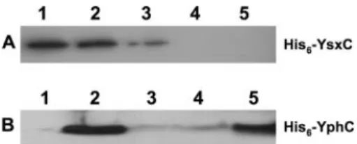
YsxC and YphC directly interact with the ribosome. To
determine if YphC or YsxC directly interacts with the ribo-some, purified ribosomes and purified His6-YsxC protein or His6-YphC protein were incubated together under a variety of guanine nucleotide conditions. Complexes were then overlaid onto sucrose cushions, pelleted ribosomes were collected, and
the presence of His6-YsxC or His6-YphC was monitored by Western blotting using a specific His-tagged antibody. Both His6-YsxC and His6-YphC were able to bind to 70S ribosomes (Fig. 4). In the case of His6-YsxC, the strength of this interac-tion was increased by the presence of either GTP or GDP (Fig. 4A). YsxC specifically interacts with individual 50S subunits but not 30S subunits, consistent with a direct role in the as-sembly of the large subunit (C. Wicker-Planquart and J.-M. Jault, unpublished data).
YphC was also found to interact with 70S ribosomes, and this interaction was stabilized by the nonhydrolyzable analog GMPPNP (Fig. 4B). Unlike YsxC, the addition of GTP (in the presence of Mg2⫹) or GDP strongly inhibited the interaction, suggesting that YphC in a GDP-bound state is unable to in-teract with the ribosome. (YphC has a potent intrinsic GTPase activity; thus, the addition of GTP will likely result in YphC existing in the GDP-bound form [A.-E. Foucher and J.-M. Jault, unpublished data].) This finding is similar to the binding of RbgA to the 50S subunit in that RbgA will interact only with
FIG. 3. Analysis of the protein content of the ribosomal interme-diates isolated from YsxC- and YphC-depleted cells. (A) A 12% SDS-PAGE gel containing purified large-subunit intermediates. Molecular masses (kDa) are indicated on the left side of the gel. Lane 1, 44.5S intermediate isolated from YsxC-depleted cells; lane 2, 45S interme-diate isolated from YphC-depleted cells; lane 3, 45S intermeinterme-diate isolated from RbgA-depleted cells; lane 4, mature 50S subunits iso-lated from wild-type cells. The bottom arrow indicates where L16 is present in the 50S subunit but absent from the intermediates. The top arrow indicates a region of the gel where the 44.5S complex differs from the 45S intermediates. The identities of the proteins are not yet known, although their sizes indicate that they cannot be ribosomal proteins. (B) A 16% SDS-PAGE gel containing purified large-subunit intermediates. Only the region of the gel at 12 kDa and smaller is shown. Lane 1, 44.5S complex from YsxC-depleted cells; lane 2, 45S complex from YphC-depleted cells; lane 3, 45S complex from RbgA-depleted cells; lane 4, mature 50S subunits from wild-type cells. Arrows indicate the positions of L36 and possibly L27, found only in the mature 50S subunit.
the mature 50S complex when bound to a nonhydrolyzable analog of GTP (24).
DISCUSSION
The results provided in this paper demonstrate that when either YphC or YsxC is depleted from B. subtilis, a defect in the assembly of the large ribosomal subunit occurs. These defects lead to a sharp decrease in functional 70S ribosomes and a marked slowing of the growth rate, as has been previ-ously described for the essential GTPase RbgA (24, 33). Both GTPases directly interact with the ribosome. These results indicate that YsxC and YphC directly participate in ribo-some biogenesis and suggest that, as has been described for eukaryotic ribosome 60S subunit biogenesis, multiple GTPases
participate in the assembly of the large ribosomal subunit in bacteria.
Interestingly, YsxC and YphC have easily identifiable ho-mologs in Escherichia coli (unlike RbgA), the organism for which most of the work on bacterial ribosome biogenesis has been performed. YihA, the E. coli YsxC homolog, has been implicated in the cell cycle and in cell division (32), and EngA, the YphC homolog in E. coli, is thought to be involved in 23S rRNA maturation (28). A ribosomal intermediate similar to the 45S or 44.5S complex that we have observed in B. subtilis has not been previously described for E. coli. Thus, it will be interesting to learn if the YsxC and YphC homologs in E. coli also yield an intermediate similar to that observed in B. subtilis or if they will control the maturation of previously described E.
coli 50S ribosomal subunit intermediates.
The ribosomal intermediate that accumulates in YsxC-de-pleted cells is a distinct complex from previously identified ribosomal intermediates.The large ribosomal subunit inter-mediate isolated from YphC-depleted cells migrates at 45S in a sucrose gradient and is indistinguishable from the 45S inter-mediate that accumulates in RbgA-depleted cells. In contrast, the large ribosomal subunit isolated from YsxC-depleted cells migrates slightly slower than 45S (44.5S), indicating an altered protein composition and/or an altered conformation. SDS-PAGE analysis of the proteins associated with the 44.5S com-plex indicates that the difference between the 44.5S and 45S complexes is likely at the level of nonribosomal proteins asso-ciated with these complexes, as each complex is missing the same three ribosomal proteins (L16, L27, and L36).
The finding that L36, in addition to L16 and L27, is also missing from the ribosomal intermediates isolated from RbgA-, YsxC-, and YphC-depleted cells is not surprising. These three proteins lie adjacent to one another in the 50S subunit, with L16 sandwiched between L27 and L36 (Fig. 5) (3, 14). L36 plays an important role in the structural organization of the 50S subunit of E. coli, and the deletion of L36 results in alterations in the RNA structure distant from the direct con-tacts between L36 and rRNA (22). L36 lies adjacent to L16 in the ribosome, and both proteins make specific contacts with helix 89 of domain V of the 23S rRNA (Fig. 5). The base of
FIG. 4. YsxC and YphC directly interact with the ribosome. (A) Analysis of His6-YsxC by Western blotting of the pellets obtained from centrifugation on a 10% sucrose cushion of the mixtures of His6-YsxC and ribosome incubated in the presence of 1 mM GDP (lane 1), His6-YsxC and ribosome incubated in the presence of 1 mM GTP (lane 2), His6-YsxC and ribosome incubated without any added nucleotide (lane 3), ribosome alone (lane 4), and His6-YsxC alone (lane 5). His6-YsxC incubated with 1 mM GTP or 1 mM GDP gave a signal that was similar to that of His6-YsxC alone (lane 1), indicating that His6-YsxC does not significantly precipitate or aggregate in the presence of nucleotides (data not shown). (B) Analysis of His6-YphC by Western blotting of the pellets obtained from centrifugation on a 10% sucrose cushion of the mixtures of His6-YphC alone (lane 1), His6-YphC and ribosome (lane 2), His6-YphC and ribosome in pres-ence of 2 mM GTP (lane 3), His6-YphC and ribosome in presence of 2 mM GDP (lane 4), and His6-YphC and ribosome in presence of 2 mM GMPPNP (lane 5). His6-YphC incubated with 2 mM GTP gave a signal that was similar to that of His6-YphC alone (lane 1), indicating that His6-YphC does not significantly precipitate or aggregate in the presence of nucleotides (data not shown).
FIG. 5. Location of L16, L27, and L36 in the 50S subunit. (A) Crown view representation of the 50S subunit from Deinococcus radiodurans (Protein Data Bank accession number 1NKW). The locations of L16 (blue), L27 (red), and L36 (yellow) in the 50S subunit are shown. The rRNA is dark gray; helix 89 and helix 38 are shown in light gray. (B) Enlarged view of L16, L27, and L36 in the ribosome. Colors are the same as in A. Figures were generated using VMD software.
helix 89 forms part of the peptidyltransferase center, and this helix makes important contacts with IF2 during subunit asso-ciation. L16 and L27 both make specific contacts with helix 38. Thus, L16, L27, and L36 lie adjacent to one another in the ribosome structure, and their incorporation appears to be a late step in the biogenesis of the B. subtilis large ribosomal subunit.
At this point, we are unable to speculate about the precise molecular functions of these GTPases in ribosome assembly. Incorporation of L27 or L36 is not likely to be the essential function(s) of RbgA, YsxC, or YphC, as L27 and L36 are dispensable for growth in both E. coli and B. subtilis (22, 23, 27). Conversely, mutants defective in L16 have not been iso-lated, suggesting that L16 is essential. One possibility for the functions of these essential GTPases is that they may control the structural organization of the ribosome, rendering the large subunit inactive until properly configured by the com-bined action of RbgA, YsxC, and YphC. In support of this idea, previous studies of L16, L27, and L36 suggest that they have functional as well as structural roles in the ribosome. L16 and L27 were previously proposed to make specific contacts with tRNA molecules that occupy the A site and the P site, respectively (4, 23). Furthermore, helix 89 likely participates in subunit association, and the lack of L16 and L36 would likely cause a defect in the secondary structure of this helix (20, 22). In addition to serving a structural role in the ribosome, L36 may also participate in elongation factor binding and/or sub-unit association (22). Taken together, the regulated incorpo-ration of L16, L27, and L36 may allow the assembling ribo-some to remain inactive for functional activities and subunit association until the subunit is fully formed. Alternatively, YsxC or YphC may participate in the assembly process after incorporation of the ribosomal proteins. Depletion of these GTPases may yield a large subunit that is unstable and loses L16, L27, and L36 upon purification. We find it unlikely that YsxC and/or YphC functions in translation initiation or elon-gation, as cells defective in these processes have large subunits that migrate at 50S in sucrose gradients (33). Attempts are under way to determine if YsxC and YphC can specifically interact with the large-subunit intermediates that accumulate upon their depletion.
The ribosomal intermediates that form in RbgA-, YsxC-, or YphC-depleted cells are similar to a Bacillus
stearothermophi-lus intermediate that forms during in vitro ribosome assembly.
When purified ribosomal components from B.
stearothermophi-lus are incubated in vitro at 37°C, a reconstitution intermediate
(RI50) is formed, which migrates much slower on a sucrose gradient than fully functional 50S subunits (13). RI50has none of the functional activities associated with 50S subunits. Heat-ing of this RI50complex to 60°C for 1 h results in the activation of the particle (designated RI50*). When the protein compo-sitions of RI50and RI50* were compared, it was found that RI50was missing three proteins and had a reduced level of a fourth protein (8, 13). One of the missing proteins is⬃16 kDa, indicating that it could be L16. Interestingly, the other two proteins that are missing are both small ribosomal proteins whose identities cannot be predicted with certainty from pre-viously published data. However, their sizes are consistent with these proteins being L27 and L36. The fact that the missing proteins in the RI50particle during in vitro assembly correlate
well with the proteins that are missing in the altered large-subunit complexes in RbgA-, YsxC-, or YphC-depleted cells is intriguing and further supports the possibility that the 45S and 44.5S complexes isolated in vivo are naturally occurring assem-bly intermediates. Indeed, Fahnestock and coworkers previ-ously speculated that the conversion of RI50 into an active ribosomal subunit would likely be carried out by a nonriboso-mal factor (13). We suggest that the Rbg GTPases could serve as such factors.
Multiple GTPases control large-subunit ribosome assembly.
Our results indicate that RbgA, YsxC, and YphC participate in the assembly of the 50S subunit in Bacillus subtilis. RbgA binds to both the 45S intermediate and to the mature 50S subunit (in the presence of GTP␥S), suggesting that RbgA participates in the final maturation step. We envision at least two possibilities for the roles of RbgA, YsxC, and YphC in ribosome assembly. First, these GTPases could function in a linear pathway with one of the GTPases acting prior to the others. As the 44.5S subunit from YsxC appears to be distinct from the 45S inter-mediates isolated from YphC of RbgA-depleted cells, it is possible that YsxC may act independently of RbgA and YphC in ribosome assembly. From our current data, we detect no significant difference between YphC- and RbgA-depleted cells, indicating that they may act at a similar step in ribosome biogenesis. Alternatively, these GTPases may all function on the same ribosomal intermediate, a protein complex similar to RI50that accumulates during in vitro assembly. An analogous situation occurs during the biogenesis of the 60S subunit in yeast in which at least three GTPases participate in the matu-ration of the pre-60S complex (12). A more detailed analysis of the 44.5S and 45S protein composition and structure may help elucidate the role of GTPases in ribosome assembly.
ACKNOWLEDGMENTS
This work was supported by an IRGP New Investigator award from Michigan State University and startup funds from Michigan State University provided to R.A.B. and by a Young Investigator ATIP grant and a PGP grant (04-020), both from the CNRS, to J.-M.J.
We thank Lucas Krause, Mark Koenigsknecht, and Brian Shy for excellent technical assistance.
REFERENCES
1. Anand, B., S. K. Verma, and B. Prakash. 2006. Structural stabilization of GTP-binding domains in circularly permuted GTPases: implications for RNA binding. Nucleic Acids Res. 34:2196–2205.
2. Arigoni, F., F. Talabot, M. Peitsch, M. D. Edgerton, E. Meldrum, E. Allet, R.
Fish, T. Jamotte, M. L. Curchod, and H. Loferer.1998. A genome-based approach for the identification of essential bacterial genes. Nat. Biotechnol.
16:851–856.
3. Ban, N., P. Nissen, J. Hansen, P. B. Moore, and T. A. Steitz. 2000. The complete atomic structure of the large ribosomal subunit at 2.4 A resolution. Science 289:905–920.
4. Bashan, A., I. Agmon, R. Zarivach, F. Schluenzen, J. Harms, R. Berisio, H.
Bartels, F. Franceschi, T. Auerbach, H. A. Hansen, E. Kossoy, M. Kessler, and A. Yonath.2003. Structural basis of the ribosomal machinery for peptide bond formation, translocation, and nascent chain progression. Mol. Cell
11:91–102.
5. Britton, R. A., B. S. Powell, S. Dasgupta, Q. Sun, W. Margolin, J. R. Lupski,
and D. L. Court.1998. Cell cycle arrest in Era GTPase mutants: a potential growth rate-regulated checkpoint in Escherichia coli. Mol. Microbiol. 27: 739–750.
6. Campbell, T. L., D. M. Daigle, and E. D. Brown. 2005. Characterization of the Bacillus subtilis GTPase YloQ and its role in ribosome function. Bio-chem. J. 389:843–852.
7. Charollais, J., D. Pflieger, J. Vinh, M. Dreyfus, and I. Iost. 2003. The DEAD-box RNA helicase SrmB is involved in the assembly of 50S ribosomal subunits in Escherichia coli. Mol. Microbiol. 48:1253–1265.
8. Cohlberg, J. A., and M. Nomura. 1976. Reconstitution of Bacillus
mophilus 50 S ribosomal subunits from purified molecular components.
J. Biol. Chem. 251:209–221.
9. Culver, G. M. 2003. Assembly of the 30S ribosomal subunit. Biopolymers
68:234–249.
10. Daigle, D. M., and E. D. Brown. 2004. Studies of the interaction of
Esche-richia coli YjeQ with the ribosome in vitro. J. Bacteriol. 186:1381–1387.
11. Dassain, M., A. Leroy, L. Colosetti, S. Carole, and J. P. Bouche. 1999. A new essential gene of the ‘minimal genome’ affecting cell division. Biochimie
81:889–895.
12. Dez, C., and D. Tollervey. 2004. Ribosome synthesis meets the cell cycle. Curr. Opin. Microbiol. 7:631–637.
13. Fahnestock, S., W. Held, and M. Nomura. 1973. The assembly of bacterial ribosomes, p. 179–217. In R. Markham et al. (ed.), Generation of subcellular structures. North Holland/American Elsevier, New York, N.Y.
14. Harms, J., F. Schluenzen, R. Zarivach, A. Bashan, S. Gat, I. Agmon, H.
Bartels, F. Franceschi, and A. Yonath.2001. High resolution structure of the large ribosomal subunit from a mesophilic eubacterium. Cell 107:679–688. 15. Hedges, J., M. West, and A. W. Johnson. 2005. Release of the export adapter,
Nmd3p, from the 60S ribosomal subunit requires Rpl10p and the cytoplas-mic GTPase Lsg1p. EMBO J. 24:567–579.
16. Himeno, H., K. Hanawa-Suetsugu, T. Kimura, K. Takagi, W. Sugiyama,
S. Shirata, T. Mikami, F. Odagiri, Y. Osanai, D. Watanabe, S. Goto, L. Kalachnyuk, C. Ushida, and A. Muto.2004. A novel GTPase activated by the small subunit of ribosome. Nucleic Acids Res. 32:5303–5309.
17. Inoue, K., J. Alsina, J. Chen, and M. Inouye. 2003. Suppression of defective ribosome assembly in a rbfA deletion mutant by overexpression of Era, an essential GTPase in Escherichia coli. Mol. Microbiol. 48:1005–1016. 18. Johnson, A. W., E. Lund, and J. Dahlberg. 2002. Nuclear export of ribosomal
subunits. Trends Biochem. Sci. 27:580–585.
19. Karbstein, K., S. Jonas, and J. A. Doudna. 2005. An essential GTPase promotes assembly of preribosomal RNA processing complexes. Mol. Cell
20:633–643.
20. La Teana, A., C. O. Gualerzi, and A. E. Dahlberg. 2001. Initiation factor IF 2 binds to the alpha-sarcin loop and helix 89 of Escherichia coli 23S ribo-somal RNA. RNA 7:1173–1179.
21. Lin, B., D. A. Thayer, and J. R. Maddock. 2004. The Caulobacter crescentus CgtACprotein cosediments with the free 50S ribosomal subunit. J. Bacteriol.
186:481–489.
22. Maeder, C., and D. E. Draper. 2005. A small protein unique to bacteria organizes rRNA tertiary structure over an extensive region of the 50 S ribosomal subunit. J. Mol. Biol. 354:436–446.
23. Maguire, B. A., A. D. Beniaminov, H. Ramu, A. S. Mankin, and R. A.
Zimmermann.2005. A protein component at the heart of an RNA machine: the importance of protein L27 for the function of the bacterial ribosome. Mol. Cell 20:427–435.
24. Matsuo, Y., T. Morimoto, M. Kuwano, P. C. Loh, T. Oshima, and N.
Ogasawara.2006. The GTP-binding protein YlqF participates in the late step of 50 S ribosomal subunit assembly in Bacillus subtilis. J. Biol. Chem.
281:8110–8117.
25. Miroux, B., and J. E. Walker. 1996. Over-production of proteins in
Esche-richia coli: mutant hosts that allow synthesis of some membrane proteins and
globular proteins at high levels. J. Mol. Biol. 260:289–298.
26. Morimoto, T., P. C. Loh, T. Hirai, K. Asai, K. Kobayashi, S. Moriya, and N.
Ogasawara.2002. Six GTP-binding proteins of the Era/Obg family are es-sential for cell growth in Bacillus subtilis. Microbiology 148:3539–3552. 27. Ohashi, Y., T. Inaoka, K. Kasai, Y. Ito, S. Okamoto, H. Satsu, Y. Tozawa, F.
Kawamura, and K. Ochi.2003. Expression profiling of translation-associated genes in sporulating Bacillus subtilis and consequence of sporulation by gene inactivation. Biosci. Biotechnol. Biochem. 67:2245–2253.
28. Owens, R. M., G. Pritchard, P. Skipp, M. Hodey, S. R. Connell, K. H.
Nierhaus, and C. D. O’Connor.2004. A dedicated translation factor controls the synthesis of the global regulator Fis. EMBO J. 23:3375–3385. 29. Ruzheinikov, S. N., S. K. Das, S. E. Sedelnikova, P. J. Baker, P. J. Artymiuk,
J. Garcia-Lara, S. J. Foster, and D. W. Rice.2004. Analysis of the open and closed conformations of the GTP-binding protein YsxC from Bacillus sub-tilis. J. Mol. Biol. 339:265–278.
30. Sato, A., G. Kobayashi, H. Hayashi, H. Yoshida, A. Wada, M. Maeda, S.
Hiraga, K. Takeyasu, and C. Wada.2005. The GTP binding protein Obg homolog ObgE is involved in ribosome maturation. Genes Cells 10:393–408. 31. Sharma, M. R., C. Barat, D. N. Wilson, T. M. Booth, M. Kawazoe, C.
Hori-Takemoto, M. Shirouzu, S. Yokoyama, P. Fucini, and R. K. Agrawal.
2005. Interaction of Era with the 30S ribosomal subunit: implications for 30S subunit assembly. Mol. Cell 18:319–329.
32. Tan, J., U. Jakob, and J. C. Bardwell. 2002. Overexpression of two different GTPases rescues a null mutation in a heat-induced rRNA methyltransferase. J. Bacteriol. 184:2692–2698.
33. Uicker, W. C., L. Schaefer, and R. A. Britton. 2006. The essential GTPase RbgA (YlqF) is required for 50S ribosome assembly in Bacillus subtilis. Mol. Microbiol. 59:528–540.
34. Wout, P., K. Pu, S. M. Sullivan, V. Reese, S. Zhou, B. Lin, and J. R.
Maddock.2004. The Escherichia coli GTPase CgtAEcofractionates with the
50S ribosomal subunit and interacts with SpoT, a ppGpp synthetase/hydro-lase. J. Bacteriol. 186:5249–5257.
35. Zalacain, M., S. Biswas, K. A. Ingraham, J. Ambrad, A. Bryant, A. F.
Chalker, S. Iordanescu, J. Fan, F. Fan, R. D. Lunsford, K. O’Dwyer, L. M. Palmer, C. So, D. Sylvester, C. Volker, P. Warren, D. McDevitt, J. R. Brown, D. J. Holmes, and M. K. Burnham.2003. A global approach to identify novel broad-spectrum antibacterial targets among proteins of unknown function. J. Mol. Microbiol. Biotechnol. 6:109–126.
36. Zhang, S., and W. G. Haldenwang. 2004. Guanine nucleotides stabilize the binding of Bacillus subtilis Obg to ribosomes. Biochem. Biophys. Res. Com-mun. 322:565–569.