HAL Id: inserm-02526607
https://www.hal.inserm.fr/inserm-02526607
Submitted on 31 Mar 2020
HAL is a multi-disciplinary open access archive for the deposit and dissemination of sci-entific research documents, whether they are pub-lished or not. The documents may come from teaching and research institutions in France or abroad, or from public or private research centers.
L’archive ouverte pluridisciplinaire HAL, est destinée au dépôt et à la diffusion de documents scientifiques de niveau recherche, publiés ou non, émanant des établissements d’enseignement et de recherche français ou étrangers, des laboratoires publics ou privés.
donors fails to detect a relation between SDF1/CXCR4
or VCAM/VLA4 genetic polymorphisms and the level of
hematopoietic progenitor cell mobilization in response
to G-CSF
Sylvain Garciaz, Patrick Sfumato, Angela Granata, Anne-Marie Imbert,
Claire Fournel, Boris Calmels, Claude Lemarie, Jacques Chiaroni, Didier
Blaise, Jean-Marie Boher, et al.
To cite this version:
Sylvain Garciaz, Patrick Sfumato, Angela Granata, Anne-Marie Imbert, Claire Fournel, et al.. Analy-sis of a large single institution cohort of related donors fails to detect a relation between SDF1/CXCR4 or VCAM/VLA4 genetic polymorphisms and the level of hematopoietic progenitor cell mobilization in response to G-CSF. PLoS ONE, Public Library of Science, 2020, 15 (3), pp.e0228878. �10.1371/jour-nal.pone.0228878�. �inserm-02526607�
RESEARCH ARTICLE
Analysis of a large single institution cohort
of related donors fails to detect a relation
between SDF1/CXCR4 or VCAM/VLA4
genetic polymorphisms and the level of
hematopoietic progenitor cell mobilization
in response to G-CSF
Sylvain Garciaz1,2, Patrick Sfumato1, Angela Granata1, Anne-Marie Imbert1, Claire Fournel1, Boris Calmels1,2,3, Claude Lemarie1,2,3, Jacques Chiaroni4,5, Didier Blaise1,2, Jean-Marie Boher1, Christophe Picard4,5, Christian Chabannon
ID1,2,3*, Julie di Cristofaro4,5
1 Institut Paoli-Calmettes, Comprehensive Cancer Center, Marseille, France, 2 Aix-Marseille Univ, Inserm, CNRS, Institut Paoli-Calmettes, CRCM, Marseille, France, 3 Centre d’Investigations Cliniques de Marseille, module Biothe´rapies, Inserm CBT, Marseille, France, 4 Etablissement Franc¸ais du Sang PACA Corse, Biologie des Groupes Sanguins, Marseille, France, 5 Aix Marseille Univ, CNRS, EFS, ADES, Marseille, France
*chabannonc@ipc.unicancer.fr,christian.chabannon@univ-amu.fr
Abstract
We studied a cohort of 367 healthy related donors who volunteered to donate their hemato-poietic stem cells for allogeneic transplantation. All donors were homogeneously cared for at a single institution, and received rhG-CSF as a mobilization treatment prior to undergoing apheresis. Peripheral blood CD34+ cell counts were used as the main surrogate marker for rhG-CSF induced mobilization. We searched whether inter-individual variations in known genetic polymorphisms located in genes whose products are functionally important for mobi-lization, could affect the extent of CD34+ mobimobi-lization, either individually or in combination. We found little or no influence of individual SNPs or haplotypes for the SDF1, CXCR4, VCAM and VLA4 genes, whether using CD34+ cell counts as a continuous or a categorical variable. Simple clinical characteristics describing donors such as body mass index, age and possibly sex are more potent predictors of stem cell mobilization. The size of our cohort remains relatively small for genetic analyses, however compares favorably with cohorts analyzed in previously published reports suggesting associations of genetic traits to response to rhG-CSF; notwithstanding this limitation, our data do not support the use of genetic analyses when the choice exists of several potential donors for a given patient.
a1111111111 a1111111111 a1111111111 a1111111111 a1111111111 OPEN ACCESS
Citation: Garciaz S, Sfumato P, Granata A, Imbert
A-M, Fournel C, Calmels B, et al. (2020) Analysis of a large single institution cohort of related donors fails to detect a relation between SDF1/CXCR4 or VCAM/VLA4 genetic polymorphisms and the level of hematopoietic progenitor cell mobilization in response to G-CSF. PLoS ONE 15(3): e0228878. https://doi.org/10.1371/journal.pone.0228878
Editor: Halvard Bo¨nig, Goethe University Frankfurt,
GERMANY
Received: July 9, 2019 Accepted: January 24, 2020 Published: March 5, 2020
Copyright:© 2020 Garciaz et al. This is an open access article distributed under the terms of the Creative Commons Attribution License, which permits unrestricted use, distribution, and reproduction in any medium, provided the original author and source are credited.
Data Availability Statement: All relevant data are
within the manuscript and its Supporting Information files.
Funding: This work was supported by Institut
Paoli-Calmettes, Inserm and by Etablissement Franc¸ais du Sang and CNRS.
Competing interests: Both Dr Sylvain Garciaz and
myself Christian Chabannon received funding from SANOFI S.A. as speakers during
industry-Introduction
Allogeneic stem-cell transplantation (ASCT) is a curative treatment for patients with severe hematological malignancies. Several sources of stem cells can be used, including bone marrow (BM), peripheral blood (PB) and umbilical cord blood. PB cell collection presents several advantages: leukapheresis is a moderately invasive and semi-automated procedure that can be performed on an outpatient basis and does not require access to the operating room nor gen-eral anesthesia; infusion of PB cells to the recipient is associated with rapid engraftment and hospital discharge, both after myeloablative and reduced intensity conditioning regimen. [1,2] Although, the relationship between the numbers of infused CD34+ cells and recipient engraft-ment and outcome remains controversial [3,4], most transplant programs expect that the col-lected cell graft will contain at least a defined minimal number of progenitors, and some will possibly cap the number of infused CD34+ cells. These objectives can be reached only when donors CD34+ cells are appropriately mobilized through the administration of granulocyte colony-stimulating factor (G-CSF). The number of CD34+ cells mobilized into PB varies sig-nificantly among donors, mainly depending on clinical factors as age, gender or weight. [5,6] Genetic susceptibility may also influence the quality of mobilization. Indeed, several BM pro-teins are involved in stem-cell homing and G-CSF-induced mobilization, including CXCR4 and its ligand SDF1 (CXCL12) [7,8] and VCAM1 and its ligand VLA4. [9,10] Interactions of these two receptor-ligand pairs are disrupted following G-CSF treatment, and structural or functional variations in these molecules may influence response to G-CSF. [11–13] Some of these inter-individual variations may be appreciated through single nucleotide polymorphisms (SNPs). In contexts other than HSCT, many studies have evidenced a relation between SNPs and the variability in response to drug administration. [14,15] There are limited evidences suggesting that similar patterns may exist for CD34+ cell mobilization in response to rhG-CSF, either for patients or healthy donors who are preparing to undergo apheresis prior to autolo-gous or allogeneic hematopoietic cell transplantation respectively. [11–13] We conducted a retrospective analysis of the association betweenSDF1/CXCR4 and VCAM/VLA4 genetic
poly-morphisms and CD34+ cells mobilization in healthy related donors.
Patients & methods
Donor selection and care
The study includes three hundred and sixty-seven adult healthy donors who donated their blood mononuclear cells to related patients who received allogeneic transplantation between 1997 and 2016 and were cared for at a single institution: Institut Paoli-Calmettes, the compre-hensive cancer center in Marseille (seeTable 1). All donors were identified, screened and col-lected in accordance with national or international regulations, institutional policies, EFI and EBMT/FACT-JACIE prescriptions, including transparent information on HLA typing, dona-tion and their rights to consent. The project was approved by the "Comite´ d’Orientadona-tion Strate´-gique" (COS; Internal Review Board) at the Direction de la Recherche Clinique et de
l’Innovation (DRCI), Institut Paoli-Calmettes. All patients and donors involved in the adult transplantation program at Institut Paoli-Calmettes provided informed written consent for the use of their personal data as per EBMT and FACT-JACIE requirements, in compliance with national and European regulations.
In preparation for apheresis, donors received daily SQ injections of rhG-CSF in the evening as per institutional procedures. Prior to September 2009, all donors received a 600μg daily dose independently of their weight. Starting in September 2009, the G-CSF daily dose was adjusted to donor’s weight to approximately match the 10μg/kg/day dosing, as recommended sponsored symposium. I also benefited of an
invitation – including financial support for traveling – to a scientific meeting as a delegate. In addition, SANOFI S.A. funded another and different clinical project on autologous stem cell mobilization and collection that was conducted at Institut Paoli-Calmettes, the Comprehensive Cancer Center in Marseille, France. This does not alter our adherence to PLOS ONE policies on sharing data and materials (as detailed online in the journal guide for authorshttp://journals.plos.org/plosone/ s/competinginterests).
per rhG-CSF label (daily doses were 480, 600, 780 or 900μg). Counseling was provided on expected side effects, particularly bone pain, and prophylactic paracetamol prescribed to reduce pain. Circulating CD34+ cells were first counted at day 5, after 4 evening injections of rhG-CSF, using a single-platform flow-cytometry based technique as previously described. [4]
No donor infectious or inflammatory manifestations were reported in the 8 days preceding donation. All CRP were negative (<6 mg/l).
VCAM rs1041163, VLA4 rs1449263 and CXCR4-rs2680880 genotyping
Genomic DNA (gDNA) was extracted from a 200-μl total blood sample using the QIAmp Blood DNA Mini kit (Qiagen, Courtaboeuf, France) according to manufacturer’s instructions.
VCAM1-rs1041163 (T>C), VLA4-rs1449263 (A>G) and CXCR4-rs2680880 (A>T) were
analyzed by direct sequencing after PCR amplification. PCR amplification was performed on 50 ng of gDNA in a final volume of 25μL containing 1x PCR buffer, 1.5 mM MgCl2, 0.2 mM of dNTPs, 0.1 unit of Taq DNA-polymerase (Invitrogen, France) and 0.16μM of each primer. Primers designed using the Primer 3 program (http://bioinfo.ut.ee/primer3-0.4.0/primer3/). TheVCAM, VLA4 and CXCR4 PCR primer sequences were respectively 5’ATTGGCCATTG
TCTTTGAGC3’ and 5’GATGCTGTTCTAGGGTGTGG3’; 5’TGCCCACTATATGCCAA
Table 1. Donors clinical and biological data.
Variable Classes Statistics All (n = 367)
Gender Male n (%) 209 (56.95)
Female n (%) 158 (43.05)
Age n 365
Mean (SD) 50.45 (12.29)
Median [Min—Max] 52.00 [18.00–78.00]
Number of missing data 2
Height n 329
Mean (SD) 169.9 (9.118)
Median [Min—Max] 170.0 [150.0–195.0]
Number of missing data 38
Weight n 334
Mean (SD) 73.87 (15.44)
Median [Min—Max] 72.00 [44.00–130.0]
Number of missing data 33
Body Mass Index n 329
Mean (SD) 25.50 (4.620)
Median [Min—Max] 25.00 [16.00–45.00]
Number of missing data 38
G-CSF n 367 Mean (SD) 674.1 (132.9) Median [Min—Max] 600.0 [480.0–960.0] G-CSF/kg n 334 Mean (SD) 9.441 (1.849) Median [Min—Max] 9.375 [4.615–15.00]
Number of missing data 33
PB CD34+ cells /microL at day 5 n 367
Mean (SD) 67.36 (41.73)
Median [Min—Max] 59.20 [4.000–237.6] https://doi.org/10.1371/journal.pone.0228878.t001
AAA3’ and 5’AGGGAGCCATCAGAGGAAAC3’; and 5’GGAAAAGATGGGGAGGAGA G3’ and 5’CACTTCCAATTCAGCAAGCA3’. Amplification was carried out as follows: 1 cycle at 95˚C for 5 min; 30 cycles at 95˚C for 30 sec, Tm (57˚C forVCAM and VLA4 and 53˚C
forCXCR4) for 30 sec, and 72˚C for 1 min; and 1 cycle at 72˚C for 7 min. PCR amplification
was checked by agarose electrophoresis and PCR fragments (lengthVLA4: 250 bp; VCAM: 227
bp;CXCR4: 159 bp) were sequenced using a Big Dye Terminator V1.1 kit (Invitrogen) with
each PCR primer according to the manufacturer’s protocol. Sequences were analyzed using the Codon Code Aligner program (Codon Code Corporation, Massachusetts) as previously described. [16–17]
CXCR4 (rs12691874, rs16832740, rs2228014) and SDF1 (rs1413519,
rs1801157, rs2297630, rs266085, rs266087) genotyping
A homemade multiplex primer extension method was used to simultaneously analyze 8 SNPs of theCXCR4 and SDF1 genes (rs12691874 A>G, rs16832740 T>C,
CXCR4-rs2228014 C>T,rs1413519 G>C, rs1801157 G>A, rs2297630 G>A,
SDF1-rs266085 C>T,SDF1-rs266087 G>A). All primers were designed using the Primer 3 program.
Multiplex PCR using primers flanking the target SNPs were performed on 100 ng of genomic DNA in a final volume of 25μL containing PCR Qiagen Master Mix (Qiagen, France) and 0.8M of each primer (Table 2). Amplification was carried out as follows: 1 cycle at 95˚C for 15 min; 30 cycles at 95˚C for 30 sec, 57˚C for 45 sec, and 72˚C for 60 sec; and 1 cycle at 72˚C for 10 min. After control on agarose gel, 5μl of PCR product was incubated with 0.5 units of ther-mosensitive alkaline phosphatase and 1 unit of exonuclease-I (Euromedex; France) for 15 min at 37˚C followed by 15 min at 80˚C to remove unincorporated primers and dNTPs. The sec-ond step was a multiplex extension reaction performed using the SNapShot kit (Invitrogen) according to manufacturer’s protocol in a final volume of 10μL containing 3 μL of the PCR product, 5μL of SNapShot mix, and extension primers (Table 3). The reaction program was 25 cycles at 95˚C for 10 seconds, 50˚C for 5 seconds, 60˚C for 30 seconds. Snap Shot extension primer data were analyzed using GeneMapper v4.0 with specific detection parameters as previ-ously described. [17]
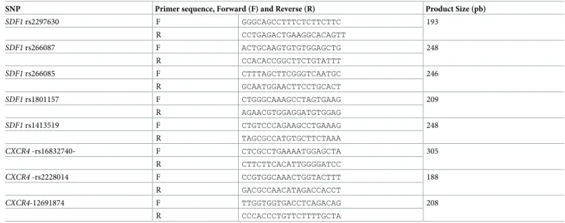
Table 2. PCR primers sequence and concentration used to co-amplify 6 PCR fragments encompassing 8 SNPs of the 8 SNPs of theCXCR4 and SDF1 genes.
SNP Primer sequence, Forward (F) and Reverse (R) Product Size (pb)
SDF1 rs2297630 F GGGCAGCCTTTCTCTTCTTC 193 R CCTGAGACTGAAGGCACAGTT SDF1 rs266087 F ACTGCAAGTGTGTGGAGCTG 248 R CCACACCGGCTTCTGTATTT SDF1 rs266085 F CTTTAGCTTCGGGTCAATGC 246 R GCAATGGAACTTCCTGCACT SDF1 rs1801157 F CTGGGCAAAGCCTAGTGAAG 209 R AGAACGTGGAGGATGTGGAG SDF1 rs1413519 F CTGTCCCAGAAGCCTGAAAG 248 R TAGCGCCATGTGCTTCTAAA CXCR4 -rs16832740- F CTCGCCTGAAAATGGAGCTA 305 R CTTCTTCACATTGGGGATCC CXCR4 -rs2228014 F CCGTGGCAAACTGGTACTTT 188 R GACGCCAACATAGACCACCT CXCR4-12691874 F TTGGTGGTGACCTCAGACAG 208 R CCCACCCTGTTCTTTTGCTA https://doi.org/10.1371/journal.pone.0228878.t002
Statistical analyses
Donor’s associated data—categorized into biological and clinical data including age, height, weight, IMC, sex, G-CSF total dose, G-CSF dose/kg and peripheral blood CD34+ cell counts— are described inTable 1.
Allelic frequencies and haplotype estimation. Missing data at a locus led to the exclusion
of the concerned sample from further analyses at the given locus. No multiple imputations were used. Allelic and two or more loci haplotype frequencies were estimated using an EM algorithm implemented in the Gene[Rate] computer tools. [12] Deviations from Hardy-Wein-berg equilibrium (HWE) were tested using a nested likelihood model. [13]
Haplotypes frequencies based on genotype of each SNPs of a same gene, i.e.SDF1 and CXCR4, were estimated by Gene[rate] computer tool package with no a priori. For allelic and
two or more loci frequency estimations, all putative homozygotes were considered either true homozygotes or heterozygotes for the observed allele, and an undefined or undetectable (‘blank’) allele as previously described. [16]
Based on this haplotype estimation, main haplotypes, with a cumulated frequency higher than 98%, werea priori encoded for each gene and genotype data were reanalyzed according
to this new nomenclature. Using an in-house computer program, data output files (.txt) were formatted into files readable by the “Phenotype” application of the Gene[rate] computer tool package.
Biological parameters statistical testing. Statistical analyses were performed using SPSS
software (SPSS 19.0 for Windows; SPSS Inc., Chicago, IL) and the R software version 3.0.3 associated to random forest SRC package [18]. The primary endpoint was influence of SNPs on peripheral blood CD34+ cell count/mL on day 5 of G-CSF treatment; CD34+ cell counts were considered as continuous and categorical variables in separate analyses. For continuous variables, median and extreme values are presented. Differences in medians have been ana-lyzed with the t-test for comparisons of two independent samples in univariate analyses or with one-way anova for multiple comparisons. t-tests were considered as significant when two-tailed p-values were < 0.05, except for clinical or molecular factors that has already been associated with a modification with mobilization in previous studies (age, gender, BMI and
VCAM1-rs1041163 CC homozygous variant). [14] In these case, given the expected influence on mobilization, we used a one-tailed p-value < .05. No adjustment for multiple tests was performed.
Linear regression. Two random forests for linear regression [19] were used to evaluate importance of clinical and molecular variables on a higher CD34+ cells count. The first model considered SNP classification as molecular variables and the second used haplotypes
Table 3. Extension primers sequence and concentration used to genotypeCXCR4-rs12691874 A>G, CXCR4-rs16832740 T>C, CXCR4-rs2228014 C>T,
SDF1-rs1413519 G>C,SDF1-rs1801157 G>A, SDF1-rs2297630 G>A, SDF1-rs266085 C>T, SDF1-rs266087 G>A.
SNP Primer sequence,
Forward (F) and Reverse (R)
Primer Size (b) Final Concentration (μM)
SDF1 rs1413519 F AGGCCCCAGAACAGAAGCTA 20 2.44 SDF1 rs2297630 F 4T-GTTCGTCTCAGTCTGCATAA 24 0.39 CXCR4 rs16832740 F 12T-GCCTGGAATTTCAATATACA 32 1.95 SDF1 rs266087 F 16T-TAAGAGAGGAAGTGGAGGGC 36 2.44 SDF1 rs266085 F 24T-TGCATCCGCTCCCCCAACAC 44 2.44 SDF1 rs1801157 F 32T-TCTCCATCCACATGGGAGCC 52 0.78 CXCR4 rs2228014 F 20T-CTGGACCGCTACCTGGCCAT 40 1.95 CXCR4 12691874 R 28T-ACAGTCCACAGGGCTCTAGG 48 1.95 https://doi.org/10.1371/journal.pone.0228878.t003
classification. The measure of the prediction accuracy of the Random Forest models was given by the mean squared error (MSE); variables importance (VIMP) was determined using permu-tation importance measure for Random forest, based on out-of-bag (OOB) estimate of predic-tion error: for a given variable, OOB cases (the original data left out from the bootstrap sample used to grow the tree; approximately 1/3 of the original sample) are randomly permuted in this variable and the prediction error is recorded. The VIMP of this variable is defined as the difference between the perturbed and unperturbed error rate averaged over all trees. The larger value this difference, the more predictive the variable.
Results
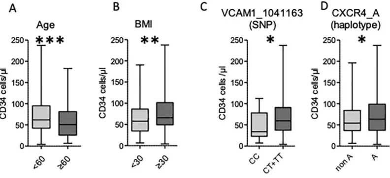
Influence of clinical characteristics on CD34+ cell mobilization
Among the 367 donors, 158 were females and 209 males. Median age and BMI were 51 (18–77) and 25 (12–44), respectively. The mean number of CD34+ cells/μl after four injections of rhG-CSF was 59.2 [4–237.6]. We first carried out a univariate analysis to study the impact of gender, age and body mass index (BMI) on CD34+ cell mobilization (Table 4). An age younger than 60 (n = 269) and a BMI �30 (n = 54) were both associated with a higher CD34+cell count (60.9 [4–237.6]vs 51.4 [6.7–183.3], p = .001 and 67.4 [4–237.6] vs 57.2 [6.5–190.6], p = .005,
respectively,Fig 1A and 1B). Men showed a trend to a better mobilization in comparison with women, although the difference was not significant (62 [7.1–237.6]vs 54.5 [4–193.9], p = 0.08).
Impact of VCAM/VLA4 and SDF1/CXCR4 SNPs on CD34 positive cell
mobilization
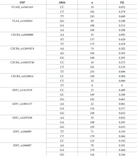
We next searched for a relation between each genetic SNP and PB CD34+ cell counts was ana-lyzed in univariate analyses. Allelic frequencies are described inTable 5. We did not observe deviations from the Hardy-Weinberg distribution. The CT or TT variants for the
VCAM1-rs1041163 SNP (n = 347) were associated with a higher CD34+ cell count than the homozy-gous CC variant (59.6 [4–237.6]vs 34 [7.4–112.3], p = 0.03) (Fig 1CandTable 6). We did not find any other significant association between SNPs and mobilization (Table 6). We also used the CD34 count as a categorical variable taking 30 CD34+ cells/μl as a cut-off, assuming it is clinically relevant in pinpointing the poor mobilizer (<30 CD34+ cells/μl). This analysis failed to find a significant association between VCAM/VLA4 or SDF1/CXCR4 polymorphism and mobilization (Table 7).
Impact of VCAM/VLA4 and SDF1/CXCR4 haplotypes on CD34+ cell
mobilization
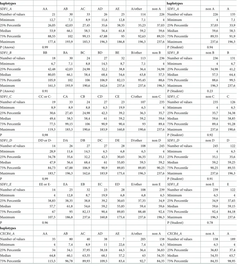
Allelic frequencies and haplotype estimation. Haplotypes were also investigated to
iden-tify better mobilizers. A haplotype is a group of gene variants that are inherited together from
Table 4. Relation between donor clinical characteristics and observed CD34+ cell mobilization after 4 injections of rhG-CSF.
Clinical characteristics Median CD34+ cells/μl range Two-tailed p value One-tailed p value
gender female (n = 158) 54.5 4–193.9 0.16 0.08 male (n = 209) 62 7.1–237.6 age (years) <60 (n = 269) 60.9 4–237.6 0.002 0.001 �60 (n = 76) 51.4 6.7–183.3 BMI <30 (n = 232) 57.2 6.5–190.6 0.01 0.005 �30(n = 54) 67.4 4–237.6 https://doi.org/10.1371/journal.pone.0228878.t004
a single parent. Haplotypic frequencies based on genotype of each SNPs of a same gene, i.e.
SDF1 and CXCR4, estimated by Gene[rate] without a priori, are described in Tables8and9. Nine and 8 haplotypes were respectively estimated for SDF1 and CXCR4, and for both genes, 5 haplotypes displayed a cumulated frequency of 99.3% and 98.5% respectively, encoded
SDF1-A to SDF1-E and CXCR4-A to CXCR4-E. Genotype data reanalyzed according to this a priori nomenclature are described in Tables10and11. With this new coding, blank haplotype represented respectively 5.8% and 6.1% forSDF1 and CXCR4.
TheCXCR4_A haplotype (either homozygous or heterozygous) was associated with a
higher mobilized CD34+ cell count, in comparison with other haplotypes (63.7 [4–237.6]vs
54.35 [6.5–196.3], p = .03) (Fig 1CandTable 12). We did not find any other significant associa-tion between haplotypes for other genes and CD34+ cells mobilizaassocia-tion (Table 10).
Clinical versus biological factors in predicting CD34+ cells mobilization
Two random forests for linear regression model were built in order to study importance of clinical and molecular variables on CD34+ cell mobilization [20,21]. As shown inFig 2, clini-cal factors were strongly associated with CD34+ cell mobilization contrary to molecular data, either taking SNP or haplotypes into account. Thus, we concluded that age and BMI data alone are sufficient to predict CD34+ cell mobilization in the context of ASCT.
Discussion
ASCT is used for the treatment of patients with a variety of severe malignant or non-malignant hematological disorders, either constitutional or acquired. A main challenge in ASCT is to rap-idly identify and select a suitable donor, from whom to collect sufficient numbers of hemato-poietic cells and progenitors; when apheresis is used to collect allogeneic peripheral blood stem cells, most transplant programs have defined minimal number of CD34+ cells to procure as a mean to ensure rapid engraftment and establish hematopoietic chimerism in the recipient.
Fig 1. Clinical and molecular factors influencing CD34+ mobilization in univariable analyses.
The extent of CD34+ cell mobilization varies significantly among donors, mainly depending on age, gender or weight, possibly also on genetic variations. [11–13] However, few published studies conducted on CD34+ cell mobilization included a multivariate analysis simultaneously considering biological and genetic variables. Here, we analyzed biological and genetic parame-ters described to influence CD34+ cell mobilization after G-CSF administration in 367 conse-cutive volunteer healthy donors that were homogeneously cared for at a single institution. Our results suggest that age, BMI, and possibly sex mostly influence the response to G-CSF.
Larger registry studies conducted in unrelated–and thus on younger—donors already iden-tified the influence of these factors on stem cell mobilization; our single-institution cohort of related donors offers the advantage of harmonized mobilization and collection procedures. In addition, published studies on unrelated donors do not include the analysis of genetic variants. In our cohort, genetic variations in genes whose products are known to play an important role in stem cell egress out of the bone marrow little affects the results of mobilization and
Table 5. Allele frequency forVCAM, VLA4, CXCR4 and SDF1 in the donor cohort.
SNP Allele n FQ VCAM_rs1041163 CC 19 0.052 CT 102 0.278 TT 245 0.669 VLA4_rs1449263 GG 69 0.188 GA 188 0.513 AA 109 0.298 CXCR4_rs2680880 AA 35 0.095 AT 157 0.428 TT 175 0.478 CXCR4_rs12691874 AA 74 0.202 AG 184 0.502 GG 108 0.295 CXCR4_rs16832740 CC 10 0.273 CT 101 0.276 TT 255 0.696 CXCR4_rs2228014 CC 330 0.904 CT 35 0.096 TT 0 0 SDF1_rs1413519 CC 15 0.409 GC 109 0.298 GG 242 0.661 SDF1_rs1801157 AA 22 0.061 GA 116 0.317 GG 228 0.622 SDF1_rs2297630 AA 19 0.052 GA 108 0.295 GG 239 0.653 SDF1_rs266085 TT 71 0.193 CT 170 0.464 CC 125 0.342 SDF1_rs266087 AA 70 0.191 GA 170 0.464 GG 126 0.344 https://doi.org/10.1371/journal.pone.0228878.t005
collection procedures used in the clinical context of ASCT. Such associations were evidenced in a much smaller cohort of 112 donors in a previously published report [11]; donors in this
Table 6. Impact ofVCAM/VLA4 and SDF1/CXCR4 SNP on CD34+ cell mobilization using CD34+ as a continuous variable.
SNP median range two-tailed p value
VCAM_rs1041163 CC (n = 19) 34 7.4–112.3 -CT (n = 102) 58,6 6.5–237.6 0,16 TT (n = 245) 60,1 4–193.9 0,04 CT+TT (n = 347) 59,6 4–237.6 0,06 VLA4_rs1449263 GG (n = 69) 53,9 7.4–103.4 -GA (n = 188) 59,9 4–237.6 0,33 AA (n = 109) 59 6.7–186.8 0,59 GA+AA (n = 297) 59 6.7–186.8 0,39 SNP median range P CXCR4_rs2680880 AA (n = 35) 53,8 21.7–162 -AT (n = 157) 59,6 6.5–186.8 0,65 TT (n = 175) 59,6 4–237.6 0,25 AT+TT (n = 332) 59,6 4–237.6 0,31 CXCR4_rs12691874 AA (n = 74) 57,1 6.8–193.3 -AG (n = 184) 58,9 6.5–237.6 0,99 GG (n = 108) 60,6 4–190.6 0,78 AG+GG (n = 292) 59,5 4–237.6 0,91 CXCR4_rs16832740 CC (n = 10) 60,6 32.2–120.9 -CT (= 101) 59,2 6.5–186.8 0,71 TT (n = 255) 59 4–237.6 0,71 CT+TT (n = 356) 59,15 4–237.6 0,79 CXCR4_rs2228014 CC (n = 330) 59,6 4–237.6 -CT (n = 35) 53,8 7.1–175.8 0,49 SNP median range P SDF1_rs1413519 CC (n = 15) 56,4 4–107.5 GC (n = 109) 59,6 6.8–237.6 0,22 GG (n = 242) 59,5 6.5–196.3 0,15 GC+GG (n = 351) 59,6 6.5–196.3 0,15 SDF1_rs1801157 AA (n = 22) 52,35 12.7–177.4 GA (n = 116) 59,2 7.1–196.3 0,4 GG (n = 228) 59,6 4–237.6 0,47 GA+GG (n = 344) 59,6 4–237.6 0,44 SDF1_rs2297630 AA (n = 19) 49,4 8.9–119.3 GA (n = 108 59,4 6.5–190.6 0,36 GG (n = 239) 59,4 4–237.6 0,31 GA+GG (n = 347 59,4 4–237.6 0,28 SDF1_rs266085 TT (n = 71) 64,8 6.7–193.9 CT (n = 170) 59,6 8.7–237.6 0,72 CC (n = 125) 57,8 4–183.9 0,4 CT+CC (n = 295) 59,1 4–237.6 0,67 SDF1_rs266087 AA (n = 70) 63,2 6.7–193.9 GA (n = 170 59,6 8.7–237.6 0,77 GG (n = 126) 58,4 4–183.9 0,46 GA+GG (n = 296) 59,15 4–183.9 0,73 https://doi.org/10.1371/journal.pone.0228878.t006
VCAM/VLA4 and SDF1/CXCR4 SNP on CD34+ cell mobilization using CD34+ as a categorical variable.
Test Classes Statistics All (n = 367) CD34+<30 (n = 66) CD34+�30 (n = 301) p-value (wilcoxon or khi2)
Gender Male n (%) 209 (56.95) 35 (53.03) 174 (57.81) 0.4778 Female n (%) 158 (43.05) 31 (46.97) 127 (42.19) Age n 365 65 300 0.012 Mean (SD) 50.45 (12.29) 54.23 (14.70) 49.63 (11.57) Median [Min—Max] 52.00 [18.00–78.00] 55.00 [21.00–78.00] 51.00 [18.00–77.00] < = 60 n (%) 291 (79.73) 43 (66.15) 248 (82.67) 0.0027 >60 n (%) 74 (20.27) 22 (33.85) 52 (17.33)
Number of missing data 2 1 1
BMI n 329 57 272 0.260
Mean (SD) 25.50 (4.620) 24.82 (4.268) 25.64 (4.685) Median [Min—Max] 25.00 [16.00–45.00] 25.00 [18.00–45.00] 25.00 [16.00–44.00]
< = 25 n (%) 179 (54.41) 33 (57.89) 146 (53.68) 0.5610
>25 n (%) 150 (45.59) 24 (42.11) 126 (46.32)
Number of missing data 38 9 29
VLA4_rs1449263 A,A n (%) 109 (29.7) 26 (39.4) 163 (54.1) 0.0867 G,A n (%) 188 (51.2) 25 (37.8) 83 (27.6)
G,G n (%) 69 (18.8) 15 (22.7) 54 (17.9)
Number of missing data 1 1
VCAM_rs1041163 T,T n (%) 245 (66.8) 42 (63.6) 203 (67.4) 0.2865
C,T n (%) 102 (27.8) 18 (27.3) 84 (27.9)
C,C n (%) 19 (5.2) 6 (9.1) 13 (4.3)
Number of missing data 1 1
SDF1_rs1413519 G,G n (%) 242 (66.12) 45 (68.18) 197 (65.67) 0.8776
G,C n (%) 109 (29.78) 18 (27.27) 91 (30.33) C,C n (%) 15 (4.098) 3 (4.545) 12 (4.000)
Number of missing data 1 1
SDF1_rs1801157 G,G n (%) 228 (62.30) 38 (57.58) 190 (63.33) 0.4433
G,A n (%) 116 (31.69) 22 (33.33) 94 (31.33) A,A n (%) 22 (6.011) 6 (9.091) 16 (5.333)
Number of missing data 1 1
SDF1_rs2297630 G,G n (%) 238 (65.21) 43 (65.15) 195 (65.22) 0.9367
G,A n (%) 108 (29.59) 19 (28.79) 89 (29.77) A,A n (%) 19 (5.205) 4 (6.061) 15 (5.017)
Number of missing data 2 2
SDF1_rs266085 C,C n (%) 125 (34.15) 19 (28.79) 106 (35.33) 0.5964
C,T n (%) 170 (46.45) 33 (50.00) 137 (45.67) T,T n (%) 71 (19.40) 14 (21.21) 57 (19.00)
Number of missing data 1 1
CXCR4_rs2680880 T,T n (%) 175 (47.68) 32 (48.48) 143 (47.51) 0.2947
A,T n (%) 157 (42.78) 31 (46.97) 126 (41.86) A,A n (%) 35 (9.537) 3 (4.545) 32 (10.63)
Number of missing data 0
CXCR4_rs12691874 G,G n (%) 108 (29.51) 19 (28.79) 89 (29.67) 0.6591
G,A n (%) 184 (50.27) 31 (46.97) 153 (51.00) A,A n (%) 74 (20.22) 16 (24.24) 58 (19.33)
Number of missing data 1 1
CXCR4_rs16832740 T,T n (%) 255 (69.67) 47 (71.21) 208 (69.33) 0.3216
C,T n (%) 101 (27.60) 19 (28.79) 82 (27.33)
C,C n (%) 10 (2.732) 10 (3.333)
Number of missing data 1 1
CXCR4_rs2228014 C,C n (%) 330 (90.16) 60 (90.91) 270 (90.00) 0.8853
C,T n (%) 35 (9.563) 6 (9.091) 29 (9.667)
T,T n (%) 1 (0.273) 1 (0.333)
Number of missing data 1 1
Table 8.SDF1 haplotypes and their frequencies (FQ) estimated by Gene[rate] based on rs1801157, rs266087, rs2297630, rs266085, rs1413519 observed polymorphisms. HAPLOTYPE FQ A~A~G~T~G 0.2158 G~A~G~T~G 0.2046 G~G~A~C~G 0.1995 G~G~G~C~C 0.1868 G~G~G~C~G 0.1862 A~G~G~T~G 0.0027 G~A~G~T~C 0.0017 G~A~G~C~G 0.0014 G~G~G~T~C 0.0014 https://doi.org/10.1371/journal.pone.0228878.t008
Table 9.CXCR4 haplotypes and their frequencies (FQ) estimated by Gene[rate] based on rs16832740, rs2228014,
rs2680880 and rs12691874 observed polymorphisms.
HAPLOTYPE FQ T~C~T~G 0.3263 T~C~T~A 0.3103 C~C~A~G 0.1587 T~C~A~A 0.1414 T~T~T~G 0.0486 T~C~A~G 0.0062 C~C~T~G 0.0047 C~C~A~A 0.0019 https://doi.org/10.1371/journal.pone.0228878.t009
Table 10. MainSDF1 haplotypes frequencies (FQ) estimated with an a priori by Gene[rate].
HAPLOTYPE RS1801157 RS266087 RS2297630 RS266085 RS1413519 FQ SDF1A A A G T G 0.2026 SDF1B G A G T G 0.1943 SDF1C G G A C G 0.1881 SDF1D G G G C G 0.1782 SDF1E G G G C C 0.1782 BLANK 0.0585 https://doi.org/10.1371/journal.pone.0228878.t010
Table 11. MainCXCR4 haplotypes frequencies (FQ) estimated with an a priori by Gene[rate].
HAPLOTYPE RS16832740 RS2228014 RS2680880 RS12691874 FQ CXCR4A T C T G 0.3074 CXCR4B T C T A 0.2933 CXCR4C C C A G 0.1524 CXCR4D T C A A 0.1376 CXCR4E T T T G 0.0482 BLANK 0.0611 https://doi.org/10.1371/journal.pone.0228878.t011
Table 12. Impact ofVCAM/VLA4 and SDF1/CXCR4 haplotypes on CD34+ cell mobilization.
haplotypes haplotypes
SDF1_A AA AB AC AD AE A/other non A SDF1_A non A A
Number of values 21 30 33 26 25 114 226 Number of values 226 135
Minimum 12,7 7,1 8,9 11,6 12,8 7,1 4 Minimum 4 7,1 25% Percentile 26,05 42,03 27,45 35,6 38,35 35,23 37,03 25% Percentile 37,03 33,9 Median 53,9 66,1 58,5 56,4 61,8 59,2 59,6 Median 59,6 58,5 75% Percentile 88,55 102 99,15 67,88 95 92,63 89,55 75% Percentile 89,55 91,9 Maximum 177,4 193,9 183,3 196,3 186,8 196,3 237,6 Maximum 237,6 196,3 P (Anova) 0.99 P (Student) 0.94
SDF1_B BB BA BC BD BE B/other non B SDF1_B non B B
Number of values 18 30 24 27 32 113 236 Number of values 236 131
Minimum 6,7 7,1 8,8 14,5 8,7 7,1 4 Minimum 4 6,7 25% Percentile 42,48 42,03 24,98 32,2 38,8 36,4 34,98 25% Percentile 34,98 41,2 Median 80,05 66,1 58,4 68,4 54,6 63,8 57,5 Median 57,5 64,4 75% Percentile 105,9 102 106 106,9 82,13 95,45 88,6 75% Percentile 88,6 99,5 Maximum 161,3 193,9 190,6 162,6 237,6 237,6 196,3 Maximum 196,3 237,6 P (Anova) 0.91 P (Student) 0.23
SDF1_C CC or C- CA CB CD CE C/other non C SDF1_C non C C
Number of values 19 33 24 27 23 107 235 Number of values 235 126
Minimum 8,9 8,9 8,8 6,5 19,9 6,5 4 Minimum 4 6,5 25% Percentile 30,6 27,45 24,98 42,3 39,2 36,3 35,7 25% Percentile 35,7 34,38 Median 49,4 58,5 58,4 61 59,2 59,2 59,6 Median 59,6 58,85 75% Percentile 77,5 99,15 106 90,9 90,4 93 89,4 75% Percentile 89,4 91,28 Maximum 119,3 183,3 190,6 183,9 160,8 190,6 237,6 Maximum 237,6 190,6 P 0,98 P (Student) 0.92
SDF1_D DD or D- DA DB DC DE D/other non D SDF1_D non D D
Number of values 14 26 27 27 28 108 245 Number of values 245 122
Minimum 28,9 11,6 14,5 6,5 6,8 6,5 4 Minimum 4 6,5 25% Percentile 34,78 35,6 32,2 42,3 30,65 36,35 35,1 25% Percentile 35,1 35,6 Median 47,9 56,4 68,4 61 55,85 59,5 59,2 Median 59,2 59,25 75% Percentile 84,73 67,88 106,9 90,9 89,85 89,85 90,25 75% Percentile 90,25 89,55 Maximum 183,7 196,3 162,6 183,9 175,4 196,3 237,6 Maximum 237,6 196,3 P 0,99 P (Student) 0.91
SDF1_E EE or E- EA EB EC ED E/other non E SDF1_E non E E
Number of values 14 25 32 23 28 108 239 Number of values 239 122
Minimum 4 12,8 8,7 19,9 6,8 6,8 6,5 Minimum 6,5 4 25% Percentile 38,83 38,35 38,8 39,2 30,65 37,35 34,9 25% Percentile 34,9 37,65 Median 57,7 61,8 54,6 59,2 55,85 59,4 59,6 Median 59,6 59,15 75% Percentile 67 95 82,13 90,4 89,85 88,48 92,4 75% Percentile 92,4 84,18 Maximum 107,5 186,8 237,6 160,8 175,4 237,6 196,3 Maximum 196,3 237,6 P 0.96 P (Student) 0.78 haplotypes
CXCR4_A AA AB AC AD AE A/other non A CXCR4_A non A A
Number of values 35 80 40 38 7 205 158 Number of values 158 189
Minimum 4 7,4 8,9 11 22,6 7,4 6,5 Minimum 6,5 4
25% Percentile 34 34,1 37,95 38,18 44,5 36,4 36,83 25% Percentile 36,83 37,4
Median 64,8 60,1 63,35 68,1 57,2 63 54,35 Median 54,35 63,7
75% Percentile 115,5 96,78 89,93 109,5 83,4 92,7 84,35 75% Percentile 84,35 98,95
study were however younger than in our cohort (38 years old vs 50 years old), which may affect the results of such studies. While the number of analyzed individuals may appear small, the cohort was large enough to allow for the confirmation of the predictive value of age and BMI, variables whose predictive value for mobilization was already demonstrated in other contexts. The random forest analysis that was performed suggests that addition of genetic factors will not add to the predictive value of the model, and that increasing the size of the cohort is unlikely to change our conclusions.
Given these uncertainties, it is unlikely that screening donors for these individual SNPs could produce relevant information to guide clinical practice. In addition, we further analyzed whether haplotypes could be associated with different levels of response to rhG-CSF, but failed to evidence such a relation. Nevertheless, we found that theVCAM1-rs1041163 CC
Table 12. (Continued)
haplotypes haplotypes
Maximum 190,6 237,6 186,8 183,9 175,8 237,6 196,3 Maximum 196,3 237,6
P 0.37 P (Student) 0,03
CXCR4_B BB BA BC BD BE B/other non B CXCR4_B non B B
Number of values 35 80 31 31 13 155 168 Number of values 168 190
Minimum 17 7,4 6,5 6,8 7,1 6,5 4 Minimum 4 6,5 25% Percentile 42,5 34,1 35,7 25,9 22,3 32,2 41,38 25% Percentile 41,38 33,03 Median 58,2 60,1 59,6 47,3 63 58,8 60,6 Median 60,6 58,5 75% Percentile 95,6 96,78 80,4 90,9 93,6 90,9 90,08 75% Percentile 90,08 91,68 Maximum 196,3 237,6 168,5 147,9 119,3 237,6 190,6 Maximum 190,6 237,6 P 0.65 P (Student) 0.31
CXCR4_C CC or C- CA CB CD CE C/other non C CXCR4_C non C C
Number of values 9 40 31 16 9 94 262 Number of values 262 103
Minimum 32,2 8,9 6,5 21,7 41,9 6,5 4 Minimum 4 6,5 25% Percentile 48,3 37,95 35,7 31,93 43,55 40,58 34,98 25% Percentile 34,98 41,63 Median 60,6 63,35 59,6 47,5 54,2 59,3 59,6 Median 59,6 59,5 75% Percentile 79,13 89,93 80,4 62,1 89,1 83,8 92,63 75% Percentile 92,63 82,6 Maximum 120,9 186,8 168,5 105,4 111 186,8 237,6 Maximum 237,6 186,8 P 0.67 P (Student) 0.29
CXCR4_D DD or D- DA DB DC DE D/other non D CXCR4_D non D D
Number of values 7 29 31 16 4 80 271 Number of values 271
Minimum 32,8 11 6,8 21,7 38 6,8 4 Minimum 4 6,8 25% Percentile 56 40,85 25,9 31,93 40,98 35,55 35 25% Percentile 35 36,3 Median 79,9 76,6 47,3 47,5 50,65 52,6 59,6 Median 59,6 56 75% Percentile 85,7 109,7 90,9 62,1 63,18 91,73 90 75% Percentile 90 90,9 Maximum 162 183,7 147,9 105,4 67,1 183,7 237,6 Maximum 237,6 183,7 P 0.31 P (Student) 0.55
CXCR4_E EE or E- EA EB EC ED E/other non E CXCR4_E non E E
Number of values 2 7 13 9 4 33 323 Number of values 323
Minimum 20 22,6 7,1 41,9 38 7,1 4 Minimum 4 7,1 25% Percentile 20 44,5 22,3 43,55 40,98 42,5 35,3 25% Percentile 35,3 41,9 Median 39 57,2 63 54,2 50,65 54,2 60,1 Median 60,1 54,2 75% Percentile 58 83,4 93,6 89,1 63,18 85,8 92 75% Percentile 92 83,4 Maximum 58 175,8 119,3 111 67,1 175,8 237,6 Maximum 237,6 175,8 P 0.80 P (Student) 0.31 https://doi.org/10.1371/journal.pone.0228878.t012
homozygous variant is associated with a lower mobilization, similarly to what was previously described in a 112 healthy individual cohort in a study by Martin-Antonio et al; [11] the authors also found that this variant was associated with a lower PB CD34+ cells mobilization after G-CSF treatment. By contrast, in a recent study on a smaller cohort of 46 patients, the fre-quency of this VCAM1 CC allele was higher in the good mobilizer group. [13] Our study also confirms previous negative findings on the influence of theSDF1-rs1801157 polymorphism in
two other cohort of 463 and 515 donors on CD34+ cell mobilization. [15,22]
CD44, another gene that encodes a molecule involved in adhesive and chemotactic interac-tions of CD34+ cells within the bone marrow niche [14], orDGKB, a crucial regulator of
gly-cerolipid metabolism [13] are also involved in stem cell retention and mobilization, and deserve further exploration.
In conclusion, our study provides additional evidences supporting a relation between clini-cal donor characteristics such as BMI, age, and possibly sex and the biologiclini-cal response to rhG-CSF used as a CD34+ cell mobilization agent in view of cell procurement for ASCT. Together with previously published work, it does not support a strong relationship between genetic polymorphisms in the sequence coding for functionally important molecules–and pharmacological targets–involved in hematopoietic progenitor cell trafficking. Since interac-tions of stem cells with the bone marrow niches involve multiple molecular actors, it is possible that a more comprehensive exploration such as GWAS could identify genetic patterns associ-ated with more or less profound response to rhG-CSF. Existing evidences however do not sup-port donor explorations for clinical applications. To ensure rapid hematopoietic recovery after HSCT, optimization of CD34+ cell collection during apheresis mostly relies on tailoring proce-dural parameters to donor characteristics, including immediate pre-apheresis measurement of CD34+ cell numbers in the peripheral blood.
Supporting information
S1 File. Table A. SDF1 haplotypes and their frequencies estimated by Gene[rate] based on
rs1801157, rs266087, rs2297630, rs266085, rs1413519 observed polymorphisms. Table B. CX4CR1 haplotypes and their frequencies estimated by Gene[rate] based on
Fig 2. Regression analyses evaluating importance of clinical and molecular variables (A: SNPs and B: haplotype for the 4 studied genes) in predicting CD34+ cells
mobilization.
rs16832740, rs2228014, rs2680880 and rs12691874observed polymorphisms.
Table C. Main SDF1 haplotypes frequencies estimated with ana priori by Gene[rate].
Table D. Main CXCR4 haplotypes frequencies estimated with ana priori by Gene[rate].
(DOCX)
Acknowledgments
The authors thank all personnel at the Hematopoietic Cell Transplant program at Institut Paoli-Calmettes for providing excellent care, and patients, donors and families for their will-ingness to permit access to their data and contribute to the research initiatives.
Author Contributions
Conceptualization: Anne-Marie Imbert, Christophe Picard, Christian Chabannon, Julie di
Cristofaro.
Data curation: Anne-Marie Imbert, Boris Calmels, Jean-Marie Boher.
Formal analysis: Sylvain Garciaz, Patrick Sfumato, Jean-Marie Boher, Christian Chabannon. Funding acquisition: Christian Chabannon.
Investigation: Angela Granata, Anne-Marie Imbert, Claire Fournel, Boris Calmels, Claude
Lemarie, Didier Blaise, Christophe Picard, Christian Chabannon, Julie di Cristofaro.
Project administration: Christian Chabannon. Resources: Christophe Picard, Christian Chabannon. Supervision: Christian Chabannon.
Validation: Jacques Chiaroni, Jean-Marie Boher, Christophe Picard, Christian Chabannon,
Julie di Cristofaro.
Writing – original draft: Sylvain Garciaz.
Writing – review & editing: Anne-Marie Imbert, Boris Calmels, Christophe Picard, Christian
Chabannon, Julie di Cristofaro.
References
1. Savani BN, Labopin M, Blaise D, Niederwieser D, Ciceri F, Ganser A, et al. Peripheral blood stem cell graft compared to bone marrow after reduced intensity conditioning regimens for acute leukemia: a report from the ALWP of the EBMT. Haematologica. 2016; 101(2):256–262.https://doi.org/10.3324/ haematol.2015.135699PMID:26565001
2. Byrne M, Savani BN, Mohty M, Nagler A. Peripheral blood stem cell versus bone marrow transplanta-tion: A perspective from the Acute Leukemia Working Party of the European Society for Blood and Mar-row Transplantation. Exp. Hematol. 2016; 44(7):567–573.https://doi.org/10.1016/j.exphem.2016.04. 005PMID:27106798
3. To¨rle´n J, Ringde´n O, Le Rademacher J, Batiwalla M, Chen J, Erkers T, et al. Low CD34 dose is associ-ated with poor survival after reduced-intensity conditioning allogeneic transplantation for acute myeloid leukemia and myelodysplastic syndrome. Biol. Blood Marrow Transplant. J. Am. Soc. Blood Marrow
Transplant. 2014; 20(9):1418–1425.
4. Collignon A, Calmels B, Harbi S, Fu¨rst S, Granata A, Faucher C, et al. Impact of CD34-positive cell dose on outcome after peripheral blood stem cell allogeneic transplantation prepared with ATG-based reduced intensity conditioning regimen. Am. J. Hematol. 2017; 92(4):E57–E59.https://doi.org/10.1002/ ajh.24661PMID:28133837
5. Suzuya H, Watanabe T, Nakagawa R, Watanabe H, Okamoto Y, Onishi T, et al. Factors associated with granulocyte colony-stimulating factor-induced peripheral blood stem cell yield in healthy donors.
6. Okano A, Ashihara E, Shimazaki C, Uchiyama H, Inaba T, Tanigchi K, et al. Predictive parameters for granulocyte colony-stimulating factor-induced peripheral blood stem cell mobilization. J. Clin.
Aphere-sis. 2008; 23(6):171–177.https://doi.org/10.1002/jca.20179PMID:18988229
7. Ponomaryov T, Peled A, Petit I, Taichman RS, Habler L, Sandbank J, et al. Induction of the chemokine stromal-derived factor-1 following DNA damage improves human stem cell function. J. Clin. Invest. 2000; 106(11):1331–1339.https://doi.org/10.1172/JCI10329PMID:11104786
8. Jo DY, Rafii S, Hamada T, Moore MA. Chemotaxis of primitive hematopoietic cells in response to stro-mal cell-derived factor-1. J. Clin. Invest. 2000; 105(1):101–111.https://doi.org/10.1172/JCI7954PMID:
10619866
9. Craddock CF, Nakamoto B, Andrews RG, Priestley GV, Papayannopoulou T. Antibodies to VLA4 integ-rin mobilize long-term repopulating cells and augment cytokine-induced mobilization in primates and mice. Blood. 1997; 90(12):4779–4788. PMID:9389694
10. Bonig H, Watts KL, Chang K-H, Kiem H-P, Papayannopoulou T. Concurrent blockade of alpha4-integrin and CXCR4 in hematopoietic stem/progenitor cell mobilization. Stem Cells Dayt. Ohio. 2009; 27 (4):836–837.
11. Martı´n-Antonio B, Carmona M, Falantes J, Gil E, Baez A, Suarez M, et al. Impact of constitutional poly-morphisms in VCAM1 and CD44 on CD34+ cell collection yield after administration of granulocyte col-ony-stimulating factor to healthy donors. Haematologica. 2011; 96(1):102–109.https://doi.org/10.3324/ haematol.2010.026401PMID:20851866
12. Lenk J, Bornhauser M, Kramer M, Ho¨ llig C, Poppe-Thiede K, Schmidt H, et al. Sex and body mass index but not CXCL12 801 G/A polymorphism determine the efficacy of hematopoietic cell mobilization: a study in healthy volunteer donors. Biol. Blood Marrow Transplant. J. Am. Soc. Blood Marrow
Trans-plant. 2013; 19(10):1517–1521.
13. Mishima S, Matsuda C, Ishihara T, Nagase M, Taketani T, Nagai A. Single nucleotide polymorphisms of the DGKB and VCAM1 genes are associated with granulocyte colony stimulating factor-mediated peripheral blood stem cell mobilization. Transfus. Apher. Sci. Off. J. World Apher. Assoc. Off. J. Eur.
Soc. Haemapheresis. 2017; 56(2):154–159.
14. Chang H-W, Chuang L-Y, Tsai M-T, Yang C-H. The importance of integrating SNP and cheminfor-matics resources to pharmacogenomics. Curr. Drug Metab. 2012; 13(7):991–999.https://doi.org/10. 2174/138920012802138679PMID:22591347
15. Chaudhary R, Singh B, Kumar M, Gakhar SK, Saini AK, Parmar VS, et al. Role of single nucleotide poly-morphisms in pharmacogenomics and their association with human diseases. Drug Metab. Rev. 2015; 47(3):281–290.https://doi.org/10.3109/03602532.2015.1047027PMID:25996670
16. Carlini F, Ferreira V, Buhler S, Tous A, Eliaou JF, Rene´ C, et al. Association of HLA-A and Non-Classi-cal HLA Class I Alleles. PloS One. 2016; 11(10):e0163570.https://doi.org/10.1371/journal.pone. 0163570PMID:27701438
17. Di Cristofaro J, Julie DC, Buhler S, Basire A, Galicher V, Baier C, et al. Linkage disequilibrium between HLA-G*0104 and HLA-E*0103 alleles in Tswa Pygmies. Tissue Antigens. 2011; 77(3):193–200.
https://doi.org/10.1111/j.1399-0039.2010.01599.xPMID:21299523
18. Ishwaran H. and Kogalur U.B. (2013). Random Forests for Survival, Regression and Classification (RF-SRC), R package version 1.4.
19. Breiman L. Random Forests. Mach. Learn. 2001; 45(1):5–32.
20. Nunes JM. Using uniformat and gene[rate] to Analyze Data with Ambiguities in Population Genetics. Evol. Bioinforma. Online. 2015; 11(Suppl 2):19–26.
21. Nunes JM, Riccio ME, Buhler S, Di D, Currat M, Ries F, et al. Analysis of the HLA population data (AHPD) submitted to the 15th International Histocompatibility/Immunogenetics Workshop by using the Gene[rate] computer tools accommodating ambiguous data (AHPD project report). Tissue Antigens. 2010; 76(1):18–30.https://doi.org/10.1111/j.1399-0039.2010.01469.xPMID:20331842
22. Schulz M, Karpova D, Spohn G, Damert A, Selfried E, Binder V et al. Variant rs1801157 in the 3’UTR of SDF-1ß Does Not Explain Variability of Healthy-Donor G-CSF Responsiveness. PLoS One. 2015 Mar 24; 10(3):e0121859.https://doi.org/10.1371/journal.pone.0121859PMID:25803672


![Table 9. CXCR4 haplotypes and their frequencies (FQ) estimated by Gene[rate] based on rs16832740, rs2228014, rs2680880 and rs12691874 observed polymorphisms.](https://thumb-eu.123doks.com/thumbv2/123doknet/14701053.564800/12.918.293.865.424.590/table-cxcr-haplotypes-frequencies-estimated-gene-observed-polymorphisms.webp)
![Table B. CX4CR1 haplotypes and their frequencies estimated by Gene[rate] based on](https://thumb-eu.123doks.com/thumbv2/123doknet/14701053.564800/15.918.66.863.111.412/table-cx-haplotypes-frequencies-estimated-gene-rate-based.webp)
