HAL Id: hal-01316670
https://hal.sorbonne-universite.fr/hal-01316670
Submitted on 17 May 2016HAL is a multi-disciplinary open access archive for the deposit and dissemination of sci-entific research documents, whether they are pub-lished or not. The documents may come from teaching and research institutions in France or abroad, or from public or private research centers.
L’archive ouverte pluridisciplinaire HAL, est destinée au dépôt et à la diffusion de documents scientifiques de niveau recherche, publiés ou non, émanant des établissements d’enseignement et de recherche français ou étrangers, des laboratoires publics ou privés.
The coeruleus/subcoeruleus complex in idiopathic rapid
eye movement sleep behaviour disorder
Mickael Ehrminger, Alice Latimier, Nadya Pyatigorskaya, Daniel
Garcia-Lorenzo, Smaranda Leu-Semenescu, Marie Vidailhet, Stéphane
Lehericy, Isabelle Arnulf
To cite this version:
Mickael Ehrminger, Alice Latimier, Nadya Pyatigorskaya, Daniel Garcia-Lorenzo, Smaranda Leu-Semenescu, et al.. The coeruleus/subcoeruleus complex in idiopathic rapid eye movement sleep be-haviour disorder. Brain - A Journal of Neurology , Oxford University Press (OUP), 2016, 139 (4), pp.1180-1188. �10.1093/brain/aww006�. �hal-01316670�
The coeruleus/subcoeruleus complex in idiopathic rapid eye movement
sleep behaviour disorder
Mickael Ehrminger*,1,2,3 Alice Latimier*,1,3,4 Nadya Pyatigorskaya3,4, Daniel
Garcia-Lorenzo3,4, Smaranda Leu-Semenescu,1,3 Marie Vidailhet,3,4 Stéphane Lehericy,3,4
Isabelle Arnulf1,3,4,5
*These authors contributed equally to this work.
1 Sleep Disorders Unit, Pitié-Salpêtrière Hospital, AP-HP, Paris, France 2 Ecole Normale Supérieure, Paris, France
3 Sorbonne University, UPMC Paris-6, Paris, France
4 Brain and Spine Institute – ICM, Center for Neuroimaging Research – CENIR, UPMC UMR 1127; Inserm U 1127; CNRS UMR 7225
5 INSERM and AP-HP, CIC-1421, Pitié-Salpêtrière University Hospital, F-75013 Paris, France.
Correspondence to: Isabelle Arnulf, Service des Pathologies du Sommeil, Hôpital
Pitié-Salpêtrière, 47-83 Boulevard de l’Hôpital, 75651 Paris Cedex 13, France
E-mail: isabelle.arnulf@aphp.fr; phone: 33 1 42 16 77 02; fax: 33 1 42 16 77 00.
Abstract
Idiopathic rapid eye movement sleep behaviour disorder is characterized by nocturnal violence, increased muscle tone during rapid eye movement sleep and the lack of any other neurological disease. However, idiopathic rapid eye movement sleep behaviour disorder can precede parkinsonism and dementia by several years. Using 3-Tesla magnetic resonance imaging and neuromelanin-sensitive sequences, we previously found that the signal intensity was reduced in the locus coeruleus/subcoeruleus area of patients with Parkinson’s disease and rapid eye movement sleep behaviour disorder. Here, we studied the integrity of the locus coeruleus/subcoeruleus complex with neuromelanin-sensitive imaging in 21 patients with idiopathic rapid eye movement sleep behaviour disorder and compared the results with those from 21 age- and gender-matched healthy volunteers. All subjects underwent a clinical examination, motor, cognitive, autonomous, psychological, olfactory and colour vision tests, and rapid eye movement sleep characterization using video-polysomnography and 3-Tesla magnetic resonance imaging. The patients more frequently had preclinical markers of alpha-synucleinopathies, including constipation, olfactory deficits, orthostatic hypotension, and subtle motor impairment. Using neuromelanin-sensitive imaging, reduced signal intensity was identified in the locus coeruleus/subcoeruleus complex of the patients with idiopathic rapid eye movement sleep behaviour. The mean sensitivity of the visual analyses of the signal performed by neuroradiologists who were blind to the clinical diagnoses was 82.5%, and the specificity was 81% for the identification of idiopathic rapid eye movement sleep behaviour. The results confirm that this complex is affected in idiopathic rapid eye movement sleep behaviour (to the same degree as it is affected in Parkinson’s disease). Neuromelanin-sensitive imaging provides an early marker of non-dopaminergic alpha-synucleinopathy that can be detected on an individual basis.
Key-words
: locus subcoeruleus; REM sleep behaviour disorder; neuromelanin-sensitive
imaging; MRI; Lewy bodiesAbbreviations:
HK-RBDQ = Hong Kong rapid eye movement sleep behaviour disorder questionnaire; RBD = rapid eye movement sleep behaviour disorder; RBDSQ = rapid eye movement sleep behaviour disorder screening questionnaire, REM = rapid eye movement; 3D = three-dimensional.
Introduction
Rapid eye movement (REM) sleep behaviour disorder (RBD) is characterized by violent, enacted dreams and nightmares that are associated with enhanced muscle tone during REM sleep (Schenck et al., 1986). Patients are referred for injuries and nocturnal motor behaviours, which often mimic aggressive scenarios (American Academy of Sleep Medicine, 2014). This condition is usually observed in middle-aged men suffering from a neurodegenerative disease, particularly alpha-synucleinopathies (i.e., multiple system atrophy, Parkinson’s disease and Lewy body dementia). The disorder also occurs in patients without any other obvious features of a neurodegenerative disease and is called idiopathic RBD. The presence of idiopathic RBD predicts the development of alpha-synucleinopathy in 20-45% patients within 5 years (Schenck et al., 1996; Iranzo et al., 2006; Postuma et al., 2009), and in up to 92% patients within 14 years after the diagnosis of RBD (Schenck et al., 2013; Iranzo et al., 2014). Several non-motor problems are associated with idiopathic RBD, including reduced odour and colour discrimination (Stiasny-Kolster et al., 2005; Postuma et al., 2009), autonomic dysfunction, including constipation, dysuria and orthostatic symptoms (Ferini-Strambi et al., 1996), and altered cognition (Gagnon et al., 2009).
The main marker of RBD is REM sleep without atonia. In normal REM sleep, muscle tone is abolished through an active brainstem network that includes the locus subcoeruleus in the pons and the magnocellular nucleus in the medulla oblongata (Luppi et al., 2013). Lesioning of the locus subcoeruleus in cats and rats causes RBD-like behaviours (Sastre and Jouvet, 1979; Luppi et al., 2013). Interestingly, this structure and the adjacent locus coeruleus (which together form the coeruleus/subcoeruleus complex) are damaged early by alpha-synuclein in human brains (Braak et al., 2003). We previously investigated
whether the coeruleus/subcoeruleus complex is damaged in Parkinson’s disease using neuromelanin-sensitive MRI sequences that identify the pigment contained in the complex (Garcia-Lorenzo et al., 2013). We found that patients with Parkinson’s disease and RBD exhibited reduced signals in the coeruleus/subcoeruleus complex, while this was not the case in patients with Parkinson’s disease without RBD or in controls. Additionally, the decrease in signal was proportional to the loss of muscular atonia during REM sleep, which suggested a direct, causal link. Based on this result, we hypothesized that patients with idiopathic RBD would exhibit a similar signal reduction in the coeruleus/subcoeruleus complex. Therefore, we evaluated the neuromelanin signal in the complexes of patients with idiopathic RBD and matched controls and correlated the loss of atonia during REM sleep with the MRI signal reduction. We also evaluated motor and non-motor signs of degeneration that are often found in synucleinopathies.
Materials and methods
Subjects
Twenty patients with idiopathic RBD were consecutively recruited in the Sleep Disorders Unit of the Pitié-Salpêtrière hospital between January and November of 2014. The patients met the international diagnostic criteria for RBD, which include a history of dream enactment with injurious or potentially injurious movements and the presence of enhanced tonic chin muscle tone during REM sleep (American Academy of Sleep Medicine, 2014). Idiopathic RBD was defined after a complete interview and neurological and cognitive examinations conducted by sleep neurologists due to the absence of definite criteria for parkinsonism (Hughes et al., 1992), early dementia (a Mini-Mental State Examination
score above 26 or a Montreal Cognitive Assessment score above 23) and other neurodegenerative disorders (Folstein et al., 1975; Nazem et al., 2009). Patients with RBD secondary to antidepressant treatment and other diseases (e.g., narcolepsy, neurodegenerative and inflammatory diseases) and those with claustrophobia or metallic implants (e.g., arterial stents) who could not be placed in the MRI scanner were not included. The controls were recruited though word of mouth among the spouses of patients, a list of healthy volunteers in the general clinical research centre (Centre d’Investigation Clinique Paris Est), the patients with adequately treated sleep apnoea who were followed in the sleep centre, and an advertisement on a research site. The exclusion criteria for the healthy volunteers included a history of neurological disorders, RBD, and a percentage of REM sleep without atonia greater than 15% (to exclude patients with possible preclinical RBD). The controls were matched for age and gender with the patients. The controls were paid for their participation. All participants provided written informed consent. The study was approved by the local ethics committee (Comité de Protection des Personnes Ile-de-France VI).
Motor, cognitive, sensory and autonomous system examinations
Parkinsonism was investigated using the revised version of the Unified Parkinson’s Disease Rating Scale sponsored by the Movement Disorder Society (MDS-UPDRS), including sections of Parts I (items 2, 5, 6, 9, 12) and II (items 3, 4, 7, 10-13) and the complete Part III (Goetz et al., 2008). Additionally, the participants performed motor tests, including the Alternate Tapping Test, which evaluate hand motor speed and coordination by alternatively tapping two counters separated by a 20-cm distance (Nutt et al., 2000). The Timed Up and Go Test was used to measure gait and transfer speed. The participants were required to rise from a chair, walk 3 meters, turn and walk back to their seat and sit
down (Podsiadlo and Richardson, 1991). The Grooved Pegboard Test was used to evaluate motor dexterity and visual-manual coordination and involved a task of inserting 25 sticks into 25 holes as rapidly as possible (Lafayette Instruments, 2002). Global cognitive functioning was evaluated with the Montreal Cognitive Assessment version 7.1 (Nasreddine et al., 2005). The Montreal Cognitive Assessment allows for the rapid evaluations of memory, spatial and temporal orientation, executive function, attention, verbal and visual-spatial abilities. The maximal score is 30. The psychological state of the participant was assessed using the Hospital Anxiety and Depression Rating Scale, which is an auto-questionnaire that provides Depression and Anxiety sub-scores, both of which range from 0 to 21 (Zigmond and Snaith, 1983). Colour vision was evaluated using the Farnsworth-Munsell 100 Hue Test (Farnsworth, 1943). The patients were required to arrange 85 scrambled coloured caps in order according to colour. The score was calculated as an error score, which enabled classification of the subjects into three categories: superior (0-16), average (20-100), and low discrimination (greater than 100). Olfaction was evaluated using Sniffin’ Sticks (Hummel et al., 1997); the subjects were required to identify 12 odours (recognition of the smell from among 4 choices). The score corresponds to the number of identified smells (0-12). Autonomic symptoms were evaluated using the questions related to bowel and urinary problems of the Multiple System Atrophy Rating Scale, which have scores that range from 0 (no problem) to 4 (very severe disorder) for constipation and urinary problems (Wenning et al., 2004). Blood pressure and heart rate were measured in the supine position after 5 minutes of rest and after standing for one, three and five minutes using an automatic device (Dynamap Ltd). The orthostatic blood pressure drop was calculated after one minute of standing up.
The participants completed the questionnaires, including the Hong Kong - RBD Questionnaire (Li et al., 2010), the RBD Screening Questionnaire (Stiasny-Kolster et al., 2007), the Pittsburgh Sleep Quality Index (Buysse et al., 1989) and the Epworth Sleepiness Scale (Johns, 1991). All participants underwent video-polysomnography, including Fp1-A2, C3-A2 and C3-O1 electro-encephalographic derivations, bilateral electro-oculography, nasal pressure and respiratory effort monitoring, tracheal sound recording, electrocardiography, pulse oximetry, and EMG recording of the levator menti and bilateral tibialis anterior muscles (Medatec, France). The night behaviours were filmed via an infrared video camera, and a microphone captured the ambient sounds. The recordings were scored (BrainNet, France) based on visual inspections of 30-second epochs according to standard criteria (Iber et al., 2007). A REM sleep epoch was considered as “REM sleep without atonia” if more than 50% of the epoch contained an enhanced EMG activity of the levator menti that was at least three times greater than the corresponding lowest activity during non-REM sleep.
Magnetic resonance imaging data acquisition
Brain imaging was performed at the Centre de Neuro-Imagerie de Recherche using a Siemens 3-Tesla whole-body TRIO 32-channel TIM system. Radiofrequency transmission was performed with a body coil, and the signal was received with a 12-channel receiver head coil. The protocol included the acquisition of whole-brain high-resolution anatomical three-dimensional (3D) T1-weighted images and T1-weighted neuromelanin-sensitive images. The neuromelanin-sensitive images were acquired using three-dimensional (3D) axial turbo spin echo T1-weighted images (repetition time/echo time/flip angle: 900 ms/15 ms/180°, three averages, voxel size: 0.4*0.4*3 mm3). Whole-brain 3D T1-weighted scans were acquired using a sagittal MP-RAGE acquisition sequence (inversion time: 900 ms,
repetition time/echo time/flip angle: 2300 ms/4.18 ms/9°, one average, voxel size: 1*1*1 mm3).
Neuromelanin-sensitive images analysis
The processing of the neuromelanin-sensitive images was identical to that described by Garcia-Lorenzo et al. (2013). In brief, to calculate the neuromelanin signal intensity in the coeruleus-subcoeruleus complex, several steps were necessary. First, we defined three anatomical regions in the International Consortium for Brain Mapping templates and resampled these regions onto the neuromelanin-sensitive T1-weighted images with rigid and non-linear transformations (Fonov et al., 2011). One of the regions corresponding to the rostral pons and mesencephalon (approximately 6200 mm3) served as a reference for the normalisation of the signal between subjects. The other two regions corresponded to the bilateral regions containing the coeruleus-subcoeruleus complexes (approximately 700 mm3 each). We considered the ten connected voxels with the brightest intensities in each of these two regions as representative of the coeruleus-subcoeruleus complex. The signal intensity was then calculated relative to the intensity in the reference region, which was given an arbitrary intensity value of 100.
Blind visual analysis of neuromelanin-sensitive signal
Additionally, the MRI scans were performed by two neuroradiologists who were blind to the participants’ statuses (idiopathic RBD or control) and performed visual analyses of the signal intensities of the locus coeruleus/subcoeruleus complexes. The presence or absence of a coeruleus/subcoeruleus complex signal was rated for each hemisphere on all neuromelanin-sensitive images. The scans were classified according to a 3-point scale as follows: 1) the coeruleus/subcoeruleus complex signal was present bilaterally, (2) the
signal was possibly present, and (3) the signal low or absent. A score of 1 indicated a healthy subject, 3 indicated a patient, and 2 indicated uncertainty. Each reader performed the analyses of all of the examinations twice in a random order. There was no time limit for the visual evaluations. After the two readings, the readers had to allocate the subjects with scores of 2 (uncertain) into one of the other two categories, i.e., healthy volunteers (scores of 1) and patients with RBD (scores of 3).
Statistical analysis
The measures were compared between groups using t-tests for quantitative measures and chi-square tests for qualitative measures. Correlations between the quantitative measures were examined using Pearson’s correlation coefficient. Differences were considered significant when p was lower than 0.05.
Results
Characteristics of the sample
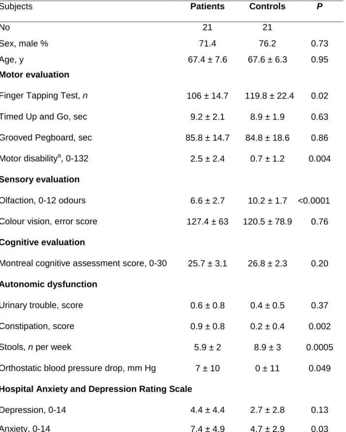
Twenty-two patients and 22 age- and gender-matched controls participated in the study. One patient who was unable to lie supine in the MRI machine and one healthy control subject (in whom a structural pons abnormality was fortuitously discovered) were excluded from the MRI analysis. Consequently, 21 patients with idiopathic RBD and 21 controls were analysed (Table 1). As expected based on the matching, there was no difference in the age or sex ratio between the two groups. The patients had higher motor disability scores on the MDS-UPDRS III and performed worse at the Finger Tapping Test relative to the controls, but no differences were noted in the other motor tests. The patients exhibited
a marked impairment in odour identification but not in colour vision compared with the controls. The cognitive performances of the patients and controls were similar. The patients exhibited more symptoms of constipation (and a lower number of stools per week) and a greater orthostatic drop in systolic arterial pressure systolic but no differences in urinary symptoms compared with the controls. The average anxiety scores (but not the depression scores) were higher in the patients than in the controls.
Sleep evaluation
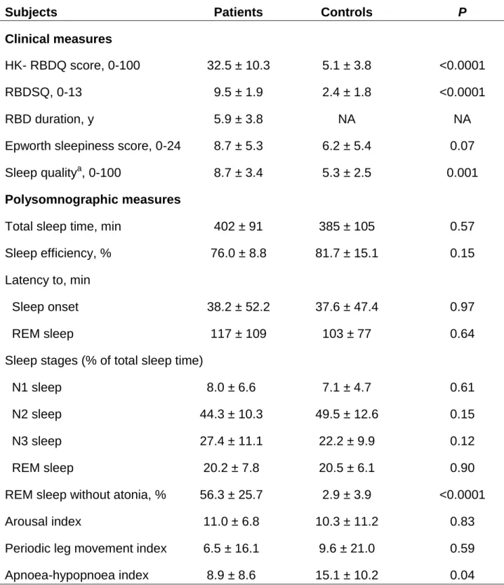
The scores for the two RBD questionnaires were higher for the patients than the controls. The global sleep quality index was lower in the patients, and additional alterations were noted in the sub-scores related to bad dreams, daytime functioning and sleep medication (Table 2). There were no differences between groups in sleep continuity, architecture or fragmentation according to the polysomnography results, with the exceptions of a greater percentage of REM sleep without atonia (as expected due to the disease) and a lower apnoea-hypopnoea index in the patients compared with the controls.
Magnetic resonance imaging data analysis
Quantitative analysis revealed that the MRI neuromelanin-sensitive signal intensities were significantly reduced in the patients compared with the controls in both the right and left coeruleus/subcoeruleus complexes, and no further differences between sides were noted (Table 3 and Fig.1). As shown in Table 4, the signal intensity correlated negatively with the percentage of REM sleep without atonia in the entire group (Fig. 2) but not with other sleep measures (data not shown) and not within the patient group. The signal intensity was also correlated with the severity of RBD symptoms, and the sleep quality score in the
entire group (but not in the patients group) but not with the motor, cognitive, sensory or psychological scores. The duration of RBD symptoms did not correlate with the loss of REM sleep atonia (r = 0.01, P = 0.96) or with motor disability (r = 0.34, P = 0.17).
Regarding the visual analyses, when the two radiologists analysed the MRI signals on a individual basis while blind to the diagnoses, only 2 subjects in the second reading of reader 1 and 3 subjects in the three other readings were classified as 2 (uncertain). After re-allocation of the uncertain scores to the categories of healthy volunteers and patients, the intra-reader agreement was 87.8% for reader 1 and 95.1% for reader 2. The inter-reader agreement was 81.7%. The mean sensitivity across the two inter-readers who performed with visual analysis for the diagnosis of idiopathic RBD was 82.5%, the specificity was 81.0%, the positive predictive value was 80.4%, and the negative predictive value was 83.4%. The patients with idiopathic RBD who had signals that overlapped with the signals of the normal controls did not differ in terms of motor or non-motor signs and symptoms from the other patients with idiopathic RBD.
Discussion
The neuromelanin-sensitive signal intensities were decreased in the
coeruleus/subcoeruleus complexes of the patients with idiopathic RBD compared with the healthy subjects. The signal intensity correlated with the percentage of REM sleep without atonia and with the severity of RBD in the 40 participants (but not in the patients
subgroup). Theindividual signal reductions as visually evaluated by radiologists were 82.5% sensitive and 81% specific for the diagnosis of RBD. The patients with idiopathic RBD more frequently exhibited preclinical markers of alpha-synucleinopathies, including constipation, olfactory deficits, orthostatic hypotension, and subtle motor impairment, as
well as higher anxiety scores and lower subjective sleep quality (despite normal sleep measures on polysomnography).
The level of signal loss in the coeruleus/subcoeruleus complexes of the idiopathic RBD patients was remarkably similar to the level observed in PD with RBD using the same technique and a 3T scanner (Garcia-Lorenzo et al., 2013). Additionally, the correlation between muscle atonia loss during REM sleep and signal loss in the brain was high, these correlations were remarkably similar between the PD patients with RBD (r = 0.49) and the entire group here (r = 0.44), but not within the idiopathic RBD subgroup. The muscle atonia loss (measured as enhanced tonic chin muscle activity) is however a continuous value, which reaches 10% at the 90th percentile of the normal population (Frauscher et al., 2014). As recently shown, isolated REM sleep without atonia heralds RBD as well as other biomarkers of neurodegeneration (Stefani et al., 2015). Based on the correlation observed here, one may suspect that the dysfunction of the REM atonia system is progressive, with subjects without RBD (aged healthy subjects and PD patients without RBD) having lost enough neurons to present 0-15% of REM sleep without atonia but not sufficiently to exhibit higher percentage of REM sleep without atonia plus RBD symptoms. Furthermore, the signal loss correlated with the clinical severity of RBD (as evaluated by two
questionnaires developed in the general population), but did not significantly correlate with other motor, cognitive or sensory tests or the psychological scores and sleep measures (with the exceptions of the impairment of sleep quality, which domain overlaps with the clinical severity of RBD). These findings provide evidence that this brainstem area drives motor atonia during REM sleep in humans. Notably, the human subcoeruleus locus is considered to be the anatomical equivalent of the sublaterodorsalis nucleus in rats (Boissard et al., 2002; Lu et al., 2006) and the peri-locus coeruleus alpha in cats (Sakai, 1991). In the rat, this nucleus contains glutamatergic neurons that activate the
motor neurons and thus induce atonia during REM sleep (Luppi et al., 2010). In human RBD, several multimodal MRI studies have found changes in the white and grey matter in the pons; however, these studies have not reached the level of precision of neuromelanin-sensitive sequences. Changes in diffusion metrics in the midbrain tegmentum and the rostral pons have been identified in patients with idiopathic RBD (Unger et al., 2010; Scherfler et al., 2011). Additionally, discrete inflammatory and vascular lesions in the midbrain and the tegmentum pontis have been associated with symptomatic RBD (Kimura et al., 2000; Boeve et al., 2007; Limousin et al., 2009). Evidently, the
coeruleus/subcoeruleus complex is influenced at an early stage of Lewy body pathology (which is associated with aggregated alpha-synuclein) in elderly brains that corresponds to the Braak stage 2 (Braak et al., 2003). Although direct causality between the loss of signal observed here and the mechanism of RBD cannot be definitively inferred based only on neuropathological examinations, the convergence of these in vivo imaging, clinical and animal-based findings strongly suggest that damage to complex is causative of RBD disorder and that such damage is one of the first steps in alpha-synuclein pathology.
Our study has several limitations. First, the measurements of muscle atonia in REM sleep were based only on single-night recordings. However, the between-night consistency of this measure is good (r = 0.55) when the tonic and not the phasic muscle tone is considered (Cygan et al., 2010). Another limitation is that the MRI signal analyses encompassed both the locus subcoeruleus (which probably contains the REM atonia system) and the locus coeruleus (which contains the noradrenergic arousal system). One cannot rule out the possibility that noradrenergic neurons co-degenerate in parallel with the atonia system in idiopathic RBD and PD-associated RBD and that such degeneration is a part of these diseases. The development of in vivo functional markers that are specific to the locus subcoeruleus, such as acetylcholine or glutamate marking (based on the
hypotheses that the locus subcoeruleus is cholinergic in humans as it is in cats or glutamatergic as it is rats) could further increase the specificity.
This study provides a measure of the MRI signal in the coeruleus/subcoeruleus complex that is based on specific sequences that target the anomalies associated with REM sleep behaviour disorder. The high sensitivities of the loss of signal at both the group calculation level and as a visual measure in a given patient could provide a diagnostic test for future patients. Additionally, follow-up of the signal intensity in the coeruleus/subcoeruleus complex will aid the determination of whether this marker further decreases over years and whether it can be used to follow the neurodegenerative process in, for example, future neuroprotective trials.
In conclusion, the signal loss in the coeruleus/subcoeruleus complex was strongly and specifically linked to the RBD symptoms, which suggests that this area drives muscle atonia in REM sleep in humans and that its loss causes RBD.
Fundings: This study was funded by a grant from the NRJ Foundation - Institute of France to IA and by the French program Investissement d’Avenir run by the Agence Nationale pour la Recherche (grants ‘IHU-A-ICM, Paris Institute of Translational
neuroscience ANR-10-IAIHU-06’ and ‘Infrastructure d’avenir en Biologie Santé - ANR-11-INBS-0006’). We thank the Clinical Investigation Centre CIC Paris-Est for its help in recruiting the control subjects.
American Academy of Sleep Medicine. The international Classification of Sleep Disorders, 3rd edition. 2014. Darien, IL: American Academy of Sleep Medicine.
Boeve BF, Silber MH, Saper CB, Ferman TJ, Dickson DW, Parisi JE, et al. Pathophysiology of REM sleep behaviour disorder and relevance to neurodegenerative disease. Brain 2007; 130: 2770-88.
Boissard R, Gervasoni D, Schmidt MH, Barbagli B, Fort P, Luppi PH. The rat ponto-medullary network responsible for paradoxical sleep onset and maintenance: a combined microinjection and functional neuroanatomical study. Eur J Neurosci 2002; 16: 1959-73.
Braak H, Del Tredici K, Rub U, de Vos RA, Jansen Steur EN, Braak E. Staging of brain pathology related to sporadic Parkinson's disease. Neurobiol Aging 2003; 24: 197-211.
Buysse D, Reynolds CI, Monk T, Berman S, Kupfer D. The Pittsburgh Sleep Quality Index: a new instrument for psychiatric practice and research. Psychiatry Res 1989; 28: 193-213.
Cygan F, Oudiette D, Leclair-Visonneau L, Leu-Semenescu S, Arnulf I. Night-to-night variability of muscle tone, movements, and vocalizations in patients with REM sleep behavior disorder. J Clin
Farnsworth D. The Farnsworth 100-hue test and dichotomous tests for color vision. J Optom Soc
Am 1943; 33: 568-78.
Ferini-Strambi L, Oldani A, Zucconi M, Smirne S. Cardiac autonomic activity during wakefulness and sleep in REM sleep behavior disorder. Sleep 1996; 19: 367-9.
Folstein MF, Folstein SE, McHugh PR. "Mini-mental state". A practical method for grading the cognitive state of patients for the clinician. J Psychiatr Res 1975; 12: 189-98.
Fonov V, Evans A, Botteron K, Almli C, McKinstry R, Collins D. Unbiased average age-appropriate atlases for pediatric studies. 54 2011; Neuroimage 313-27.
Frauscher B, Gabelia D, Mitterling T, Biermayr M, Bregler D, Ehrmann L, et al. Motor events during healthy sleep: a quantitative polysomnographic study. Sleep 2014; 37: 763-73, 73A-73B.
Gagnon JF, Vendette M, Postuma RB, Desjardins C, Massicotte-Marquez J, Panisset M, et al. Mild cognitive impairment in rapid eye movement sleep behavior disorder and Parkinson's disease. Ann
Garcia-Lorenzo D, Longo-Dos Santos C, Ewenczyk C, Leu-Semescu S, Gallea C, Quattrocchi G, et al. The locus coeruleus/subcoeruleus complex in rapid eye movement sleep behavior disorders in Parkinson’s disease: a 3T MRI study. Brain 2013; 136: 2120-9.
Goetz CG, Tilley BC, Shaftman SR, Stebbins GT, Fahn S, Martinez-Martin P, et al. Movement
Disorder Society-sponsored revision of the Unified Parkinson's Disease Rating Scale (MDS-UPDRS): scale presentation and clinimetric testing results. Mov Disord 2008; 23: 2129-70.
Hughes A, Daniel S, Kilford L, Lees A. Accuracy of clinical diagnosis of idiopathic Parkinson's disease: a clinico-pathological study of 100 cases. J Neurol Neurosurg Psychiatry 1992; 55: 181-4.
Hummel T, Sekinger B, Wolf S, Pauli E, Kobal G. Sniffing Sticks: olfactory performance assessed by the combined testing of odor identification, odor discrimination and olfactory threshold. Chem
Senses 1997; 22: 39-52.
Iber C, Ancoli-Israel S, Chesson A, Quan S. The AASM Manual for the Scoring of Sleep and
Associated Events: Rules, Terminology and Technical Specifications, 1rst Ed.2007. Westchester, IL: American Academy of Sleep Medecine.
Iranzo A, Fernandez-Arcos A, Tolosa E, Serradell M, Molinuevo JL, Valldeoriola F, et al.
Neurodegenerative disorder risk in idiopathic REM sleep behavior disorder: study in 174 patients.
PLoS One 2014; 9: e89741.
Iranzo A, Molinuevo JL, Santamaria J, Serradell M, Marti MJ, Valldeoriola F, et al. Rapid-eye-movement sleep behaviour disorder as an early marker for a neurodegenerative disorder: a descriptive study. Lancet Neurol 2006; 5: 572-7.
Johns MH. A new method for measuring daytime sleepiness: the Epworth Sleepiness Scale. Sleep 1991; 14: 540-5.
Kimura K, Tachibana N, Kohyama J, Otsuka Y, Fukazawa S, Waki R. A discrete pontine ischemic lesion could cause REM sleep behavior disorder. Neurology 2000; 55: 894-5.
Li SX, Wing YK, Lam SP, Zhang J, Yu MW, Ho CK, et al. Validation of a new REM sleep behavior disorder questionnaire (RBDQ-HK). Sleep Med 2010; 11: 43-8.
Limousin N, Dehais C, Gout O, Heran F, Oudiette D, Arnulf I. A brainstem inflammatory lesion causing REM sleep behavior disorder and sleepwalking (parasomnia overlap disorder). Sleep Med 2009; 10: 1059-62.
Lu J, Sherman D, Devor M, Saper CB. A putative flip-flop switch for control of REM sleep. Nature 2006; 441: 589-94.
Luppi PH, Clement O, Sapin E, Gervasoni D, Peyron C, Leger L, et al. The neuronal network
responsible for paradoxical sleep and its dysfunctions causing narcolepsy and rapid eye movement (REM) behavior disorder. Sleep Med Rev 2010; 15: 153-63.
Luppi PH, Clement O, Valencia Garcia S, Brischoux F, Fort P. New aspects in the pathophysiology of rapid eye movement sleep behavior disorder: the potential role of glutamate,
gamma-aminobutyric acid, and glycine. Sleep Med 2013; 14: 714-8.
Nasreddine ZS, Phillips NA, Bedirian V, Charbonneau S, Whitehead V, Collin I, et al. The Montreal Cognitive Assessment, MoCA: a brief screening tool for mild cognitive impairment. J Am Geriatr
Soc 2005; 53: 695-9.
Nazem S, Siderowf AD, Duda JE, Have TT, Colcher A, Horn SS, et al. Montreal cognitive assessment performance in patients with Parkinson's disease with "normal" global cognition according to mini-mental state examination score. J Am Geriatr Soc 2009; 57: 304-8.
Nutt J, Lea E, Van H, Schuff R, Sexton G. Determinants of tapping speed in normal control subjects and subjects with Parkinson’s disease: differing effects of brief and continued practice. Mov Disord 2000; 15: 843-9.
Podsiadlo D, Richardson S. The timed “Up & Go”: a test of basic func- tional mobility for frail elderly persons. J Am Geriatr Soc 1991; 39: 142–8.
Postuma RB, Gagnon JF, Vendette M, Fantini ML, Massicotte-Marquez J, Montplaisir J. Quantifying the risk of neurodegenerative disease in idiopathic REM sleep behavior disorder. Neurology 2009; 72: 1296-300.
Postuma RB, Gagnon JF, Vendette M, Montplaisir JY. Markers of neurodegeneration in idiopathic rapid eye movement sleep behaviour disorder and Parkinson's disease. Brain 2009; 132: 3298-307.
Sakai K. Physiological properties and afferent connections of the locus coeruleus and adjacent tegmental neurons involved in the generation of paradoxical sleep in the cat. Prog Brain Res 1991; 88: 31-45.
Schenck CH, Boeve BF, Mahowald MW. Delayed emergence of a parkinsonian disorder or dementia in 81% of older males initially diagnosed with idiopathic REM sleep behavior disorder (RBD): 16year update on a previously reported series. Sleep Med 2013; 14: 744-48.
Schenck CH, Bundlie SR, Ettinger MG, Mahowald MW. Chronic behavioral disorders of human REM sleep: a new category of parasomnia. Sleep 1986; 9: 293-308.
Schenck CH, Bundlie SR, Mahowald MW. Delayed emergence of a parkinsonian disorder in 38% of 29 older men initially diagnosed with idiopathic rapid eye movement sleep behaviour disorder.
Neurology 1996; 46: 388-93.
Scherfler C, Frauscher B, Schocke M, Iranzo A, Gschliesser V, Seppi K, et al. White and gray matter abnormalities in idiopathic rapid eye movement sleep behavior disorder: a diffusion-tensor imaging and voxel-based morphometry study. Ann Neurol 2011; 69: 400-7.
Stefani A, Gabelia D, Hogl B, Mitterling T, Mahlknecht P, Stockner H, et al. Long-Term Follow-up Investigation of Isolated Rapid Eye Movement Sleep Without Atonia Without Rapid Eye Movement Sleep Behavior Disorder: A Pilot Study. J Clin Sleep Med 2015. In press.
Stiasny-Kolster K, Doerr Y, Moller JC, Hoffken H, Behr TM, Oertel WH, et al. Combination of 'idiopathic' REM sleep behaviour disorder and olfactory dysfunction as possible indicator for
alpha-synucleinopathy demonstrated by dopamine transporter FP-CIT-SPECT. Brain 2005; 128: 126-37.
Stiasny-Kolster K, Mayer G, Schafer S, Moller JC, Heinzel-Gutenbrunner M, Oertel WH. The REM sleep behavior disorder screening questionnaire--a new diagnostic instrument. Mov Disord 2007; 22: 2386-93.
Unger MM, Belke M, Menzler K, Heverhagen JT, Keil B, Stiasny-Kolster K, et al. Diffusion tensor imaging in idiopathic REM sleep behavior disorder reveals microstructural changes in the brainstem, substantia nigra, olfactory region, and other brain regions. Sleep 2010; 33: 767-73.
Wenning GK, Tison F, Seppi K, Sampaio C, Diem A, Yekhlef F, et al. Development and validation of the Unified Multiple System Atrophy Rating Scale (UMSARS). Mov Disord 2004; 19: 1391-402.
Zigmond AS, Snaith RP. The hospital anxiety and depression scale. Acta Psychiatr Scand 1983; 67: 361-70.
Figure legends
Figure 1 Axial T1-weighted neuromelanin-sensitive images of the
coeruleus/subcoeruleus complex in a healthy volunteer (A) and a patient with idiopathic
rapid eye movement sleep behaviour disorder (B). The locus area (arrows) is visible as an area of increased signal intensity.
Figure 2 Correlation between the neuromelanin-sensitive signal intensity and the percentage of rapid eye movement (REM) sleep without atonia in the patients with
idiopathic REM sleep behaviour disorder (plain diamonds) and the control subjects (empty diamonds). Note that the correlation is significant only when including both controls and patients.
Table 1 - Clinical characteristics of the patients with idiopathic REM sleep behaviour
disorder and the age- and sex-matched healthy controls
Subjects Patients Controls P
No 21 21
Sex, male % 71.4 76.2 0.73
Age, y 67.4 ± 7.6 67.6 ± 6.3 0.95
Motor evaluation
Finger Tapping Test, n 106 ± 14.7 119.8 ± 22.4 0.02
Timed Up and Go, sec 9.2 ± 2.1 8.9 ± 1.9 0.63
Grooved Pegboard, sec 85.8 ± 14.7 84.8 ± 18.6 0.86
Motor disabilitya, 0-132 2.5 ± 2.4 0.7 ± 1.2 0.004
Sensory evaluation
Olfaction, 0-12 odours 6.6 ± 2.7 10.2 ± 1.7 <0.0001
Colour vision, error score 127.4 ± 63 120.5 ± 78.9 0.76
Cognitive evaluation
Montreal cognitive assessment score, 0-30 25.7 ± 3.1 26.8 ± 2.3 0.20
Autonomic dysfunction
Urinary trouble, score 0.6 ± 0.8 0.4 ± 0.5 0.37
Constipation, score 0.9 ± 0.8 0.2 ± 0.4 0.002
Stools, n per week 5.9 ± 2 8.9 ± 3 0.0005
Orthostatic blood pressure drop, mm Hg 7 ± 10 0 ± 11 0.049
Hospital Anxiety and Depression Rating Scale
Depression, 0-14 4.4 ± 4.4 2.7 ± 2.8 0.13
Anxiety, 0-14 7.4 ± 4.9 4.7 ± 2.9 0.03
a evaluated using the part III of the Movement Disorder Society - United Parkinson’s
Table 2 - Clinical and polysomnographic sleep measures of the patients with
idiopathic REM sleep behaviour disorder (RBD) and the healthy controls
Subjects Patients Controls P
Clinical measures
HK- RBDQ score, 0-100 32.5 ± 10.3 5.1 ± 3.8 <0.0001
RBDSQ, 0-13 9.5 ± 1.9 2.4 ± 1.8 <0.0001
RBD duration, y 5.9 ± 3.8 NA NA
Epworth sleepiness score, 0-24 8.7 ± 5.3 6.2 ± 5.4 0.07
Sleep qualitya, 0-100 8.7 ± 3.4 5.3 ± 2.5 0.001
Polysomnographic measures
Total sleep time, min 402 ± 91 385 ± 105 0.57
Sleep efficiency, % 76.0 ± 8.8 81.7 ± 15.1 0.15
Latency to, min
Sleep onset 38.2 ± 52.2 37.6 ± 47.4 0.97
REM sleep 117 ± 109 103 ± 77 0.64
Sleep stages (% of total sleep time)
N1 sleep 8.0 ± 6.6 7.1 ± 4.7 0.61
N2 sleep 44.3 ± 10.3 49.5 ± 12.6 0.15
N3 sleep 27.4 ± 11.1 22.2 ± 9.9 0.12
REM sleep 20.2 ± 7.8 20.5 ± 6.1 0.90
REM sleep without atonia, % 56.3 ± 25.7 2.9 ± 3.9 <0.0001
Arousal index 11.0 ± 6.8 10.3 ± 11.2 0.83
Periodic leg movement index 6.5 ± 16.1 9.6 ± 21.0 0.59
Apnoea-hypopnoea index 8.9 ± 8.6 15.1 ± 10.2 0.04
a
Table 3 - MRI signal intensity in the coeruleus/subcoeruleus complex
Patients Controls P
Left side, mean ± SD 123.7 ± 5.7 130.1 ± 6.1 <0.001
Left side, median 123.8 130.4 <0.001
(Q1; Q3) (121.0; 128.6) (126.8; 132.3)
Right side, mean ± SD 120.6 ± 5.2 125.2 ± 4.8 0.003
Right side, median 121.8 125.0 0.009
(Q1;Q3) (116.5; 123.9) (122.3; 126.9)
Difference right/left (p-value)
0.09 0.01
Mean of both sides 122.2 ± 5.3 128.2 ± 5.2 0.001
Median of both sides 122.4 128.1 0.002
(Q1; Q3) (119.1; 126.4) (124.5; 142.0)
Table 4 - Correlations between the coeruleus/subcoeruleus signal intensity and
motor and non-motor signs and symptoms in all subjects, in patients with idiopathic
rapid eye movement sleep behaviour disorder (IRBD) and controls
All subjects IRBD Controls
R P R P R P Age, y 0.01 0.93 0.23 0.33 0.22 0.36 RBD duration, y NA NA 0.06 0.82 NA NA Hong Kong - RBD Questionnaire, 0-100 0.52 0.007 0.36 0.12 0.20 0.39 RBD Screening Questionnaire, 0-13 0.47 0.002 0.11 0.65 0.04 0.86 Sleep measures
Percentage of REM sleep without atonia, 0-100%
0.44 0.004 0.05 0.85 0.06 0.81
Epworth sleepiness score, 0-24
0.26 0.1 0.05 0.82 0.26 0.26 Sleep quality,a, 0-100 0.35 0.03 0.35 0.15 0.08 0.74
Motor tests
Finger Tapping Test, n 0.02 0.88 0.02 0.93 0.31 0.19
Timed Up and Go, sec 0.11 0.51 0.13 0.59 0.17 0.47
Grooved Pegboard, sec 0.12 0.45 0.21 0.39 0.18 0.44
Motor disabilityb, 0-132 0.22 0.17 -0.08 0.75 0.20 0.39
Sensory tests
Olfaction, 0-12 odours 0.37 0.05 -0.06 0.81 0.21 0.37
Colour vision, error score 0.31 0.05 -0.32 0.16 0.41 0.07
Cognitive scorec, 0-30 0.18 0.26 0.01 0.96 0.20 0.40
Autonomous tests
Stools, n per week 0.13 0.44 0.18 0.46 0.22 0.38
Orthostatic blood pressure drop, mm Hg 0.04 0.80 0.09 0.71 0.33 0.17 Psychological tests Depression, 0-14 0.10 0.52 0.10 0.68 0.11 0.64 Anxiety, 0-14 0.18 0.27 0.02 0.91 0.07 0.76 a
evaluated using the Pittsburgh sleep quality inventory; b evaluated using the part III of the Movement Disorder Society - United Parkinson’s disease rating scale; c
evaluated using the Montreal cognitive assessment; R= Pearson correlation coefficient; p= level of significance of the correlation. The significant values are in bold.