Abstract — In the post genomic era, proteomics has enormous potential in biology and medicine. Among the various bioanalytical tools developed, protein microarray is one of the recent advancements which offer high throughput profiling of cellular proteins to provide insights into the mechanisms of biological processes. Fundamentally, the protein microarray involves the immobilization of interacting elements, proteins, on a few square microns of a solid support and in principle, it is capable of detecting analytes with a higher sensitivity than conventional macroscopic immunoassays. Here in the present report we delineates the design, fabrication and functional analysis of protein microarray using semi-synthetic ssDNA tagged-proteins as capturing moiety as well as address on a solid support. Optimization of the platform has been carried out by investigating various parameters such as surface chemistry, signal amplification, and conditions for homogenous liguid phase protein-protein interaction.
Index Terms — Proteomics, Protein micro array, Dendrimer, glass slide, ssDNA-antibody
I. INTRODUCTION
uring the last decade DNA microarray has been instrumental in probing the global transcriptome of the cells.1-3 Followed by protein microarray which has emerged as another complimentary approach for high throughput analysis to understand the complex cellular system.4 Currently, there are many different strategies for the fabrication and functional analysis of protein microarray.5-6 Fundamentally these microarray formats stems on different immobilization chemistry, detection methodology or capturing of the analyte. Nevertheless, all of the protein arrays can be classified as either forward phase (antibody or any capturing entity immobilized onto surface) or reverse phase array (antigens immobilized onto the surface and probe the respective antibody). Many different types of proteins could be simultaneously detected on the same chip. Despite these successes of protein microarray in profiling the proteome of the cell, it has many inherent limitations which are still unresolved and prevent protein microarray technology from reaching its full potential.7
Some of these limitation includes the generation of
content ie probe for complete proteome analysis, the conservation of protein functionality during the immobilization, as well as the provision of the required absolute and relative sensitivity.
Even though protein and nucleic acid microarrays were technologically similar in many aspects, the most significant differences are found in the physical properties and stability of soluble proteins and DNA at interfaces. Unlike DNA, proteins are chemically and physically heterogeneous, have a three-dimensional structure which is critical to their function, and have no analogous amplification technique. Many proteins are also known to adsorb non-specifically to commonly used substrates and pose a constant problem in generating false positive signals. These factors can compromise the performance of protein microarrays and limit its specificity and sensitivity, particularly when complex biological fluids are used. In addition, surface modifications provide homogenous and efficient activation of the entire surface of the substrate used. Thus, the surface chemistries must be chemically and physically robust. Herein, we describe the design, fabrication and use of a PAMAM modified glass surface for the generation of a robust, homogenous chemically activated and high density substrate which is useful for protein microarrays. In addition, we designed and fabricated a new platform, spatially addressable protein array (SAPA), by exploring the specificity of ssDNA hybridization for a self-assembly of semi-synthetic ssDNA-antibody conjugates which captured the antigen from complex biological samples.
II. EXPERIMENTAL SECTION
Materials and instrumentations. The
carboxyl-terminated PAMAM (10% solution in methanol), N-hydroxysuccinimide (NHS), succinic anhydride, 4-hydroxy-3-nitrobenzenesulfonic acid (HNSA), polyethylene oxide (PEO, 100,000 Mwt) and Sephadex G50 were obtained from the Sigma and 1-Ethyl-3-(3-Dimethylaminopropyl)carbodiimide hydrochloride (EDC) were purchased from Pierce Chemicals. The diamino polyethylene glycole (PEG, 3400 Mwt) was obtained from Nektar Therapeutics, CA, USA. Cy5 fluorescent mono-reactive dye was purchased from Amersham Biosciences. The amine and epoxy slides were from Genetix in UK. The
Design, fabrication and functional analysis of a new
protein array based on ssDNA-based assembly
Parayil Kumaran AJIKUMAR1, Jin Kiat NG1, Yew Chung TANG1, Jim Yang LEE1,2,
Gregory STEPHANOPOULOS1,3, Heng-Phon TOO1,4 1
MEBCS, Singapore-MIT Alliance, National University of Singapore, Singapore
2
Chemical and Biomolecular Engineering, National University of Singapore
3
Department of Chemical Engineering, MIT, Cambridge, Massachusetts 02139, USA
4
Department of Biochemistry, Kent Ridge Crescent, National University of Singapore
mouse monoclonal anti-EGFP (Molecular Probes) and TRITC-labelled rabbit IgG (DakoCytomation, USA) were purchased and purified using a Pierce protein A/G column according to manusfacturer’s instructions. eGFP was obtained from Upstate, USA. Laser scanning was carried out on the GenePix 4000B (Axon Instruments CA, USA). Fluorescence spectrophotometry was carried out on the Fluorostar Optima (BMG Labtechnologies GmbH, Germany) and atomic force microscopy using the Nanoscope III (Digital Instrument Int.)
Preparation of carboxyl-terminated dendrimer-functionalized glass slides. Carboxyl-terminated PAMAM
dendrimer slides were prepared using amino-silylated glass slides. The carboxylic acid of the PAMAM dendrimer was activated with a solution of EDC/NHS (1:1, 100 mM) in 0.1 M MES (pH 6.3), layered onto the amine slides and incubated for 2 h. The slides were then washed with double distilled water, ethanol and air dried. The slides were then kept in vaccuo until use. The carboxyl-terminated PAMAM slides were further activated by EDC/HNSA or EDC/NHS active ester (1:1; 100 mM) in 0.1 M MES, 0.5 M NaCl (pH 6.3) buffer. The activated slides were washed with double distilled water, ethanol and air dried by centrifugation. The slides were stored in desiccators under nitrogen at room temperature until use
.
Cell culture and lysate extraction. The Neuro 2A cell
line (CCL-131) was obtained from ATCC. The cells were grown at 37 °C in a 5% CO2 atmosphere in DMEM medium (Gibco/Life Technologies) supplemented with 10% fetal calf serum, 100 units/mL penicillin, and 100 µg/mL streptomycin. At 90% confluency, the cells were washed twice with 1x phosphate buffered saline (1x PBS) and lysed with 1x PBS, 0.5% Triton X-100. The lysate was subsequently cleared by centrifugation and the supernatant containing the cellular proteins recovered for labeling.
Labeling of cell lysate and eGFP. The cell lysates or
eGFP were labeled with Cy5 mono-reactive dye according to the recommended procedure by the manufacturer. The protein samples were diluted to 0.5 mg/mL in labeling buffer (0.1 M NaCO3, 50 mM NaCl, pH 9). The diluted protein solution was incubated with Cy5-NHS dye (4 µg/µl in DMSO) with a protein-dye ratio of 1:1 (w/w) with intermittent shaking at at 25 oC for 30 min. The reaction was then quenched using Tris-HCl (0.1 M, pH 8) for 10 min. Free dye was removed by size exclusion chromatography (Sephadex G50) pre-equilibriated in 1x PBS, pH 7.4. Concentration of the eluted protein was measured using the microBCA kit from Pierce.
Immobilization of antibodies onto slides and capturing of labeled antigen. Antibodies in the printing buffer (1%
glycerol, 5 mM EDTA in 1x PBS at pH 8) were spotted (500 nL each) onto the pre-activated PAMAM or epoxy slides. The coupling reaction was allowed to proceed for 4 hours at 250C in a humid chamber. The arrayed slides were then washed 3x in buffer containing 1x PBS and 0.05% Tween 20 (wash buffer), each for 10 min. The arrayed slides were then incubated in blocking buffer (1x PBS
containing 3% Tween 20, 5% Trehalose and 0.1% NaN3) for 30 min followed by incubation with 20 µL of varying concentrations of eGFP-Cy5 or cell lysates in 1x PBS, 0.1% TX-100, 1% or 10% non- milk solution in a humidity chamber at 250C for 1 h in the dark. The slides were then incubated in wash buffer for 10 minutes (3 times) and spun dried. The slides were then imaged and results analyzed using GenePix 3.0 software (Axon Instruments, Union City, CA).
Immobilization of H2N-ssDNA over the activated
dendrimer and succinamic acid slides. For the covalent
attachment of the H2N-ssDNA, typically 500 nl of a solution of the 5′-amino-modified oligonucleotide in 0.1 M NaHCO3 (pH 8.5) was arrayed on to the HNSA-activated slides and incubated for 3 h in a humid chamber. The slides were washed with water and subsequently incubated in solution of 1 M Tris buffer (pH 9) for another 1 h at 42 C to quench all the remaining activated groups. After a thorough wash with water, the slides were dried by spinning in a centrifuge with a slide holder (500 rpm 5 min at 27 C) and further dried under a stream of nitrogen.
Synthesis of antibody conjugates.
ssDNA-antibodies conjugates were synthesized using a modified reported procedure.8 The sulfhydryl group of a modified FAM-labeled oligo 5′FAM-(CH2)6-(T)20-3′(CH2)6-SH was linked using a bifunctional cross-linker, sulfo-SMCC, to the amine group on the surface of the antibody. To optimize and quantify each step TRITC-labeled rabbit anti-IgG and the FAM-labeled ssDNAs were used. To generate a free sulfhydryl group the protected thiol-modified oligonucleotides were decapped by reduction of the disulfide bond (10 mM DTT) and purified by precipitation with acetone. The purified TRITC-labeled rabbit anti-IgG was reacted with the sulfo-SMCC cross-linker at a 1000 molar excess in phosphate buffered saline (1xPBS, pH 7.4) for 30 min at room temperature. Unreacted cross-linker was removed using a Pierce protein desalting column. The yield of the cross linker attached antibody was quantified by measuring the fluorescence of the TRITC (~70%). Purified products were reacted with thiol-modified oligonucleotide in a 1:10 molar equivalent (1 h, 4 οC). Further incubation at 37 οC (20 min) was performed to reduce the non-specific binding of antibody onto the appendorf tube. The excess oligonucleotides were separated from the conjugates by non-denaturing PAGE (20% 19:1) at 120 V for 10 min. The conjugates which had not migrated into the gel were recovered from the well. These conjugates were diluted in 1x PBS (pH 8) solutions and stored at 4°C. Product yields were quantified by measuring the fluorescence intensity of the FAM in the oligo and TRITC in the antibody. Approximately 4 molecules of ssDNAs were conjugated to one molecule of the antibody by using FAM-labeled oligo to quantify the oligo incorporation at the final stage and micro BCA to quantify the antibody.
Investigation of the loading capacity of the slides by immobilization of Cy5-labeled oligo and hybridization of ssDNA-TRITC labeled IgG antibody conjugate. To
quantify and compare the loading capacity of the oligo address, on PAMAM-carboxyl and succinamic acid derivatized slides, amino-modified Cy5-labeled ssDNA (5′NH2-(CH2)6-(T)20-3′ (CH2)6-Cy5) with concentrations of 10, 20, 30, 40, 60, 80 µmole/L (0.1 M Na2CO3, pH 9) were spotted onto a pre-activated glass slide and incubated for 2 hrs in a humid chamber and processed as described in the oligo immobilization procedure. The loading of the labeled oligos were checked by measuring the fluorescence intensity using a GenePix 4000, Axon laser scanner. To quantify and compare the amount of captured ssDNA-tagged antibody, the slides were prepared by loading with different concentrations (10, 20, 30, 40, 60, 80 µmole/L) of complimentary (3′NH2-(CH2)6-(2T)20-5′) and non-complimentary (5′NH2-(CH2)6-(2A)20-3′) ssDNA. After quenching and washing, 5’FAM-(CH2)6 -(2T)20-3(CH2)6SH-oligo tagged TRITC labeled IgG antibody in 1xPBS pH 7.4 with 0.1% milk and 0.1% TX 100 at pH 7.4 (20µL, 2 pmole) is incubated using a cover slip. Incubated for 1 hr to capture the ssDNA- IgG over the complimentary ssDNA spotted glass slide, washed with 0.1% TX 100 in 1xPBS pH 7.4 at pH 7.4 (30 min), 1xPBS pH 7.4 (1 min) and were dried by spinning in a centrifuge with a slide holder (500 rpm 5 min at 27 οC) and further dried under a stream of nitrogen. The fluorescence intensity of the hybridized antibody was measured using a GenePix 4000, Axon laser scanner.
III. RESULTSANDDISCUSSION
Surface chemistry. In the protein/antibody micro array the
major objective is the study of interaction partners. Thus, the key requirements are high binding capacity of interaction entity yet with low nonspecific protein background and low variability. There have been numerous studies to identify an optimal surface protein array. The surfaces used for protein array were divided as three classes, (i) two-dimensional plain glass slides which are activated with a variety of coupling chemistries such as aldehyde, epoxy or carboxylic esters,9 (ii) three dimensional (3D) gel or membrane-coated surfaces such as polyacrylamide, agarose and nitrocellulose110-13 and (iii) surface coatings, such as PEG, BSA, avidin and dendrimer over the 2D glass slides.14 In 2D glass surfaces proteins or antibodies bind either by electrostatic interactions or through covalent linkages which offers strong attachment combined with low variation. However, the limitations such as rapid evaporation of the liquid environment as well as the close surface contact may lead to the denaturation of native structure of the protein. Where as in the 3D gel or membrane surfaces, proteins bind through physical adsorption and have better preservation of the native protein conformation. However, due to the lack of specific binding of the proteins often leads to leaching and large variations in the signal intensity. In principle the third group of surface coatings has the advantages of both 2D and 3D surfaces mentioned above. Although the 3D structures are not visible, the coating of large macromolecules generated supramolecular structure on the
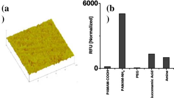
surfaces provides an interface which is more compatible for minimizing the protein denaturation and specific binding on the surfaces. Among the various macromolecule coated surfaces, dendrimers is a unique class of polymer that differs from linear polymers in that they don't have entangled chains and at the same time have numerous chain-ends that can be easily functionalized. Previously, poly(amidoamine) (PAMAM) dendrimers with highly uniform and compact amine linkers have been exploited for various applications in high density DNA microarrays and protein immobilization studies.15-16 In the present study we explored the single-step chemical coupling of a PAMAM-carboxyl dendrimer (3.5G, 64 carboxyl groups) to generate quasi three dimensional nanostructured monolayer on amino-silylated glass slides (Fig. 1a). It is anticipated that these newly fabricated surfaces are homogenous, with a high density of carboxyl functional groups. A comparison of different surfaces was performed for the nonspecific protein adsorption as well as protein immobilization. As expected the, the non-specific protein adsorption on carboxyl terminated PAMAM slides is almost same as to a PEG modified linear linker such as epoxy surface (Fig. 1b).
Fig. 1: (a) Tapping mode AFM height images (10 × 10 µm)
of the dendrimer coated slides. (b) Nonspecific protein adsorption of the Cy5 dye labelled cell lysate over various chemically modified glass slides
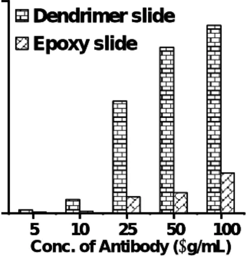
Besides the provision of an optimum surface which has low nonspecific protein adsorption, the optimal immobilization of probe protein/antibody on the surface and the optimization of assay conditions and the detection sensitivity of analytes is another key parameter of every micro array technology. In order to validate the performance of the carboxyl terminated PAMAM slides, the slide surface was further pre-activated with EDC/HNSA and immobilized with different concentration (1-100 µg/mL) of monoclonal anti-eGFP antibody. Similar concentrations of the antibodies were immobilized over a linear linker 2D epoxy slide for comparison. The antigen, eGFP labeled with Cy5 (0.2 pmol/µl), was then incubated and captured on the arrays. The signal intensity was significantly greater on the PAMAM slides than the epoxy slides (Fig. 2). The result is consistent with the proposition that dendrimers not only increased the density of functional sites but was also highly accessible to analytes which is
0 6000 RFU [ N o rma lized ] P A MA M-C O OH P A MA M-N H2 PE G Su c c en a m ic A c id Am in e 0 6000 RFU [ N o rma lized ] P A MA M-C O OH P A MA M-N H2 PE G Su c c en a m ic A c id Am in e
(a
)
(b
)
more comparable to the properties of 3D gel or membrane coated slides.
Dendrimer slide
Epoxy slide
5 10 25 50 100 Conc. of Antibody (µg/mL)Dendrimer slide
Epoxy slide
5 10 25 50 100Dendrimer slide
Epoxy slide
5 10 25 50 100 Conc. of Antibody (µg/mL)Fig. 2: Comparison of the antigen captured by antibody
immobilized on a PAMAM carboxyl slides and epoxy slides
ssDNA-tagged antibody and addressable protein array.
The high loading capacity of ssDNA on carboxyl terminated PAMAM slides was further evaluated. In order to compare the efficiency of hybridization linear linker succinamic acid slides was used for comparison. Initially, a doubly functionalized ssDNA was serially diluted and arrayed (500 nl, 5′NH2-(CH2)6-(T)20-3′ (CH2)6-Cy5 in 0.1M Na2CO3, pH 8-8.5) onto pre-activated PAMAM carboxyl and succinamic acid slides. After 3 hours of incubation in a humid chamber, the unbound ssDNA was removed by washing in water and the remaining activated groups quenched by incubating the slides in 1 M Tris for 1 h. The observed fluorescence intensity showed that dendrimer-coated slides have higher loading capacity of ssDNA compared to the linear linker succinamic acid slides. 0 20 40 60 80 100 0 65000 RFU Conc. of H2N-ssDNA Dendrimer slide Succenamic slide
Fig. 3: The amount of 20-mer ssDNA (5′NH2-(CH2)6 -(T)20-3′ (CH2)6-Cy5) immobilized on to PAMAM carboxyl and succinamic acid surfaces.
The DNA-tagged antibodies were prepared using a bifunctional linker Sulfo-SMCC attached to the surface amine group of TRITC-labeled polyclonal Swine anti-rabbit IgG (ARIgG), followed by electrophilic addition of
the thiol group in a 20-mer modified ssDNA, 3′SH-(CH2)6 -(T)20-(CH2)6 FAM 5′, to the maleimides in the SMCC. Excess ssDNA was removed and the conjugate was purified by non-denaturing PAGE (20% 19:1). Purity of the ssDNA-tagged antibodies was further confirmed by loading into an agarose gel (1%). The calculated yield using the FAM-labeled ssDNA incorporated onto the antibody showed that on average, 4 molecules of ssDNA were conjugated to a molecule of IgG. The amount of protein was quantified by microBCA analysis and the final yield of the product was found to be ~60%, which is 5-6 times more than the yield achieved by previously reported procedures.
Fig. 4: Schematic representation of bifunctional cross
linker SMCC mediated conjugation of ssDNA to TRITC-labeled polyclonal Swine anti-rabbit IgG (ARIgG).
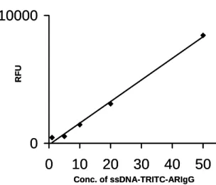
The hybridization of ssDNA-tagged antibodies were investigated using ssDNA (3′NH2-(CH2)6-(T)20-5′ and 3′NH2-(CH2)6-(A)20-5′) (10, 20 30 40 and 50 µM) arrayed on carboxyl terminated PAMAM slides using ssDNA (3′HS-(CH2)6-(T)20-(CH2)6 FAM5′) conjugated TRITC -ARIgG. The observed results showed that the amount of ssDNA-TRITC-ARIgG hybridized on the complimentary ssDNA arrays increased with increasing concentration of the oligonucleotide on the dendrimer slides and saturated at 30 µM of oligonucleotides spotted on the slides (data not shown). This indicates that steric hindrance from the ssDNA-IgG conjugate would influence the efficiency of hybridization onto the complementary ssDNA arrayed on the slides. In addition the concentration dependent hybridization of ssDNA-TRITC-ARIgG (5, 10, 20, 50 µg/mL) showed that that the hybridization was increasing linearly with concentration and did not reach saturation even at a concentration of 50 µg/mL of the conjugate (Fig. 4).
0
31000
0
10
20
30
40
50
RFU Conc. of ssDNA-TRITC-ARIgG0
31000
0
10
20
30
40
50
RFU Conc. of ssDNA-TRITC-ARIgGFig. 5: The amount of ssDNA-ARIgG hybridized from 5,
10, 20 and 50 µg/mL ssDNA-TRITC_ARIgG on a 30 µM
SH
SH
+
+
ssDNA arrayed slides.
The amount of ssDNA-TRITC-ARIgG hybridized on surface decreased dramatically as the conjugate concentration dropped below 10 µg/mL. This indicates that the hybridization of the ssDNA-antibodies has a significant influence on the signal intensity when compared to the ssDNA-antibodies to antigens ratio in the capturing medium. From the aforementioned observation, we fixed the concentration of the ssDNA-antibody capturing probe at 20 µg/mL for subsequent experiments. In order to optimize the concentration of conjugates in the solution, we titrated different concentrations of ssDNA-IgG (1, 5, 10, 20, 50 µg/mL) against 0.1 µg/mL rabbit IgG (labeled with Cy5) in the capturing medium (Fig. 5). The observed results showed that very good signal intensity with high signal to noise was observed even at very low antigen 0.1 µg/mL antigen concentrations which indicated the combined effect of the higher efficiency of the probes hybridizing onto the immobilized ssDNA on the high density dendrimer substrate (Fig. 5).
0
10000
0
10
20
30
40
50
RFU Conc. of ssDNA-TRITC-ARIgG0
10000
0
10
20
30
40
50
RFU Conc. of ssDNA-TRITC-ARIgGFig. 5: Capturing efficiency of 0.1 µg/mL antigen in
different concentrations of ssDNA-TRITC-ARIgG in the capturing solution (1, 5, 10, 20, 50 µg/mL) on a 30 µM oligo spotted slide.
We have performed a detection sensitivity assay for the newly designed array format by capturing antigen Cy5-labeled rabbit IgG of different concentrations (50, 500, 5000, 5000 pM) using a ssDNA-tagged swine anti-rabbit IgG (SAR-IgG) (20 µg/mL) on complimentary ssDNA-arrayed slides (Fig 6). The observed detection limit was 1 pM of the antigen with the signal-to-noise ratio of ~30. This shows the efficiency of the new approach of DNA-directed assembly (DDA) in the detection of low amounts of antigen (50 pM) with significantly lower concentrations of the capture reagent (0.001 µM). Recently, similar studies explored the DNA direct immobilization by Wacker et al.6 reported highest signal intensities with best spot homogeneity and reproducibility as well as the lowest consumption of antibodies. However, the additional effort involving multistep preparation of ssDNA tagged streptavidin and preparation of conjugate for each antibody
will result in lower yield. With DDI, immobilization and capturing of antigen method still has the problems of denaturation of immobilized antibody and the antibody orientation on array surface which are common hurdle in the antibody immobilized arrays also. In contrast, our new array system offers the capturing of the antigen in solution, followed by the highly efficiently assembly conjugated over a highly compatible dendrimer modified slides.
RFU Conc. of Antigen (pM) 10 10000 10 100 1000 10000 100000 RFU Conc. of Antigen (pM) 10 10000 10 100 1000 10000 100000
Fig. 6. Different concentrations of antigen-rabbit IgG,
captured with 10 µg/mL ssDNA-SARIgG, using 30 µM oligo spotted slide.
IV. CONCLUSIONS
In summary, the present study described a simple, efficient fabrication of a novel antibody micro array format. The performance of newly fabricated surface and array format for antibody microarray was evaluated in terms specificity and sensitivity.
V. ACKNOWLEDGEMENT
N.J.K, T.Y.C. and PKA acknowledges the financial support provided by the Singapore-MIT (SMA) Alliance.
REFERENCES
1. M. Schena, D. Shalon, R. W. Davis, and P. O. Brown, "Quantitative monitoring of gene expression patterns with a complementary DNA microarray," Science, 270, 467–470, 1995.
2. R. Ekins, and F. W. Chu, "Microarrays: their origins and applications," Trends Biotechnol., 17, 217–218, 1999.
3. T. Ideker, V. Thorsson, J. A. R. Ranish, Christmas, J. Buhler, J. K. Eng, R. Bumgarner, D. R. Goodlett, R. Aebersold, and L. Hood, "Integrated genomic and proteomic analyses of a systematically perturbed metabolic network," Science, 292, 929–934, 2001. 4. G. MacBeath, and S. L. Schreiber, "Printing proteins as
microarrays for high-throughput function determination," Science, 289, 1760–1763, 2000.
5. W. Kusnezow, A. Jacob, A. Walijev, F. Dieh, and J. D. Hoheisel, "Antibody microarrays: an evaluation of production parameters," Proteomics, 3, 254–264, 2003.
6. R. Wacker, H. Schröder, and C. M. Niemeyer, "Performance of antibody microarrays fabricated by either DNA-directed immobilization, direct spotting, or streptavidinbiotin attachment: a comparative study," Anal. Biochem., 330, 281–287, 2004.
7. P. Angenendt, “Progress in protein and antibody microarray technology” DDT 10, 503-511, 2005. 8. (a) C. M. Niemeyer, T. Sano, C. L. Smith, and C. R.
Cantor, “Oligonucleotide-directed self-assembly of proteins: semisynthetic DNA-streptavidin hybrid molecules as connectors for the generation of macroscopic arrays and the construction of
supramolecular bioconjugates,” Nucleic Acids Res., 22, 5530-5539, 1994.
9. J. Sobek, and R. Schlappbach, “Substrate architecture and functionality defining the properties and
performance of DNA, peptide, protein and
carbohydrate microarrays,” Pharmagenomics, 32-44, 2004.
10. P. Arenkov, A. Kukhtin, A. Gemmell, S. Voloshchuk, V. Chupeeva, and A. Mirzabekov, “Protein microchips: use for immunoassay and enzymatic reactions,” Anal. Biochem., 278, 123–131, 2000.
11. A. Y. Rubina, E. I. Dementieva, A. A. Stomakhin, E. L. Darii, S. Pan'kov, V. V. E. Barsky, S. M. Ivanov, E. V. Konovalova, and A. D. Mirzabekov, “Hydrogel-based protein microchips: manufacturing, properties, and applications,” Biotechniques, 34, 1008–1014, 2003. 12. V. Afanassiev, V. Hanemann, and S. Wölfl,
“Preparation of DNA and protein micro arrays on glass slides coated with an agarose film,” Nucleic Acids Res., 28, E66, 2000.
13. B. Kersten, A. Possling, F. Blaesing, E. Mirgorodskaya, J. Gobom, and H. Seitz, “Protein microarray
technology and ultraviolet crosslinking combined with mass spectrometry for the analysis of protein-DNA interactions,” Anal. Biochem., 331, 303–313, 2004. 14. P. Angenendt, J. Glökler, D. Murphy, H. Lehrach, and
D. J. Cahill, “Toward optimized antibody microarrays: a comparison of current microarray support materials,” Anal. Biochem. 309, 253–260, 2002,.
15. R. Benters, C. M. Niemeyer, and D. Wöhrle,
“Dendrimer-Activated Solid Supports for Nucleic Acid and Protein Microarrays,”Chembiochem, 2, 686-694, 2001.
16. S. Pathak, A. K. Singh, J. R. McElhanon, and P. M. Dentinger, “Dendrimer-Activated Surfaces for High Density and High Activity Protein Chip Applications,” Langmuir, 20, 6075-6079, 2004.