HAL Id: hal-01583904
https://hal.sorbonne-universite.fr/hal-01583904
Submitted on 8 Sep 2017
HAL is a multi-disciplinary open access archive for the deposit and dissemination of sci-entific research documents, whether they are pub-lished or not. The documents may come from teaching and research institutions in France or abroad, or from public or private research centers.
L’archive ouverte pluridisciplinaire HAL, est destinée au dépôt et à la diffusion de documents scientifiques de niveau recherche, publiés ou non, émanant des établissements d’enseignement et de recherche français ou étrangers, des laboratoires publics ou privés.
Evaluating the potential of poly(beta-amino ester)
nanoparticles for reprogramming human fibroblasts to
become induced pluripotent stem cells
Nupura S. Bhise, Karl J. Wahlin, Donald J. Zack, Jordan J. Green
To cite this version:
Nupura S. Bhise, Karl J. Wahlin, Donald J. Zack, Jordan J. Green. Evaluating the potential of poly(beta-amino ester) nanoparticles for reprogramming human fibroblasts to become induced pluripo-tent stem cells. International Journal of Nanomedicine, Dove Medical Press, 2013, 8, pp.4641-4658. �10.2147/IJN.S53830�. �hal-01583904�
International Journal of Nanomedicine
Dove
press
O r I g I N a l r e s e a r c h
open access to scientific and medical research Open access Full Text article
evaluating the potential of poly(beta-amino
ester) nanoparticles for reprogramming
human fibroblasts to become induced
pluripotent stem cells
Nupura s Bhise1,* Karl J Wahlin2,* Donald J Zack2–4 Jordan J green1,2 1Department of Biomedical
engineering, Translational Tissue engineering center, and Institute for Nanobiotechnology, 2Department of
Ophthalmology, The Johns hopkins University school of Medicine, Baltimore, MD, 3solomon h snyder
Department of Neuroscience, Department of Molecular Biology and genetics, and Institute of genetic Medicine, The Johns hopkins University school of Medicine, Baltimore, MD, Usa; 4Institut de la
Vision, Paris, France
*These authors contributed equally to this work
correspondence: Jordan J green Department of Biomedical engineering, Translational Tissue engineering center, and Institute for Nanobiotechnology, The Johns hopkins University school of Medicine, Baltimore, MD 21218, Usa Phone +1 410 614 9113
Fax +1 443 287 6298 email green@jhu.edu
Background: Gene delivery can potentially be used as a therapeutic for treating genetic
diseases, including neurodegenerative diseases, as well as an enabling technology for regen-erative medicine. A central challenge in many gene delivery applications is having a safe and effective delivery method. We evaluated the use of a biodegradable poly(beta-amino ester) nanoparticle-based nonviral protocol and compared this with an electroporation-based approach to deliver episomal plasmids encoding reprogramming factors for generation of human induced pluripotent stem cells (hiPSCs) from human fibroblasts.
Methods: A polymer library was screened to identify the polymers most promising for gene
delivery to human fibroblasts. Feeder-independent culturing protocols were developed for nanoparticle-based and electroporation-based reprogramming. The cells reprogrammed by both polymeric nanoparticle-based and electroporation-based nonviral methods were characterized by analysis of pluripotency markers and karyotypic stability. The hiPSC-like cells were further differentiated toward the neural lineage to test their potential for neurodegenerative retinal disease modeling.
Results: 1-(3-aminopropyl)-4-methylpiperazine end-terminated poly(1,4-butanediol
diacry-late-co-4-amino-1-butanol) polymer (B4S4E7) self-assembled with plasmid DNA to form nanoparticles that were more effective than leading commercially available reagents, including Lipofectamine® 2000, FuGENE® HD, and 25 kDa branched polyethylenimine, for nonviral
gene transfer. B4S4E7 nanoparticles showed effective gene delivery to IMR-90 human pri-mary fibroblasts and to dermal fibroblasts derived from a patient with retinitis pigmentosa, and enabled coexpression of exogenously delivered genes, as is needed for reprogramming. The karyotypically normal hiPSC-like cells generated by conventional electroporation, but not by poly(beta-amino ester) reprogramming, could be differentiated toward the neuronal lineage, specifically pseudostratified optic cups.
Conclusion: This study shows that certain nonviral reprogramming methods may not necessarily
be safer than viral approaches and that maximizing exogenous gene expression of reprogram-ming factors is not sufficient to ensure successful reprogramreprogram-ming.
Keywords: poly(beta-amino ester) nanoparticles, reprogramming, human fibroblasts, induced
pluripotent stem cells
Introduction
Retinitis pigmentosa is a heterogeneous group of inherited retinal degenerative diseases that result in vision loss due to the dysfunction and death of retinal photoreceptor cells.1
Most forms of retinitis pigmentosa are currently untreatable. The lack of an effective
International Journal of Nanomedicine downloaded from https://www.dovepress.com/ by 134.157.80.157 on 08-Sep-2017
For personal use only.
Number of times this article has been viewed
This article was published in the following Dove Press journal: International Journal of Nanomedicine
Dovepress
Bhise et al
cure to reverse vision loss has led to a heightened interest in regenerative medicine approaches using stem cells as therapeutic agents.2 Cell transplantation therapy to repair or
replace damaged and lost photoreceptors has the potential to effectively treat some forms of retinitis pigmentosa.
Reprogramming human differentiated cells into induced pluripotent stem cells (iPSCs) could potentially enable a patient to receive a customized cell therapy of any cell type that is a perfect genetic match.3,4 Moreover, these
cells can help recapitulate the disease condition in vitro for studying the pathophysiology of the disease as well as drug screening. The use of viral vectors raises serious clinical safety concerns, as has been tragically demonstrated in several gene therapy clinical trials.5–8 Recent efforts have
thus focused on nonviral methods as a potentially safer alternative. iPSCs have been nonvirally generated using electroporation of nonintegrating episomal plasmids,4,9,10
small molecule modulators,11 minicircle DNA vectors,12
recombinant proteins,13,14 and synthetic messenger (m)RNA
technology.15
Although promising, these approaches report low repro-gramming efficacy, and protocols involving animal-origin feeder cells and conditioned media containing serum may raise safety concerns for translation.4 In addition, some of
these methods have only been demonstrated in easily ame-nable somatic cell types,10 and in some studies the resulting
cells were incompletely reprogrammed.16 New biomaterials
that can safely and effectively deliver nucleic acids could potentially enable the generation of cells expressing defined factors in defined amounts for transplantation therapy to repair/replace a malfunctioning gene. This technology could potentially enable the reprogramming of human adult pri-mary cells into human pluripotent stem cells that may offer a replenishable source of autologous cells for drug testing, disease research, and cell therapy.
Commercially available nonviral reagents such as FuGENE® HD (Roche, Basel, Switzerland) and
Lipofectamine® 2000 (Invitrogen, Carlsbad, CA, USA) are
routinely used in cell biology research, yet often exhibit lower efficacy and higher cytotoxicity than desired.17–19
Cationic polymers condense DNA into nanoparticles via electrostatic interaction with the negatively charged DNA backbone. Poly(beta-amino ester)s (PBAEs) are a newer class of biodegradable polymeric vectors20 that can complex
with DNA, promote cellular uptake, facilitate endosomal escape, and allow for DNA release in the cytoplasm and subsequent transport to the nucleus. We have synthesized new PBAEs and used them to develop highly effective nonviral
nanoparticle-based gene delivery systems.21 For some human
primary cells, these polymers are comparable with viruses in terms of gene delivery efficacy.22
Polymeric nanoparticle-based nonviral gene delivery technology has the potential to offer a safe and effective means of generating genetically matched human iPSCs from human fibroblasts in a feeder-independent manner. These reprogrammed human cells could then potentially be used as new stem cell lines for research purposes including in vitro disease modeling that would offer a platform to study drugs that could target the cellular dysfunction itself. Because these cells would be created without the use of viruses, they may be safer for cell transplantation and clinical use.
In this study, we explored the use of polymeric nano-particle-based nonviral gene delivery to achieve feeder-independent reprogramming of human fetal lung fibroblasts using a combination of three episomal plasmids, with the goal of utilizing the resulting cells for disease modeling of retini-tis pigmentosa. A library of next-generation biodegradable cationic polymers useful for creating effective gene delivery nanoparticles was developed to transfect human fibroblasts. Subsequently, a polymeric nanoparticle reprogramming pro-tocol was developed and optimized, and the resulting propro-tocol was then compared with an electroporation-based protocol in terms of reprogramming efficacy and the neuronal differen-tiation capacity of the generated human iPSC-like cells.
Materials and methods
cell culture
Human fibroblasts
IMR-90 human fetal lung fibroblast cells (American Type Culture Collection, Manassas, VA, USA) were propagated following protocols and reagents recommended by the American Type Culture Collection. Specifically, IMR-90 cells were grown in Eagle’s Minimum Essential Medium (American Type Culture Collection) supplemented with 10% fetal bovine serum (American Type Culture Collection) and 100 units of penicillin and streptomycin. IMR-90 cells were fed every other day, subcultured upon confluence, and used prior to passage 8. Patient-derived dermal fibroblasts were donated with informed consent for this research, which was approved by The Johns Hopkins Medicine institutional review board. Adult dermal fibroblasts derived from a patient with retinitis pigmentosa were obtained from a skin biopsy sample by digestion in medium comprised of Dulbecco’s Modified Eagle’s Medium (DMEM) with 20% fetal bovine serum, 0.25% collagenase type I, 0.25% Trypsin, 0.05% DNAse I, and 1% penicillin- streptomycin. Thereafter, cells were grown
International Journal of Nanomedicine downloaded from https://www.dovepress.com/ by 134.157.80.157 on 08-Sep-2017
Dovepress Reprogramming human fibroblasts to become iPSCs
in DMEM containing 20% fetal calf serum, 1× nonessential amino acids, 1 mM pyruvate, and 1× antibiotic-antimycotic (Life Technologies, Grand Island, NY, USA). A week prior to electroporation or PBAE transfection, fibroblasts were grown on Matrigel-coated tissue culture flasks and subcultured at a high density to maintain them in a log phase of cell division. To maintain a maximal growth rate, the cells were passaged prior to reaching confluence.
human iPsc-like cells
Human iPSC-like cells were maintained in mTeSR1 (Stem Cell Technologies, Vancouver, BC, Canada) medium on feeder-independent growth factor-reduced Matrigel sub-strate. The cells were pretreated with blebbistatin to inhibit myosin II, a downstream effector in the Rho-associated protein kinase (ROCK) pathway, and passaged as a single-cell suspension following 5–7 minutes of treatment with Accutase® (Sigma-Aldrich, St Louis, MO, USA). The cells
were subsequently rinsed in DMEM:F12, resuspended in mTeSR1, and plated at a density of 1 × 10⁴ per 35 mm dish.
Blebbistatin was used to increase cell survival during the first 24 hours. Care was taken to reduce exposure of the mTeSR1 medium to light and high temperature.
reporter plasmids for transfection
studies and episomal plasmids
for reprogramming studies
The two reporter plasmids used in this study, ie, plasmid with luciferase gene controlled by cytomegalovirus promoter (CMV-Luc) and enhanced green fluorescent protein plasmid (EGFP-N1, 4.7 kb), were obtained from Elim Biopharmaceu-ticals (Hayward, CA, USA). The CMV-Luc reporter plasmid contains the coding sequence of firefly luciferase driven by a CMV promoter, and the EGFP-N1 plasmid contains the cod-ing sequence for green fluorescent protein driven by a CMV promoter. The three Epstein-Barr nuclear antigen-1/plasmid origin of replication (EBNA-1/oriP) episomal plasmids used in this study, pEP4EO2SEN2K (16.2 kb), pEP4EO2SET2K (17.5 kb), and pCEP4-M2L (12.8 kb), were obtained from Addgene (Cambridge, MA, USA) and originally developed by the Thomson Group.4 The pEP4EO2SEN2K plasmid
con-tains the coding sequence of Oct-4, Sox-2, Nanog, and Klf-4, the pEP4EO2SET2K plasmid contains the coding sequence of Oct-4, Sox-2, SV40-T, and Klf-4, and the pCEP4-M2L plasmid contains the coding sequence of c-Myc and Lin28. The bacterial stabs of the three episomal plasmids were pur-chased from Addgene and the plasmids were amplified by Aldevron (Fargo, ND, USA). The three plasmids were used
at a stoichiometric ratio of 3:3:2 and are referred to hereafter as the T3 plasmid combination.
Biodegradable PBae nanoparticles
for nonviral gene delivery
The acrylate monomers (Bx), amino-alcohol monomers (Sy), and end-capping groups (Ez) used for polymer syn-thesis are shown in Figure 1. The number “x” following the acrylate monomers “B” refers to the number of carbons between acrylate groups in the monomer, the number “y” following the amino-alcohol monomers “S” refers to the number of carbons between the amine group and the alcohol group in the sidechain, and the number “z” following the end-capping groups “E” refers to an arbitrary number used to designate a particular end-capping group. A PBAE library was synthesized using a two-step procedure (Figure 1) as we have recently described.21,23–25 Briefly, acrylate-terminated
polymers were first synthesized at different acrylate (Bx) to amine (Sy) monomer molar ratios (1:1, 1.05:1, 1.1:1, and 1.2:1) by conjugate addition for 48 hours at 40°C or for 24 hours at 90°C. As a second step, amine-containing small molecules (Ez) were individually conjugated to the ends of each base polymer (BxSy) to obtain the end-capped versions (BxSyEz). The final polymer stock solutions were stored in dimethyl sulfoxide at 100 mg/mL with desiccant at 4°C.
The top-performing polymer, ie, 1-(3-aminopropyl)-4-methylpiperazine end-terminated poly(1,4-butanediol diacry-late-co-4-amino-1-butanol)(B4S4E7), from the PBAE library was synthesized in a large batch to avoid batch-to-batch varia-tion in synthesis and ether-purified to remove any residual monomers by slight modification of the synthesis procedure. Five grams of the base polymer (B4S4) were synthesized at a molar ratio of 1.2:1 as described above. Five grams of B4S4 were then dissolved in 30 mL of tetrahydrofuran. A 1 M solution of E7 amine end-capping group was prepared in 10 mL of tetrahydrofuran and this amine solution was added to the base polymer solution. After stirring the solution at room temperature in the dark for 24 hours, the end-capped polymer (B4S4E7) was washed twice with anhydrous ethyl ether and dried in a desiccator connected to vacuum for 2 days. The polymer was dissolved in dimethyl sulfoxide at 100 mg/mL to form the polymer stock solution. The stock solution was stored at −20°C in 1 mL aliquots. The molecu-lar weight of the PBAEs was analyzed using gel permeation chromatography (Waters, Milford, MA, USA).
Polymer stock solutions and DNA stock solutions (1 mg/mL in deionized water) were separately diluted in
International Journal of Nanomedicine downloaded from https://www.dovepress.com/ by 134.157.80.157 on 08-Sep-2017
Dovepress
Bhise et al
sodium acetate (NaAc) buffer (25 mM, pH 5) to concentra-tions required to synthesize particles at different polymer to DNA weight ratio (wt/wt). The two solutions were mixed by vortexing and particles formed by self-assembly.
cell transfection assay
The library of PBAE polymers was screened for nonviral gene delivery in IMR-90 fibroblasts using a high-throughput 96-well transfection assay as described previously using
Untreated
Lipofectamine
® 2000
FuGENE
® HD
B4S5E5 40°C 1.05:1 B4S5E5 40°C 1.2:1 B4S5E6 40°C 1.05:1 B4S5E6 40°C 1.2:1 B4S5E7 40°C 1.05:1 B4S5E7 40°C 1.2:1 B4S5E8 40°C 1.05:1 B4S5E8 40°C 1.2:1 B4S5E9 40°C 1.05:1 B4S5E9 40°C 1.2:1 B4S5E5 90°C 1.1:1 B4S5E6 90°C 1.1:1 B4S5E7 90°C 1.1:1 B4S5E8 90°C 1.1:1 B4S5E5 90°C 1.2:1 B4S5E6 90°C 1.2:1 B4S5E7 90°C 1.2:1 B4S5 40°C 1.2:1 B4S5 40°C 1.1:1 B4S5 40°C 1.05:1 B4S5 40°C 1:1 B4S5 40°C 1:1.05 B4S5 40°C 1:1.1 B4S5 40°C 1:1.2
B4S4E6 90°C 1.2:1 B4S4E7 90°C 1.2:1
B3S4E4 90°C 1.05:1 B3S4E5 90°C 1.05:1 B3S4E6 90°C 1.05:1 B3S4E7 90°C 1.05:1 B3S4E8 90°C 1.05:1 B3S4E9 90°C 1.05:1 B5S3E1 90°C 1.05:1 B5S3E3 90°C 1.05:1 B5S3E4 90°C 1.05:1 B5S3E5 90°C 1.05:1 B5S3E6 90°C 1.05:1 B5S3E7 90°C 1.05:1 B5S3E8 90°C 1.05:1 B5S3E9 90°C 1.05:1 B6S3E1 90°C 1.05:1 B6S3E3 90°C 1.05:1 B6S3E4 90°C 1.05:1 B6S3E5 90°C 1.05:1 B6S3E6 90°C 1.05:1 B6S3E7 90°C 1.05:1 B6S3E8 90°C 1.05:1 B6S3E9 90°C 1.05:1 PEI 10,000 20 wt/wt 40 wt/wt 60 wt/wt 100 wt/wt 1,000 100 10 1 0.1
Relative light units (RLU)
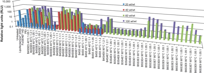
Figure 2 High-throughput screening data for gene delivery efficacy of PBAE polymers to IMR90 cells. Luciferase-encoding DNA is delivered and expression is measured as
rlU. ratios 1.05:1, 1.1:1, 1.2:1, 1:1, and 1:1.05 refer to polymerization conditions and wt/wt is the weight ratio of polymer to DNa. graphs show the mean, n$4.
Notes: acrylate monomers (Bx), amino-alcohol monomers (sy), and end-capping groups (ez). The number “x” following the acrylate monomers “B” refers to the number of
carbons between acrylate groups in the monomer, the number “y” following the amino-alcohol monomers “s” refers to the number of carbons between the amine group and the alcohol group in the sidechain, and the number “z” following the end-capping groups “e” refers to an arbitrary number used to designate a particular end-capping group.
Abbreviations: PBae, poly(beta-amino ester); rlU, relative light units; PeI, polyethylenimine.
O O
O O
1,3-propanediol diacrylate 4-amino-1-butanol (PEO)4-bis-amine
2-(3-aminopropylamino)ethanol 1-(3-aminopropyl)-4-methylpiperazine 1-(3-aminopropyl)pyrrolidine 4-aminophenyl disulfide 5-amino-1-pentanol Propane-1,3-diamine Pentane-1,3-diamine 2-methylpentane-1,5-diamine 1,4-butanediol diacrylate 1,5-pentanediol diacrylate 1,6-hexanediol diacrylate 3-amino-1-propanol O O O O O O O O O O OH H2N H2N H2N H2N H2N H2N H2N H2N H2N H2N H2N NH2 NH2 NH2 NH2 NH2 OH OH OH O O O O O O O O O O NH2 N H N H N N N S S N H R" R" O O O O O OH n N OH n N NH2 R' R' R' OH + + R B3 B4 B5 B6 S3 S4 E1 E5 E6 E7 E8 E9 E3 E4 S5 R R R" R R
Figure 1 PBae synthesis scheme and structure of monomers used for synthesis.
Notes: acrylate monomers (Bx), amino-alcohol monomers (sy), and end-capping groups (ez). The number “x” following the acrylate monomers “B” refers to the number of
carbons between acrylate groups in the monomer, the number “y” following the amino-alcohol monomers “s” refers to the number of carbons between the amine group and the alcohol group in the sidechain, and the number “z” following the end-capping groups “e” refers to an arbitrary number used to designate a particular end-capping group.
Abbreviation: PBae, poly(beta-amino ester).
International Journal of Nanomedicine downloaded from https://www.dovepress.com/ by 134.157.80.157 on 08-Sep-2017
Dovepress Reprogramming human fibroblasts to become iPSCs
CMV-Luc reporter plasmid.26 FuGENE HD, Lipofectamine
2000, and 25 kDa polyethylenimine were used as controls according to the manufacturer’s protocol. Forty-eight hours post-transfection, gene expression was measured using the
Bright-Glo luminescence assay (Promega, Madison, WI, USA) and a Synergy 2 (Bio-Tek, Winooski, VT, USA) multiplate reader (Figure 2). The top performing polymers were further optimized using EGFP-N1 reporter plasmid in
5 M 101 102 103 104 105 Dead cells 2.55 Live cells 97.45 FL3-H:FL3-H 106 107 10 M 15 M 101 102 103 104 105
EGFP + live cells 68.23 FL3-H:FL3-H 106 107 101 102 103 104 105 106 107 B C 40 30 20 10 0 Untreated B4S5E5 40°C 1.05:1 60 wt/w t B4S5E5 40°C 1.05:1 100 wt/w t B4S5E5 90°C 1.1:1 60 wt/w t B4S5E5 90°C 1.1:1 100 wt/w t B4S5E8 90°C 1.1: 1 100 wt/w t B4S4E7 90°C 1.2:1 60 wt/w t B4S4E8 90°C 1.2:1 100 wt/w t B5S3E5 90°C 1.05:1 100 wt/w t B5S3E7 90°C 1.05:1 100 wt/w t Lipofectamine ® 2000 % positive A
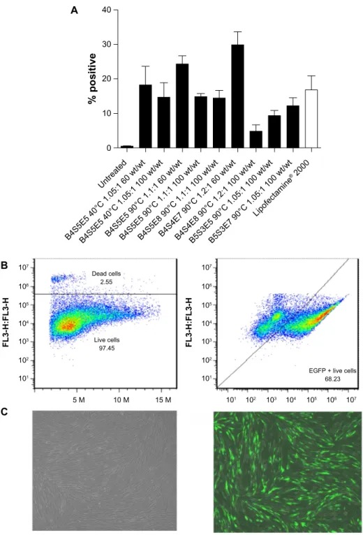
Figure 3 Flow cytometry data for IMR-90 cells transfected with EGFP using the leading polymers. Transfection efficacy expressed as percent green fluorescent
protein-positive live cells (A) following a single dose and (B) after three doses of B4s4e7 (68%). graphs show the mean ± standard error of the mean, n$5. (C) Fluorescence
microscopy images of untreated (left) and transfected (right) IMR-90 fibroblasts, showing good viability in both cases.
Notes: acrylate monomers (Bx), amino-alcohol monomers (sy), and end-capping groups (ez). The number “x” following the acrylate monomers “B” refers to the number of
carbons between acrylate groups in the monomer, the number “y” following the amino-alcohol monomers “s” refers to the number of carbons between the amine group and the alcohol group in the sidechain, and the number “z” following the end-capping groups “e” refers to an arbitrary number used to designate a particular end-capping group.
Abbreviation: EGFP, enhanced green fluorescent protein.
International Journal of Nanomedicine downloaded from https://www.dovepress.com/ by 134.157.80.157 on 08-Sep-2017
Dovepress Bhise et al B4S4E7 60 wt/w t B4S4E7 40 wt/w t B4S4E7 20 wt/w t 120 EGFP plasmid particles Episomal plasmid particles 100 80 60 40 Diameter (nm ) 20 0 4 µg, 198 wt/wt 8 µg, 99 wt/w t 13.2 µ g, 60 wt/w t 15 µ g, 53 wt/wt 20 µ g, 40 wt/wt 24 µ g, 33 wt/wt EGFP DN A DsRed DN A A B4S4E7 60 wt/w t B4S4E7 40 wt/w t B4S4E7 20 wt/w t 40 NaAc 25 mM, pH 5 PBS 30 20 Zeta potential (mV) 10 0 B C
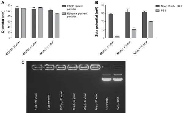
Figure 5 (A) Particle sizing data for PBae nanoparticles complexed with egFP plasmid (∼5 kb) and episomal plasmid (∼16 kb). graph shows the mean ± standard error of
the mean, n$3. (B) Zeta potential data for egFP/B4s4e7 nanoparticles. graphs show the mean ± standard error of the mean, n=5. (C) gel retardation assay data for egFP/
B4s4e7 nanoparticles.
Notes: acrylate monomers (Bx), amino-alcohol monomers (sy), and end-capping groups (ez). The number “x” following the acrylate monomers “B” refers to the number of
carbons between acrylate groups in the monomer, the number “y” following the amino-alcohol monomers “s” refers to the number of carbons between the amine group and the alcohol group in the sidechain, and the number “z” following the end-capping groups “e” refers to an arbitrary number used to designate a particular end-capping group.
Abbreviations: EGFP, enhanced green fluorescent protein; PBAE, poly(beta-amino ester); PBS, phosphate buffered saline; NaAc, sodium acetate. 120 100 80 % viability 60 40 20 0 Untreated B4S4E7 90°C 1.2:1B4S5E5 40°C 1.05: 1 B4S5E5 90°C 1.1: 1 B4S5E4 90°C 1.2: 1 B5S3E7 90°C 1.05: 1 B4S5E6 40°C 1.05: 1 B6S3E5 90°C 1.05: 1 B6S3E5 90°C 1.05: 1 Lipofectamine ® 2000 FuGENE ® HD B4S5E7 40°C 1.05: 1 B4S5E7 90°C 1.1: 1 60 wt/wt 100 wt/wt
Figure 4 cell viability data for IMr-90 cells transfected with egFP/PBae nanoparticles. graphs show the mean ± standard error of the mean, n$3.
Notes: acrylate monomers (Bx), amino-alcohol monomers (sy), and end-capping groups (ez). The number “x” following the acrylate monomers “B” refers to the number of
carbons between acrylate groups in the monomer, the number “y” following the amino-alcohol monomers “s” refers to the number of carbons between the amine group and the alcohol group in the sidechain, and the number “z” following the end-capping groups “e” refers to an arbitrary number used to designate a particular end-capping group.
Abbreviations: EGFP, enhanced green fluorescent protein; PBAE, poly(beta-amino ester).
scaled-up 24-well and six-well protocols, and transfection efficacy was analyzed using flow cytometry and fluores-cence microscopy 48 hours post-transfection (Figure 3). The percent viability of the top performing polymers was
determined using the CellTiter 96 AQueous One Solution Cell Proliferation Assay (Promega, Figure 4). The particle size of the top performing polymer was analyzed with nanoparticle tracking analysis using a NanoSight LM10-HS
International Journal of Nanomedicine downloaded from https://www.dovepress.com/ by 134.157.80.157 on 08-Sep-2017
Dovepress Reprogramming human fibroblasts to become iPSCs A 60 50 40 30 20 10 0 0 µg
% EGFP positive live cell
s 12 µg, 60 wt/wt 24 µg, 33wt/wt 48 µg, 16.5wt/wt IMR-90 fibroblasts RP1 fibroblasts 36 µg, 22 wt/wt C 1.35 1.40 1.45 1.30 1.25 1.15 1.20 1.10 1.00 1.05 0.95 0.85 0.90 0.80 0.75 0.65 0.70 AU 0.60 0.55 0.50 0.45 0.40 0.35 0.20 0.25 0.30 0.15 0.10 0.05 0.00 T3 13 µg 40 wt/w t T3 13.2 µg 60 wt/w t T3 13.2 µg 80 wt/w t T3 15 µg 52.8 wt/w t T3 20 µg 39.6 wt/w t T3 24 µg 33 wt/wt T3 4 µg 198 wt/w t T3 8 µg 99 wt/w t T3 untreate d B 475 450 425 400 375 350 325 300 275 250 225 AU 200 175 150 125 100 75 50 25 0 EGFP untre ated EGFP -N1 1 3.2 µg 40 w t/wt EGFP -N1 1 3.2 µg 60 w t/wt EGFP -N1 1 3.2 µg 80 w t/wt EGFP -N1 24 µg 40 wt/w t EGFP -N1 24 µg 60 wt/w t EGFP -N1 24 µg 80 wt/w t EGFP -N1 4 µg 19 8 wt/w t EGFP -N1 8 µg 99 wt/w t EGFP Oct4
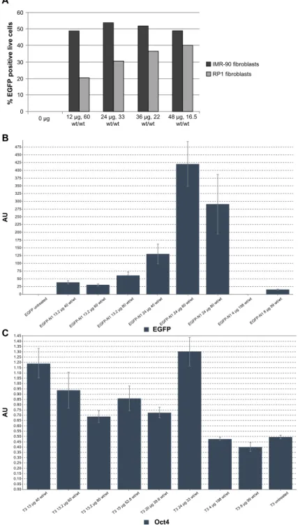
Figure 6 (A) Dose response curve for fibroblasts transfected with EGFP/B4S4E7 nanoparticles. (B) qPcr analysis data showing egFP messenger (m)rNa expression
levels in RP patient-specific adult dermal fibroblasts transfected with B4S4E7/EGFP nanoparticles. Graph shows the mean ± standard deviation, n=3. (C) qPcr analysis data showing Oct4 mRNA expression levels in RP patient-specific adult dermal fibroblasts transfected with B4S4E7/T3 nanoparticles. Graph shows the mean ± standard deviation, n=3.
Notes: acrylate monomers (Bx), amino-alcohol monomers (sy), and end-capping groups (ez). The number “x” following the acrylate monomers “B” refers to the number of
carbons between acrylate groups in the monomer, the number “y” following the amino-alcohol monomers “s” refers to the number of carbons between the amine group and the alcohol group in the sidechain, and the number “z” following the end-capping groups “e” refers to an arbitrary number used to designate a particular end-capping group.
Abbreviations: EGFP, enhanced green fluorescent protein; qPCR, quantitative polymerase chain reaction; RP, retinitis pigmentosa; T3, three reprogramming plasmids.
(NanoSight, Salisbury, UK) and the zeta potential was analyzed by dynamic light scattering using a Zetasizer NanoZS (Malvern Instruments, Malvern, UK; Figure 5). The transfection conditions were optimized for high efficacy
and low cytotoxicity by tuning the polymer/DNA wt/wt, nanoparticle incubation time, and DNA dose per well using flow cytometry and quantitative polymerase chain reaction (qPCR) analysis (Figure 6).
International Journal of Nanomedicine downloaded from https://www.dovepress.com/ by 134.157.80.157 on 08-Sep-2017
Dovepress Bhise et al 80 70 60 50 40 30 20 10 0 Untreated
% EGFP positive live cell
s
1500 V 1600 V 1700 V
A B
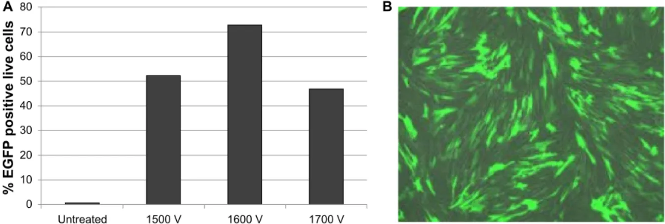
Figure 7 (A) Flow cytometry data for IMr-90 cells electroporated with egFP-N1 plasmid at different voltages, ie, 1,500 V, 1,600 V, and 1,700 V. (B) Fluorescence
microscopy image of IMr-90 cells electroporated with egFP-N1 plasmid at 1,500 V.
Abbreviation: EGFP, enhanced green fluorescent protein.
Nonviral gene delivery
by electroporation
One million IMR-90 fibroblast cells were electroporated using a 100 µL tip-type in a Neon® transfection system
and MP-100 microporator (Life Technologies, Figure 7). Total plasmid DNA (EGFP-N1 reporter plasmid or T3 reprogramming plasmids) was used at 10–12 µg per electroporation experiment. The parameters used for electroporation were optimized for IMR-90 fibroblasts, and the final settings used were: pulse voltage 1,500 V, pulse length, 30 msec, and pulse number 1.
Specifically, IMR-90s were trypsinized for 3 minutes with 1× TrypLE or 0.05% trypsin-ethylenediaminetetraacetic acid (Life Technologies) and neutralized with fibroblast culture medium. The cells were spun at 80× g for 5 minutes at room temperature and resuspended in 4 mL of fibroblast culture medium. The cells were then counted and the cell solution corresponding to one million cells was transferred to a micro-centrifuge tube, and the cells were spun at 50× g for 5 minutes in a table-top microcentrifuge. The cells were resuspended in 112 µL of solution consisting of 100 µL of electroporation buffer R and 12 µL of T3 plasmids. The MP-100 micropora-tor parameters were set for IMR-90 fibroblasts, and a 15 mL centrifuge tube was filled with 8 mL of fibroblast culture medium. The cell suspension was pipetted up and down with a p100 pipette tip and carefully transferred to a 100 µL microporator tip while avoiding any bubbles. The micropora-tor tip, which acts as a transfection chamber, was immersed in electrolytic buffer E2, and the cells were electroporated and added to a 15 mL conical tube containing 8 mL of fibroblast culture medium. The cells were mixed gently and the total cell suspension was transferred into four wells of a six-well plate to achieve a plating density of about 250,000 cells per well.
Nonviral feeder-independent
reprogramming strategy
PBae nanoparticle-based reprogramming
The PBAE nanoparticle-based reprogramming strategy is summarized in Figure 8A. The detailed protocol is as follows.
Day 0: early passage (,p8) IMR-90 fibroblasts were plated at a density of 180,000–250,000 cells/well in a six-well tissue culture-treated plate. Cells were maintained at 37°C, 21% O2, and 5% CO2 (normoxia conditions) in human fibroblast culture medium.
Day 1: two wells with a cell density of 250,000 cells/well were transfected per nanoparticle formulation. Cells were transfected with 15 µg of T3 plasmid combination per well using B4S4E7 1.2:1 90°C polymer complexed at a polymer to DNA weight ratio of 60 wt/wt. Cells were incubated with nanoparticles for 4 hours at normoxia conditions in human fibroblast culture medium. Fresh human fibroblast medium was added after the incubation period and cells were main-tained at normoxia conditions.
Day 2: cells were transferred to 37°C, 3% O2, and 10% CO2 (hypoxia conditions) and maintained in mTeSR1 medium supplemented with 20–50 ng/mL basic fibroblast growth factor and a cocktail of small molecules consisting of 0.25 mM sodium butyrate and 5 µM PS48. Cells were maintained in this reprogramming medium until day 11.
Day 3: cells were transfected with the same conditions of PBAE nanoparticles and the T3 plasmid combination as on day 1, except in mTeSR1 reprogramming medium.
Day 4: cells were passaged using 0.05% ethylenediaminetetraacetic acid and transferred to a Matrigel-coated six-well plate at 150,000 cells/well.
International Journal of Nanomedicine downloaded from https://www.dovepress.com/ by 134.157.80.157 on 08-Sep-2017
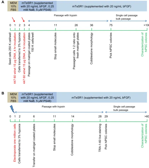
Dovepress Reprogramming human fibroblasts to become iPSCs MEM 10% FBS mTeSR1 (supplemented with 20 ng/mL bFGF, 0.25
mM NaB, 5 µM PS48) mTeSR1 (supplemented with 20 ng/mL bFGF)
Passage with trypsin Single cell passage bulk passage 0
Seed cells 250 K cell/well
447 60 wt/wt 15
µg DNA_4 hr incubation
447 60 wt/wt 15
µg DNA_4 hr incubation
Cells transferred to 3% hypoxia
Passage on matrigel coated plates
150 K cells/well
Stop small molecules
Passaged cells 1:2 ratio onto
fresh matrigel plates
Cobblestone morphology
Pick hiPSC colonies Characterization of
hiPSC colonies 1 2 3 4 11 26 36 75 >100 A MEM 10% FBS mTeSR1 (supplemented with 20 ng/mL bFGF, 0.25
mM NaB, 5 µM PS48) mTeSR1 (supplemented with 20 ng/mL bFGF)
Passage with trypsin Single cell passage
bulk passage 0
Electroporate one million cells Cells transferred to 3% hypoxia
Transfer on matrigel coated plates
Stop small molecules
Cobblestone morphology
TRA-1-60 live staining Pick hiPSC colonies
Characterization of
hiPSC colonies
1 2 6 11 14 28 29 >60
B
Figure 8 (A) PBae-nanoparticle based feeder-independent reprogramming strategy. (B) electroporation based feeder-independent reprogramming strategy. Notes: The green bar represents days. red arrows show the day of polymer-based transfection; blue arrows show the day of electroporation.
Abbreviations: PBAE, poly(beta-amino ester); MEM, minimum essential medium; FBS, fetal bovine serum; bFGF, basic fibroblast growth factor; hiPSC, human induced
pluripotent stem cell; NaB, sodium butyrate.
Day 11: small molecules were withdrawn from the reprogramming medium. Cells were now maintained in mTeSR1 medium supplemented with 20 ng/mL basic fibro-blast growth factor.
Days 25–30: cells were passaged using trypsin and trans-ferred at a 1:2 ratio to fresh Matrigel plates.
Day 36: first signs of embryonic stem cell-like morpho-logic changes were observed.
Day 75: small human iPSC-like colonies were visualized and referred to as EP2 lines. Thirteen iPSC-like colonies were
manually isolated from a starting population of 500,000 cells and gently transferred to a new dish. The two most promising representative colonies (EP2.1 and EP2.2) were propagated further using a single-cell passaging approach with Accutase and blebbistatin and characterized by karyotyping, immunos-taining, and differentiation.
Day .100: characterization of the pluripotency poten-tial of the nonvirally derived human iPSC-like colonies by Tra-1-60 live staining (Millipore, Billerica, MA, USA), immu-nocytochemistry, karyotyping, and in vitro differentiation.
International Journal of Nanomedicine downloaded from https://www.dovepress.com/ by 134.157.80.157 on 08-Sep-2017
Dovepress
Bhise et al
electroporation-based reprogramming
strategy
The electroporation-based reprogramming strategy is sum-marized in Figure 8B. The protocol used was as follows.
Day 0: early passage (,p8) IMR-90 fibroblasts were grown on a Matrigel-coated T175 cm2 cell culture flask at
37°C, 21% O2, and 5% CO2 (normoxia conditions) in human fibroblast culture medium.
Day 1: cells were trypsinized and one million cells were electroporated with 12 µg of T3 plasmid combina-tion as described earlier. Cells were plated at a density of 200,000–250,000 cells/well in six wells of a six-well plate in fresh human fibroblast medium and maintained at normoxia conditions.
Day 2: the cells were transferred to 37°C, 3% O2, and 10% CO2 (hypoxia conditions) and maintained in mTeSR1 medium supplemented with 20–50 ng/mL basic fibroblast growth fac-tor and a cocktail of small molecules consisting of 0.25 mM sodium butyrate and 5 µM PS48. Cells were maintained in this reprogramming medium until day 11.
Day 6: cells were passaged using 0.05% trypsin-ethylene-diaminetetraacetic acid and transferred to a fresh Matrigel-coated six-well plate at 150,000 cells/well.
Day 11: small molecules were withdrawn from the reprogramming medium. Cells were now maintained in mTeSR1 medium supplemented with 20 ng/mL basic fibro-blast growth factor.
Day 14: first signs of embryonic stem cell-like cobble-stone morphologic changes were observed.
Days 25–30: cells were stained using a Tra-1-60 live stain-ing protocol. Tra-1-60-positive human iPSC-like colonies were picked for propagation and referred to as EP1 lines.
Days 30–60: eleven human iPSC-like colonies were manually selected by gently transferring individual colo-nies to separate dishes. One representative colony (EP1.1) was propagated further using a single-cell passaging approach with Accutase and blebbistatin at a density of 10–20,000 cells/well in a 35 mm dish.
Day .60: characterization of the pluripotency poten-tial of the nonvirally derived human iPSC-like cells was accomplished by immunostaining, karyotyping, and in vitro differentiation to neuronal lineage.
Mechanical isolation of human
iPsc-like colonies
Early-passage human iPSC-like colonies were mechani-cally isolated when they reached a size just smaller than one field of a 10× objective (about 1,000 µm). Reduced growth
factor Matrigel-coated plates were prepared 8–30 hours before passaging. A finely drawn out glass Pasteur pipette was used to dissect the colonies into four quarters and each quarter was gently lifted with a p200 pipette tip and transferred to a Matrigel-coated well filled with mTeSR1 medium.
single-cell passage of human
iPsc-like colonies
Late-passage human iPSC-like colonies were single-cell pas-saged when they reached a size just smaller than one field of a 10× objective (about 1,000 µm). Reduced growth factor Matrigel-coated plates were prepared 8–30 hours prior to pas-saging. One milligram of blebbistatin was dissolved in 340 µL of dimethyl sulfoxide to make a 10 mM (2,000×) stock solution, and 10 µL aliquots of this solution were stored at −80°C pro-tected from light. For each experiment, 10 µL of the blebbistatin stock solution was added to each 20 mL of mTeSR1 to make a fresh single-use working medium. The cells were pretreated for 10 minutes with 5 µM blebbistatin in mTeSR1 and then treated with 1 mL of prewarmed Accutase per 35 mm dish for 5–7 min-utes. Cells were dissociated by gentle trituration, quenched with 2 mL of mTeSR1, and pelleted at 80× g for 5 minutes at room temperature. Cells were resuspended in 1 mL of mTeSR1 with blebbistatin at a density of 10,000–20,000 cells per 35 mm dish or per well of a six-well plate. The media was switched to mTeSR1 without blebbistatin 48 hours after passaging.
Immunostaining
After 5 minutes of fixation in 4% paraformaldehyde in phos-phate-buffered saline containing 5% sucrose, the cells were blocked and permeabilized for 30 minutes in 2% normal donkey serum, 0.1% Triton X-100 in phosphate-buffered saline, and then incubated overnight at 4°C with the following antibodies: rabbit anti-Oct4 1/500 (Abcam Inc, Cambridge, MA, USA), rabbit anti-Nanog 1/1000 (Millipore), rabbit anti-Sox2 1/1000 (Mil-lipore), mouse anti-SSEA4 1/100 (Mil(Mil-lipore), rabbit polyclonal anti-Pax6 (Covance, Princeton, NJ, USA), and mouse monoclo-nal anti-Tuj1 1/1000 (Covance). After three rinses in phosphate-buffered saline, the cells were incubated for 1 hour with the corresponding secondary antibody conjugated to Alexa-488 or Alexa-647 (Invitrogen), and counterstained with the nuclear dye Hoechst 33342 (Invitrogen).
Karyotype analysis
The human iPSC-like colonies were karyotyped at Cell Line Genetics (Madison, WI, USA). The EP1 line was karyotyped at passage number 14, and the EP2.1 and EP2.2 lines were both karyotyped at passage number 16.
International Journal of Nanomedicine downloaded from https://www.dovepress.com/ by 134.157.80.157 on 08-Sep-2017
Dovepress Reprogramming human fibroblasts to become iPSCs
In vitro neural differentiation assay
iPSC-like cells maintained in mTeSR1 were incubated for 5 min-utes in 1 mg/mL dispase and clump-passaged to form embryoid bodies according to a modified serum or knockout serum replacement-free protocol adapted from Meyer et al.27 Briefly,
over a 4-day period, the embryoid bodies were transitioned from mTeSR1 to neural induction medium comprised of DMEM:F12 (1:1) containing 2 mM glutamine, 1% N2 supplement, 1× nones-sential amino acids, and heparin sulfate. At day 5, the embryoid bodies were plated onto dishes coated with reduced growth factor Matrigel to allow for neural rosette formation and further cultured in nonessential medium until day 13, at which time rosettes were mechanically lifted from the plate by passing a strong stream of medium over the neural rosettes which preferentially lifted off the plate. Neurospheres were grown at low density as floating spheres on uncoated polystyrene dishes for several weeks, at which time cultures were enzymatically dissociated into small clumps and single cells, and reattached onto Matrigel-coated dishes.
qPcr analysis
EGFP and Oct4 expression was quantified by qPCR analysis in dermal fibroblasts derived from a patient with retinitis pig-mentosa post-transfection with varying conditions of B4S4E7/ EGFP-N1 nanoparticles and B4S4E7/T3 nanoparticles, respectively. The transfection conditions were optimized by varying the DNA dose and polymer to DNA weight ratio (wt/ wt). The mRNA was isolated from the cells using RNeasy Mini Kit (Qiagen, Venlo, The Netherlands). Oct4 primers used were hOct4(120) forward ATTCTCCAGGTTGCCTCTCA and hOct4(120) reverse GTGGAGGAAGCTGACAACAA. EGFP primers used were EGFP(235) for CACATGAAGCAG-CACGACTT and EGFP(235) reverse CGGCCATGATATA-GACGTTG. Three housekeeping genes were used: CREB-BP (CREBBP forward GAGAGCAAGCAAACGGAGAG and CREBBP reverse AAGGGAGGCAAACAGGACA),
SRP72 (SRP72 forward
TCTGCCTCTACAAGTAACAT-CAT and SRP72 reverse CTTCTGCCTCTACAAGTAACAT-CATCACCAGCCACCTT) and
FBX12 (FBX12 forward GCCTTGGTCATATCATCAG and FBX12 reverse TTCTTCATCCGTCCTGTT).28 The qPCR
analysis was done on a BioRad PCR station (BioRad, Hercules, CA, USA) and the qPCR data were analyzed using qbase PLUS software (Biogazelle, Zwijnaarde, Belgium).
Results and discussion
Biodegradable PBae nanoparticles
for nonviral gene delivery to fibroblasts
A library of cationic biodegradable PBAEs useful for nonvi-ral gene delivery to hard-to-transfect human fibroblasts was
developed. The polymers were synthesized via a Michael addition reaction by using various combinations of the diacrylate (Bx), amino-alcohol (Sy) and end-capping group (Ez) monomers shown in Figure 1. In addition to varying the polymer backbone chemical structure, the synthesis condi-tions were also varied, including temperature (40°C versus 90°C) and diacrylate to amine monomer ratio, to expand the structures in the library. Upon electrostatic self-complexation with the reporter plasmids CMV-Luc and EGFP-N1, the PBAEs formed gene delivery nanoparticles with particle diameters of approximately 100 nm (Figure 5A). Using the particle size distribution data from nanoparticle tracking analysis measurements, the number of plasmids associated with a single PBAE-based nanoparticle was quantified.21 It
was observed that 30–100 plasmids were encapsulated in each nanoparticle depending on polymer structure.
To identify polymers from the PBAE library with the ability to transfect human fibroblasts in an efficacious, rapid, and high-throughput manner, the PBAE library was screened on IMR-90 fetal lung fibroblasts using a 96-well plate assay.26 PBAE/CMV-Luc nanoparticles were synthesized
by varying the polymer to DNA weight ratio from 20 wt/wt to 100 wt/wt. Figure 2 shows the compiled transfection effi-cacy data reported as relative luminescence units per well. Several PBAE nanoparticle conditions had higher transfec-tion efficacy than the commercial controls, ie, FuGENE HD, Lipofectamine 2000, and polyethylenimine. In general, the transfection efficacy increased with increasing wt/wt ratio (20 wt/wt ,100 wt/wt) and with increasing synthesis temperature (40°C ,90°C). The top performing polymers, determined from the high-throughput 96-well plate screen-ing, were further optimized using EGFP-N1 reporter plasmid in 24-well and six-well plate transfection assays, and their transfection efficacy was quantified using flow cytometry (Figure 3). The high performers included B4S5E5 1.05:1 90°C, B4S5E5 1.1:1 90°C, B4S5E8 1.1:1 90°C, B4S4E7 1.2:1 90°C, B4S4E8 1.2:1 90°C, B5S3E5 1.05:1 90°C, and B5S3E7 1.05:1 90°C. Nanoparticles were formed at 60 wt/wt. The mean transfection efficacy of the best performing poly-mer, B4S4E7 1.2:1 90°C, was 30%±4% (n=8), which was about two-fold higher than the mean transfection efficacy of Lipofectamine 2000, ie, 17%±4% (n=10). Interestingly, the PBAE with the highest transfection efficacy, B4S4E7, had about 100 plasmids associated with a single nanoparticle as reported by Bhise et al,21 which is more than two-fold the
number of plasmids per nanoparticle calculated for B5S3E7 1.05:1 90°C, a PBAE with low transfection efficacy of 12%±4% (n=10). Both of these PBAE structures have similar
International Journal of Nanomedicine downloaded from https://www.dovepress.com/ by 134.157.80.157 on 08-Sep-2017
Dovepress
Bhise et al
hydrophobicity and the same end-capping group consisting of tertiary amines, but have different transfection efficacies, which may be partly due to differences in their plasmids per particle counts. Due to the high amount of plasmids per particle, we have found that B4S4E7 is effective at coex-pression of exogenously delivered genes as is needed for reprogramming.21
Figure 5B shows the mean zeta potential (mV) of B4S4E7/EGFP nanoparticles formed at 20, 30, and 60 wt/wt in phosphate-buffered saline and NaAc buffers. For all three wt/wt values, the mean zeta potential of B4S4E7 nanoparti-cles formulated in NaAc (25 mM, pH 5) buffer is greater than the mean zeta potential of B4S4E7 formulated in phosphate-buffered saline. For example, the mean zeta potential of 60 wt/wt particles is 32±1 mV (n=5) in NaAc and 20±5 mV (n=5) in phosphate-buffered saline. The greater positive surface charge indicates that more amines in the PBAE backbone are positively charged in NaAc buffer at pH 5 than in phosphate-buffered saline at pH 7. This increased cationic density allows PBAEs to better complex the nega-tively charged plasmids in NaAc buffer. The zeta potential of B4S4E7 nanoparticles in NaAc buffer is about 30 mV for all three wt/wt formulations, whereas in phosphate-buffered saline the mean zeta potential increased slightly with increas-ing wt/wt. The NaAc buffer maintains the stability of B4S4E7 nanoparticles even at a low wt/wt formulation (20 wt/wt). The stability of B4S4E7 nanoparticles was also tested using a gel retardation assay (Figure 5C).
The percent viability determined using the CellTiter cell proliferation assay for the top performing PBAE, B4S4E7, was .90%, demonstrating low cytotoxicity (Figure 4). The transfection efficacy of the best performing polymer, B4S4E7, after a single dose was further boosted by serially transfecting on alternate days for a total of three doses. The transfection efficacy after three doses with B4S4E7 was 68% on day 6 following the first transfection dose (Figure 3B). To maintain an appropriate confluency of cells for the third transfection dose, cells were passaged a day prior to the third transfection dose on day 3. Prolonging the expression of transfected factors is particularly important for successful reprogramming, since it is a gradual process that requires expression of reprogramming factors for a duration of 1–3 weeks.29
A dose-response curve was generated to determine the optimal dose of EGFP-N1 DNA (µg) per well of a six-well plate by varying the DNA dose per well from 12 µg to 48 µg while keeping a constant polymer amount (792 µg/well, Figure 6A). In the case of IMR-90 fetal fibroblasts, there
was no significant increase in percent transfection efficacy as the dose was increased from 12 µg to 48 µg. For the adult fibroblasts derived from a patient with retinitis pigmentosa, however, there was a general trend of increasing transfec-tion efficacy with increasing DNA dose. In additransfec-tion to the dose-dependent increase in the number of transfected cells per condition, there was a dose-dependent increase in the EGFP mRNA expression level in fibroblasts derived from a patient with retinitis pigmentosa, as quantified by qPCR analysis (Figure 6B).
Nonviral feeder-independent
reprogramming strategy
The B4S4E7 polymer was used to deliver T3 episomal plas-mids containing coding sequences for the reprogramming factors to IMR-90 cells in a six-well protocol. A combination of three episomal plasmids (T3) was used to deliver six fac-tors (Oct4, Sox2, NANOG, LIN28, KLF4, and c-Myc). The nonviral reprogramming process, which was performed in a feeder-independent manner, was optimized by modifying the polymer/DNA weight ratio (wt/wt), dose of DNA per well, number of repeat transfections, timing of transfections, and density of cells. For consistency across reprogramming experiments, the top-performing polymer B4S4E7 was syn-thesized in large scale at 5 mg and purified by precipitation. Particle size was determined using nanoparticle tracking analysis and a gel retardation assay21 was performed to verify
effective complexation of the episomal plasmids, which were larger in size (∼16 kb) as compared with the reporter plasmids (∼5 kb). We formed 100 nm B4S4E7 particles with a 16 kb T3 episomal plasmid similar to the nanoparticles formed with the 5 kb reporter EGFP plasmid (Figure 5A). B4S4E7 nanoparticles can also be stored long-term using a freeze-drying process that we have recently described.25
The ability of PBAE nanoparticles to deliver the episomal plasmids was verified by qPCR. Figure 6C shows qPCR data quantifying the expression levels of Oct4 mRNA from patient-derived fibroblasts transfected with the B4S4E7-based nanoparticles (DNA dose per well ranged from 4 µg to 24 µg, and polymer to DNA wt/wt ratios ranged from 40 to 80 wt/ wt). In the case of Oct4 expression, the 4 µg and 8 µg dose per well conditions did not show any increase in Oct4 levels over the untreated control condition. As the dose was increased beyond 8 µg, there was a detectable increase in Oct4 levels above the untreated control condition. Interestingly, for the 13.2 µg per well condition, Oct4 mRNA expression decreased with an increase in amount of polymer per well from 40 wt/ wt to 80 wt/wt. This suggests that increasing the amount
International Journal of Nanomedicine downloaded from https://www.dovepress.com/ by 134.157.80.157 on 08-Sep-2017
Dovepress Reprogramming human fibroblasts to become iPSCs
of polymer may have an inhibitory effect on T3 plasmid expression. We observed that the fibroblasts often appeared stressed, showing reduced proliferation in the wells treated with B4S4E7 amounts .792 µg/well. Thus, for B4S4E7/T3 transfections in a six-well plate format, the optimal amount of polymer per well was determined to be about 792 µg and the optimal amount of DNA per well was determined to be .13.2 µg per well.
T3 episomal plasmids are well suited for transient exog-enous expression due to their ability to self-replicate; this characteristic results in longer-term persistence of multiple episome copies in transfected cells as compared with other extrachromosomal plasmids.30 Most of the nonviral
repro-gramming protocols use electroporation to deliver the plas-mids to somatic cells. In this study, the electroporation-based nonviral reprogramming protocol originally developed by the Thomson Group was adapted to generate human iPSC-like cells from IMR-90 fibroblasts.4 As in the case of PBAE
nanoparticles, the electroporation conditions were optimized for IMR-90 fibroblasts by varying the electroporation param-eters, specifically the electroporation voltage (Figure 7).
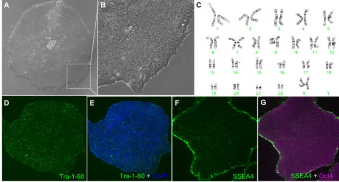
We used an electroporation-based approach and a nano-particle-based approach to develop EBNA-1/oriP episomal plasmid based feeder-independent reprogramming protocols to generate iPSC-like cell lines from human fibroblasts (Figure 8). The EP1 line generated by the electroporation
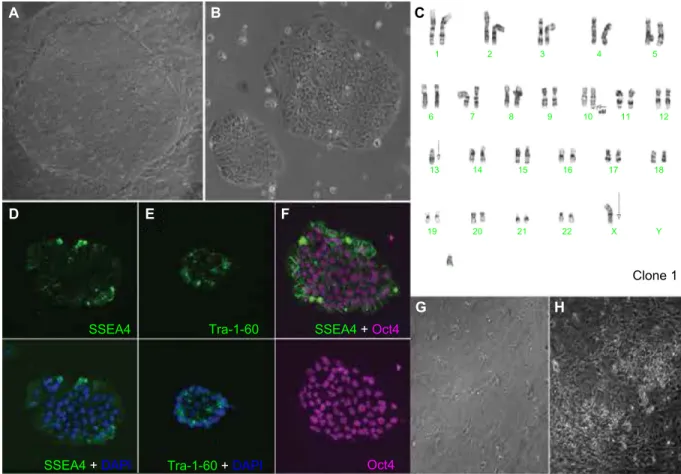
approach was positive for pluripotency markers (Oct4, SSEA4, and Tra-1-60) and showed a normal female karyo-type (Figure 9). The two EP2 lines generated by the nanopar-ticle-based reprogramming approach that were characterized by karyotyping were positive for pluripotency markers (Oct4, SSEA4, and Tra-1-60); however, they acquired significant karyotypic abnormalities (Figure 10). The EP1 colonies emerged as early as 30 days post-electroporation as deter-mined by Tra-1-60 live staining protocol, following which time we switched from trypsin to Accutase for routine cell passaging. Tra-1-60 live staining has been reliably used to identify pluripotent iPSC-like cells.31 Nanoparticle-derived
Tra-1-60-positive EP2 colonies emerged only around day 75 after the first transfection dose of T3/B4S4E7 nano-particles (15 µg and 60 wt/wt dose per well). Therefore, the EP2 colonies, which were slower to reprogram, were exposed to trypsin for a longer period of time, ie, around 2.5 months, compared with EP1 colonies that were exposed to trypsin for about 1 month. Reprogramming stress induced by oncogenic factors and long-term exposure to trypsin during in vitro culture have been shown to increase the susceptibility of fibroblasts to chromosomal mutations.32,33
Trypsin passaging may contribute to preferential selection of karyotypically abnormal cells within the culture, which are initially rare but over time acquire a growth advantage over normal cells due to mutations that favor their survival
A B C
D E F G
SSEA4
Tra-1-60 Tra-1-60 +DAPI SSEA4 +Oct4
1 2 3 4 5 6 7 8 9 10 11 12 13 14 15 16 17 X 22 21 20 19 18 Y
Figure 9 characterization of eP1-hiPsc-like cell line generated by electroporation of nonintegrating episomal plasmids. Normal morphology is observed at low (A) and high
(B) magnification. A normal karyotype is seen in (C). Immunolabeling with markers for pluripotency are shown (D–G). Abbreviation: hiPsc, human induced pluripotent stem cells; DaPI, 4′,6-diamidino-2-phenylindole.
International Journal of Nanomedicine downloaded from https://www.dovepress.com/ by 134.157.80.157 on 08-Sep-2017
Dovepress Bhise et al A B C D E F G H SSEA4 Tra-1-60 Tra-1-60 +DAPI SSEA4 +DAPI SSEA4 +Oct4 1 2 3 4 5 6 7 8 9 10 11 12 13 14 15 16 17 X 22 21 20 19 18 Oct4 Clone 1 Y
Figure 10 characterization of eP2-iPsc-like cell line generated by PBae nanoparticles. Normal morphology is observed in (A) pre-purified and (B) post-purified iPSC-like
cells. an abnormal karyotype is seen in (C) (arrows). Immunolabeling with pluripotency markers is shown (D–F). Partial neuronal differentiation of eP2-iPsc-like cell line with
regional variation (G and H).
Abbreviations: iPsc, induced pluripotent stem cells; PBae, poly(beta-amino ester); DaPI, 4′,6-diamidino-2-phenylindole.
and growth during reprogramming. The effect of long-term exposure of human pluripotent stem cells to Accutase has not been widely investigated, but a recent study by Stover and Schwartz reported no abnormalities in the karyotype after .20 passages.34 The lower reprogramming efficacy
with B4S4E7/T3 nanoparticles, despite their high transfec-tion efficacy with the B4S4E7/EGFP reporter nanoparticles, suggests that increasing the number of transfected cells is not sufficient for successful reprogramming.
In vitro neural differentiation assay
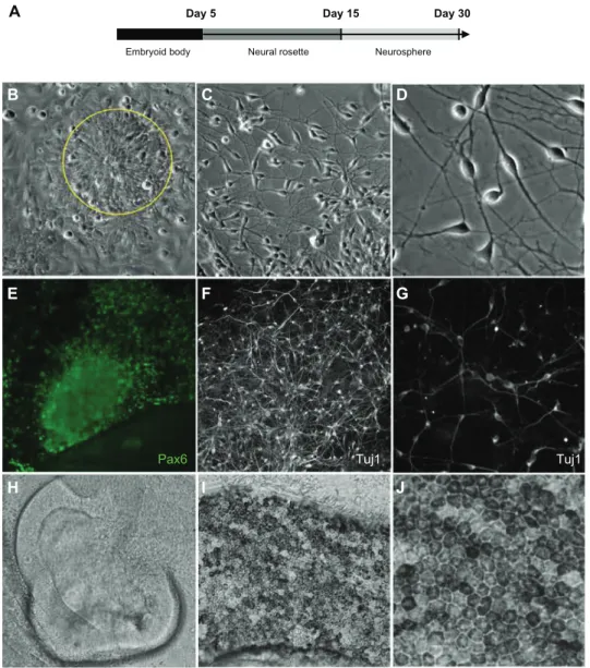
We next tested the ability of our iPSC-like lines to generate neuronal cell lineages. We subjected the iPSC-like lines to a stepwise neuronal induction protocol adapted from Meyer et al, that gives rise to retinal neurons and higher order forebrain-like neurons,27 because we were interested in
modeling retinal degeneration (Figure 11A). The EP1 and EP2 colonies were clump-passaged in mTeSR1 containing blebbistatin and transitioned to a neural induction medium containing DMEM:F12 (1:1), N2 supplement, 1 × nones-sential amino acids, and heparin sulfate. After 5 days
in suspension in non-tissue culture (TC)-coated dishes, embryoid bodies were plated onto Matrigel-coated dishes for an additional 7–9 days. During this time, conspicuous rosettes formed spontaneously in the EP1 electroporation-derived iPSC-like line (Figure 11B) but failed to appear in either the EP2.1 or EP2.2 polymer-derived lines (Figure 10F and G), indicating a failure to differentiate into neurons. On days 13–15, EP1 neural rosettes were mechanically lifted and maintained as neurospheres in DMEM:F12 (3:1), B27 and 1× nonessential amino acids in non-TC coated dishes. Fol-lowing dissociation of 1-month-old neurospheres, dissociated neurons acquired a highly branched neuronal morphology, with neural connections, and expressed multiple neuronal markers, including Pax6 and Tuj1 (Figure 11E–G). Such cells were never observed in the polymer-derived EP2.1 and EP2.2 lines. In addition to the more general neuronal markers, retinal structures were also seen, including pseudostratified neuroepithelial optic cups (Figure 11H) and retinal pigment epithelium (Figure 11I and J).
The presence of the oncogene, cMyc, in the reprogram-ming cocktail limits the clinical use of derived human
International Journal of Nanomedicine downloaded from https://www.dovepress.com/ by 134.157.80.157 on 08-Sep-2017
Dovepress Reprogramming human fibroblasts to become iPSCs B C D E F G H I J Tuj1 Tuj1 Pax6 A
Embryoid body Neural rosette Neurosphere
Day 5 Day 15 Day 30
Figure 11 Neuronal differentiation of eP1-iPsc-like cell line generated by electroporation (A). Neural rosettes (B) (indicated by yellow circle) and mature neurons (C)
low and (D) high magnification can be generated from these cells. Immunostaining with markers for neuronal lineages, Pax6 and Tuj1, are shown in (E), (F) low, and (G)
high magnifications. Retinal structures including pseudostratified neuroepithelial optic cups (H) and retinal pigment epithelium low (I) and high (J) magnifications can also be
generated.
Abbreviation: iPsc, induced pluripotent stem cells.
iPSC-like cells.35 Moreover, sustained expression of any
residual transgenic factors in derived human iPSC-like colonies post-reprogramming may hinder the downstream differentiation capacity of these cells.36,37 Montserrat et al
have shown that other PBAE-based nanoparticles can be used to reprogram somatic cells to human iPSCs, but their approach was feeder-dependent, which is undesirable for clinically relevant applications, and they reported integration of the transgene in the backbone, which introduces the risk of insertional mutagenesis and tumorigenicity.38 Their study did
not compare the PBAE-based method with the standard elec-troporation approach used for nonviral reprogramming.
Although our results demonstrate some of the potential limitations of nanoparticle-based generation of human
iPSC-like cells, there may be modifications of the approach that will be more amenable to successful generation of human iPSCs. Some of the problems we noted might be, in part, due to the slow reprogramming process with PBAEs. Thus, a faster protocol such as conversion of human patient-specific pluripotent stem cells and fibroblasts directly to retinal pho-toreceptors by forced expression of photoreceptor-expressed transcription factors might be more suitable to the use of these nanoparticles. Cord blood cells and keratinocytes have been shown to be more amenable for reprogramming, in terms of speed and efficiency, than skin fibroblasts.39,40 Cord
blood cells have a high population of progenitor stem cells, and the keratinocyte is an epithelial cell type similar to the embryonic stem cell, whereas dermal fibroblasts (especially
International Journal of Nanomedicine downloaded from https://www.dovepress.com/ by 134.157.80.157 on 08-Sep-2017
Dovepress
Bhise et al
adult) are differentiated mesenchymal cells that are close to senescence,41,42 can possess somatic mutations,43,44 and
have to undergo mesenchymal to epithelial transition during reprogramming. Although in this study PBAE nanoparticles were developed to transfect fetal as well as adult fibroblasts, similar PBAE nanoparticles have the potential to transfect epithelial cell types as well as blood cells.23,45–47 Increased
reprogramming efficacy with these more amenable cell types would help to reduce the in vitro culture time and thereby reduce the risk of elevating the chromosomal mutation rate during nanoparticle-based reprogramming. PBAEs spe-cifically designed for transporting nucleic acid cargo to the cytoplasm could potentially be used to deliver mRNA coding for the reprogramming factors to fibroblasts or umbilical cord endothelial cells.46 mRNA delivery is safer than gene delivery
because there is no risk of insertional mutagenesis associated with the cytoplasmic delivery of mRNA. Synthetic mRNA designed to escape the cell’s innate anti-RNA response allows high expression of reprogramming factor proteins in human fibroblasts. Significantly better reprogramming efficacy may be achieved with the mRNA technique, which has been reported to be more efficient and twice as fast as the gene delivery technique.15
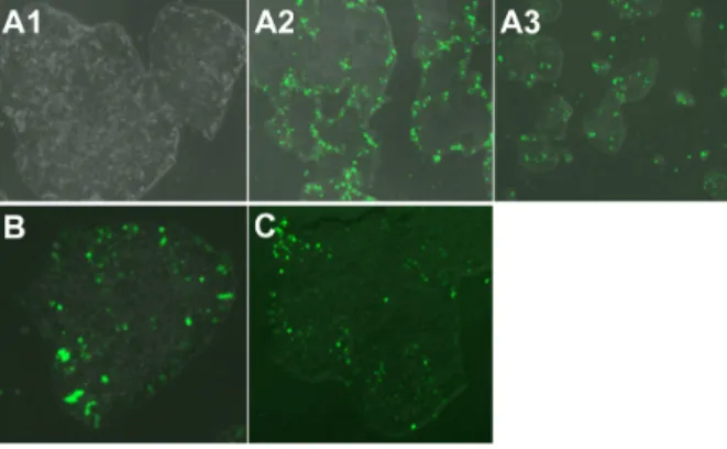
The B4S4E7/EGFP nanoparticles were able to transfect human fetal lung fibroblasts, dermal fibroblasts derived from a patient with retinitis pigmentosa, and end-stage iPSC-like populations (Figure 12). With further development,
B4S4E7-based nanoparticles may potentially be used for programming studies to differentiate these target populations to photoreceptors. The ability to create pools of customized photoreceptors could facilitate drug screening efforts target-ing new human models for retinitis pigmentosa.
Conclusion
This study demonstrates that nanoparticles formed by self-assembly of 1-(3-aminopropyl)-4-methylpiperazine end-terminated poly(1,4-butanediol diacrylate-co-4-amino-1-butanol) polymer (B4S4E7) with episomal plasmid DNA are more effective than leading commercially available reagents, including Lipofectamine 2000, FuGENE HD, and 25 kDa branched polyethylenimine, for nonviral gene transfer to IMR-90 human primary fibroblasts and dermal fibroblasts derived from a patient with retinitis pigmentosa. The fibroblasts were reprogrammed with both polymeric nanoparticle-based and electroporation based nonviral gene delivery methods. Although human iPSC-like cells derived by both methods stained positively for the pluripotency mark-ers Tra-1-60, SSEA4, and Oct4, the electroporation method generated EP1-iPSC-like cells with a normal karyotype in a shorter time period, whereas the nanoparticle-based repro-gramming method took longer and generated EP2-iPSC-like cells with gross karyotypic abnormalities. These results sug-gest that certain nonviral reprogramming methods may pose safety risks and be less efficacious than viral approaches. The karyotypically normal EP1-human iPSC-like cells generated by conventional electroporation, but not by PBAE reprogram-ming, were successfully differentiated in vitro toward the early retinal lineage for disease modeling and drug screening to study retinal degeneration.
Author contributions
NSB, KJW, DJZ, and JJG designed the study. DJZ and JJG supervised the project. NSB and KJW performed the reprogramming experiments and pluripotency analysis. NSB performed the nanoparticle synthesis and characterization experiments and KJW performed the neuronal differentia-tion experiments. NSB, KJW, DJZ, and JJG wrote and edited the paper.
Acknowledgments
This work was supported by the Technology Develop-ment Corporation Maryland Stem Cell Research Fund (2009-MSCRFE-0098-00), the National Institutes of Health (1R01EB016721 and R01EY009769), a Foundation Fighting Blindness Wynn-Gund Translational Acceleration Program
Figure 12 Fluorescence microscopy images (10×) of hiPsc-like cells transfected with
a single dose of B4s4e7/egFP nanoparticles. Images show healthy morphology post-transfection (A1), an edge effect in larger colonies (A2), and middle-transfected +
edge-transfected cells in smaller colonies (A3). Image (20×) of hiPsc-like colony
transfected with (B) two doses of B4s4e7 and egFP complexes and (C) one dose
with B4s4e7 and PiggyBac 2 plasmid system complexes.
Notes: acrylate monomers (Bx), amino-alcohol monomers (sy), and end-capping
groups (ez). The number “x” following the acrylate monomers “B” refers to the number of carbons between acrylate groups in the monomer, the number “y” following the amino-alcohol monomers “s” refers to the number of carbons between the amine group and the alcohol group in the sidechain, and the number “z” following the end-capping groups “e” refers to an arbitrary number used to designate a particular end-capping group.
Abbreviations: hiPsc, human induced pluripotent stem cells; egFP, enhanced
green fluorescent protein.
International Journal of Nanomedicine downloaded from https://www.dovepress.com/ by 134.157.80.157 on 08-Sep-2017