HAL Id: inserm-00456546
https://www.hal.inserm.fr/inserm-00456546
Submitted on 15 Feb 2010
HAL is a multi-disciplinary open access
archive for the deposit and dissemination of
sci-entific research documents, whether they are
pub-lished or not. The documents may come from
teaching and research institutions in France or
abroad, or from public or private research centers.
L’archive ouverte pluridisciplinaire HAL, est
destinée au dépôt et à la diffusion de documents
scientifiques de niveau recherche, publiés ou non,
émanant des établissements d’enseignement et de
recherche français ou étrangers, des laboratoires
publics ou privés.
vasopressin neurons.
Jean-Marc Israel, Dominique Poulain, Stéphane Oliet
To cite this version:
Jean-Marc Israel, Dominique Poulain, Stéphane Oliet. Glutamatergic inputs contribute to phasic
activity in vasopressin neurons.. Journal of Neuroscience, Society for Neuroscience, 2010, 30 (4),
pp.1221-32. �10.1523/JNEUROSCI.2948-09.2010�. �inserm-00456546�
Cellular/Molecular
Glutamatergic Inputs Contribute to Phasic Activity in
Vasopressin Neurons
Jean-Marc Israel,
1,2Dominique A. Poulain,
1,2and Ste´phane H. R. Oliet
1,21Inserm Unite´ 862, Neurocentre Magendie, and2Universite´ de Bordeaux, 33077 Bordeaux, France
Many neurons in the CNS display rhythmic patterns of activity to optimize excitation–secretion coupling. However, the mechanisms of
rhythmogenesis are only partially understood. Magnocellular vasopressin (VP) neurons in the hypothalamus display a phasic activity
that consists of alternative bursts of action potentials and silent periods. Previous observations from acute slices of adult hypothalamus
suggested that VP cell rhythmicity depends on intrinsic membrane properties. However, such activity in vivo is nonregenerative. Here, we
studied the mechanisms of VP neuron rhythmicity in organotypic slice cultures that, unlike acute slices, preserve functional synaptic
connections. Comparative analysis of phasic firing of VP neurons in vivo, in acute slices, and in the cultures revealed that, in the latter, the
activity was closely related to that observed in vivo. It was synaptically driven, essentially from glutamatergic inputs, and did not rely on
intrinsic membrane properties. The glutamatergic synaptic activity was sensitive to osmotic challenges and !-opioid receptor activation,
physiological stimuli known to affect phasic activity. Together, our data thus strongly suggest that phasic activity in magnocellular VP
neurons is controlled by glutamatergic synaptic inputs rather than by intrinsic properties.
Introduction
Vasopressin (VP) plays a key role in body fluid and
cardiovas-cular homeostasis. VP is synthesized in magnocellular
neu-rons that accumulate in the supraoptic nucleus (SON) and
paraventricular nucleus of the hypothalamus. These neurons
project to the neurohypophysis where VP is released into
the blood. In normally hydrated, unstimulated rats, most VP
neurons display a slow irregular pattern of electrical activity.
Nevertheless, !10% display a characteristic phasic pattern
comprised of bursts of firing interspersed by silent periods
(Poulain and Wakerley, 1982; Hussy et al., 1997; Gouze`nes et
al., 1998). Hyperosmotic stimulation initiates (in
nonspontane-ously phasic neurons) or reinforces (in spontanenonspontane-ously phasic
neurons) this phasic activity (Poulain et al., 1977; Wakerley et al.,
1978), thus optimizing the efficiency of stimulus–secretion
cou-pling (Dutton and Dyball, 1979). The interspike intervals in a
burst of action potentials (APs) and the silent intervals between
bursts are important determinants for neuropeptide release
(Cazalis et al., 1985), and understanding the mechanisms
under-lying this specific pattern of activity is of crucial importance in the
study of neurosecretion.
Most of our knowledge in this domain arises from
experi-ments performed in acute hypothalamic slices or from explants,
in which the phasic activity is seen to be entirely supported by
nonsynaptic depolarizing afterpotentials (DAPs) after single
spikes (Andrew and Dudek, 1984; Armstrong et al., 1994;
Ghamari-Langroudi and Bourque, 1998) whose summation
me-diates a plateau-like potential (Andrew and Dudek, 1983). Bursts
appear to result, therefore, from a regenerative mechanism
at-tributable to the proximity of spike threshold (Roper et al., 2004;
Brown and Bourque, 2006). Accordingly, blockade of synaptic
transmission in acute slices did not alter phasic firing (Hatton,
1982). However, in this preparation, synaptic activity is mostly
impaired and usually reduced to miniature synaptic currents/
potentials (Wuarin and Dudek, 1993; Brussaard et al., 1996, 1997;
Kabashima et al., 1997; Kombian et al., 2000), probably because
axon terminals are severed from their glutamatergic cell bodies.
In vivo, application of glutamate receptor antagonists like
ket-amine (Nissen et al., 1994),
D(")-2-amino-5-phosphonopentanoic
acid (
D-AP-5) (Nissen et al., 1995; Moos et al., 1997),
6-cyano-7-nitroquinoxaline-2,3-dione (CNQX) (Nissen et al., 1995), or
kynurenic acid (Brown et al., 2004) blocks phasic activity in VP
neurons, suggesting that glutamatergic inputs play a key role in
governing phasic activity. It is still not clear why this is not the
case in vitro.
In the present study, we compared phasic activity in VP
neu-rons in three different preparations: in lactating females in vivo,
in acute hypothalamic slices from adult females, and in
organo-typic slice cultures in which glutamatergic inputs are preserved
(Jourdain et al., 1999). We found that VP neuron phasic activity
in organotypic cultures is more closely related to that displayed in
vivo than the phasic pattern observed in acute slices.
Further-more, the rhythmic behavior displayed by VP neurons in
orga-notypic cultures does not depend on DAPs but is driven by
glutamatergic synaptic activity. Our observations thus question
the importance of intrinsic properties as the basis of phasic
activ-ity in VP neurons and highlight the importance of extrinsic
glu-tamatergic drive in promoting such activity.
Received June 22, 2009; revised Nov. 12, 2009; accepted Nov. 21, 2009.
This work was supported by grants from Inserm, Universite´ de Bordeaux, and Conseil Re´gional d’Aquitaine. We thank Drs. P. Ciofi, D. T. Theodosis, and D. L. Voisin for helpful discussion and critical reading of this manuscript, Prof. M. C. Lombard for her expertise in statistical analysis, and N. Dupuy for technical assistance.
Correspondence should be addressed to Jean-Marc Israel, Inserm Unite´ 862, Neurocentre Magendie, 146, rue Le´o-Saignat, 33077 Bordeaux, France. E-mail: [email protected].
DOI:10.1523/JNEUROSCI.2948-09.2010
Materials and Methods
Organotypic slice cultures. Organotypic slice cultures that included the
SON were obtained from 5- to 9-d-old rat hypothalami. The cultures were prepared using the roller tube method, as described in detail by Jourdain et al. (1996). Briefly, Wistar rat pups were anesthetized with 5% isoflurane (95% O2) for 1 min and decapitated. Brains were removed and tissue blocks that included the hypothalamus were quickly dissected and sectioned (400 "m) with a McIlwain tissue slicer. Frontal slices contain-ing the SON were cut into two parts along the third ventricle, and each part was placed on a glass coverslip coated with heparinized chicken plasma. Thrombin was added to coagulate the plasma and permit adhe-sion of the slice to the coverslip. The coverslip was inserted into a plastic flat-bottom tube (Nalge Nunc International) containing 750 "l of me-dium, pH 7.4 (290 –295 mOsm), composed of 50% Eagle’s basal medium (Invitrogen), 25% heat-inactivated horse serum (Invitrogen), and 25% HBSS (Invitrogen) enriched with glucose (7.5 mg/ml);L-glutamate (Se-romed) was added at a concentration of 2 mM. No antibiotics were used. The tubes were tightly capped and inserted in a roller drum and rotated !15 turns/h. The medium was replaced twice a week.
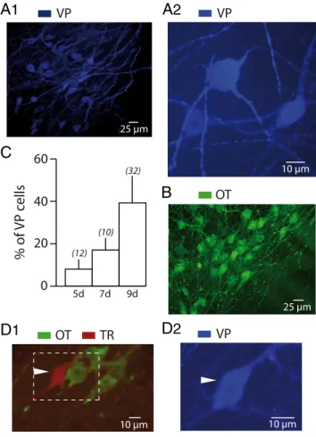
Using immunocytochemistry, we confirmed previous observations (Wray et al., 1991; Jourdain et al., 1996) showing that slice cultures
derived from 5-d-old pups are poor in VP neurons (8 # 4% of the total number of magnocellular cells; n $ 12) (Fig. 1C). However, their num-bers increase in slice cultures from older pups: 17 # 6% in slices (n $ 10) from 7-d-old rats and 39 # 13% in slices (n $ 32) from 9-d-old pups (Fig. 1 A1,A2,C). We therefore performed our recordings in 4- to 10-week-old slice cultures from hypothalamus of 9-d-old rats. Just before a recording session, a slice was transferred from the incubator to a temperature-controlled chamber (36 # 0.2°C) fixed to the stage of an inverted microscope (Diaphot; Nikon). Magnocellular neurons were vi-sualized by their large size. The microelectrode was positioned close to the cell to be recorded using oleic micromanipulators (Narishige).
For recording, the slices were perfused (0.7 ml/min) with Yamamoto’s solution (in mM: 125 NaCl, 3 KCl, 1 MgSO4, 1.25 KH2PO4, 5 NaHCO3, 2 CaCl2, 5 glucose, 10 HEPES, pH 7.25, 293–295 mOsm). Hyperosmolarity was produced by addition of mannitol, whereas hypo-osmotic medium was obtained by diminishing HEPES from 10 to 5 mM. The following were added to the medium when required: CNQX (RBI),D-AP-5 (RBI), tetrodotoxin (TTX), flufenamic acid (FFA), dynorphine (Sigma-Aldrich), and nor-binaltorphimine (BNI) (Tocris Bioscience). Intracel-lular microelectrodes were pulled from borosilicated glass capillaries (outer diameter, 1 mm; inner diameter, 0.5 mm; Sutter Instrument) with
A
1
C
D
1
D
2
B
A
2
Figure 1. Vasopressin neurons in hypothalamic organotypic cultures. A, Example of an
or-ganotypic slice culture from the hypothalamus of a 9-d-old rat after 2 months in culture show-ing numerous VP (blue fluorescence) neurons at low (A1) and high magnification (A2). The magnocellular neurons generally possessed an ovoid or a round soma from which arose two to fourthickdendritic-likeprocesses.B,Organotypicslicecultureobtainedfromthehypothalamus of a 9-d-old rat after 2 months in culture. Immunocytochemical labeling revealed that these cultures are also enriched with OT (green fluorescence) neurons. C, Summary histogram depict-ing the percentage of VP-immunoreactive neurons in organotypic slice cultures obtained from 5- (5d), 7- (7d), and 9-d-old (9d) rat hypothalami, after a minimum of 40 d in culture. The number of experiments is indicated in brackets. D, Example of a magnocellular neuron identi-fied with biocytin (TR) (red fluorescence) after intracellular recording. Subsequent immunocy-tochemistry revealed that it displayed immunoreactivity for VP (blue fluorescence) (D2) but not for OT (green fluorescence) (D1).
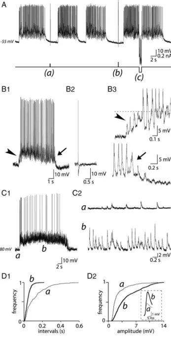
Figure 2. Basal electrical properties of VP neurons in organotypic slice cultures. A1, The top
trace shows an intracellular recording from a VP neuron in a 10-week-old culture from a 9-d-old rat. The neuron displayed a slow irregular activity varying from 0.2 to 3 Hz as indicated by the sequentialdistributionhistogramofAPdischarge(bottomtrace).InadditiontoAPs,asustained synaptic activity was clearly visible (A2). B, Example showing the progressive duration increase (broadening) of APs during a burst (B1). The broadening was also frequency dependent as illustrated in B2 in which APs were triggered by positive current pulses (%50 pA, 50 ms; in the presence of CNQX) at increasing frequencies (0.5, 10, and 15 Hz). C, Injection of a depolarizing current pulse (%70 pA, 100 ms) (data not shown) applied at resting potential ("60 mV) triggered a burst of APs. Note the presence of an afterhyperpolarizing potential (AHP) after the train of APs (arrow). When the cell was hyperpolarized to "90 mV, a notch (arrowhead) delayed the occurrence of APs. D, Two examples of spontaneous phasic activity characterized by successive bursts of APs and silent periods (D1, D2, top traces). The bottom traces are the corresponding sequential distribution histogram of APs, typical of phasic activity. Note the synaptic activity visible between bursts in D2.
a Flaming/Brown P87 puller (Sutter Instrument) and filled with 1M potassium acetate with 1% biocytin (Sigma-Aldrich). Electrode resis-tance varied from 150 to 250 M&. Intracellular potentials from neurons were recorded through a single microelectrode using an Axoclamp-2A (Molecular Devices), which also permitted injection of currents.
Acute slice preparation. Wistar rats were anesthetized with isoflurane
(5% isoflurane–95% O2) and killed by decapitation using a guillotine. Immediately after opening the skull, the brain was removed and im-mersed in ice-cold oxygenated (95% O2–5% CO2) medium for 1 min. The composition of the perfusion medium was as follows (in mM): 125 NaCl, 3 KCl, 1.24 MgSO4, 1.3 KH2PO4, 25 NaHCO3, 2 CaCl2, and 11 glucose. The brain was cut into a small cube that included the SON. The block was then fixed with cyanoacrylic glue on the top of the holder of a vibroslicer (MacIlwain; Campden Instruments). Three coronal sections (400 "m) were made and transferred onto a filter paper (optic lens neutral cleaner) in contact with a ramp-style interface recording chamber (Israel and Poulain, 2000). The medium was perifused using a peristaltic pump (Gibson) at a rate of 1 ml ! min"1and at constant temperature (32 # 0.5°C). The slices were constantly oxygenated with humidified 95% O2–5% CO2. The electrophysiological session began after a 1.5 h equilibration period. The microelectrode was positioned into the SON with a micromanipulator (Microcontroˆle) under visual control using a dissecting microscope.
Comparison of phasic activity in slices obtained from adult female (240 –260 g), lactating female (7–20 d of lactation), and adult male (250 – 280 g) rats did not yield any differences in intraburst frequency (IF), burst duration (BD), and silent duration (SD) (supplemental Fig. S1, available at www.jneurosci.org as supplemental material). We thus per-formed our experiments on adult female rats.
In vivo experiments. Experiments were performed on lactating female Wistar rats (280 –350 g) using a conventional procedure for recording extracellular action potentials in the SON (Poulain et al., 1977). Briefly,
animals were first lightly anesthetized with isoflurane (5% isoflurane–95% O2), and then deeply anesthetized with urethane (1.3 g ! kg"1) and set in a stereotaxic frame (Nar-ishige). The surface of the brain was exposed and the sagittal sinus ligatured; a bipolar con-centric stimulating electrode (Rhodes Medical Instruments) was then introduced via a dorsal approach into the pituitary stalk for anti-dromic identification of neurosecretory cells. Extracellular recordings of APs were obtained with glass micropipettes connected to a con-ventional electrophysiological apparatus as de-scribed previously (Poulain et al., 1977). Cells were tested during suckling and considered to be vasopressinergic when they did not react to suckling, contrary to the other cells, classified as oxytocinergic. The characteristics of the electrical activity were analyzed off-line from tape recordings.
Analyses of electrical activity. Electrical
sig-nals were visualized on a digital oscilloscope (Tektronix TDS 2012B), recorded directly on a recorder (Gould thermal array recorder TA 11; Gould), and stored on a hard disk through pClamp 9 software (Molecular Devices) through a Digidata 1300 interface (Molecular Devices). Stored electrical activity was re-played, and sequential frequency histograms were calculated over 1 s integration periods and plotted versus time using an appropriate program from pClamp9. Values are expressed as means # SD. Data were analyzed using with the nonparametric Kruskal–Wallis test. Cu-mulative frequency or amplitude distributions of glutamatergic and GABAergic synaptic po-tentials were obtained from off-line analysis of unitary potentials using Axograph (Molecular Devices). Data were compared statistically with the paired Student test.
Identification of recorded neurons in vitro. To identify the phenotype of
recorded cells in acute slice preparations and organotypic slice cultures, we used a method described in detail in our previous studies (Jourdain et al., 1996, 1999). The recording electrodes contained 1% biocytin (Sigma-Aldrich). At the end of the intracellular recording session, neurons were filled with biocytin using hyperpolarizing current pulses ("0.5 nA, 0.4 s, 2 Hz, 10 –20 min). Slices were then fixed in 4% paraformaldehyde and 0.15% picric acid for 2 h at room temperature and rinsed in 4% parafor-maldehyde (two times; 20 min each time). Biocytin was visualized with streptavidin-conjugated Texas Red fluorescence (Biosys; diluted 1:400) with appropriate filters (Leitz DMR microscope; Leica). Slices then underwent double immunofluorescence using a mixture of primary antibodies, one being a monoclonal mouse Ig raised against oxytocin (OT)-related neurophysin [offered by H. Gainer, University of Toledo, To-ledo, OH, and characterized in the study by Ben-Barak et al. (1985)], and the other, a polyclonal rabbit serum raised against VP-associated neuro-physin [offered by A. Robinson, David Geffen School of Medicine at UCLA, Los Angeles, CA, and characterized in the study by Roberts et al. (1991)]. After mounting with Fluoromount (Vectashield; Vector Laboratories), preparations were examined with epifluorescence with appropriate filters.
Results
Basal electrical properties of VP neurons in organotypic
slice cultures
The electrical properties of 147 VP neurons were studied in
hy-pothalamic cultures from 9-d-old rats after 5–10 weeks in vitro. A
majority of VP neurons (67%) displayed a slow irregular or
con-tinuous firing pattern, varying from 0.3 to 12 Hz (4.2 # 3.6 Hz;
n $ 12) (Fig. 2A1). Briefly, they had a mean resting membrane
Figure 3. Electrical activity of VP neurons in acute hypothalamic slices and in vivo. A, A typical phasic activity recorded in a VP
neuron from an acute slice, characterized by sequences of firing and silent periods (top trace). The bottom trace is the correspond-ing sequential stimulus histogram. B, Example of a silent neuron in an acute slice at restcorrespond-ing potential ("57 mV). Stimulation with a positive current pulse (bottom trace; black arrowhead) triggered a DAP (arrow). This DAP was not sufficient to trigger a plateau to support sustained firing. When the neuron was depolarized to "50 mV by injection of a sustained positive current, an identical stimulation induced firing (APs are truncated). Note that the activity ceased spontaneously despite sustained depolarization.
C, Injection of brief positive currents (100 ms, 40 pA; black arrowheads) in a silent cell at resting potential ("50 mV) triggered a
sustained firing activity, which was interrupted by passing a negative current (400 ms, "120 pA; empty arrowheads). D, An example of electrical activity in a VP neuron recorded extracellularly in vivo (top trace). The bottom trace is the corresponding sequential distribution histogram of APs.
potential of "64.7 # 3.6 mV (n $ 20). They displayed APs with
a mean amplitude of 59.8 # 8.9 mV and a mean duration of
1.98 # 0.47 ms (means from 310 APs recorded from 10 cells). In
addition to APs, a sustained synaptic activity was clearly visible
(Fig. 2A2). In addition, the majority of VP neurons displayed
endogenous properties typical of magnocellular neurons recorded
in acute slices of rat hypothalamus and explants, such as a
hyperpo-larizing afterpotential after single spikes, an afterhyperpolarization
(AHP) after bursts of APs (Fig. 2C, arrow), a frequency-dependent
broadening of APs during a train of depolarizing pulses (Fig.
2B1,B2), and the notch delaying the occurrence of firing at
hyper-polarized potentials (Fig. 2C, arrowhead). One-third of VP cells
dis-played a DAP (see Fig. 7A2,C2,E) after a single or a few spikes.
Forty-seven VP neurons (36% of total) spontaneously
dis-played a phasic behavior characterized by alternative bursts of
APs and periods of silence (Fig. 2D). BD varied considerably
from one cell to another (mean duration, 22.1 # 15.7 s) (n $ 352
from 69 neurons) (Fig. 2D1,D2) and for a single cell during the
recording session. The mean burst amplitude was 165 # 243 APs
(n $ 250 from 34 neurons) and the mean IF also varied from cell
to cell (9.1 # 6.3 Hz; n $ 69). As previously described in vivo
(Poulain and Wakerley, 1982), the shape of bursts displayed a
great variability, with a mean peak frequency of 19.4 # 11.0 Hz.
Silent periods (SD) also varied markedly from cell to cell, from 2
to 75 s (mean, 18.7 # 17.1 s; n $ 69) (Fig. 2D) and, to a lesser
extent, between successive bursts in a single neuron. As was
re-ported in vivo (Poulain and Wakerley, 1982), some neurons
dis-played a few isolated APs during silent periods.
A few VP neurons (3%) displayed a mixed activity consisting
of a phasic activity interrupted by intermittent brief (5–9 s)
high-frequency (20 – 60 Hz) discharges of APs, typical of oxytocinergic
neurons (Jourdain et al., 1998), an activity reminiscent of that
previously described in vivo (Moos and Ingram, 1995).
Comparison of phasic activity in vitro and in vivo
To compare the rhythmic behavior displayed by VP neurons in
organotypic slice cultures with that described previously, we
re-corded and analyzed such activity in acute slices of adult
hypo-thalamus and in adult rats in vivo. Seventy-eight identified VP
neurons were recorded from acute hypothalamic slices, among
which 53% (n $ 41) displayed a phasic firing pattern (Fig. 3A). In
these cells, DAPs were able to trigger bursting activity when the
membrane potential was sufficiently depolarized (Fig. 3B,C).
Furthermore, injection of a brief negative current interrupted the
bursts (Fig. 3C). These observations are similar to those reported
in previous studies that first described the mechanism underlying
this rhythmic behavior (Andrew and Dudek, 1984). In vivo, 55
neurons identified as vasopressinergic displayed a phasic activity
(Fig. 3D). Their characteristics were as follows: IF, 9.2 # 3.0 Hz;
BD, 28.7 # 22.0 s; and SD, 17.0 # 10.8 s (Fig. 4A), values
equiv-alent to those reported previously [IF, 4 –14 Hz; BD, 10 –25 s; SD,
5–20 s in the study by Poulain et al. (1988)]. We next used these
parameters to compare phasic activity among the three
experi-mental models.
We found that IF was not statistically different (Kruskal–Wallis
test, p ' 0.05) in the three preparations (9.1 # 6.3 Hz, n $ 69, and
8.0 # 3.2 Hz, n $ 41, for organotypic and acute slices,
respec-tively) (Fig. 4A1). BD and SD, however, differed statistically
be-tween organotypic and acute slices. Values for organotypic
cultures were similar ( p ' 0.05) to those measured in vivo (see
above), whereas they were significantly increased ( p ( 0.05) in
acute slices (75.4 # 55.4 and 49.3 # 43.9 s; n $ 41) (Fig. 4A2).
These parameters characterizing phasic activity are summarized
in Figure 4B for the three preparations. The analysis revealed that
the rhythmic firing pattern in VP neurons recorded from
orga-notypic slice cultures is closely similar to the phasic activity
re-corded in vivo, whereas it differed markedly from that rere-corded in
acute hypothalamic slices.
Phasic activity is regulated by extracellular osmolarity
It has been shown previously that acute hyperosmotic
stimula-tion triggers or accelerates phasic activity in vivo (Brimble and
Dyball, 1977; Bourque and Renaud, 1984) and depolarizes VP
neurons in vitro (Bourque, 1989; Oliet and Bourque, 1992,
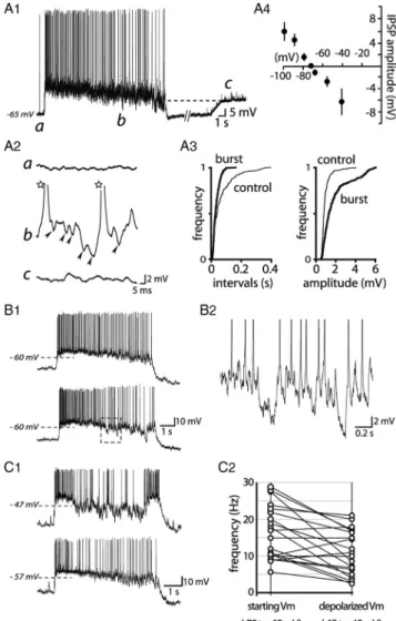
Figure 4. Comparison of phasic activity in vitro and in vivo. A1, Summary histogram showing the mean IF recorded extracellularly in vivo (gray columns; n $ 55) or intracellularly in organotypic
slice cultures (black columns; n $ 69) and acute slices (empty columns; n $ 41). Results are expressed as mean # SD. A2, Summary histogram recapitulating the mean BD and the mean SD for phasic activity in the three preparations. B, Statistical distribution of mean IF, BD, and SD in the three groups of neurons. The vertical dotted line in each histogram represents the median value.
1993a,b). Conversely, hypotonic media hyperpolarize
magnocel-lular neurons (Oliet and Bourque, 1993a,b). This prompted us to
test the effect of hypertonic (320 mOsm) and hypotonic (275
mOsm) stimuli on VP neurons in organotypic slice cultures. The
hyperosmotic challenge rapidly (10 –20 s) and progressively
in-creased AP frequency in neurons firing at a low frequency (0.1– 4
Hz; n $ 7) before triggering phasic activity in six of seven cells
(Fig. 5A). The effect was totally reversible after washing.
Hyper-osmotic stimulation was also tested on 13 neurons displaying
spontaneous phasic activity. In four cells, it caused an increase in
BD (%88 # 31%; p ( 0.05) without significantly changing SD, so
that the Q coefficient (the proportion of bursting time spent by a
phasic cell during a fixed time) (Wakerley et al., 1975) increased
from 0.19 # 0.03 to 0.41 # 0.05 ( p ( 0.05). In six cells,
hyper-osmolarity reduced both BD and SD (Fig. 5B1), consequently
increasing Q from 0.26 # 0.12 to 0.81 # 0.26 (Fig. 5B2).
Con-comitantly, we noticed a slight but significant increase in IF
(127 # 16%; p ( 0.05) and in peak frequency (119 # 10%; p (
0.05). Finally, in the remaining three phasic neurons,
hyperos-molarity depolarized membrane potential (9.3 # 2.7 mV)
accompanied by an increase in membrane conductance,
lead-ing to a sustained firlead-ing that terminated the rhythmic activity
(data not shown).
Reducing the osmolarity to 275 mOsm
had an opposite action on electrical
activ-ity since it inhibited firing in VP neurons,
turning the phasic pattern into a slow
ir-regular AP discharge, decreasing spike
frequency by 86 # 7% (n $ 4) (Fig.
5C1,C2). This effect was also completely
reversible when the osmolarity was
re-turned to 295 mOsm (Fig. 5C2).
Control of phasic activity by !-opiates
The !-opioid peptide dynorphin is
synthe-sized and copackaged with VP in the same
neurosecretory granules (Watson et al.,
1982; Shuster et al., 2000). In vivo and in
vitro studies established that activation of
!-opioid receptors on VP neurons inhibited
phasic behavior (Brown et al., 1998)
through the inhibition of DAP (Brown and
Bourque, 2004). Conversely, antagonizing
!-opioid receptors was shown to facilitate
phasic firing, revealing the key role played
by endogenous dynorphin in regulating
rhythmic activity (Brown and Bourque,
2004). We thus tested whether dynorphin
would have a similar effect on the phasic
ac-tivity displayed by VP neurons in
organo-typic slice cultures. We first found that
dynorphin (1 "
M) decreased the amplitude of
DAP from 4.7 # 0.8 to 2.9 # 0.5 mV [n $ 3;
p ( 0.05, a value similar to that reported in
explants (Brown and Bourque, 2004)]. As
ex-pected, in six VP cells, dynorphin reduced
phasic activity, decreasing Q from 0.57 # 0.19
to 0.13 # 0.07. In five other VP cells,
dynor-phin switched the activity from phasic to a
slowirregularfiring(Fig.6A),decreasingspike
discharge by 75.4 # 12.7% of control values.
In the three remaining VP neurons,
dynor-phin did not have any effect on firing activity.
We then applied the specific !-opioid
re-ceptor antagonist BNI on phasically firing neurons to assess the role
of endogenous !-opiates on phasic activity. As clearly shown in
Fig-ure 6B, BNI (1 "
M) reduced SD and dramatically increased BD,
therefore increasing Q from 0.35 # 0.10 to 0.78 # 0.18 (n $ 4; p (
0.05). BNI also increased IF (%101 # 47.1%; n $ 4; p ( 0.05).
Together, the results obtained with the agonist and antagonist of
!-opiate receptors indicate that phasic activity in organotypic slice
cultures is regulated by the endogenous release of dynorphin, as is
the case in hypothalamic explants (Brown and Bourque, 2004;
Brown et al., 2006) and in vivo (Brown et al., 1998, 2006).
The effects of extracellular osmolarity and !-opiates on
gluta-matergic transmission were further analyzed in a TTX (1 "
M)-containing medium. A hypertonic (%25 mOsm) medium did not
modify the frequency of miniature EPSPs (mEPSPs) (%4.3 #
13.6%; n $ 5; p ' 0.05) (supplemental Fig. S3A1–A3, available at
www.jneurosci.org as supplemental material) but unexpectedly
slightly increased their amplitude (%5.2 # 2.9%; n $ 5; p ( 0.05)
(supplemental Fig. S3A2,A3, available at www.jneurosci.org as
supplemental material). Dynorphin and BNI decreased ("30.9 #
11.7%; n $ 4; p ( 0.05) (supplemental Fig. S3B1–B3, available at
www.jneurosci.org as supplemental material) and increased
(%25.7 # 9.8%; n $ 7; p ( 0.05) (supplemental Fig. S3C1–C3,
Figure 5. Phasic activity in VP neurons in organotypic slice cultures is regulated by extracellular osmolarity. A1, The
spontane-ous slow irregular activity in a VP neuron (top trace) progressively accelerated after application of a hyperosmotic stimulus (%25 mOsm); the cell finally displayed a phasic activity (bottom trace) (A2; n $ 6). B1, Example of a neuron displaying a spontaneous phasic activity (sequential distribution histogram of APs) maintained under normal iso-osmotic (top trace, control) or hyperos-motic conditions (bottom trace, %25 mOsm). B2, Summary histogram showing the value of the activity quotient Q before (empty column) and after (black columns) application of the hyperosmotic stimulus (triangle). The osmotic stimulus dramatically in-creased Q, which progressively returned to its initial value during washing out (number of experiments is indicated in brackets).
C1, Spontaneous phasic activity recorded in a VP neuron (top trace). A hypo-osmotic stress ("20 mOsm) progressively decreased
activity (medium trace), thereby unmasking synaptic activity during a burst; this ultimately arrested (bottom trace) firing.
available at www.jneurosci.org as
supple-mental material) the frequency of
mEP-SPs, respectively, without alteration of
mEPSP amplitude ("2.5 # 4.1%, n $ 4,
p ' 0.05; and %1.3 # 4.1%, n $ 7, p '
0.05, respectively) (supplemental Fig.
S3B,C, available at www.jneurosci.org as
supplemental material).
DAP and phasic activity
DAP was shown to play a key role in the
phasic activity displayed by VP neurons in
hypothalamic acute slices (Andrew and
Dudek, 1983, 1984) and explants
(Ghamari-Langroudi and Bourque, 1998).
Further-more, the regulation of phasic activity by
!-opiate receptors was seen to be mediated
via a modulation of DAP (Brown and
Bourque, 2004). It was thus of paramount
importance to assess the presence and
the role of DAP in the phasic activity of
VP neurons in organotypic slice
cul-tures. Among VP neurons displaying a
spontaneous phasic activity, only 18 of
46 neurons (39%) expressed a DAP after
the induction of a single or a couple of
APs (Fig. 7A1,A2,C1,C2,E). This
sug-gested that DAPs were not necessarily
involved in phasic activity (Fig. 7B1,B2).
We therefore tested the effect of FFA, a
blocker of cationic currents previously
shown to inhibit DAP in explants
(Ghamari-Langroudi
and
Bourque,
2002). In phasically active VP neurons displaying DAPs, FFA
completely blocked DAPs, as expected, but it did not
signifi-cantly alter the phasic pattern (Fig. 7C,D). FFA did not affect
the rhythmic pattern of firing in phasically firing neurons that
did not exhibit DAPs (Fig. 7D).
We next checked whether DAPs were able to trigger and
main-tain plateau-like potentials supporting AP firing in organotypic
slice cultures. This role was first demonstrated in acute slices and
explants with brief positive and negative current injections that
were able to induce and terminate phasic activity, respectively
(Andrew and Dudek, 1984; Armstrong et al., 1994; Li and Hatton,
1997; Ghamari-Langroudi and Bourque, 1998). We were able to
reproduce such results in acute hypothalamic slices, as illustrated
in Figure 3, B and C. However, using identical protocols in
orga-notypic slice cultures, neither positive nor negative currents were
able to trigger or to stop bursts (Fig. 8A). Furthermore, in
cul-tures treated with CNQX (15 "
M) to inhibit EPSPs, neither
de-polarizing membrane potential nor increasing the amplitude of
the current pulse triggering DAPs was effective at generating
sustained plateau potentials (n $ 12) (Fig. 7E).
Role of glutamatergic input in phasic activity
As has been suggested by several groups (Andrew and Dudek,
1984; Brown et al., 2004; Sabatier et al., 2004), synaptic activity
may be an alternative process to generate phasic activity. Detailed
analysis of the recordings obtained from cells in the organotypic
slice cultures revealed that bursts were often preceded by an
in-creased EPSP activity, which summated to form a plateau-like
potential (Fig. 8B1–B3), independent of the presence of DAPs
(Fig. 8B2). On the contrary, it was obvious that the termination
of the plateau was concomitant with a decrease of both the
am-plitude and the frequency of EPSPs (Fig. 8B3). The role for
syn-aptic activity was highlighted further when a slight
hyperpolarization of membrane potential was imposed on the
neurons, unmasking an abundant excitatory synaptic activity
during the bursts (Fig. 8C1). Under such conditions, which
per-mit detection and quantification of synaptic events before and
during bursts (Fig. 8C), we noted that both EPSP frequency
(%177 # 75% of control values; n $ 8) and amplitude (%123 #
39%; n $ 8) were mostly increased during active periods (Fig.
8D). Confirmation of the key role played by glutamatergic
activ-ity in the regulation of phasic firing of VP neurons was obtained
after application of the specific antagonists of AMPA-kainate and
NMDA receptors, CNQX and
D-AP-5 (30 "
M), respectively. In
phasically firing VP neurons (Fig. 9A1), application of 15 "
MCNQX completely blocked AP firing (Fig. 9A2). Membrane
de-polarization restored AP discharge without generating phasic
ac-tivity (Fig. 9A2) (n $ 3). Similarly, application of a brief positive
current failed to trigger a rhythmic behavior in CNQX-treated
preparations (Fig. 9B2). The inhibitory effect of CNQX on phasic
firing was dose dependent and tightly correlated to the inhibition
of EPSP activity (supplemental Fig. S2, available at www.
jneurosci.org as supplemental material), suggesting a link
be-tween glutamatergic transmission and VP neuron rhythmic
behavior. In a different series of experiments, we saw that
D-AP-5
switched the activity from phasic to a very slow irregular firing
(Fig. 9C), decreasing AP frequency by "87.5 # 8.7% (n $ 4).
Since phasic activity is regulated by external osmolarity and
!-opiate receptor activation, we analyzed effects on EPSP activity
after modification of the external osmolarity or application of
Figure 6. Phasic activity in VP neurons from organotypic slice cultures is controlled by !-opiates. A, Spontaneous phasic activity
(control) was progressively altered by application of the opiate dynorphin (DYN) (1 "M). After an 8 min treatment, the cell displayed a slow, irregular firing activity. B, The specific !-receptor antagonist BNI dramatically accelerated phasic activity by simultaneously increasing the duration of the bursts and reducing the extent of the silent periods.
dynorphin. Hyperosmolarity increased significantly the
fre-quency (%90 # 49%; p ( 0.05; n $ 7) and amplitude (%43 #
24%; p ( 0.05; n $ 7) of glutamatergic events (Fig. 10A).
Decreasing the osmolarity to 275 mOsm reduced their frequency
("49 # 10%; n $ 4) and their amplitude ("56 # 4%; n $ 4)
(Fig. 10B). In agreement with their respective action on phasic
activity, BNI increased both the frequency (%166 # 81%; n $ 4;
p ( 0.05) and amplitude (%147 # 48%; n $ 4) (Fig. 10C),
whereas dynorphin decreased both the frequency ("56.4 #
9.5%; n $ 8; p ( 0.05) and the amplitude ("58.8 # 10.2; n $ 8;
p ( 0.05) of EPSPs (Fig. 10D).
Role of GABAergic inputs in phasic activity
We examined the presence of GABA
Areceptor-induced IPSPs
during silent periods and bursts (Fig. 11A1,A2). Because the
rest-Figure 7. Phasic activity in VP neurons is not primarily linked to DAP. A1, B1, Spontaneous
phasic activities recorded in two VP neurons. A2, B2, In the presence of CNQX, a positive current pulse (bottom trace) triggered APs, which were followed (A2) or not (B2) by a marked DAP (A2, arrowhead). C, Spontaneous phasic activity (C1, control) recorded in a neuron in which APs triggered DAPs (C2, in the presence of CNQX). C2, DAP was triggered by a burst of two APs (truncated) elicited by a positive current pulse (%20 pA, 100 ms). DAP was blocked by FFA (100 "M) (C2, black trace). C3, Electrical activity recorded in the same neuron in the presence of FFA.
D, Summary histogram showing that FFA did not significantly alter either the duration (BD) or
amplitude (number of APs within a burst) (BA) of bursts in neurons that displayed (gray col-umns; n $ 4) or not (black colcol-umns; n $ 4) DAPs. E, CNQX abolished firing activity (resting potential, "55 mV). Injection of a positive current pulse (bottom trace: a, %60 pA, 100 ms) triggered a single AP that evoked a DAP (arrowhead). The cell was depolarized to the potential reached during the previous DAP ("52 mV; dashed line) by injection of a positive constant current (a, bottom trace, %80 pA; arrow). An identical current pulse (b) triggered two APs (APs were truncated). The resulting DAP (arrowhead) had a larger amplitude, sufficient to trigger a single AP (asterisk). Successive currents of larger amplitude (c, %100 pA; d, 130 pA) and dura-tion (d, 300 ms) triggered more robust DAPs, which triggered only very brief plateau potentials.
Figure 8. Glutamatergic afferents govern phasic activity in VP neurons. A, Example of a
spontaneous phasic activity at resting membrane potential ("55 mV) recorded from a VP neuron in an organotypic slice culture. Injection of positive currents (bottom trace, a, %0.15 nA, 100 ms; b, %0.2 nA, 100 ms) during silent periods did not trigger any burst. Conversely, application of a negative current (bottom trace, c, "0.2 nA, 1 s) during a burst did not interrupt this burst. B1, A single burst from a recording obtained in a VP neuron displaying spontaneous phasic activity which did not express DAPs (B2). Higher magnifi-cation (B3) revealed that the beginning of the plateau (B1, arrowhead) consisted of the summation of EPSPs (top trace, arrowhead), which led to a plateau potential (dotted line; APs were truncated). On the contrary, the cessation of the plateau (B1, arrow) is attrib-utable to the cessation of EPSPs (bottom trace, arrow). C1, Example of a burst recorded at a hyperpolarized membrane potential ("80 mV). This unmasked a robust synaptic activ-ity occurring during the burst. C2, Samples of electrical activactiv-ity displayed at higher am-plification showing EPSP activity during the silent period (a) and within a burst (b) of phasic activity. D, Cumulative frequencies of EPSP intervals (D1) and amplitudes (D2) during a silent period (a, thin traces) and within a burst (b, thick traces). The inset represents the mean amplitude of EPSPs obtained from records in C2a (thin trace) and
ing membrane potential is close to the
equilibrium potential for chloride ions
(approximately "70 mV) (Fig. 11A4), we
had to depolarize the membrane to
un-mask IPSP activity during silent periods.
This depolarization matches the
mem-brane potential reached during the phasic
plateau (Fig. 11A1). In all the neurons
tested (n $ 22), IPSP amplitude was
dra-matically increased (%172 # 107%)
dur-ing the bursts, whereas a rise in frequency
was observed in only 64% of neurons (n $
14; %87 # 90%; p ( 0.05) (Fig. 11A2,A3).
Careful inspection of successive bursts
during phasic activity of a single cell (Fig.
11B1) showed that giant IPSPs could
oc-cur during the burst (Fig. 11B2), causing
an inhibition of firing frequency. To study
the impact of IPSPs on phasic activity, we
compared within the same cells intraburst
AP frequency as a function of membrane
potential during bursts (Fig. 11C1). At
de-polarized potentials, AP firing rate should
increase while, at the same time, IPSP
am-plitude will be augmented. Thus, the
im-pact of GABAergic transmission on
intraburst firing should be stronger. We
therefore examined bursts in 20 neurones
in which membrane potential was
depolar-ized (from 10 to 15 mV) at one point during
the recording session. Analyzing firing
fre-quency versus membrane potential showed
that depolarization increased IPSP
ampli-tude (Fig. 11C1) while decreasing AP firing
frequency ("23 # 32%; p ( 0.05) in 16 cells
(Fig. 11C1,C2).
Discussion
We here investigated mechanisms
in-volved in generating phasic activity in
magnocellular VP neurons of the
hypo-thalamus. Comparison of intraburst
fre-quency, duration of bursts, and of silent
periods clearly showed that the activity recorded in organotypic
cultures was similar to the phasic activity displayed by VP
neu-rons in vivo. Moreover, our recordings revealed that such activity
took place in the absence of DAPs and was strongly dependent on
glutamatergic synaptic activity. Together, our results highlight
the importance of synaptic drive versus intrinsic membrane
properties to account for the rhythmic behavior of magnocellular
VP neurons.
Phasic activity of VP neurons in vitro and in vivo
VP neurons in organotypic slice cultures from postnatal
hypo-thalamus possess passive and active electrical properties similar
to those described in VP neurons in acute slices and explants
from adult hypothalamus (Renaud and Bourque, 1991). Under
normal conditions in the adult in vivo, only a fraction of VP
neurons exhibits a phasic pattern of firing (Poulain and
Waker-ley, 1982). This also occurs in organotypic slice cultures in which
a few VP neurons spontaneously displayed a phasic activity.
In our experiments, values for parameters like IF, BD, and SD
in organotypic cultures (IF, 9.1 Hz; BD, 22 s; SD, 18 s) were
similar to those in vivo (IF, 9.2 Hz; BD, 29 s; SD, 17 s), and in
agreement with those reported in vivo (Poulain and Wakerley,
1982; Nissen et al., 1995; Moos et al., 1997; Gouze`nes et al., 1998).
In contrast, VP neurons in acute slices displayed longer bursts (75
s) and silent periods (49 s). This has also been noted in previous
studies of VP activity in acute slices [BD, 41 s; SD, 35 s in the study
by Sabatier et al. (2004)] and in explants [BD, 47 s; SD, 44 s in the
study by Ghamari-Langroudi and Bourque (2000)]. Thus, the
phasic activity of VP neurons in organotypic slice cultures
ap-pears more similar to that observed in vivo than in acute slices.
As already described in acute slices and in vivo (for review, see
Brown and Bourque, 2006), the phasic activity expressed by VP
neurons in organotypic cultures was facilitated and inhibited by
hypertonic and hypotonic stimuli, respectively. As expected
(Brown et al., 1998; Brown and Bourque, 2004), this activity was
dependent on the endogenous release of dynorphin whose action
on !-opiate receptors regulated burst duration.
Role of DAPs in phasic activity
Studies in acute slice preparations (Andrew and Dudek, 1984)
revealed an intrinsic property, described as DAP, that has been
Figure 9. Phasic activity in VP neurons is blocked by glutamatergic receptor antagonists. A, Example of spontaneous phasic
activity recorded in a VP neuron in an organotypic slice culture (A1), which was totally abolished by CNQX (A2, left part). A positive current (%0.15 nA) (data not shown) was injected (arrowhead) to bring the cell to a membrane potential close to the potential reached during the burst (dashed lines a and b in A1). The cell then displayed a continuous, low firing activity. B, CNQX completely blocked phasic activity induced by hyperosmotic stimulation (%25 mOsm) (B1, B2). Injection of a positive current (%0.15 nA, 3 s) only triggered APs (star). C, Example of a spontaneous phasic activity in a VP neuron (control; C1) inhibited by the NMDA gluta-matergic receptor antagonist,D-AP-5 (C2). Only rapid EPSPs persisted, some of them triggering APs.
considered essential for phasic activity. Furthermore,
com-pounds affecting DAP amplitude, such as FFA
(Ghamari-Langroudi and Bourque, 1998), dynorphin, and BNI (Brown
and Bourque, 2004) alter phasic activity, which argues in favor
of a tight relationship between the two processes.
In organotypic slice cultures, however, we could not establish
such a link. Only 40% of phasically active cells displayed DAPs,
and when DAPs were present, their suppression by FFA had no
effect on phasic activity. In addition, we found that DAP per se
was not sufficient to support phasic activity, as has been noted in
recordings from isolated magnocellular neurons (Oliet and
Bourque, 1992). Conversely, we reproduced the actions of
dynor-phin and BNI on phasic firing, but these effects were mediated
through the modulation of excitatory synaptic activity. Our data
therefore argue against an essential role for DAPs in the
gen-eration of phasic pattern in VP neurons. An alternative
possi-bility, already proposed in previous reports (Andrew and
Dudek, 1984; Nissen et al., 1994, 1995; Moos et al., 1997;
Brown et al., 2004), is that such phasic activity is controlled by
synaptic activity from glutamatergic inputs.
Role of glutamatergic inputs in phasic activity
Glutamate synapses represent '20% of the synaptic input
imping-ing on VP cells (El Majdoubi et al., 1996). In vivo experiments
un-ambiguously showed that glutamate receptor blockers reversibly
interrupted phasic activity in VP neurons (Nissen et al., 1994,
1995; Moos et al., 1997), whereas glutamatergic receptor agonists
triggered (Nissen et al., 1995) or accelerated (Moos et al., 1997)
phasic activity. Glutamate was shown to be the sole source of
EPSPs in coronal slices in vitro (van den Pol et al., 1990; Wuarin
and Dudek, 1993; Yang et al., 1994; Richard and Bourque, 1995;
Jourdain et al., 1998). In our cultures, blockade of EPSPs
sup-pressed phasic activity even when firing was triggered and
main-tained with a depolarizing current indicating that glutamatergic
EPSPs are required for generating rhythmic bursts. In addition,
when AP firing was arrested by hyperpolarization, summation of
bursts of synaptic potentials ultimately reached plateau-like
depo-larizations. These observations are similar to those of Yang et al.
(1994) that showed that stimulation of the organum vasculosum
Figure 10. EPSPs in VP neurons are regulated by osmolarity and opiates. A1, B1, C1, D1,
Samples of recordings of VP neurons in organotypic slice cultures under control conditions (top traces) or during stimulation by a hyperosmotic (%25 mOsm) (A1) or a hypo-osmotic medium ("20 mOsm; B1), application of BNI (C1) or dynorphin (DYN) (D1). A2, B2, C2, D2, Respective cumulative frequencies for EPSP intervals (left traces) and amplitudes (right traces) (C, control).
A3, B3, C3, D3, Mean amplitude of EPSPs obtained during control (C) (thin traces), or
hyperos-motic (A3, thick trace), hypo-oshyperos-motic (B3, thick trace) conditions, or application of BNI (C3, thick trace) or dynorphin (DYN) (D3, thick trace), respectively. Hyperosmotic stimulation and BNI application significantly increased both frequency and amplitude of EPSPs, whereas hypo-osmotic stimulation and dynorphin significantly decreased EPSP amplitude and frequency.
Figure 11. IPSP activity during bursts. A1, Left hand, Example of a burst of APs during phasic
activity. At the end of the burst, a positive current was injected into the cell to bring the mem-brane potential close to the value reached during the plateau (c; dashed line). A2, Extracts of the trace shown in A1 at different periods (a– c). IPSPs were clearly detected within the burst (b; arrowheads; stars indicate truncated APs). A3, Cumulative distribution for IPSP intervals and amplitude during silent (control; thin trace) and bursting (thick trace) periods. A4, Relationship between IPSP amplitude and membrane potential revealing a reversal potential for IPSPs near "70 mV. B1, Example of two bursts recorded in the same neuron at the same membrane potential ("75 mV). The dashed lines indicate the mean membrane potential during the pla-teau ("60 mV). In the bottom trace, large hyperpolarizations occurred during the burst (dotted square), which are shown at higher magnification in B2. C1, Example of two bursts recorded in the same neuron that reached different average potentials during the plateau. C2, Mean fre-quency of APs (ordinates) within bursts recorded at starting membrane potential (starting Vm) and at a more depolarized potential (depolarized Vm), in 20 neurons.
lamina terminalis (OVLT) triggered a slow depolarizing envelop
resulting from EPSP summation. Whereas glutamatergic activity
appeared to be driving the phasic bursts, GABAergic transmission
was also enhanced during these active periods, thereby
attenuat-ing AP firattenuat-ing rate. This could be an important process to prevent
excessive firing discharge and the associated fatigue of
vasopres-sin secretion (Ingram et al., 1982).
Exposure of the neurons to osmotic challenges modified both
the frequency and amplitude of EPSPs, while affecting to a less
extent mEPSP amplitude. These findings are in agreement with
an action of osmotic stimuli on glutamatergic neurons impinging
on VP cells, as previously described (Richard and Bourque,
1995). Therefore, in any case, blocking glutamatergic synaptic
transmission with CNQX abolished the phasic firing triggered by
a hyperosmotic stimulus (Fig. 9B1), as has been noted in explants
of adult hypothalamic tissue (Richard and Bourque, 1995). This
is in agreement with in vitro data indicating that the
osmosensi-tive OVLT sends a glutamatergic synaptic input to SON neurons
(Richard and Bourque, 1995; Inenaga et al., 1997). Although the
distant OVLT is not included in our hypothalamic organotypic
slice cultures, another osmosensitive area, the median preoptic
nucleus (Bourque et al., 1994), closer to the SON, is probably
present and could account for the osmotic modulation of EPSP
activity in VP neurons.
In our study, in addition to a slight decrease in DAP amplitude
(Brown and Bourque, 2004), we found that dynorphin
dramati-cally reduced both glutamatergic EPSPs and phasic activity in VP
neurons. In TTX-containing medium, we found that dynorphin
decreased the frequency of mEPSPs, confirming its presynaptic
ac-tion as demonstrated previously (Iremonger and Bains, 2009).
Pre-synaptic modulation of glutamatergic input by dynorphin or its
agonists has been noted in other structures, including the nucleus
accumbens (Hjelmstad and Fields, 2003), hippocampus (Wagner
et al., 1993), nucleus tractus solitarius (Rhim et al., 1993), and
spinal cord (Randic´ et al., 1995).
The respective contribution of intrinsic properties like DAPs
or osmosensitivity, and extrinsic factors like synaptic
transmis-sion in regulating phasic activity remains to be determined. DAPs
appear to play a critical role in preparations in which synaptic
afferents are severed from their soma. In acute slices of adult
tissue, what remains is essentially miniature postsynaptic
activ-ity. Sabatier et al. (2004) proposed that the amplitude of DAPs,
and thus their impact on phasic activity, is likely to be larger in
acute slices than in vivo. It should be noted that in explants
(Arm-strong et al., 1994) and in acute slices (J.-M. Israel, unpublished
results), not only do VP and OT neurons share basic intrinsic
properties, but they both can display DAPs as well as phasic firing.
In contrast, magnocellular neurons in vivo, as noted above,
re-ceive a strong glutamatergic synaptic input. In acute slices from
immature animals, synaptic events were sensitive to TTX,
indi-cating that local glutamatergic neurons do project on
magnocel-lular neurons (Wuarin and Dudek, 1993). This is also the case in
organotypic slice cultures originating from postnatal animals in
which glutamatergic afferents are preserved (Jourdain et al.,
1999).
Another intrinsic property likely to affect phasic activity is the
osmosensitivity of the VP neurons themselves. Although isolated
cells do not display phasic activity in the absence of synaptic
input, hyperosmotic and hypo-osmotic media upregulate and
downregulate, respectively, a mechanosensitive cationic
conduc-tance that modulates membrane potential (Oliet and Bourque,
1993a,b). In our organotypic cultures, maintaining cells in a
hyper-osmotic medium depolarized some VP neurons via an increase in
membrane conductance. This could constitute, therefore, an
addi-tional mechanism allowing the cell to finely tune its response to
afferent input.
A local glutamatergic network supports phasic activity
It has been established that most direct innervations of SON neurons
derives from interneurons in the region adjacent to the nucleus
(Le´-ra´nth et al., 1975). Both anatomical (Tribollet and Dreifuss, 1981;
Oldfield et al., 1992; Roland and Sawchenko, 1993; Csa´ki et al.,
2002) and electrophysiological (Chaudhry et al., 1989; Wuarin
and Dudek, 1993; Boudaba et al., 1997) observations in vivo
clearly showed that local glutamatergic synaptic circuits exist in
the hypothalamus. We showed that glutamatergic neurons
sur-vive in organotypic slice cultures from postnatal hypothalamus
(Jourdain et al., 1999). Without excluding a role for
extrahypo-thalamic glutamate afferents, for example from the OVLT, our
observations highlight the importance of a local,
intrahypotha-lamic pool of glutamatergic neurons driving the electrical activity
of VP neurons. Whether the rhythmic pattern exhibited by
gluta-mate afferents reflects an intrinsic property of glutagluta-mate neurons,
or an emerging network behavior, is open to question. In vivo, it is
well known that phasic activity between neighboring neurons is not
synchronized, which implies that there is no synchronization
of glutamate neurons when they are bursting, or that VP
neu-rons receive input from different glutamate neuneu-rons exhibiting
different patterns. This would be a major difference with the pool of
glutamatergic neurons that govern the synchronized bursting
activity of magnocellular OT neurons (Jourdain et al., 1998,
1999; Israel et al., 2003, 2008).
References
Andrew RD, Dudek FE (1983) Burst discharge in mammalian neuroendo-crine cells involves an intrinsic regenerative mechanism. Science 221:1050 –1052.
Andrew RD, Dudek FE (1984) Analysis of intracellularly recorded phasic bursting by mammalian neuroendocrine cells. J Neurophysiol 51:552–566.
Armstrong WE, Smith BN, Tian M (1994) Electrophysiological characteris-tics of immunochemically identified rat oxytocin and vasopressin neu-rones in vitro. J Physiol 475:115–128.
Ben-Barak Y, Russell JT, Whitnall MH, Ozato K, Gainer H (1985) Neuro-physin in the hypothalamo-neurohypophysial system. I. Production and characterization of monoclonal antibodies. J Neurosci 5:81–97. Boudaba C, Schrader LA, Tasker JG (1997) Physiological evidence for local
excitatory synaptic circuits in the rat hypothalamus. J Neurophysiol 77:3396 –3400.
Bourque CW (1989) Ionic basis for the intrinsic activation of rat supraoptic neurones by hyperosmotic stimuli. J Physiol 417:263–277.
Bourque CW, Renaud LP (1984) Activity patterns and osmosensitivity of rat supraoptic neurones in perfused hypothalamic explants. J Physiol 349:631– 642.
Bourque CW, Oliet SH, Richard D (1994) Osmoreceptors, osmoreception, and osmoregulation. Front Neuroendocrinol 15:231–274.
Brimble MJ, Dyball RE (1977) Characterization of the responses of oxytocin- and vasopressin-secreting neurones in the supraoptic nucleus to osmotic stimulation. J Physiol 271:253–271.
Brown CH, Bourque CW (2004) Autocrine feedback inhibition of plateau potentials terminates phasic bursts in magnocellular neurosecretory cells of the rat supraoptic nucleus. J Physiol 557:949 –960.
Brown CH, Bourque CW (2006) Mechanisms of rhythmogenesis: insights from hypothalamic vasopressin neurons. Trends Neurosci 29:108 –115. Brown CH, Ludwig M, Leng G (1998) !-Opioid regulation of neuronal
ac-tivity in the rat supraoptic nucleus in vivo. J Neurosci 18:9480 –9488. Brown CH, Bull PM, Bourque CW (2004) Phasic bursts in rat
magnocellu-lar neurosecretory cells are not intrinsically regenerative in vivo. Eur J Neurosci 19:2977–2983.
of supraoptic nucleus kappa-opioid receptors terminates spontaneous phasic bursts in rat magnocellular neurosecretory cells. J Neurophysiol 95:3235–3244.
Brussaard AB, Kits KS, de Vlieger TA (1996) Postsynaptic mechanism of depression of GABAergic synapses by oxytocin in the supraoptic nucleus of immature rat. J Physiol 497:495–507.
Brussaard AB, Kits KS, Baker RE, Willems WP, Leyting-Vermeulen JW, Voorn P, Smit AB, Bicknell RJ, Herbison AE (1997) Plasticity in fast synaptic inhibition of adult oxytocin neurons caused by switch in GABAA receptor subunit expression. Neuron 19:1103–1114.
Cazalis M, Dayanithi G, Nordmann JJ (1985) The role of patterned burst and interburst interval on the excitation-coupling mechanism in the iso-lated rat neural lobe. J Physiol 369:45– 60.
Chaudhry MA, Dyball RE, Honda K, Wright NC (1989) The role of inter-connection between supraoptic nucleus and anterior third ventricular region in osmoregulation in the rat. J Physiol 410:123–135.
Csa´ki A, Kocsis K, Kiss J, Hala´sz B (2002) Localization of putative glutama-tergic/aspartatergic neurons projecting to the supraoptic nucleus area of the rat hypothalamus. Eur J Neurosci 16:55– 68.
Dutton A, Dyball RE (1979) Phasic firing enhances vasopressin release from the rat neurohypophysis. J Physiol 290:433– 440.
El Majdoubi M, Poulain DA, Theodosis DT (1996) The glutamatergic in-nervation of oxytocin- and vasopressin-secreting neurons in the rat su-praoptic nucleus and its contribution to lactation-induced synaptic plasticity. Eur J Neurosci 8:1377–1389.
Ghamari-Langroudi M, Bourque CW (1998) Caesium blocks depolarizing after-potentials and phasic firing in rat supraoptic neurones. J Physiol 510:165–175.
Ghamari-Langroudi M, Bourque CW (2000) Excitatory role of the hyperpolarization-activated inward current in phasic and tonic firing of rat supraoptic neurons. J Neurosci 20:4855– 4863.
Ghamari-Langroudi M, Bourque CW (2002) Flufenamic acid blocks depo-larizing afterpotentials and phasic firing in rat supraoptic neurones. J Physiol 545:537–542.
Gouze`nes L, Desarme´nien MG, Hussy N, Richard P, Moos FC (1998) Vaso-pressin regularizes the phasic firing pattern of rat hypothalamic magno-cellular vasopressin neurones. J Neurosci 18:1879 –1885.
Hatton GI (1982) Phasic bursting activity of rat paraventricular neurones in the absence of synaptic transmission. J Physiol 327:273–284.
Hjelmstad GO, Fields HL (2003) Kappa opioid receptor activation in the nucleus accumbens inhibits glutamate and GABA release through differ-ent mechanisms. J Neurophysiol 89:2389 –2395.
Hussy N, Deleuze C, Pantaloni A, Desarme´nien MG, Moos F (1997) Agonist action of taurine on glycine receptors in rat supraoptic mag-nocellular neurones: possible role in osmoregulation. J Physiol 502:609 – 621.
Inenaga K, Cui L-N, Nagatomo T, Honda E, Ueta Y, Yamashita H (1997) Osmotic modulation in glutamatergic excitatory synaptic inputs to neu-rons in the supraoptic nucleus of rat hypothalamus in vitro. J Neuroen-docrinol 9:63– 68.
Ingram CD, Bicknell RJ, Brown D, Leng G (1982) Rapid fatigue of neu-ropeptide secretion during continual electrical stimulation. Neuroen-docrinology 35:424 – 428.
Iremonger KJ, Bains JS (2009) Retrograde opioid signaling regulates gluta-matergic transmission in the hypothalamus. J Neurosci 29:7349 –7358. Israel JM, Poulain DA (2000) 17#-Oestradiol modulates in vitro electrical
properties and responses to kainate of oxytocin neurones in lactating rats. J Physiol 524:457– 470.
Israel JM, Le Masson G, Theodosis DT, Poulain DA (2003) Glutamatergic input governs periodicity and synchronization of bursting activity in oxy-tocin neurons in hypothalamic organotypic cultures. Eur J Neurosci 17:2619–2629.
Israel JM, Poulain DA, Oliet SHR (2008) Oxytocin-induced postinhibitory rebound firing facilitates bursting activity in oxytocin neurons. J Neurosci 28:385–394.
Jourdain P, Poulain DA, Theodosis DT, Israel JM (1996) Electrical proper-ties of oxytocin neurons in organotypic cultures from postnatal rat hypo-thalamus. J Neurophysiol 76:2772–2785.
Jourdain P, Israel JM, Dupouy B, Oliet SH, Allard M, Vitiello S, Theodosis DT, Poulain DA (1998) Evidence for a hypothalamic oxytocin-sensitive pattern-generating network governing oxytocin neurons in vitro. J Neurosci 18:6641–6649.
Jourdain P, Dupouy B, Bonhomme R, Poulain DA, Israel JM, Theodosis DT (1999) Visualization of local afferent inputs to magnocellular oxytocin neurons in vitro. Eur J Neurosci 11:1960 –1972.
Kabashima N, Shibuya I, Ibrahim N, Ueta Y, Yamashita H (1997) Inhibition of spontaneous EPSCs and IPSCs by presynaptic GABAB receptors on rat supraoptic magnocellular neurons. J Physiol 504:113–126.
Kombian SB, Mouginot D, Hirasawa M, Pittman QJ (2000) Vasopressin preferentially depresses excitatory over inhibitory synaptic transmission in the rat supraoptic nucleus in vitro. J Neuroendocrinol 12:361–367. Le´ra´nth C, Za´borszky L, Marton J, Palkovits M (1975) Quantitative studies
on the supraoptic nucleus in the rat. I. Synaptic organization. Exp Brain Res 22:509 –523.
Li Z, Hatton GI (1997) Ca2%release from internal stores: role in generating depolarizing after-potentials in rat supraoptic neurones. J Physiol 498:339 –350.
Moos FC, Ingram CD (1995) Electrical recordings of magnocellular supraoptic and paraventricular neurons displaying both oxytocin- and vasopressin-related activity. Brain Res 669:309–314.
Moos FC, Rossi K, Richard P (1997) Activation of N-methyl-D-aspartate receptors regulates basal electrical activity of oxytocin and vasopressin neurons in lactating rats. Neuroscience 77:993–1002.
Nissen R, Hu B, Renaud LP (1994) N-Methyl-D-aspartate receptors antag-onist ketamine selectively attenuates spontaneous phasic activity of supraoptic vasopressin neurons in vivo. Neuroscience 59:115–120. Nissen R, Hu B, Renaud LP (1995) Regulation of spontaneous phasic firing
of rat supraoptic vasopressin neurones in vivo by glutamate receptors. J Physiol 484:415– 424.
Oldfield BJ, Hards DK, McKinley MJ (1992) Neurons in the median preop-tic nucleus of the rat with collateral branches to the subfornical organ and supraoptic nucleus. Brain Res 586:86 –90.
Oliet SH, Bourque CW (1992) Properties of supraoptic magnocellular neu-rones isolated from the adult rat. J Physiol 455:291–306.
Oliet SH, Bourque CW (1993a) Mechanosensitive channels transduce os-mosensitivity in supraoptic neurons. Nature 364:341–343.
Oliet SH, Bourque CW (1993b) Steady-state osmotic modulation of cat-ionic conductance in neurons of rat supraoptic nucleus. Am J Physiol 265:R1475–R1479.
Poulain DA, Wakerley JB (1982) Electrophysiology of hypothalamic mag-nocellular neurones secreting oxytocin and vasopressin. Neuroscience 7:773– 808.
Poulain DA, Wakerley JB, Dyball RE (1977) Electrophysiological differen-tiation of oxytocin- and vasopressin-secreting neurones. Proc R Soc Lond B Biol Sci 19:367–384.
Poulain DA, Brown D, Wakerley JB (1988) Statistical analysis of patterns of electrical activity in vasopressin and oxytocin-secreting neurones. In: Pulsatility in neuroendocrine systems (Leng G, ed), pp 119 –154. Boca Raton, FL: CRC.
Randic´ M, Cheng G, Kojic L (1995) !-Opioid receptor agonists modulate excitatory transmission in substantia gelatinosa neurons of the rat spinal cord. J Neurosci 15:6809 – 6826.
Renaud LP, Bourque CW (1991) Neurophysiology and neuropharmacol-ogy of hypothalamic magnocellular neurons secreting vasopressin and oxytocin. Prog Neurobiol 36:131–169.
Rhim H, Glaum SR, Miller RJ (1993) Selective opioid agonists modulate afferent transmission in the rat nucleus tractus solitarius. J Pharmacol Exp Ther 264:795– 800.
Richard D, Bourque CW (1995) Synaptic control of rat supraoptic neurones during osmotic stimulation of the organum vasculosum lamina termina-lis in vitro. J Physiol 489:567–577.
Roberts MM, Robinson AG, Hoffman GE, Fitzsimmons MD (1991) Vaso-pressin transport regulation is coupled to the synthesis rate. Neuroendo-crinology 53:416 – 422.
Roland BL, Sawchenko PE (1993) Local origins of some GABAergic projec-tions to the paraventricular and supraoptic nuclei of the hypothalamus in the rat. J Comp Neurol 332:123–143.
Roper P, Callaway J, Armstrong W (2004) Burst initiation and termina-tion in phasic vasopressin cells of the rat supraoptic nucleus: a com-bined mathematical, electrical, and calcium fluorescence study. J Neurosci 24:4818 – 4831.
Sabatier N, Brown CH, Ludwig M, Leng G (2004) Phasic spike patterning in rat supraoptic neurones in vivo and in vitro. J Physiol 558:161–180. Shuster SJ, Riedl M, Li X, Vulchanova L, Elde R (2000) The kappa opioid
receptor and dynorphin co-localize in vasopressin magnocellular neu-rosecretory neurons in guinea-pig hypothalamus. Neuroscience 96:373–383.
Tribollet E, Dreifuss JJ (1981) Localization of neurones projecting to the hypothalamic paraventricular nucleus area of the rat: a horseradish per-oxidase study. Neuroscience 6:1315–1328.
van den Pol AN, Wuarin JP, Dudek FE (1990) Glutamate, the dominant exci-tatory transmitter in neuroendocrine regulation. Science 250:1276–1278. Wagner JJ, Terman GW, Chavkin C (1993) Endogenous dynorphins inhibit
excitatory neurotransmission and block LTP induction in the hippocam-pus. Nature 363:451– 454.
Wakerley JB, Poulain DA, Dyball RE, Cross BA (1975) Activity of phasic neurosecretory cells during haemorrhage. Nature 258:82– 84.
Wakerley JB, Poulain DA, Brown D (1978) Comparison of firing patterns in
oxytocin- and vasopressin-releasing neurones during progressive dehy-dration. Brain Res 16:425– 440.
Watson SJ, Akil H, Fischli W, Goldstein A, Zimmerman E, Nilaver G, van wimersma Griedanus TB (1982) Dynorphin and vasopressin: common localization in magnocellular neurones. Science 216:85– 87.
Wray S, Kusano K, Gainer H (1991) Maintenance of LHRH and oxytocin neurons in slice explants cultured in serum-free media: effects of tetrodo-toxin on gene expression. Neuroendocrinology 54:327–339.
Wuarin JP, Dudek FE (1993) Patch-clamp analysis of spontaneous synaptic currents in supraoptic neuroendocrine cells of the rat hypothalamus. J Neurosci 13:2323–2331.
Yang CR, Senatorov VV, Renaud LP (1994) Organum vascularosum lamina terminalis-evoked postsynaptic responses in rat supraoptic neurones in