Nanomaterials and the human lung: what is
known and what must be deciphered to realise
their potential advantages?
Corinne Jud
a, Martin J.D. Clift
a, Alke Petri-Fink
a,b, Barbara Rothen-Rutishauser
a,c aAdolphe Merkle Institute, University of Fribourg, Marly, SwitzerlandbChemistry Department, University of Fribourg, Fribourg, Switzerland cRespiratory Medicine, Bern University Hospital, Inselspital, Bern, Switzerland
Summary
Due to the constant expansion within the nanotechnology
industry in the last decade, nanomaterials are omnipresent
in society today. Nanotechnology-based products have
nu-merous different applications ranging from electronic (e.g.,
advanced memory chips) to industrial (e.g., coatings or
composites) to biomedical (e.g., drug delivery systems,
diagnostics). Although these new nanomaterials can be
found in many “everyday” products, their effects on the
hu-man body have still to be investigated in order to identify
not only their risk, but also their potential benefits towards
human health. Since the lung is commonly thought to be
the main portal of entry into the human body for
nanoma-terials released within the environment, this review will
at-tempt to summarise the current knowledge and
understand-ing of how nanomaterials interact with the respiratory tract.
Furthermore, the advantages and disadvantages of
differ-ent experimdiffer-ental model systems that are commonly used to
study this exposure route to the human body will be
dis-cussed.
Key words: nanomaterials; lung cell-interaction; lung
models; in vitro; epithelial airway barrier; endocytosis;
risk assessment
Introduction
Throughout history major technical progress has always
been accompanied by a fundamental change in the way
of life of humans, including all the positive and negative
factors that accompany it. With the invention of the
com-puter, for example, the world moved from the industrial to
the information age. The next technology that might have
the potential to move humanity into a new age is
nanotech-nology, which explores the unique properties of
nanoma-terials. These differ tremendously from their larger sized
counterparts with respect to their physical, chemical and
mechanical properties [
1
]. Thanks to nanotechnology, the
secret behind the lotus effect was solved [
2
] and
self-clean-ing bioinspired products (e.g. paints, sprays) have been
marketed. In addition, to mention only a few other
ex-amples, the particular physicochemical characteristics of
nanomaterials allowed the development of lighter and
stronger construction materials [
3
], longer-lasting,
biocom-patibile medical implants [
4
], and highly sensitive sensors
[
5
,
6
]. However, apart from all the benefits that may be
gained from such materials their potential risk to human
health and the environment must also be considered.
Nanomaterials are usually defined as objects having at least
one of their three dimensions between 1 and 100 nm;
nano-particles have all three dimensions in the nanoscale [
7
].
Ac-cording to their origin, nanoparticles can be subdivided
in-to three major groups: naturally occurring, unintentionally
produced and engineered (manufactured for a specific
pur-pose) [
8
]. Natural nanoparticles, for example soil colloids
(e.g., silicate clay material), viruses, volcanic ash or
air-Figure 1
The number of publications in the field of nanotechnology is increasing exponentially. The dark grey area in the main graph depicts the publications per year listed in the ISI Web of Knowledge database (all databases) for the search term “nano”. In the inset, the light grey area shows the search result for the keyword “nanotoxicology” and the black area the hits for “nanotoxicology AND lung”.
borne nanocrystals of sea salt are abundant in the
environ-ment. Unintended nanoparticles are most often generated
as by-products of man-made processes such as grinding or
combustion. Prominent members of this class are diesel
ex-haust, welding fumes or soot. Engineered nanoparticles are
tailor-made materials that can be further subdivided into
four categories: carbon-based (e.g., fullerene, carbon
nan-otubes), metal-based (e.g., metal oxides, nanosized metals,
quantum dots), dendrimers (branched nanosized polymers
with a high potential for medical applications) and
com-posites (e.g., gold or titanium dioxide functionalised carbon
dots) [
9
].
Switzerland plays a prominent role in the field of
nanotech-nology and Swiss industry produces various nanoparticles
in considerable quantities. The following nanoparticles are
the ones that are currently used in largest amounts by Swiss
companies (>1000 kg per year): Ag, Al-Ox, Fe-Ox, SiO
2,
TiO
2and ZnO [
10
]. Worldwide, the volume of
nanomater-ials made up of different maternanomater-ials – so called
nanocom-posites – used in 2011 was estimated to be about 140,000
metric tons and is expected to reach about 330,000 metric
tons in 2016 [
11
]. Annually, the global production of
ul-trafine TiO
2accounts for about 50,000 metric tons [
12
],
and Swiss industry processes about 435 metric tons per
year [
10
]. Hence, Switzerland uses about 0.87% of TiO
2produced worldwide, which is a considerable quantity,
con-sidering the size of the country. This overall
nanotechno-logy boom is also well reflected in the exponential increase
in scientific publications on the subject over the last 20
years, with a total of 90,719 hits for the search term “nano”
between 1979 and 2011 (fig. 1). During the same period
only 484 articles were published about “nanotoxicology”
in general. Since the term “nanotoxicology” was coined
only in 2004, early research that studied the interactions
of ultrafine particles (which is the term usually used for
combustion-derived nanoparticles) with the lung was not
included in these hits. Between the years 2004 and 2011,
the search term “nanotoxicology and lung” yields only 114
hits on the ISI Web of Knowledge database (inset fig. 1).
Hence, this literature analysis clearly shows that there is a
great need for research into potential health effects of
nan-omaterials.
Because of their use in a plethora of applications,
inter-action between nanomaterials and humans is inevitable –
be it during their manufacturing, use or disposal. The
spe-cific routes by which nanomaterials may enter the human
body, and potentially elicit adverse effects, are the lung via
inhalation, the gastrointestinal tract via ingestion, and the
skin and the bloodstream via intravenous injection. If
nan-oparticles manage to enter the blood stream, they can reach
secondary organs which leads to their systemic distribution
[
8
,
13
,
14
]. Since the lung is considered to be the most
im-portant area of interaction between nanomaterials that are
released into the environment and the human body [
8
], this
review attempts to summarise the current knowledge in this
research field. Furthermore, it will give an overview on
current in vitro lung models that are a promising alternative
to in vivo and ex vivo experiments.
The human lung – from the trachea to
the alveoli
The main task of the lung and other biological
compart-ments is to act as a barrier between the “outside” and the
“inside” [
15
]. Owing to its extensive internal surface area
(>150 m
2) and very thin air-blood tissue barrier (<1 μm)
[
16
,
17
], the lung is perfectly designed for optimal gas
ex-change by diffusion of oxygen and carbon dioxide between
the air and the blood.
The respiratory tract consists of three structurally and
func-tionally distinct areas (fig. 2) [
17
,
18
]. The air enters the
respiratory tract via the most proximally located,
ex-trathoracic region which consists of the nasal cavity, the
mouth, the pharynx and the larynx. At the end of the
ex-trathoracic zone, the air enters the tracheobronchiolar
re-gion which includes the trachea, the main bronchi, the
bronchi, the bronchioles and the terminal bronchioles.
Dur-ing its passage through this first section (the extrathoracic
and the tracheobronchiolar regions), the incoming air is
humidified and temperature conditioned. Moreover, larger
particulate material can be removed from the air by
depos-ition in the airways and by subsequent mucociliary
activ-ity (fast particle clearance); it has been shown recently that
nanomaterials can be trapped in human mucus [
19
]. The
proximal part of the alveolar-interstitial region is composed
of the respiratory bronchioli with only a few adjacent
al-veoli. Its task is air conduction, slow clearance of
partic-ulate material and a small amount of gas exchange. From
there, the air reaches the third area, which is the distal part
of the alveolar-interstitial region. This zone consists of the
most peripheral airways, the alveolar ducts whose walls
are completely covered with alveoli entrances, the
alveol-ar sacs (alveolalveol-ar ducts with alveoli closing the end of the
terminal ducts) and the interstitial connective tissue. This
area is mainly dedicated to the exchange of oxygen and
carbon dioxide between the incoming air and the blood
[
20
]. Particles that enter this deepest region of the lung are
cleared very slowly [
20
].
Figure 2
The human respiratory tract can be subdivided into three main structurally and functionally distinct areas. The air enters the thoracic region and continues to the tracheobronchiolar region (1, light grey). From there the air is conducted to the proximal part of the alveolar-interstitial region (2, white) before it reaches the distal part of the alveolar-interstitial region (3, dark grey).
With every breath, humans inhale not only air, but also
millions of particles that deposit in the lung in a
size-dependent manner [
21
,
22
]: the smaller the particles, the
deeper they can penetrate into the lung [
8
]. However, the
respiratory tract is protected from both dangerous and
in-offensive particulate matter by a series of structural and
functional barriers [
23
]. The first barrier that particles
en-counter is a thin film of surfactant [
24
,
25
] followed by an
aqueous surface-lining layer including the mucociliary
es-calator [
26
]. The surface-active lipoprotein complex
(sur-factant) is predominantly produced by epithelial type II
cells and consists of about 85–90% phospholipids [
27
] and
about 10% surfactant proteins A, B, C and D [
28
,
29
].
Its main function is to reduce the alveolar surface tension
[
17
], but it also plays a crucial role in particle displacement
in the lung because it wets particles by means of surface
forces and then displaces them into the liquid phase
(hy-pophase) [
25
,
30
,
31
]. This suggests that the particle
sur-face is altered by components of the surfactant which can
modify their effects on lung cells [
28
,
30
–
33
]. A recent
ex vivo study performed on rat lungs showed that particles
suspended in 0.9% NaCl affect lung compliance by simply
adsorbing surfactant. Particles precoated with surfactant,
on the other hand, did not lower the maximal expiratory
volume flow of the explanted lungs [
34
]. It has been shown
that treating nanoparticles with surfactant protein A
resul-ted in a higher uptake by macrophages [
35
]. Another study
demonstrated a less efficient uptake of nanoparticles
in-to alveolar macrophages and lung dendritic cells isolated
from surfactant protein D deficient mice, compared with
the uptake of these cells isolated from wild type mice.
In a nutshell, their findings indicated an enhanced uptake
of nanoparticles due to surfactant protein D adsorption on
the particle surface, which in turn led to particle
aggrega-tion [
36
]. Furthermore, it was also observed that precoating
of multiwalled carbon nanotubes with pulmonary
surfact-ant significsurfact-antly influences their potential to cause
oxidat-ive stress, cytokine/chemokine release and apoptosis [
28
].
Despite the relevance of surfactant proteins for the
up-take of nanomaterials into phagocytic cells, they are only
a minor component of surfactant. The major constituents
of pulmonary surfactant are phospholipids. Recent findings
demonstrated that surfactant lipids play an important role
in modulating the interactions of nanoparticles with
macro-phages that are mediated by surfactant protein A or D [
37
].
Furthermore, phosphatidylserine-coated single-walled
car-bon nanotubes (SWCNT) were ingested at a significantly
higher rate by alveolar macrophages upon their exposure
to mice via pharyngeal aspiration as compared with
non-coated SWCNT [
38
]. Nonetheless, additional studies are
needed to elucidate how this interplay between surfactant
proteins and lipids affects the interactions of nanomaterials
with cells.
Nanomaterials that reach the hypophase can interact with
the next lung barrier level, which is composed of
macro-phages (professional phagocytes) [
39
,
40
], the epithelial
cellular layer with tight junctions as well as adherens
junc-tions between the cells [
41
,
42
], and a network of dendritic
cells inside and underneath the epithelium [
43
,
44
]. The
role of the dendritic cells is to maintain the fragile
equi-librium between raising an active immune response against
a potentially dangerous pathogen and inducing tolerance
against inoffensive substances. Hence, their main task is
to engulf foreign material and to present antigen-derived
peptides to T-cells. In their immature state, dendritic cells
have a high endocytic activity but a low potential to
stim-ulate T-cells. Once they have endocytosed an antigen, they
transform into mature dendritic cells with a low capacity
to uptake further pathogens and a high potential to
stim-ulate T-cells. The activated dendritic cells then migrate to
the draining lymph nodes along a chemokine gradient [
45
,
46
]. In the lymph node, they interact with naïve T-cells
by presenting them antigen-derived peptides. This
stimula-tion results in clonal expansion of T-cells and their
differ-entiation into various kinds of effector T-cells [
47
]. Some
of these effector T-cells might then leave the lymph node
and migrate to the inflamed tissue where they will fight the
pathogen that triggered the immune response [
48
].
Nano-materials could interact with dendritic cells in two ways:
either the material could be taken up by macrophages and
subsequently presented to dendritic cells in an
antigen-like manner, or they could interfere with the presentation
of another antigen. Although the uptake of fluorescently
labelled ovalbumin into monocyte-derived dendritic cells
was not altered by the presence of polyvinyl alcohol –
supramagnetic oxide nanoparticles (PVA-SPIONs),
subse-quent antigen-processing and antigen-presentation were
down-regulated. As a consequence, CD4
+T-cell activation
was reduced and cytokine profiles were altered, suggesting
that particle exposure reverted dendritic cells to a more
immature-like state [
49
].
After the lung epithelial cells, the structural barriers are
completed by the basement membrane [
50
], connective
tis-sue [
51
] and capillary endothelium [
52
,
53
].
Toxicokinetics and toxicodynamics of
nanomaterials
There is published evidence that nanomaterials are able to
cross the air-blood barrier in the lung in animals, which
gives them access to the circulatory system [
54
,
55
]. In
humans, only one study so far has described a rapid and
significant
translocation
of
inhaled
nanoparticles
(
99mTechnetium-labeled carbon) to the systemic blood
cir-culation and their subsequent translocation to other organs
[
56
]. In contrast, most other studies could detect only a low
degree of translocation for iridium [
57
] or carbonaceous
nanoparticles [
58
,
59
]. Once the nanoparticles crossed the
air-blood barrier, they were transported via the circulation
to secondary organs such as the liver and the heart [
57
,
60
]. Besides the translocation of nanomaterials through the
blood stream, there are also indications that inhaled
nan-omaterials can reach the brain [
61
] along or inside
neur-ones that project from the nasal epithelium [
62
]. Indeed,
recent results obtained from whole body exposures in the
rat confirm the existence of neuronal translocation
path-ways: it was shown that inhaled ultrafine manganese oxide
particles (30 nm) could translocate along the neuronal
ol-factory route to the olol-factory bulb and other regions of the
central nervous system. It was deduced from a predictive
particle deposition model that around 11.5% of the amount
deposited on the olfactory mucosa reached the olfactory
bulb [
63
]. Similar results were obtained for the neuronal
translocation of ultrafine elemental
13C particles (36 nm).
More than 50% of these nanoparticles were deposited in the
nasopharyngeal region during nasal breathing. From this
fraction, about 20% reached the olfactory bulb in rats [
61
].
These findings are also in line with early results obtained
in nonhuman primates for the translocation of polioviruses
(30 nm) and silver-coated gold nanoparticles (50 nm) to the
olfactory bulb [
64
–
66
].
As mentioned above, nanomaterials can be taken up by
various cell types [
67
–
70
]. Once they have penetrated into
the cell, they may elicit several biological responses
ran-ging from the enhanced expression of proinflammatory
cy-tokines [
71
] to the generation of reactive oxygen species
(ROS) [
72
] or DNA strand breaks [
73
]. Oxidative stress
has been widely reported to play a key role in the
mech-anisms that underlie the adverse health effects related to
exposure to particulate matter [
74
]. Moreover, oxidative
stress has been linked to inflammatory responses that are
also known to result in decreased cellular functions
[
75
–
77
], which in turn is strongly associated with the onset
of adverse health effects after exposure to ultrafine
particles [
78
,
79
]. Prior to understanding such effects
however, it is essential to understand the lethal dose (LD
50)
as well as the inhibitory and effective doses (IC
50and EC
50,
respectively) of any nanomaterial in the biological system
used, so as to determine whether or not a nanomaterial
may elicit an effect that is separate from a cytotoxic
re-sponse (IC
50) [
80
–
82
]. Ultimately, these effects have been
associated with the onset of genotoxicity which might lead
to cancer [
76
,
83
], which is known to be a consequence
of accumulated alterations in the genetic code. Hence
as-says measuring genotoxicity have been introduced to
in-vestigate the potential carcinogenic risk of poorly soluble
particles such as engineered nanomaterials and diesel
ex-haust particles [
84
].
Although a great deal of effort has been dedicated to
un-ravelling why and how nanomaterials may elicit adverse
health effects, the precise mechanisms of nanomaterial
tox-icology are still not fully understood. Further research into
realistic nanomaterial exposure scenarios is still urgently
needed to assess the factors highlighted previously. Today,
many investigations into nanomaterials are based on the
hypothesis that oxidative stress [
83
] underlies the adverse
cellular effects they elicit. However, besides oxidative
stress, the fibre paradigm [
85
] and the theory of
genotox-icity [
86
] have also been suggested to play an important
role in toxicological effects of nanomaterials. A recent
study focusing on the fibre paradigm showed that long,
straight and stiff multiwalled carbon nanotubes injected
in-to the periin-toneal cavity of mice elicited increased
granu-loma (small areas of tissue inflammation) formation in
vivo. Most importantly, these lesions were phenotypically
similar to those caused by long, straight and stiff asbestos
fibres [
87
]. Nevertheless, as already indicated by its name,
the fibre paradigm can only be applied to nanofibres and
in particular to high aspect ratio nanomaterials (i.e., long,
narrow and biopersistent) [
88
]. Therefore, this paradigm
is not approprate for the large number of spherical
nan-oparticles. The theory of genotoxicity, on the other hand,
fits both spherical and fibrous nanoparticles [
89
]. However,
the weak point of this theory is that it is based to a large
extent on studies that used particles bigger than 100 nm.
Only a few genotoxicity testing strategies are based on
nan-oparticles (<100 nm). Hence much more work is needed to
decipher and understand the genotoxic, mutagenic and
car-cinogenic potential of nanomaterials.
Where and how do nanomaterials
interact with lung cells?
Once nanomaterials have passed the surfactant film and are
located in the aqueous lining layer, they come in to close
contact with cellular structures, such as the outer plasma
membrane. The plasma membrane can be seen as a
check-point because it segregates the cytoplasm from the
extra-cellular environment, and it coordinates the entry and exit
of variously sized molecules. Since the cell entry
mechan-isms that will be addressed here are common and not
spe-cific to lung cells, this section will be more general and not
only focused on the respiratory tract.
Small molecules (e.g., ions, carbohydrates, amino acids)
are essentially able to cross the plasma membrane through
the action of channels or pumps that span the membrane.
Macromolecules (e.g., proteins, polysaccharides), on the
other hand, have to be internalised via an active form of
up-take by the cell, known as endocytosis [
90
]. To form the
Figure 3
Possible mechanisms for cellular uptake of nanomaterials and their subsequent intracellular trafficking. Nanomaterials may be actively incorporated via phagocytosis (1), macropinocytosis (2), clathrin-dependent endocytosis (3), clathrin- and caveolae-inclathrin-dependent endocytosis (4) or caveolae-mediated endocytosis (5). Particles that were internalised via active uptake are commonly transported in vesicular structures that then fuse to phagolysosomes or endosomes (1–5). Sometimes, they might be exocytosed upon macropinocytosis (2). Alternatively, they may also be carried to the cytosol, or be transported via caveosomes to the endoplasmic reticulum, or cross the cell as part of transcytotic processes (5). Besides active transport, nanoparticles may also enter the cell passively via diffusion through the plasma membrane (6). From the cytoplasm they may then gain access to subcellular compartments such as the nucleus and mitochondria (6). However, further research is needed to clarify if a particular entering mechanism or a certain intracellular localisation elicit specific cellular responses. Figure and figure legend have been modified from reference [101].
endocytic vesicles, the plasma membrane first invaginates,
engulfing the macromolecules, and then detaches from the
remaining plasma membrane. Two main types of
endocyt-osis can be distinguished: pinocytendocyt-osis (“cell drinking”) and
phagocytosis (“cell eating”). Pinocytosis is the ingestion of
extracellular fluid and small molecules. During this process
only very small vesicles with a diameter up to about 0.15
µm are formed [
91
]. Phagocytosis, on the other hand, is
dedicated to the uptake of large particles such as
microor-ganisms and cell debris. It is actin-dependent and often
re-ceptor mediated [
92
]. In contrast to pinocytosis,
phagocyt-osis forms large vesicles (so-called phagosomes) that have
in general a diameter of about 0.25 µm [
91
]. Another
dif-ference between the two uptake mechanisms is that
phago-cytosis is performed mainly by specialised phagocytic cells
such as macrophages or dendritic cells, whereas
pinocytos-is occurs continuously in all eukaryotic cells [
90
].
Figure 3 summarises different possible mechanisms for
cellular entry and intracellular trafficking of nanomaterials.
Professional phagocytes such as macrophages engulf
particles because this serves as a defence mechanism that
removes foreign material from the organism. However
some studies show that phagocytosis is not the only
pos-sible way for particles to enter the cells. The uptake of
fine polystyrene particles (1 μm) by macrophages could be
blocked by cytochalasin D, but this was not the case for
polystyrene nanoparticles (78 nm). Since cytochalasin D is
a potent inhibitor of actin polymerization, these findings
indicate that nanoparticles can also cross the macrophage
membrane via actin-independent processes [
67
].
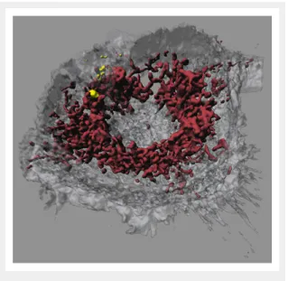
Figure 4
Confocal laser scanning microscopy image of a macrophage that had taken up iron-platinum nanoparticles. Fluorescent labelling of mitochondria using MitoTracker (bordeaux) reveals that the fluorescently labelled polymer-coated iron-platinum nanoparticles (yellow) do not colocalise with mitochondria in macrophages (white, transparent).
Image adapted and reproduced with permission from: Lehmann AD, Parak WJ, Zhang F, Ali Z, Röcker C, Nienhaus GU, et al. Fluorescent–magnetic hybrid nanoparticles induce a dose-dependent increase in proinflammatory response in lung cells in vitro correlated with intracellular localization. Small.
2010;6(6):753-62 [100].
Another uptake mechanism is caveolae-mediated
endocyt-osis. As the name implies, one of the characteristics of this
type of endocytosis is the presence of caveolin proteins in
the 50–100 nm omega-shaped invaginations of the plasma
membrane [
93
,
94
]. Once assembled, caveolar membrane
microdomains remain stable during vesicular trafficking
[
95
]. Clathrin-dependent endocytosis is a type of
pino-cytosis that occurs in virtually all mammalian cells. This
receptor-mediated process is very well studied and leads to
vesicles of about 100 nm in diameter, which are coated by
a protein complex that mainly consists of clathrin. In
con-trast to caveolin-mediated transport, the vesicle coat does
not remain stable during clathrin-dependent endocytosis.
Once the vesicles have detached from the plasma
mem-brane, the clathrin coat is disassembled and the clathrin
triskelia are recycled back to the plasma membrane where
they assemble again around a new vesicle bud [
96
]. The
importance of these two processes for nanomaterial
up-take needs further investigation. By means of inhibition of
specific endocytotic pathways, the uptake of nanomaterials
can be studied in detail and it could be shown that caveolin,
as well as clathrin-dependent uptake, are the main
mechan-isms for pegylated-gold nanoparticles (15 nm) [
97
]. Again,
the mechanism depends on cell as well as particle type and
needs to be investigated in more detail.
The endocytic pathways described above have one feature
in common: particles are localised in intracellular vesicles
after internalisation. However, several studies indicate the
existence of alternative pathways for particles to enter the
cells that allow nanomaterials to remain nonmembrane
bound [
67
,
68
,
98
]. The authors of these studies suggest,
among other mechanisms, passive or receptor-mediated
diffusion of nanomaterials through membrane pores, as
well as so-called adhesive interactions that are mediated by
van der Waals or steric interactions [
99
].
Although a lot of research in recent years has been directed
toward gaining a better understanding of the cellular uptake
of nanomaterials, many questions remain that need to be
answered both in vitro and in vivo. The particular chemical
and physical properties of the nanomaterial and of the
membranes that are responsible for the translocation of
nanomaterials into the cells and subsequent compartments
(e.g., nucleus, mitochondria) need to be elucidated.
Intra-cellular trafficking studies using quantitative transmission
electron microscopy have shown that the preferred
local-isation for pegylated-gold nanoparticless (15 nm) in A549
alveolar epithelial cells are vesicles of different sizes [
97
].
Another study using laser scanning microscopy combined
with digital image restoration showed that polymer-coated
gold and iron oxide nanoparticles co-localised with
lyso-somes but not with mitochondria or the cell nuclei (fig. 4)
[
100
].
In recent years, several reviews have discussed different
mechanisms by which nanomaterials can be taken up by
cells and their resultant mode of intracellular trafficking
[
101
–
103
]. The speed of these processes seems to be
strongly dependent on the surface properties of the
nano-materials and on their in vivo surface modifications (e.g.,
by endogenous proteins or lipids found in surfactant or
plasma) [
8
]. These observations led to the formulation of
the “corona” theory, which states that, in a biological
en-vironment (e.g., surfactant, blood, mucus), the particle
sur-face is covered by biological macromolecules (e.g.,
pro-teins, lipids). This corona can be further subdivided into
a “soft” and “hard” part. The former is characterised by a
dynamic exchange of macromolecules between the particle
surface and the biological surrounding, whereas the latter
consists of biological molecules that are strongly attached
to the particle [
37
,
104
,
105
]. Although it is clear that the
surface properties of nanoparticles are essential for their
in-teraction with cells, it is still debated as to which
character-istics are leading to which cellular responses. In addition,
the cell types investigated also differ with regard to uptake
and defence mechanisms [
100
,
106
]. A general conclusion
that all cells may react similarly after exposure to the same
nanomaterial should be avoided.
Models used to assess the interactions
of nanomaterials with the lung: in
vivo, ex vivo and in vitro
So far, three approaches have been used to study the effects
of particles on the respiratory tract under controlled
con-ditions: in vivo experiments on animals, ex vivo studies on
biopsies or isolated lungs and in vitro experiments using
more or less complex cell culture systems [
107
]. Of course,
all three strategies have advantages and disadvantages that
must be considered. Moreover, they all require profound
methodological knowledge and an interdisciplinary
ap-proach in order to assess the risk of nanomaterials and their
interactions with cells.
In vivo exposure procedures with nanomaterials can be
subdivided into three main categories: whole body, head/
nose/mouth-only or lung-only exposures [
108
–
110
].
Dur-ing whole body exposure, animal sufferDur-ing is clearly lowest
compared with the other two methods, and it mimics
en-vironmental, occupational or intended exposure most
real-istically. Since this exposure type is less stressful for the
animals because it requires neither anaesthesia nor surgery,
it is ideally suited for studies of chronic exposure. The
quality of the results obtained using whole body exposure,
however, depends strongly on an equal distribution of the
particles in the exposure chamber. Moreover, relatively
large amounts of test material are needed to fill the volume
of the exposure chamber, which might limit its usefulness
when expensive materials have to be tested.
Head/nose/mouth-only exposures are more stressful for
an-imals because their food and water supplies are cut off
dur-Figure 5
Two chamber cell culture system.
A conventional two chamber cell culture system in a six-well culture dish. Cells are grown on a porous polyethylene terephthalate (PET) membrane. For reference, the well on the left does not contain medium. B/C Triple cell co-culture model of the human air-liquid barrier consisting of macrophages (light gray, top), epithelial cells (white, middle) and dendritic cells (dark grey, bottom). The cells can be kept submerged (B) or at the air-liquid interface (C).
ing the exposure. Nonetheless, the big advantage of this
delivery method is that the particle administration is very
efficient and doses can be well controlled. As with whole
body exposure, head/nose/mouth-only exposure does not
require anaesthesia and surgery [
111
–
113
].
The third way to study the effects of inhaled aerosolised
nanomaterials is lung-only exposure. This method is
tech-nically much more challenging because it requires
intub-ation or tracheotomy for intratracheal or orotracheal
in-stillation, respectively. With this exposure technique very
precise dosages can be administered but the significance
of the results may be obscured by the lack of reaction of
the systemic and autonomous nervous systems due to
an-aesthesia. Moreover, this technique can lead to local tissue
damage and uneven distribution of the applied substances
in the lung [
111
,
114
,
115
].
Although lung-only exposure is frequently used to study
the toxicology of nanomaterials, it should not be regarded
as an adequate substitute for inhalation studies because it
is not representative of environmental and occupational
ex-posure scenarios [
111
]. In a comparison study in mice,
inhalation of single-walled carbon nanotubes (SWCNTs)
elicited a stronger inflammatory response and increased
oxidative stress than instillation of an equivalent mass.
Al-though the trends were similar in both exposure models,
in-halation of the dry powder was more potent for SWCNTs
than instillation of the suspension [
116
]. In rats the opposite
has been observed: inhaled ultrafine TiO
2particles (21 nm)
led to a decreased pulmonary response compared with a
similar dose of instilled particles. These results might be
explained by differences between the two methods in
particle distribution, dose rate, or clearance [
117
].
However, another study in rats comparing the two
admin-istration routes for TiO
2particles gave consistent toxicity
data for inhalation and instillation [
118
]. Hence, under
cer-tain circumstances and for specific materials, instillation
might be a cost-effective procedure for initial safety
screen-ing.
In vivo, temperature, humidity and pressure in the lung
are tightly controlled.. Thus, it is important to remember
this in order to obtain meaningful data from ex vivo
ex-periments. The most common ex vivo method is the use
of isolated perfused lungs. Separating the lung from the
body has the advantage that experimental parameters can
be better controlled and monitored. However, it has the
disadvantage that it is difficult to maintain physiological
conditions ex vivo, which limits the lifespan of the lung
under artificial conditions to only a few hours. Moreover,
this approach is technically very demanding and requires
a profound knowledge of surgery [
108
,
119
]. Isolated
per-fused lungs have not only been used to study the transport
of pharmaceutically relevant substances through the
air-blood barrier, but also to study the translocation of
nan-oparticles through this barrier [
120
,
121
]. A study
invest-igating the transport of 18 nm iridium particles through
isolated rat lungs has shown that they do not translocate to
the perfusate under normal conditions. However,
pretreat-ing the lungs with either H
2O
2(to mimic oxidative stress)
or histamine allowed particles to translocate across the
air-blood barrier [
121
]. These results confirm earlier findings
where no translocation of ultrafine polystyrene particles
(24, 110 or 190 nm) through isolated rabbit lungs could be
measured under physiological conditions [
120
]. Although
these findings confirm each other, they should be
inter-preted carefully because they conflict with certain in vivo
results. These include the finding that albumin (80 nm,
coated with
99mTc) [
54
] and ultrafine carbon particles (18
nm) [
122
] translocate across the lung barrier in vivo. These
contrasting findings were explained by the absence of the
lymph flow, haemodynamic factors and inflammatory cells
in the explanted lungs [
120
].
Besides isolated perfused lungs, precision-cut lung slices
can also be used as an ex vivo model. Murine lung slices,
for example, have been used to study the suitability of solid
lipid nanoparticles as a drug delivery system [
123
].
Although both in vivo and ex vivo models are valuable tools
for the study of the effects and toxicokinetics of
nanomater-ials in the lung, they have certain limitations. To understand
the toxicodynamics, i.e., how nanomaterials interact with
individual cells and which pathways they influence inside
cells, a zoom-in is required. Because of the complex nature
of the lung architecture, in vitro models are more suited
to the study of these complex parameters in a simplified
set-up. It goes without saying that a good in vitro model
should mimic as many characteristics of the corresponding
region in the respiratory tract as possible. Culturing human
or animal cells according to strict standardised protocols
and following guidelines for good cell culture practice
al-lows results to be obtained that are much more
reprodu-cible than in vivo data [
124
–
126
]. This high data
consist-ency, the relatively low costs and the short experimental
time span mean that in vitro methods are particularly
suit-able for high throughput screening. However, in vitro data
may differ from in vivo results because cell culture
sys-tems are isolated from the physiological context. Hence, at
a certain point, in vitro findings have to be confirmed in
an-imals and humans. Moreover, species differences might be
more significant than often assumed. Disappearance
kinet-ics of hydrophilic molecules, for example, differ quite
tre-mendously between species [
108
,
127
,
128
]. Certain drugs,
might be effective in animals but not in humans. For
ex-ample, in mice, 3-methyladenine (3-MA) has been shown
to reduce acute lung injury triggered by polyamidoamine
dendrimers (a nanomaterial developed for clinical
applic-ations) [
129
]. In humans, however, this autophagy
inhibit-or is not stable [
130
]. Additionally, there is evidence that
there is an important difference between the aquaporin
dis-tribution in human and rodent airways [
131
]. Thus a big
ad-vantage of cell culture experiments is that they can be
per-formed using cells from human origin, yielding data that
are eventually more meaningful for humans than those
ob-tained from animal experiments. For this reason, and
be-cause most studies in the field of nanotoxicology have used
human cells [
110
], we will focus here on human in vitro
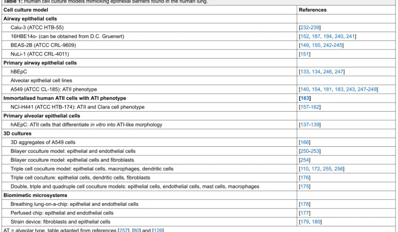
systems only. Table 1 summarises the currently available
cell culture models mimicking the human lung.
Cells used for in vitro experiments can stem either from a
continuous cell line (secondary cultures) or freshly isolated
tissues (primary cultures). Cell lines have the advantage
that they are very homogenous. If they are used properly,
they yield very reproducible results. However, they retain
only little phenotypic differentiation compared with the
ini-tial cell type in vivo. Primary cultures, on the other hand,
are very heterogeneous, consisting of several cell types
with cells at various stages of differentiation. Besides, they
need fairly complex media to be maintained in culture.
Since these cells undergo senescence quite quickly in vitro,
they are only viable for a few passages. Moreover, each
tis-sue isolate is unique owing to donor variation. This makes
them difficult to standardise, which causes a higher
vari-ability of the results [
124
,
132
]. Other limiting factors for
primary cultures are that healthy human airway tissue is not
easily available and that only a few cells are obtained per
isolation [
110
].
Over the last three decades, protocols for the isolation of
primary epithelial cells from both the tracheobronchiolar
[
133
–
136
] and the alveolar region [
137
–
141
] have been
established. Because of the ease of use of cell lines and
the limitations of primary cells discussed above, most
nan-otoxicology studies have been performed using
immor-talised epithelial cells. The most popular
tracheobronchi-olar cell lines are Calu-3 [
142
], 16HBE14o- [
143
] and
BEAS-2B [
144
]. These three cell lines are not only
fre-quently used for drug absorption studies [
145
], but also
to assess particle-cell interactions [
146
] and to investigate
the toxicity of particulate matter [
147
] or nanoparticles
[
148
–
150
]. Permeability values observed in Calu-3 and
16HBE14o- cell lines appear to be predictive of absorption
properties within intact lungs [
108
]. Besides these cell
lines, NuLi-1 [
151
] seems to be another promising
candid-ate for future nanotoxicity studies on human airway
epi-thelial cells [
108
,
152
]. The most widely used and
best-characterised in vitro model of the human alveolar
epithe-lium is the A549 cell line, which has many features of
alveolar epithelial type II (ATII) cells [
153
–
155
]. However,
there are marked differences in morphology and
transepithelial electrical resistance (TEER) between
primary human alveolar epithelial cells (hAEpCs) and
A549 cells [
156
]. NCI-H441 is another in vitro model of
the alveolar epithelium that was obtained from a human
lung adenocarcinoma. This cell line has been described as
having significant TEER values [
157
,
158
] and the
char-acteristics of not only ATII [
159
,
160
] cells but also Clara
cells (i.e., bronchiolar exocrine cells) [
161
,
162
]. Recently,
primary human ATII cells have been immortalised and
used for latex particle (50 nm – 1 µm) uptake studies. This
cell line displays an ATI phenotype and the immortal cells
no longer express alkaline phosphatase, surfactant
pro-tein C and thyroid transcription factor-1, but they do
ex-press increased calveolin-1 and the receptor for advanced
glycation end products. This in vitro model of ATI cells
might help us to understand the importance of this cell type
for the translocation of particles [
163
] as compared with
AT2 cells.
The majority of studies in the field of nanotoxicology have
been performed with monocultures grown as monolayers
on impermeable surfaces. Several studies have shown that
cells that are grown this way after their isolation from the
tissue undergo dedifferentiation and lose their specialised
functions [
164
]. This might be because they lose their
ha-bitual three-dimensional (3D) environment and also their
neighbours of different cell types. Since in vivo cells
con-tinuously crosstalk through intercellular signalling to
main-tain homeostasis and to coordinate immune responses
[
165
], the absence of these neighbouring cells might
influ-ence the experimental outcome. Recent studies have shown
that adding a third dimension to the cell’s environment
[
166
–
168
], or co-culturing different cell types,
signific-antly influences cellular characteristics, behaviour and
re-sponses to stimuli [
169
,
170
]. Including these additional
parameters into an in vitro model creates a greater
simil-arity between the artificial system and the natural situation
in the human body, which ultimately leads to more
relev-ant results. Our research group has recently developed an
in vitro model of the human airway barrier consisting of
three different cell types. In our triple cell co-culture
sys-tem, monolayers of either A549 [
153
], 16HBE14o- [
152
]
or primary epithelial type I cells (hAEpCs) [
171
] are grown
on a microporous membrane in a two-chamber system (fig.
5A). Once the monolayer is confluent, macrophages and
dendritic cells derived from human blood monocytes are
added to the apical and basal side of the epithelium,
re-spectively (fig. 5B). After thorough evaluation, this model
has already been successfully used to study cellular
inter-play and signalling, as well as the cellular responses of
epi-thelial cells, macrophages and dendritic cells to airborne
or suspended particles of different sizes (≤1 μm) and
ma-terials (polystyrene, titanium dioxide, gold, cerium oxide)
[
69
,
110
,
146
,
172
–
174
]. Particle translocation and
cellu-lar localisation were studied in parallel, and it could be
shown that translocation of nanoparticles into the
differ-ent cell types is differdiffer-ent from their larger particle
coun-terparts [
69
]. In addition to the triple co-culture described
above, a quadruple co-culture model consisting of
epitheli-al cells, macrophages, mast cells and endotheliepitheli-al cells has
been established [
175
]. Another triple co-culture model of
the human airways was made up of fibroblasts,
monocyte-derived dendritic cells and epithelial cells [
176
]. In these
three models, the cells are not just cultured together: they
are built up on the porous support in such a way that the
in vitro architecture reflects the specific in vivo
surround-ings that the model is mimicking. To come even closer to
the lung environment in our body, dynamic microsystems
have recently been developed that simulate blood
circula-tion [
177
] and even breathing [
178
] or bronchoconstriction
[
179
,
180
]. It was shown that cells that were subjected to
mechanical strain took up significantly more polystyrene
nanoparticles (100 nm) than static cells [
178
].
A realistic in vitro model must be
combined with an appropriate
exposure system
Not only do in vitro models have to reflect the natural
situation as closely as possible, the method of exposure
to the nanomaterials must also be chosen carefully. To
date, most experiments studing interactions between
nan-omaterials and lung cells have used nanomaterial
suspen-sions that were applied to submerged cell cultures [
69
,
148
,
181
–
183
]. In vivo, however, lung epithelial cells are
sep-arated from the air by only a thin aqueous lining layer
with a surfactant film at the air-liquid interface [
24
,
25
]. As
a consequence, the first barrier particles encounter in the
lung after deposition on the epithelium is surfactant. The
two-chamber system described earlier not only leads to
in-creased differentiation of the epithelial cells [
168
], but it
also allows them to be kept at the air-liquid interface. The
medium can be removed from the upper chamber without
any harm to the cells because they continue to be fed from
the bottom (fig. 5C) [
184
–
188
]. The advantage of this
tech-nique is that the cells are still covered by a thin liquid film
Table 1: Human cell culture models mimicking epithelial barriers found in the human lung.
Cell culture model References Airway epithelial cells
Calu-3 (ATCC HTB-55) [232-239]
16HBE14o- (can be obtained from D.C. Gruenert) [152,187,194,240,241]
BEAS-2B (ATCC CRL-9609) [149,150,242-245]
NuLi-1 (ATCC CRL-4011) [151]
Primary airway epithelial cells
hBEpC [133,134,246,247]
Alveolar epithelial cell lines
A549 (ATCC CL-185): ATII phenotype [140,154,181,183,243,247-249]
Immortalised human ATII cells with ATI phenotype [163]
NCI-H441 (ATCC HTB-174): ATII and Clara cell phenotype [157-162]
Primary alveolar epithelial cells
hAEpC: ATII cells that differentiate in vitro into ATI-like morphology [137-139]
3D cultures
3D aggregates of A549 cells [166]
Bilayer coculture model: epithelial and endothelial cells [250-253]
Bilayer coculture model: epithelial cells and fibroblasts [254]
Triple cell coculture model: epithelial cells, macrophages, dendritic cells [110,172,255,256] Triple cell coculture: epithelial cells, dendritic cells, fibroblasts [176]
Double, triple and quadruple cell coculture models: epithelial cells, endothelial cells, mast cells, macrophages [175]
Biomimetic microsystems
Breathing lung-on-a-chip: epithelial and endothelial cells [178]
Perfused chip: epithelial and endothelial cells [177]
Strain device: fibroblasts and epithelial cells [179,180]
which is much closer to the in vivo situation. However, to
mimic the natural situation it is not sufficient to remove the
medium from the upper chamber only shortly before
apply-ing test substances, because lung epithelial cells need time
to secrete surfactant [
110
,
186
]. Taking these aspects into
account, several recent studies investigated the effects of
nanomaterials on the lung at the air-liquid interface using
newly developed exposure systems [
174
,
189
–
194
].
For safety reasons and to prevent contamination, air-liquid
exposures are usually in a closed system. Nanomaterials
are either directly produced in the vicinity of the cell
cul-ture dish (e.g., flame spray synthesis [
174
], combustion
en-gine [
192
]) or they are nebulied in an exposure chamber
and allowed to settle on the cells [
186
,
191
,
195
–
197
]. For
a complete review on the currently available air-liquid
ex-posure systems see Müller et al. [
198
] and Paur et al. [
199
].
In summary, air-liquid exposure is not only more
physiolo-gical, but it is also mimics more realistically nanomaterial
morphology encountered by the lung in the real world. In
suspension, particle agglomeration and hence deposition
behaviour might be changed by characteristics of the
dis-persion medium (e.g., pH [
200
,
201
], ionic strength [
200
],
protein content [
202
]). Hence, to get most meaningful
res-ults from in vitro systems one has to combine the proper
cell culture model with the appropriate exposure method.
What needs to be done in the future
for a better understanding of the
interactions?
Although researchers worldwide have put a lot of effort
in-to elucidating nanomaterial-(lung) cell interactions, the
un-derlying mechanisms are still poorly understood. To shed
further light on this, more systematic and interdisciplinary
approaches are needed to gain a maximum of information
about a specific nanomaterial. First of all, nanomaterials
that are to be tested on a biological system have to be
fully characterised and these data have to be made available
to other scientists in publications, for later comparison.
Particle characterisation should not be limited to classic
parameters such as size, surface charge, surface structure,
coating, chemical composition or particle shape, but should
also include information about contaminants (e.g.,
endo-toxins such as lipopolysaccharide or adjuvants) or their
col-loidal stability in the environment used later during the
ex-periment [
81
,
203
]. The impact of particle characteristics
on cell uptake and cytotoxicity is an entire field of research,
which has grown tremendously in recent years. In
particu-lar, the effects of particle size and surface charge on cellular
uptake, cytotoxicity, and biodistribution have been studied
extensively. For example, Chithrani and colleagues studied
the uptake of gold nanoparticles and showed that uptake
velocity and concentration varied with size [
204
]. They
also showed that spheres were more readily and efficiently
internalised than rods of the same size. However, at present
it is very difficult to compare published data since not only
materials, surfaces and cells, but also protocols,
concen-trations, controls or methods vary substantially. In
addi-tion, it is well known that biological fluids usually have
a high ionic strength, which might screen possible
repuls-ive forces between nanomaterials (owing to their identical
charge). In consequence, nanomaterials might change their
colloidal behaviour and agglomerate (be loosely bound)
or even aggregate be firmly bound or even fused) in cell
culture medium, for example. Eventually, however, they
might also be more stable in a biological environment. In
any case, changes in their colloidal stability will
influen-ce influen-cellular uptake mechanisms and subsequent influen-cellular
re-sponses [
205
]. Moreover, to avoid misinterpretation of the
results due to experimental artefacts it is crucial that
sci-entists of different fields, such as chemistry, physics and
biology, collaborate closely [
206
]. The importance of an
interdisciplinary approach is illustrated by a study that
in-vestigated the effects of gold nanoparticles (7 nm) on
hu-man dendritic cells. During a first attempt they found that
in conventional laboratory surroundings, gold spheres
ac-tivated dendritic cells. However, when the nanoparticles
were sterile and endotoxin free, they had no maturation
ef-fect on dendritic cells. Combining the knowledge of
ma-terial and biomedical scientists revealed that the observed
activation of the dendritic cells during the first experiment
was due to lipopolysaccharide contamination of the gold
spheres [
207
]. This study also showed that standardisation
of certain experimental procedures between laboratories is
important because this greatly enhances the
comparabil-ity of the results. For instance, choosing a certain
num-ber of methods to measure biochemical markers (e.g.,
re-active oxygen species production) would allow the effects
of different nanomaterials to be compared and hence for
them to be ranked by their potential adverse effects [
208
,
209
]. Moreover, in order to double-check the results, at
least two complementary techniques should be used to
as-sess one parameter [
203
]. However, before a method or a
kit is chosen, it is crucial to test whether the nanoparticles
(or buffer components, etc.) interfere with it or not [
210
].
Hence, the probability of obtaining positive or
false-negative effects can be minimised. Of course it is good
laboratory practice to include appropriate positive and
neg-ative controls in each experiment [
80
,
211
].
So far, it is still unclear as to which particle parameters
de-termine which cell entry mechanism and which cellular
ef-fects. Hence it is of primary importance to perform
system-atic studies that change only one parameter step-by-step
(e.g. size [
212
], exposure time [
174
], coating [
97
]). To be
relevant, the range should be carefully chosen to mimic
realistic exposure. In the long run, such studies will help
to modulate the biological effects caused by nanoparticles
by allowing fine-tuning of some of their parameters. In
our daily life, however, we are not exposed to one kind
of well-defined monodispersed particles but to a mixture
of various particles that we inhale with every breath. As
a consequence, future studies should also address this
as-pect. Again, a systematic approach would be helpful to
decipher the contribution of individual components to the
overall biological impact of the mixture [
213
]. Hence, as
a first step all the constituents would have to be
identi-fied and characterised. Then they should be tested one by
one for their toxicological potential before mixing them
again stepwise. It is needless to say that such an approach
is only feasible for relatively simple mixtures with only
a few components. To overcome the huge amount of data
that would have to be combined for complex systems,
com-puter modelling could be an asset.
Conclusion
Despite various concerns about nanomaterials, they have
various beneficial effects. They potentially offer not only
the possibility to target (cancer) cells but might also help
to fight other diseases such as Alzheimer’s disease [
214
] or
illnesses caused by antibiotic-resistant bacteria [
215
].
Nan-omaterials can also serve as an effective tool in
diagnos-is (e.g., increased contrast for magnetic resonance
ima-ging, detection of pathogens or proteins) and life science
research (e.g., fluorescent labels, purification of biological
molecules and cells) [
216
]. However, risk assessment
should not be neglected in the light of their great
advant-ages.
In a nutshell, inhaled nanomaterials might cause
inflamma-tion or other potentially adverse cellular effects, depending
on their properties and their rate of clearance from the
res-piratory tract [
217
]. Nanomaterials can be removed from
the lung by mucocilliary clearance within the conducting
airways, by macrophage and/or dendritic cell
phagocytos-is, or by translocation through the air-blood tissue
barri-er [
218
]. Over the last decade, there has been a great deal
of research into the effects of nanomaterials on the
respir-atory tract. However, to obtain meaningful data it is
im-portant to use well characterised nanomaterials, a realistic
exposure scenario system, and an appropriate and
valid-ated lung model. In vitro studies have many advantages
over in vivo or ex vivo studies. For instance, they can be
used for high-throughput screening, which allows the
ana-lysis of the effects of a large number of nanomaterials on
the respiratory tract in a short time. Moreover, species
dif-ferences can be ruled out when cells of human origin are
used. Unfortunately, cell culture systems often do not
ex-hibit all the characteristics of the corresponding native
tis-sue. However, this issue can be minimized using 3D and
co-culture models [
186
]. If an appropriate cell culture
sys-tem is chosen – carefully weighing its advantages and
lim-itations – in vitro models of the human lung are a powerful
tool for addressing specific scientific questions. Ultimately,
the development and use of more sophisticated and
well-validated cell culture lung models will help to reduce
anim-al experiments, which is desirable both ethicanim-ally and
finan-cially. Yet, with all efforts taken, the perfect model for lung
risk assessment of nanomaterials does not yet exist. Hence,
data obtained in in vitro and animal studies, should always
be compared with each other and, more importantly, with
epidemiological and/or clinical studies and vice versa [
219
]
because they all provide different pieces of the same
re-search puzzle [
220
]. The relation between air pollution and
adverse health effects has, for example, been reported in
epidemiological studies [
221
–
229
], but animal and, more
importantly, in vitro studies helped to pinpoint the cellular
pathways that are activated by ultrafine particles [
230
]. In
addition, animal as well as epidemiological studies allow
the investigation of the consequences of chronic exposures.
This is fundamental to the adequate risk assessment of
non-biodegradable and non-excreted nanomaterials [
231
]. New
models to study long term outcomes such as, for example,
oncogenicity of nanomaterial exposure are still crucially
needed.
In conclusion, further research is required in order to
un-derstand the potential adverse effects of nanomaterials on
the respiratory tract and, via systemic distribution, on the
human body in general. In the meantime researchers and
workers should take all possible precautions to minimise
their exposure to nanomaterials until their specific hazard
potential has been clarified.
Funding / potential competing interests: The authors wish to
thank the Swiss National Foundation (NRP64, #320030_138365/1, and PP00P2_123373), the German Research Foundation (SPP1313), the Lunge Zürich, the Adolphe Merkle Foundation, FriMat and the University of Fribourg for financial support. Moreover they would like to thank Christian Heinzmann for his help in taking pictures. The authors declare no conflict of interest.
Correspondence: Dr. Corinne Jud, Adolphe Merkle Institute,
University of Fribourg, Route de l'Ancienne Papeterie, CH-1723 Marly, Switzerland,corinne.jud[at]unifr.ch
References
1 Rao C, Cheetham A. Materials Science at the Nanoscale. Nanomaterials Handbook. London, New York: Taylor Francis CRC Press; 2006. 2 Barthlott W, Neinhuis C. Purity of the sacred lotus, or escape from
con-tamination in biological surfaces. Planta. 1997;202:1-8.
3 Lee J, Mahendra S, Alvarez PJJ. Nanomaterials in the Construction In-dustry: A Review of Their Applications and Environmental Health and Safety Considerations. ACS Nano. 2010;4(7):3580-90.
4 Liu H, Webster TJ. Nanomedicine for implants: A review of studies and necessary experimental tools. Biomaterials. 2007;28(2):354-69. 5 Yogeswaran U, Chen SM. A review on the electrochemical sensors and
biosensors composed of nanowires as sensing material. Sensors-Basel. 2008;8(1):290-313.
6 Choi KJ, Jang HW. One-Dimensional Oxide Nanostructures as Gas-Sensing Materials: Review and Issues. Sensors-Basel. 2010;10(4):4083-99.
7 ISO/TS 80004-1. Nanotechnologies – Vocabulary – Part 1: Core terms. Geneva: International Standards Organization; 2010.
8 Oberdorster G, Oberdorster E, Oberdorster J. Nanotoxicology: an emer-ging discipline evolving from studies of ultrafine particles. Environ Health Perspect. 2005;113(7):823-39.
9 Ruffini Castiglione M, Cremonini R. Nanoparticles and higher plants. Caryologia. 2009;62(2):161-5.
10 Schmid K, Riediker M. Use of nanoparticles in Swiss Industry: a tar-geted survey. Environ Sci Technol. 2008;42(7):2253-60.
11 Parker PM. The 2011 Report on Nanoparticles in Composites: World Market Segmentation by City. San diego: ICON Group International; 2011.
12 Ultrafine Titanium Dioxide. [22.06.2012]; Available from: ht-
tp://www.cristalglobal.com/ProductResources/Ul-trafine%20Titanium%20Dioxide%20-%20info.pdf.
13 Semmler-Behnke M, Kreyling WG, Lipka J, Fertsch S, Wenk A, Takenaka S, et al. Biodistribution of 1.4- and 18-nm Gold Particles in Rats. Small. 2008;4(12):2108-11.
14 Kreyling WG, Hirn S, Schleh C. Nanoparticles in the lung. Nat Biotech. [10.1038/nbt.1735]. 2010;28(12):1275-6.
15 Stern ST, McNeil SE. Nanotechnology safety concerns revisited. Tox-icol Sci. 2008;101(1):4-21.
16 Gehr P, Bachofen M, Weibel ER. The normal human lung: ultrastructure and morphometric estimation of diffusion capacity. Respir Physiol. 1978;32(2):121-40.