HAL Id: hal-02379658
https://hal.archives-ouvertes.fr/hal-02379658
Submitted on 25 Nov 2019HAL is a multi-disciplinary open access archive for the deposit and dissemination of sci-entific research documents, whether they are pub-lished or not. The documents may come from teaching and research institutions in France or abroad, or from public or private research centers.
L’archive ouverte pluridisciplinaire HAL, est destinée au dépôt et à la diffusion de documents scientifiques de niveau recherche, publiés ou non, émanant des établissements d’enseignement et de recherche français ou étrangers, des laboratoires publics ou privés.
Interaction of a Model Peptide on Gram Negative and
Gram Positive Bacterial Sliding Clamps
Christophe André, Isabelle Martiel, Philippe Wolff, Marie Landolfo, Bernard
Lorber, Cyrielle Silva da Veiga, Annick Dejaegere, Philippe Dumas, Gilles
Guichard, Vincent Olieric, et al.
To cite this version:
Christophe André, Isabelle Martiel, Philippe Wolff, Marie Landolfo, Bernard Lorber, et al.. Interaction of a Model Peptide on Gram Negative and Gram Positive Bacterial Sliding Clamps. ACS Infectious Diseases, American Chemical Society, 2019, 5 (6), pp.1022-1034. �10.1021/acsinfecdis.9b00089�. �hal-02379658�
1
Interaction of a Model Peptide on Gram Negative and Gram Positive
Bacterial Sliding Clamps.
Christophe André¤ +
, Isabelle Martielᵻ +, Philippe Wolff§ +, Marie Landolfo§, Bernard Lorber§, Cyrielle Silva da Veiga§, Annick Dejaegere£, Philippe Dumas±, Gilles Guichard¤
, Vincent Oliéricᵻ, Jérôme Wagner# and Dominique Y. Burnouf§.
§Université de Strasbourg, CNRS, Architecture et Réactivité de l’ARN, UPR 9002, Institut de Biologie
Moléculaire et Cellulaire du CNRS, 15 rue René Descartes, F-67000 Strasbourg, France
ᵻSwiss Light Source (SLS), Paul-Scherrer-Institute (PSI), Villigen, Switzerland.
¤ Institut Européen de Chimie et Biologie, Université de Bordeaux-CNRS UMR 5248, CBMN, 2, rue Robert
Escarpit, 33607 Pessac, France.
£
Institut de Génétique et de Biologie Moléculaire et Cellulaire (IGBMC), Département de Biologie Structurale et Génomique, 1 rue Laurent Fries, BP10142, 67404 Illkirch, France
#
CNRS UMR7242, ESBS, Université de Strasbourg, BP 10413, 67412 Strasbourg Cedex, France
±IGBMC, 1 rue Laurent Fries, BP 10142, 67404 Illkirch CEDEX
+
: these authors equally contribute to this work.
Keywords: sliding clamp, ITC, kinITC.
SUMMARY.
Bacterial sliding clamps control the access of DNA polymerases to the replication fork and are appealing molecular targets for antibacterial drugs development. To this end, it is critical to decipher the polymerase-clamp binding mode across various bacterial species. We showed previously that synthetic peptides targeting the clamp binding pocket of Gram- bacteria poorly interact with that of Gram+ homologous proteins. Here we analyzed the interaction of a reference peptide with several E. coli and B. subtilis clamp variants. For both Gram- and Gram+ pockets, the peptide binds through an induced-fit process but the complex stability
2
varies according to a pocket specific network of interactions. Thermodynamic and molecular dynamics analyses identify a strategic position in the pocket where a mobile residue is necessary for an efficient peptide interaction. A residue at another position modulates the folding dynamics of the pocket upon ligand binding in E. coli, while in B. subtilis, this residue is essential for polymerase activity and might thus be a Gram+ specific molecular marker.
3
INTRODUCTION.
Bacterial resistance to antibiotics is a major threat for human health. According to several official reports, a return to the pre-antibiotic era during the 21st century is a realistic possibility1. In order to tackle this challenge, one strategy aims at identifying new bacterial molecular targets and at developing efficient molecules that will block their physiological functions. The bacterial processivity factor, also referred to as the sliding clamp (SC), has been previously identified as a potential drugable new target 2 3 4 5 6. The ultimate proof of concept was recently brought by the natural cyclic peptides griselimycins that bind to Mycobacterium tuberculosis sliding clamp (MtSC) and display good in vivo anti-bacterial activity in animal models7. However, the molecular mechanisms that govern the SC/ligand interaction are not well understood4589.
In bacteria, SC is a homodimer that encircles and slides along double stranded DNA10.
It binds the replicative DNA polymerase, thus conferring a high processivityto the resulting
holoenzyme 1112. It also serves as a molecular hub on which all the other DNA polymerases
bind (polI, II , IV and V in E. coli) 13 14 as well as other enzymes involved in DNA metabolism 15 and this interaction is required for these proteins to fulfill their functions 13. Remarkably, in all cases, SC-protein interaction is mediated by a short peptide segment
which encompasses the consensus sequence (QL[S/D]LF) 16. This peptide portion binds SC
within a hydrophobic pocket located between domains II and III of SC 173 and formed by two
sub-sites joined by a shallow groove: sub-site 1, also referred to as the leucine-rich pocket, interacts with the C-terminal part of the peptide, whereas sub-site 2 binds the highly conserved Q residue (SI.1).
We have previously observed that short synthetic peptides designed to bind to Escherichia coli SC (EcSC) with an increased affinity also interact efficiently with SC from
other Gram- bacteria such as Pseudomonas aeruginosa5. However, they show a lower
interaction with MtSC and fail to interact efficiently with SC from other Gram+ strains such as Staphylococcus aureus and Bacillus subtilis (BsSC), suggesting that peptide binding on SC
occurs through a different process in Gram+ and Gram- bacteria 18.
A careful analysis of the peptide binding process on EcSC revealed that a fully efficient binding pocket folds upon peptide binding 3519. Notably, two residues, S346 and M362, seem to
4
undergo a large concerted movement that allow the opening of a groove in which the linear peptide can lie 5 (SI.1). Interestingly enough, these two residues are conserved in Gram- strains but not in Gram+ bacteria, where they are essentially replaced by proline and leucine/threonine, respectively. Noteworthy, in MtSC only the residue corresponding to EcSC S346 is changed into proline, whereas the methionine residue (MtSC M396) is conserved. Thus,
as far as these two residues are concerned, MtSC (P362, M396) stands as an intermediate between Ec
SC (S346, M362) and BsSC (P357, L373) (Table 1). These observations, in correlation with
peptide binding analyses18, suggest that the mode of peptide interaction might be linked to the gating function of these two residues.
In this work, we evaluate the respective contribution of residues EcSC S346 and EcSC
M362 and their corresponding residues in MtSC and BsSC, to peptide binding. For each SC,
every residue was mutated (ML, SP, LM and PS), leading to a series of single and
double mutants (Table 1). The interaction of peptide P7 (AcQXDLF, X = cyclohexylalanyl,
Cha) (SI.2)5 with each of these SCs was analyzed by ITC, X-Ray crystallography, molecular
dynamics and in vitro replication assays. Our results reveal the prominent role of these residues for the peptide interaction with Gram- and Gram+ SC, and will have strong implications for the design of strain-specific anti-replicative molecules.
MATERIAL AND METHODS.
Construction of mutant SC, expression and purification of processivity factors.
Plasmids pET15b containing the dnaN genes from E. coli, M. tuberculosis or B. subtilis
18
were site specifically mutagenized using the Quikchange® procedure (Stratagene). Oligonucleotides were from IDT. Selected recombinant plasmids were sequenced (GATC, Kontanz, Ge) and transformed in BL21 (DE3) pLys E. coli strains. For expression of dnaN proteins, cells were grown in LB at 37°C to OD 0.5, then induced by IPTG (0.1mM) at 28°C overnight. dnaN proteins fractions were first enriched on a Ni-NTA column, eluted with an histidine step (300 mM) and further purified on a Source Q column in buffer containing 20 mM Tris HCl pH 7.5, 0.5 mM EDTA and 10% (v/v) glycerol, using a gradient from 0 to 0.5 M NaCl. After a final ultracentrifugation (45K, 1 h, 20°C), soluble proteins were concentrated on a Centricon 30K (Millipore) in the same buffer and stored at 4°C in 2 M ammonium sulfate. Buffer exchange was performed on Centrikon 10K at 4°C before use and protein quality was assessed by DLS analysis (SI.3).
5
Peptide synthesis.
Procedure for the synthesis of peptide P7 has been described elsewhere 5. The chemical
formula is shown in supporting information (SI.2)
Isothermal Titration Calorimetry.
ITC was performed using an iTC200 (Microcal Malvern Panalytical) or a PEAQ-ITC
instrument (Microcal Malvern Panalytical). Peptides (300 or 600 µM) were titrated at different temperatures (15, 25 and 34°C) by sequential injections (usually 2 µl each) into a SC solution (30 or 60 µM). Data were corrected for heat of injection by subtracting the signal of the titration of peptides into protein-free buffer solution (Hepes 10 mM pH 7.4, NaCl 0.15 M, EDTA 3 mM) (SI.4). Each titration was performed at least twice. Analyses of experimental data were performed following a classical treatment with the AFFINImeter software (https://www.affinimeter.com; S4S, Santiago de Compostela, Spain). Both types of analyses yield similar data (data not shown). Non classical methods were also used. First a global thermodynamic treatment (GTT, see below) was performed. In addition, kinetic information
was obtained in some cases with kinITC 20 32 as implemented in the software AFFINImeter. A
comparison of GTT and classical treatment analyses is presented in SI.5. All thermodynamic data are provided in SI.6, representative titration curves are presented in SI.7 and all thermodynamic profiles are presented in SI.8.
Global thermodynamic treatment (GTT) of ITC experiments.
This procedure has been described in 20. Briefly, the method consists in using a single reduced set of parameters to fit at once all experimental titration curves obtained at different temperature. The free parameters are Kd0 and H0, the values of Kd and H at a reference
temperature T0 (the mean temperature of all experiments) and Cp = H/T governing the
evolution of H with the temperature. These three parameters alone, have to replace 2 Nexp
parameters (one Kd and one H for each experiment). This GTT yields theoretical curves
describing the evolution of all thermodynamic parameters with the temperature. These curves are shown with the points corresponding to the results from the usual individual treatments: it is important to stress that they do not result from a fit of these points (see Figure 1). A comparison of the data obtained by GTT and classical treatment is presented in SI.5. When the peptide interaction with SC was weak, the deviation between the points from the
6
individual treatments and the curve from GTT was large (see Figure 1B and SI.5 B). In these cases, the reported results are from the individual treatments. Of course, the discrepancy just mentioned is a mark of poor quality.
Crystallogenesis, X-ray diffraction, data collection and processing.
Crystals of EcwtSC/P7 and EcMtSC/P7 complexes were obtained by screening the
crystallization reagent kit PEG/Ion HT from Hampton Research. 200 nL sitting drops were
prepared on plates (Greiner XTL round) by mixing 50 or 100 nL of preformed SC/P7
complexes (protein concentration from 6 to 12 mg/ml; protein/peptide ratio: 1.5) with 150 or 100 nL of crystallization solution, using a Mosquito pipetting station (TTP Labtech). Plates were incubated at 20°C (EcSC) or 4°C (MtSC). Crystals were obtained in several crystallization conditions and were frozen in liquid ethane. Crystals with the higher resolution were obtained
in the following conditions: EcwtSC/P7: MES 50 mM pH 6, CaCl2 50 mM PEG400 30% (m/v)
+ PEG/Ion HT kit condition E6 (0.2 M sodium malonate pH 6, PEG 3350 20% (m/v)).
EcM1
SC/P7: MES 50 mM pH 6, CaCl2 50mM, PEG400 28% (m/v) + PEG/Ion HT kit
condition B3 (0.2 M lithium nitrate, PEG 3350 20% (m/v), pH 7.1). MtwtSC/P7: Acetate de Na
3.2 M pH 6.9, PEG 3350 18% (m/v) + PEG/Ion HT kit condition A2 (0.2 M potassium
fluoride, PEG 3350 20% (m/v), pH 7.3). MtM1SC/P7: Acetate de Na 3.2 M pH 6.9, PEG 3350
18% (m/v) + PEG/Ion HT kit condition B8 (Magnesium formate dihydrate 0.2 M; PEG 3350 20% (m/v), pH 7).
The data were collected at the X06DA PXIII beamline at the Swiss Light Source (Villigen, Switzerland), equipped with a PILATUS 2M detector and the multiaxis PRIGo goniometer 21. The wavelength was 1.00 Å, with a flux at full transmission of 4·1011
photons·s-1. The data were collected with two rotations of 360° at χ=0° and χ=30° (0.2° and
0.1s per frame). The crystal-to-detector distance was 300 mm. The data were processed and
merged using the software autoPROC version 1.0.5 22 and STARANISO (Cambridge, United
Kingdom: Global Phasing Ltd) 23. The structures were solved by molecular replacement using
the software MOLREP version 11.4.04 24 and search models 1OK7 for the E. coli cases and
4TR7 for the M. tuberculosis cases. Refinement was performed with BUSTER version 2.10.3
(Cambridge, United Kingdom: Global Phasing Ltd) and phenix.refine version 1.10.1-2155 25,
with NCS constraints for all structures and TLS for both higher resolution E. coli structures. Secondary structure restraints were additionally imposed in phenix.refine for the Mtwt-P7 structure. 5% of reflections were set aside for the Rfree test set.
7
Data processing and refinement statistics are available in SI.9 The 4 structures EcwtSC/P7,
EcM1
SC/P7, MtwtSC/P7 and MtM1SC/P7 are deposited in the Protein Data Bank (PDB) under the
respective accession codes 6FVL, 6FVM, 6FVN and 6FVO. No complex structure could be solved for any BsSC.
The solvent accessible surface area was calculated using AREAIMOL in CCP4 26. The
difference in accessible surface area of a binding pocket with and without P7 was obtained by
considering the structures from this work and from Wolff et al.18 (PDB codes 1OK7 for
Ecwt
SC and 4TR7 for MtwtSC).
Replication assays.
Escherichia coli Pol III* purification. E. coli strain BLR transformed with pHOC2.6.1 (kindly provided by C. McHenry), coding for all the 8 polIII subunits, were grown at 37°C in LB buffer supplemented with 100 µg/ml carbenicillin to reach an OD600nm of about 0.65, transferred to 30°C and then expression of Pol III* subunits was started by adjunction of 1 mM IPTG and extended for 3 h. Cell pellets (16.8 g) were stored at -20°C overnight. Purification of PolIII* was conducted essentially as described previously 27. Briefly, pellets thawed at 4°C for 30 min were resuspended in 50 ml of 50 mM Hepes-KOH pH 7.5, 10% (m/v) sucrose, 10 mM DTT. 5 ml of lysis buffer (50 mM Hepes-KOH pH 7.5, 10% (m/v) sucrose, 2 M NaCl, 0.3 M spermidine) were added to the cell suspension and Tris base 2 M was used to adjust to pH 8. Lysis was performed on ice for 1 h after addition of lysosyme to 0.2 mg/ml. After centrifugation at 20 000 rpm for 1 hour at 4°C ( Beckmann 70 Ti rotor), proteins were precipitated by slow addition of 0.226 g/ml ammonium sulfate and incubation 30 min at 4°C with agitation. After centrifugation (45 min, 16 000 rpm, 4°C), pellet was successively extracted with 3.275 ml of buffer A (25 mM Hepes-KOH pH 7.5, 5% (v/v) glycerol, 100 mM KCl, 1 mM EDTA, 5 mM DTT) supplemented with 0.2 g/ml ammonium sulfate, centrifuged, and further extracted with 1.2 ml of buffer A with 0.7 g/ml ammonium sulfate. After centrifugation (20 min, 30, 000 rpm, 4°C), the pellet was resuspended in buffer A without KCl and conductivity was adjusted to the one of buffer B (25 mM Hepes-KOH pH 7.5, 5% (v/v) glycerol, 20 mM KCl, 1 mM EDTA, 5 mM DTT), then loaded onto a SP sepharose column (1 ml, GE Healthcare) equilibrated with buffer B and developed with a linear gradient of 0 to 30 % buffer C (25 mM Hepes-KOH pH 7.5, 5% (v/v) glycerol, 1 M KCl, 1 mM EDTA, 5 mM DTT) on AKTA smart chromatography system (GE Healthcare).
8
Finally, pooled fractions were buffer exchanged for buffer D (25 mM Hepes-KOH pH 7.5, 5% (v/v) glycerol, 20 mM KCl, 5 mM DTT) on a PD10 column (GE Healthcare) and concentrated by ultra-filtration on Amicon Ultra centrifugal units (MWCO 10K), adjusted to 50% (v/v) glycerol and to 0.21 mg of protein per ml and stored at -80°C.
Bacillus subtilis minimal replicase (BsP) was reconstitued by assembly of purified proteins (kindly provided by Charles McHenry and Paul R. Dohrmann) at ratio of 1 PolC, 5 tau, 1.25 delta and 1.25 delta' to provide a 1.25 µM enzyme complex in buffer HD (40 mM Tris acetate pH=7.8, 10 mM Mg acetate, 3 µM zinc sulfate and 0.015 % (v/v) IGEPAL CA-630).
In vitro primer extension assays. Single-stranded pUC118 DNA was annealed to a 5' radiolabeled 40 mer oligonucleotide (5'GCTGGCGAAAGGGGGATGTGCTGCAAGGCGA TTAAGTTGG3') in 50 mM NaCl and 15 mM Tris acetate (pH=7.8). All DNA synthesis assays were pre-assembled on ice in RB buffer (HD buffer supplemented with 2 mM DTT, 150 mM K glutamate, 160 µg/ml BSA, 0.5 % (m/v) PEG 8000, 250 µM ATP and 100 µM each dNTP). Unless specified, each reaction contains 40 fmoles of primed DNA, 100 fmoles of polymerase complex and 100 fmoles, as a dimer, of beta sliding clamp (SC). DNA synthesis assay were performed for 6 min. at 37°C before addition of 15 µl of 95% formamide dye loading buffer and heating at 95°C for 5 minutes. Radioactive reaction products were analysed by electrophoresis on 6 to 8 % denaturing (8 M urea) polyacrylamide gels followed by imaging using a Typhoon FLA 9500 device (GE Healthcare).
P7 primer extension inhibition assays were performed essentially as above except that mixtures of SC (100 fmoles in HD buffer, 1µl) and P7 peptide (diluted in HD buffer, 1µl) were first incubated for 5 min. at RT (20-25 °C) before adding 8 µl of primed DNA plus polymerase complex in RB buffer.
Molecular Dynamics.
Initial Structures:
Experimental X-Ray structures of EcwtSC were used as a starting point for the molecular
dynamics simulations of the EcwtSC in complex with a de-acetylated form of P7 (QXDLF, X =
Cha). Starting structure for the EcM2SC (M362L) was constructed from the EcwtSC by side-chain
modification using PyMol. B. Subtilis simulations were constructed from the BswtSC structure
9
mutant BsM2SC (L373M). For both wtSC and mutant M2SC, the peptide was positioned by
superimposition onto P1 (PDB 1OK7) and P1 was subsequently removed.
Free-energy perturbation calculation:
The relative affinities of the wtSC and M2SC for the peptide were estimated using free energy
perturbation implemented in NAMD28 in the following thermodynamic cycle 2930:
Figure 1: Thermodynamic cycle used in computing the free energy change in the virtual mutation of the wtSC into the mutant M2SC in the unbinding form (ΔGu) and in the binding form (ΔGb).
These relative affinities, ΔΔG, can be calculated from the following equation:
(1)
Gu and Gb are calculated by transforming the Hamiltonian of the initial states (either the
apo WT (Gu) or the holo WT (Gb)) into the final states (either the apo mutant M2 (Gu) or
the holo mutant M2 (Gb)). The transformation is modeled by introducing a state variable, λ,
which can vary from 0 (initial state) to 1 (final state). The free-energy calculation is split into small windows, each one involving a small variation of λ. A new residue is designed to simulate the transformation (for example of the residue M362 into L362). In that way, we have
built a new residue, name M2L, which includes the atoms of the side chains of a methionine
and of a leucine. The structure was then generated by using the force field (FF) charmm 27 31
and was immersed in a tetragonal box filled with TIP3 water molecule and 24 Na+ ions to neutralize the total charge of the full system. The simulation was then heated at 300K and equilibrated before the free-energy perturbation calculation.
10
We have previously observed that peptides initially designed to bind EcSC with high affinities poorly interact with MtSC and BsSC, although the binding pockets of all SC are structurally highly homologous18. In order to decipher the molecular basis of these different binding processes, we determined the thermodynamic parameters of the interaction between a reference ligand, namely peptide P7 (SI.2 and SI.10) and different variants of EcSC, MtSC and
Bs
SC (Table 1).
Peptide P7 differentially interacts with natural SC from different bacteria.
We first determined the thermodynamic profiles at different temperatures of P7
binding on natural SC, namely EcwtSC, MtwtSC and BswtSC, (Figure 1). For each SC/P
7
interaction, global thermodynamic treatment (GTT) 32 of all experiments at all temperatures yielded theoretical curves describing the evolution of each thermodynamic parameters with temperature (Figure 1, left column). Thermodynamic profiles are inferred from these curves (Figure 1, right column). The profiles observed with the three natural SC are very different (Figure 1, see also SI.5 for a comparison between GTT and individual analysis of ITC raw data and SI.6 for numerical data).
The EcwtSC/P7 interaction is enthalpy driven, with a significant increase of the
enthalpic contribution from -10 to -14 kcal/mole when temperature rises from 15 to 34°C. This enthalpic contribution is much weaker for the MtwtSC (-4 to -8 kcal/mol), and for BswtSC (-6 to -3 kcal/mol) over the same range of temperature (Figure 1 right column, SI.5). Nevertheless, the binding process is still enthalpy-driven for MtwtSC and BswtSC. The ΔCp (ΔH/T) values are -210 cal/mol/deg for P7 binding onto EcwtSC, -180 cal/mole/deg with
Mtwt
SC. The ΔCp for BswtSC was not reliably determined. P7 interaction with EcwtSC shows an
unfavorable entropic contribution (-TΔS) which increases with temperature (Figure 1A and
SI.5). For BswtSC, this contribution is weaker and becomes favorable above 25°C (Figure 1B),
while for. MtwtSC, it evolves in an opposite way (Figure 1C). The Gibb's free energy, ΔG, also
varies significantly between the different complexes. For EcwtSC, it reaches -9 kcal/mole and
slightly increases with higher temperatures (Figure 1 and SI.5). It reaches about -5 and -7 kcal/mole for BswtSC and MtwtSC, respectively, indicative of a weaker interaction between P7
and these two SC, and in both cases, it varies very slightly over the range of temperature. Interestingly, ΔG reaches a maximum at 18°C for BswtSC/P7 and a minimum at 29°C for
Mtwt
11
column), implying that around these temperatures, the ΔG variation is minimal. For EcwtSC/P7,
extrapolation of the curve ΔS= f(T) indicates that ΔS = 0 at T < 0°C.
E. coli M. tuberculosis B. subtilis
Name Amino acids Name Amino acids Name Amino acids wt EcwtSC S346; M362 Mtwt SC P362; M396 Bswt SC P357; L373 M1 EcM1SC P346; M362 MtM1SC S362; M396 BsM1SC S357; L373 M2 EcM2SC S346; L362 MtM2SC P362; L396 BsM2SC P357; M373 M3 EcM3SC P346; L362 - - BsM3SC S357; M373 G1 EcG1SC G346; M362 - - BsG1SC G357; L373 G2 EcG2SC S346; G362 - - BsG2SC P357; G373
Table 1: Names and sequences characteristics of the different sliding clamps used in this study. Ec:
Escherichia coli; Mt: Mycobacterium tuberculosis; Bs: Bacillus subtilis; wt: wild type sliding clamps; M1, M2, M3, G1 and G2: mutant sliding clamps.
The dissociation constant measured for EcwtSC/P7 interaction increases from 100 to 450 nM
between 15 and 34°C (Table 2, Figure 1A and SI.6). For the MtwtSC/P7 interaction, the Kd is
about 20 to 40 times lower (between 4 and 7 µM), while the lowest affinity is observed for
Bswt
SC, with Kd values ranging from 40 to 80 µM range.
All these data confirm our previous observations and show that P7 binding greatly
vary with the SC bacterial origin18. To investigate the molecular basis of these differences, we
constructed several mutants of each SC where the residues corresponding to EcwtSC S346 and
Ecwt
SC M362 are singly (M1, M2, G1 and G2 respectively) or both (M3) mutated (Table 1).
The interaction of P7 with these SC variants was analyzed using ITC, crystal structures, in
12 Figure 1: Thermodynamic analyses of P7 interaction with
Ecwt
SC (A), BswtSC (B) and MtwtSC (C) at different temperatures. Global thermodynamic treatment (GTT) (left column): The curves represent the functions obtained by treating simultaneously all experimental ITC data at three temperatures. Dots represent the results of a classical treatment of each experimental ITC data processed individually. For weak interaction, such as BswtSC/ P7, the gap between individual treatment and the theoretical curve obtained by GTT is more important.
Thermodynamics profiles (right column) displaying ΔH, -TΔS and ΔG at three temperatures were obtained from the GTT derived curves. ΔH, -TΔS and ΔG (all in kcal/mol) are white, grey and dark bars, respectively. All ITC data are presented in SI.6 and typical ITC titration curves are shown in SI.7. ITC control experiments at three temperatures are presented in SI.4.
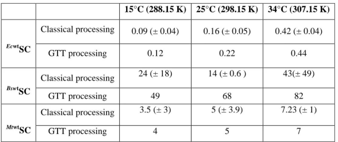
13 15°C (288.15 K) 25°C (298.15 K) 34°C (307.15 K) Ecwt SC Classical processing 0.09 (± 0.04) 0.16 (± 0.05) 0.42 (± 0.04) GTT processing 0.12 0.22 0.44 BswtSC Classical processing 24 (± 18) 14 (± 0.6 ) 43(± 49) GTT processing 49 68 82 MtwtSC Classical processing 3.5 (± 3) 5 (± 3.9) 7.23 (± 1) GTT processing 4 5 7
Table 2: Dissociation constants (KD: µM) of the P7 interaction with the three natural SC at different
temperatures. The large standard deviations observed for BswtSC and MtwtSC are indicative of a poor interaction. Independent experiments were performed two to four times. All results were obtained through classical or GTT processing of experimental data, as indicated. Full thermodynamic data are in SI.6 and examples of ITC titration curves are presented in SI.7.
Mutations S346P and M362L in EcwtSC drastically modify the thermodynamics of SC/P7
interaction.
Complete thermodynamic profiles of P7 interaction with the various SC mutants are
presented in SI.8 and SI.11. Figure 2 presents a comparison of the thermodynamics profiles of the P7 interaction with EcwtSC and the double mutant EcM3SC. Details on the specific effects
induced by single mutations are developed in SI.11. Introduction of the S346P and M362L
mutations in the EcwtSC binding pocket results in a large reduction of |ΔH| of about +6
kcal/mol at 15°C and +10 kcal/mol at 34°C. The resulting ΔCp drops from -210 (EcwtSC) to
-50 cal/mol/deg for EcM3SC, indicative of a change in the mode of interaction33. As opposed to
what is observed with EcwtSC, the entropic component (-TΔS) becomes favorable (≈ -2 to -3
kcal/mol) to the EcM3SC/P7 interaction at all temperatures. The resulting free energy of
interaction, ΔG, is stabilized around -6 to -7 kcal/mol, i.e. about 2-3 kcal/mol higher that those observed for EcwtSC, at all temperatures. Consequently, the mutations S346P and M362L in Ecwt
SC trigger a drastic change in the P7 mode of interaction, underlying the significant
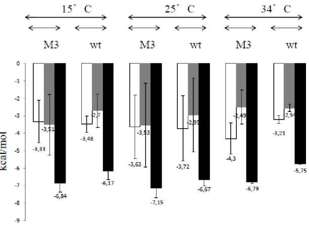
14 Figure 2: Thermodynamic profiles of P7 interactions with
Ecwt
SC (wt) and EcM3SC (M3) at different temperatures. ΔH, -TΔS and ΔG ( all in kcal/mol) are white, grey and dark bars, respectively. Data are means of at least two independent experiments. For the sake of comparison, all results are obtained through classical processing of experimental data (SI.6).
EcM3
SC and BswtSC present similar thermodynamic profiles upon P7 interaction.
Because the two mutations introduced in EcM3SC are equivalent to the natural residues
found in BswtSC (Table 1), we also compared the thermodynamic profiles obtained for P7
interaction with EcM3SC and BswtSC (Figure 3). These profiles are very similar at all temperatures, confirming that the introduction of S346P and M362L mutations in EcwtSC is
sufficient to convert the E. coli specific thermodynamic profile into that observed with BswtSC. For both types of interactions, the enthalpic contribution is equivalent, ranging from -3 to -4 kcal/mole, and in both cases, the entropic contributions are favorable to the interaction, around -3 kcal/mol. The large variations of both parameters result from the poor efficiency of the peptide binding. The resulting free energy of interaction is slightly more favorable for
EcM3
15
residues, the local environment of the EcSC pocket stabilizes the peptide interaction more significantly than that of BswtSC.
Figure 3: Thermodynamic profiles of P7 interactions with EcM3SC (M3) and BswtSC (wt) at different
temperatures. ΔH, -TΔS and ΔG (all in kcal/mol) are white, grey and black bars, respectively. Data are means of at least two independent experiments. Results are obtained through classical processing of experimental data (SI.6).
Mutations P357S and L373M residues in BswtSC improve P7 interaction.
We also analyzed the effects of the introduction of Ec natural residues (S346 and M362)
in the Bs context (Figure 4, SI.8 B and SI.11 B for details on single mutations effects on P7
interaction). As opposed to the major deleterious effect on complex formation triggered by mutations introduced in EcwtSC (Figure 2), the mutations of the corresponding residues in
Bs
SC improve the efficiency of the interaction with P7, as indicated by the systematic increase
of ΔG values with BsM3SC as compared to BswtSC (Figure 4). However, the effect is limited as
these ΔG values remain about 2 kcal/mole lower than those measured for EcwtSC. This is in
agreement with the above mentioned observation that the BsSC pocket might be globally less
favorable to P7 binding. Consequently, as observed for the Ec context (SI.11 A) but with
16
of the interaction (SI.11 B). At all temperatures, the P357S mutation (mutant BsM1SC) triggers a
reduction of the ΔH values, as compared to those measured in BswtSC, and an increase of the
entropic factor favorable contribution. Alternatively, the L373M mutation (mutant BsM2SC)
increases the ΔH value by -3 to -5 kcal/mol at each temperature and drastically reduces the entropic contribution, which becomes unfavorable at high temperature.
Figure 4 : Thermodynamic profiles of P7 interactions with BswtSC (wt) and BsM3SC (M3) at different
temperatures. ΔH, -TΔS and ΔG (all in kcal/mol) are white, grey and black bars, respectively. Data are means of at least two independent experiments. Results are obtained through classical processing of experimental data (SI.6).
The added contribution of both mutations, measured with mutant BsM3, combine the effects of each individual mutation. As a result, the ΔH evolution becomes strongly dependent on temperature and the entropic contribution turns to be unfavorable at 34°C for both M2 and M3 mutants (SI.11 B), but already at 25°C for the double mutant, which suggests a
synergistic effect of both mutations (Figure 4). Indeed, the ΔCp value determined for BsM3SC
(-270 cal/mole/deg) is closed to that measured for EcwtSC (-210 cal/mole/deg), whereas the values calculated for BsM1SC and BsM2SC are -10 and -40 cal/mol/deg, respectively. These
17
observations show that the two mutations, P357S and L373M, induce a complete change of the
thermodynamic profile of the BsSC/P7 interaction. Moreover, in the two different pocket
environments, Ec and Bs, the drastic changes in thermodynamic profiles are triggered by the M2 mutations (Figure SI.11 A and B) thus identifying a strategic position for what concerns the peptide interaction. The position of M1 mutations is not that crucial for the complex formation.
Specific contributions of the strategic residues to the P7 interaction.
To further analyze the specific contributions of the S346 (or P346) and M362 (or L362)
residues to the P7 interaction, we compared the thermodynamic data obtained for wtSC (or
M1
SC or M2SC) with those obtained with G1SC and G2SC (Table 1), where the natural residues are replaced by glycine (Figure 5 and SI.12). Note that thermodynamic data obtained with the G mutants directly provide the whole pocket contribution to the interaction, excluding the S/P and M/L side chain effects. The specific contributions of the S/P and M/L residues to each thermodynamic parameter are obtained by the difference between the parameter measured for
wt
SC and that measured for the GnSC (SI.12). The results are presented in Figure 5.
In the Ec context, the S346 residue does not contribute significantly to the P7
interaction, as indicated by the weak ΔG value, particularly at high temperatures (Figure 5A,
left). The ΔH value reveals an endothermic contribution, while the favorable entropic
contribution increases with temperature and might be linked, at least partly, to water release upon peptide interaction, as observed in crystal structures5. A very different profile is observed for the M362 residue (Figure 5A, right), with essentially an exothermic specific
contribution, no entropic effect and a constant Gibb's free energy (-1 kcal/mole) over the temperature range. Thus this residue contributes for about one tenth of the overall interaction
of the peptide (see Figure 1A). Mutation S346P (Figure 5B, left) presents a similar profile as
that observed for the natural residue, with a very weak contribution to binding. In contrast, the
profile observed for the M362L mutation (Figure 5B, right) reveals how detrimental is the L
residue at this position to the P7 interaction in the Ec context. All together, these residue
specific profiles reveal that position 362 in EcSC significantlycontributes to the binding, while position 346 does not. In addition, the nature of the residue, M vs L, at this very position appears to be crucial for the binding.
18 Figure 5: Specific contribution to the P7 interaction of residues at positions 346 and362 in ECSC. (A):
contribution of natural residues S346 (left) and M362 (right). The residue specific profiles result from the
difference between thermodynamic values for Ecwt and EcG1 (left) or EcG2 (right). (B): contribution of non natural residues P346 (left) and L362 (right). The residue specific profiles result from the difference between
thermodynamic values for EcM1 and EcG1 (left) or EcM2 and EcG2 (right) (SI.12). ΔH, -TΔS and ΔG (all in kcal/mol) are white, grey and black bars, respectively. ΔH and -TΔS numerical data are retrieved from GTT curves (SI.5), except for EcM2 and EcG2 (SI.6).
A similar approach aiming at defining the specific contributions of the same residues in the Bs context was hardly feasible as the large variations in thermodynamic data obtained with BsSC (Figure 1 and 4, SI.6) preclude one to retrieve any reliable conclusion on the contribution of the P/S and L/M residues in this context. Nevertheless, the thermodynamic profiles obtained with BsG1SC and BsG2SC provide information on the contribution of the
19
exchanged residue (Figure SI.12 B). As for the S346 residue in the Ec context, the P357 residue
does not seems to contribute to the P7 interaction as indicated by the similarity of profiles
observed with BswtSC and BsG1SC (Figure SI.12 B). On the contrary, the profiles obtained for
the BsG2SC/P7 interaction reveal that the L residue, as observed for the M362 residue in the Ec
context, occupies a strategic position to impede the efficient interaction of P7 with BswtSC, as
indicated by the fact that the L373G mutation restores an interaction profile similar to that
observed for EcSC (Figure SI.12B). Indeed, the ΔCp value calculated for the BsG2SC/P7
interaction is -226 cal/mol/deg, equivalent to that measured with EcwtSC.
Specific contribution of the binding pocket to the P7 interaction.
Beside these strategic positions, the whole pocket largely contributes to an efficient ligand binding, as revealed by the thermodynamic profiles obtained with G1 or G2 mutants (SI.12). In the Ec context, the pocket-specific thermodynamic profiles are only slightly modulated by the S346G or M362G mutation (Figure SI.12 A), while in the Bs context, the
profiles obtained with G1 and G2 are very different (Figure SI.12 B). The G1 profile characterizes a weak interaction similar to that observed with BswtSC. In contrast, the G2 profile, similar to those observed for the Ec context, reveals that the Bs pocket environment is also suitable for peptide binding, provided the L373 side chain is removed. This observation
confirms the strategic position of the Bs373 (or Ec362) residue for the control of peptide
interaction and the deleterious effect of L for P7 binding. However, as indicated by the KD
values (SI.6), P7 interaction with BsG2SC remains weaker than with EcG2SC (SI.12). Moreover,
the introduction of a M373 residue (BsM2SC) fails to restore an interaction as strong as that
observed in the Ec context (Figure 4 and SI.11), which may suggest that the side chain of this M residue is not adapted to the Bs pocket environment and that subtle pocket residue to residue interactions determine an optimal ligand interaction18.
In conclusion, thermodynamic analyses reveal that, for both Ec and Bs contexts, the same strategic position, corresponding to EcM362 or BsL373, controls the efficient interaction
of the peptide with the SC binding pocket. Residues at position corresponding to EcS346 ( or
BsP357) are not actively contributing to the binding. Finally, both pockets contribute to the
20
Mode of peptide interaction as deduced from the ΔCp analysis.
ΔCp analysis provides insights into the peptide mode of interaction with SC. It has been previously shown that a ΔCp ≠ 0 is partly linked to a variation in the hydration pattern and to a variation in the solvent accessible surface area (ASA) of the protein34. A ΔCpcalc value can be retrieved by measuring the ASA variation from the crystal structure of the
Ecwt
SC/P7 complex (SI.9), as compared to the structure of the peptide free EcwtSC (PDB 1OK7
chain A). This approach characterizes the ΔCp value resulting from the burying of amino acid residues upon peptide binding and is calculated using the empiric formula: ΔCpcalc = 0.27 ΔAaromatic + 0.40 ΔAnonaromatic35. We also took into account the specific contribution of the free
P7 by considering that the residues Q, L and F were fully buried and Cha, which stacks on the
platform {Wolff, 2011 #60} was only 50% hidden after binding. As indicated by the crystal structures, the D residue, which points toward the solvent, does not contribute to the interaction. Accessible surfaces area values for each P7 residue were determined according to {Lins, 2003 #122}. A value of -276 cal/mole/deg is calculated for EcwtSC/P7 which is close to
theΔCpITC (-213cal/mol/deg)considering theapproximations on P7. Similar close values are
obtained for EcM1SC/P7 (ΔCpITC= -240 cal/mole/deg and ΔCpcalc= -281 cal/mole/deg).
In absence of any BsSC/P7 complex structure, such information is not available for the
Bs context. However, the profiles obtained with BsG2SC (SI.12 B) yields a ΔCp value of -226 cal/mol/deg, very close to that measured with EcwtSC. It suggests that this mutated pocket binds P7 according to an induced-fit process and that the low ΔCp values calculated for BswtSC
and BsM1SC is more indicative of a weak interaction, due to the presence of the L residue, than
of a different binding mode. Interestingly, the ΔCp value measured for BsM2SC (-42
cal/mole/deg) remains weak, despite the L373M mutation. Only for the double mutant BsM3SC
is the ΔCp value (-269 cal/mole/deg) in the range of that measured for BsG2SC. This may reflect the fact that, in contrast to G373, introducing a single M373 residue in the BsSC pocket
does not trigger a large structural change, presumably because of some steric hindrance between the M373 side chain and the P357 residue (or others). Only the double mutant BsM3SC
allows this structural change, as indicated by the corresponding ΔCp value, because the S357
and M373 side chains move along, as observed in EcwtSC. This observation reveals that a subtle
network of residue to residue interactions operate specifically in each pocket, as we noted before18, to ensure an optimal binding process.
21
Structure analysis of mutant SC/P7 complexes.
We solved the structures of different complexes, namely EcwtSC/P7 (PDB ID: 6FVL),
EcM1
SC/P7 (PDB ID: 6FVM), MtwtSC/P7 (PDB ID: 6FVN) and MtM1SC/P7 (PDB ID: 6FVO)
(SI.9). Until now, no structure of a SC/P7 complex has been obtained using EcM2SC, EcM3SC or
any BsSC. Although crystals were obtained with these SC, no density corresponding to the peptide was observed in the electron density map. This observation reflects the weak peptide interaction, in agreement with thermodynamic and biochemical data, and suggests that P7 is
mobile in these specific complexes, or that the crystallization process has selected the peptide-free SC. The overlay of several complexes structures is presented in SI.13. EcwtSc/P7 and
EcM1
SC/P7 complexes align nicely, yielding an rmsd = 0.37 Å over 327 C atoms (Figure
SI.13 A). MtwtSC/P7 and MtM1SC/P7 complexes also superimpose correctly, with an rmsd= 0.89
Å over 330 C atoms (Figure SI.13 C). Not surprisingly, EcwtSC/P7 and MtwtSC/P7 do not
readily superimpose as indicated by an averaged rmsd = 1.9 Å over 280 C atoms (Figure
SI.13 B). Nevertheless, the two P7 peptides superimpose correctly. In all cases, we observed a
full overlap of the EcwtS346 and EcwtM362 or their corresponding residues in MtwtSC, with their
cognate residues in the M1 mutant SC. This indicates that the conversion of this residue (from S to P, or the opposite) does not alter the pocket conformation, in agreement with the ITC and biochemical data for the Ec replicative system (SI.11A and Figure 11 below).
Kinetics analysis of the P7 binding onto SC.
The treatment of the ITC injection curves by the kinITC program 20 32 provides the kinetic rate constants that govern the peptide interaction (Figure SI.14 A and B). Figure 6 shows the Arrhenius plots for the association and dissociation constants obtained at different temperatures for the interaction of P7 with EcwtSC and EcM1SC. No kinetic information could
be obtained, neither for EcM2SC/P7 and EcM3SC/P7 complexes formation (SI.14 C and D), nor
for BswtSC/P7 and MtwtSC/P7 (data not shown), due to too weak signals.
The Arrhenius plots describing the EcSC/P7 interaction (Figure 6) reveal a significant
difference between the two complexes: an unusual negative ΔH‡on is observed for the EcM1
SC/P7 complex formation because the ON-rate decreases upon temperature increase. This
22
details in two seminal papers on DNA helix formation. A two steps process has also been proposed previously for the ligand-SC system. According to this model, an initial but labile binding of P7 by one of its two anchors, the N-ter Q residue or the C-ter LF region, is
followed by a fast step, conditioned by the first one, and corresponding to the full binding of the peptide into the SC pocket. Therefore, the observed kon is a global rate constant for the
two successive steps, which explains that the global ΔH‡on may be negative. A smaller effect
of the S346P mutation is also observed for the ΔH‡off which is reduced four times with EcM1SC
as compared to EcwtSC. All together, these effects suggest that the EcwtSC/P7 interaction
proceeds through an induced-fit mechanism, in agreement with the ΔCp analysis, and that the mutation affects the binding-induced dynamics of the pocket but not the overall efficiency of binding, as indicated by the similar thermodynamic data obtained with EcwtSC and EcM1SC (SI.6).
23 Figure 6: Arrhenius plots for the association and dissociation constants of EcwtSC/P7 and EcM1SC/P7
complexes. kon and koff values measured for the EcwtSC/P7 interaction are 3.4, 5.4 and 7.1 104 M-1.s-1 and 4.8, 15
and 47 10-3 s-1 at 15, 25 and 34°C, respectively. Values measured for the EcM1SC/P7 interaction are 3.2, 2.4 and
2.1 104 M-1.s-1 and 5.3, 6.7 and 13 10-3 s-1.
Molecular modeling studies.
Molecular dynamics simulations were performed to elucidate how the nature of residues EcM/L362 and BsL/M373 modulate the ligand interaction, as indicated by the ITC
analyses. Table 3 compares the results of ITC-derived experimental and calculated ΔΔG
values. The simulation data are in good agreement with the experimental values and reveal that the EcL362 residue is highly detrimental to the interaction while the BsM373 residue
slightly favors the complex formation, in full agreement with our ITC analyses. The simulated structures of the different complexes (BswtSC/P7 andEcM2SC/P7 on one hand and EcwtSC/P7 and
BsM2
SC/P7 on the other hand)were superimposed (SI.15). In both cases, the peptides are nicely
aligned (rmsd (C) = 0.38 Å), while the two SC chains present a looser alignement (rmsd =
1.33 Å over 305 atoms). Nevertheless, the superimposition reveals that in EcM2SC and BswtSC,
the L362/373 residue positions immediately below the peptide's third residue (SI.15 A). This
position blocks the path where the peptide can lie and results in a poor interaction, as observed by ITC for EcM2SC and BswtSC. In contrast, the M373 residue of BsM2SC adopts
Data for peptide binding ΔΔG EcM2SC vs EcwtSC ΔΔG BsM2SC vs BswtSC
Experimental (ITC) 2,8 1.1 -0,3 0.5
Calculated 3,7 0,7 -0,4 0,04
Table 3: Comparison of calculated and experimental ΔΔG values. ΔΔG (kcal/mole) results from the comparison between ΔGM2SC and ΔGwtSC (namely, ΔG M2SC - ΔG wtSC) at 25°C. When positive, binding to
mutant is less favorable. In EcSC, M2 corresponds to a M362L mutation (Table 1). In BsSC, M2 corresponds to a
L373M mutation (Table 1).
a position similar to the M362 position in EcwtSC (SI.15 B), i.e a shift of its side chain that clear
the way for the peptide to lie within the resulting groove, thus allowing an improved interaction.
24
Biochemical analysis of the various SC activities.
We used a primer elongation assay to analyze the biochemical properties of the various EcSC and BsSC to promote SC dependent polymerization by in vitro reconstituted cognate and non-cognate replicative DNA polymerase complexes. In this assay, the interaction between the replicase and the clamps is mediated by the polymerase specific peptide, namely QADMF for the E. coli polIII holoenzyme36 and QLSLF for the B. subtilis PolC16. The ability of P7 to interact with these various SC in this biochemical tests is
evaluated in competition assays.
As shown in Figure 7, both EcwtSC and BswtSC associate with their cognate in vitro reconstituted E. coli (EcP) and B. subtilis (BsP) DNA polymerases to promote a SC dependent primer elongation. The reaction is concentration dependent and saturation is reached at equimolar concentrations of SC and polymerases (100 fmole/reaction). The elongation profiles show distinct bands of arrest specific for each type of polymerase, which may result from a different sensitivity of each complex for DNA sequences or secondary structures. Neither the E. coli nor the B. subtilis replicative complexes (comprised of at least the DNA polymerase catalytic subunit and the SC loading complex) are able to form productive complexes with the non cognate SC (SI.16), as previously observed by others
when E. coli (Gram-) and S. pyogenes (Gram+) replicative machineries were analyzed37. This
underlines the specificity of interaction between the partners, mediated by the specific
peptides but also others polymerase-SC contacts such as described for Ec PolIV17 {Beuning,
25
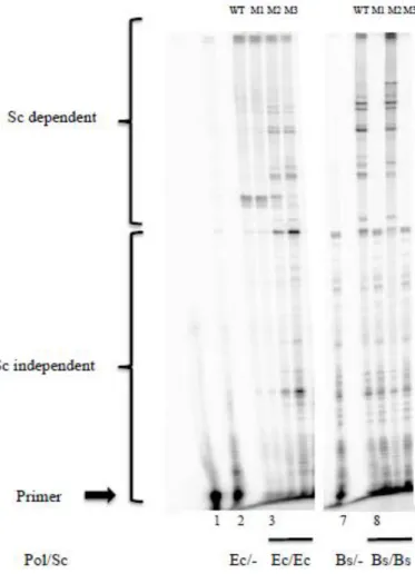
Figure 7 : Elongation profiles obtained with reconstituted E. coli and B. subtilis holoenzymes. The E.
coli and B. subtilis polymerases (EcP or BsP, 100 fmoles) are supplemented with various amount of
the cognate wtSC (from 0 to 400 fmoles). The labeled lanes (*) correspond to 100 fmoles wtSC. The primer position is indicated by the arrow.
Then we analyzed how EcP and BsP behave when associated with the different
cognate mutant SC (Figure 8). Holoenzyme formed between EcP and EcM1SC (Figure 8, lane
4) displays the same primer elongation profile as that observed using EcwtSC (Figure 8, lane
3). This indicates that the natural EcP peptide interaction is not altered by the S346P mutation,
nor in terms of elongation efficiency, as indicated by the equal bands intensities in lanes 3 and 4, nor in terms of specificity as indicated by their similar elongation profiles. This result is in
agreement with the P7 interaction data obtained in ITC experiments (Figure SI. 8 A and S.I
11 A). In contrast, holoenzymes formed by association of EcP with EcM2SC or EcM3SC (Figure
26
which can be accounted for by a decrease in processivity and/or modifications of polymerase-SC interaction dynamics. This is in agreement with the ITC data and indicates that, regardless
of the peptide, the M362L mutation is deleterious to the interaction. The combination of both
mutations (EcM3SC, lane 6) shows a synergistic effect, as revealed by the decrease in full length band intensity in lane 6 as compared to lane 5. Surprisingly enough, these profiles, and
particularly that obtained with EcM3SC, are very similar to that obtained with the natural BsP
holoenzyme (Figure 8, lane 8).
Figure 8: Elongation assays with the different EcSc and BsSc. EcP or BsP reconstituted polymerases (100 fmoles) were each tested with the various cognate SC (lanes 3 to 6 and lanes 8 to 11 for Ec and Bs polymerases, respectively) (100 fmoles) (Table 1). The primer position is indicated by the arrow (lane 1). Lanes 2 and 7 display the activity of Ec and Bs polymerases in absence of the cognate SC.
We observed that only BsM2SC associates productively with BsP and yields a similar pattern of elongation as the natural BsP holoenzyme (Figure 8, lane 10), while nor BsM1SC
27
the P357S mutation appears to be highly deleterious to the polymerase-SC interaction, as
opposed to what is observed with the EcSC, while the L373M mutation is harmless and even
favorable to the interaction, as indicated by the increased band intensities of the full length products (lane 10). The lack of elongation with BsM3SC is in contradiction with ITC data
showing that the combination of the two mutations in BsM3SC improves the P7 interaction as
compared to BsM1SC/P7 or BsM2SC/P7 interactions (Figure 4). In the elongation assay, the
effect of both mutations is not cumulative but rather reflects the effect of the P357S mutation
alone. These differences between the two approaches reveals that, in the Bs context, the S357
residue blocks the interaction of the PolC polymerase natural peptide with BsSC, but not that
of P7. Alternatively, the BsP357 position could have some unknown specific contribution to the
whole polymerase function. This effect is not observed in the Ec context and could thus reveal this position as a specific marker for Gram+ SC/polymerase interaction. Finally, as observed for both natural SC, none of the mutants SC form a productive heterologous complex with their non cognate polymerase (data not shown).
To make sure that the effects observed with BsP reflect the polymerase activity and not the SC loading process, we performed the same assay using a linear template (SI.17). SC dependent elongation products are detected with the BswtSC and BsM2SC, as observed when using circular templates, but none with BsM1SC, ruling out any defect in the SC loading process. Noteworthy, in this assay with linear templates, we observed BsM3SC dependent products, as opposed to what we obtained using circular DNA (Figure 8, lane 11). This suggests that the observed increased elongation activity brought by the L373M mutation
compensate the deleterious effect of the P357S mutation for what concerns the polymerase
interaction, but not the SC loading process. Elucidation of this point will require the study of the respective natural peptides with these SC variants.
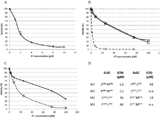
Then, we challenged the different reconstituted holoenzymes for their primer elongation activity with increasing concentrations of P7. The elongation profiles are displayed
in Figure 9, and the data are analyzed in Figure 10. In accordance with our previous observations in Figure 8 (lanes 3 and 4) showing that EcwtSC and EcM1SC are similarly efficient in driving EcP SC-dependent elongation, equal concentrations of P7, pre-incubated
with EcwtSC or EcM1SC, equally inhibit the SC dependent primer elongation activity of EcP, yielding a IC50 of 1.5 µM (Figure 10 A, D). These values are close to our previous results
28
experiments confirm that the S346P mutation has no, or a limited influence on the peptides
binding, either the natural peptide (QADMF) or P7. We note a 4-fold difference between these
biochemical results and those measured by ITC at 34°C (SI. 6), but both approaches describe the same trend despite their different sensitivity, as observed before 5.
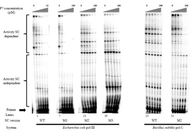
Figure 9 : Inhibition of primer elongation by P7. EcP and BsP are complemented with their cognate
wt or mutants SC pre-incubated with increasing concentrations of P7 peptide (0, 1, 2, 4, 8 and 10 µM for EcwtSC and 0, 2, 8, 25 and 75 µM for all the other SC). Lanes 1 to 6: EcP-EcwtSC. Lanes 7 to 12: EcP-EcM1SC. Lanes 13 to 18: EcP-EcM2SC. Lanes 19 to 24: EcP-EcM3SC. Lanes 25 to 30: BsP-BswtSC. Lanes 31 to 36: BsP-BsM2SC.
The same approach using EcM2SC or EcM3SC yield a much higher IC50 value of 46 µM,
indicating a much weaker interaction of these SC with P7 (Figure 10 B, D), in agreement with
our ITC data (SI.6). All the results from different analytical approaches indicate that for EcSC, the introduction of a S346P mutation is innocuous, whereas the M362L mutation is strongly
29
Finally, we challenged the natural BsP holoenzyme and its BsM2SC supplemented
version by P7 (Figure 10 C, D). While the interaction of P7 with BswtSC is weak (IC50 = 68
µM), in agreement with our ITC data (SI.2), a 5 fold (IC50 = 14 µM) improvement is
measured by introduction of the L373M mutation in BsSC, in good agreement with ITC and
MD data.
Figure 10: Quantification of EcP and BsP holoenzymes inhibition by P7. A: EcwtSC (diamonds); EcM1SC (crosses). B: ECM1SC (diamonds, dotted line); EcM2SC (squares, black line); EcM3SC (triangles, dotted line). C: BswtSC (diamonds); BsM2SC (squares, dotted line). D: Determination of IC50 from the curves in A, B and C .
CONCLUSION.
The interaction of all DNA polymerases with the replicative processivity factor (or SC) is central for their activities and is mediated by a conserved peptide sequence which binds into a hydrophobic pocket located at the SC surface. In this piece of work, we analyzed the interaction of a reference peptide, P7, with natural and mutant SC from E. coli, B. subtilis and
M. tuberculosis. In particular, we focused on the contribution of two residues of the EcSC binding pocket, namely S346 and M362, and their corresponding residues in BsSC, P357 and L373.
30
ITC experiments confirm the differential interaction of P7 with the different natural
SC. Single mutant analyses identify a strategic position in the EcSC binding pocket: a single
M362L mutation is sufficient 1) to turn the thermodynamic profile of P7 interaction into that
observed with BswtSC , 2) to inhibit the SC dependant activity of the Pol III holoenzyme and 3) to reduce the competitive inhibitory effect of P7 on this enzyme activity. Converesely, the
L373M substitution in the BsSC pocket strongly modifies the thermodynamic profile which
becomes almost similar to that observed in the natural Ec context. All these results are in line with molecular dynamics data indicating that, contrary to M residues that, in both Ec and Bs contexts, shift upon peptide interaction, L residues remain in place, immediately below the peptide, providing a structural rationale for the peptide binding inhibition in EcM2SC and in
Bswt
SC. ΔCp analyses reveal that all binding pockets endow a large structural change upon peptide binding, suggesting that P7 interacts through an induced fit process33. In the case of
Bs
SC, this process is observed only with G2 and M3 mutants, because they lack the static L373
residue which blocks peptide interaction. This indicates that the weak ΔCp values calculated
for BswtSC, EcM2SC and others, reveal a poor peptide binding and not necessarily a change in
binding mode.
Thermodynamic analyses suggest that the nature of the Ec346 residue (S or P) is not as
important for the binding as the M362 residue, as deduced from the closely similar
thermodynamic profiles obtained with EcwtSC, EcG1SC and EcM1SC. However, kinetics analyses
reveal that the S346P mutation strongly reduces the pocket dynamics. This suggests that the
residue composition of each binding pocket is highly specific and has evolved to ensure an optimized binding process through a complex and specific network of interaction, as observed previously 18. Another example is illustrated in the Bs context where biochemical analyses highlighted the P357 residueas a Bs specific strategic position for productive elongation. These
data underline the interest of a multidisciplinary approach in defining the binding characteristics of each binding pocket and will help in the future design of SC targeting compounds, with either large spectrum or strain specific activities.
AUTHORS INFORMATIONS Corresponding Author
31
E-mail: d.burnouf@ibmc-cnrs.unistra.fr. phone: 33 3 88 417 002; fax: 33 3 88 602 218
Author Contributions
+ These authors contributed equally.
ACKNOWLEDGMENTS
This work was supported by a grant IMMI n° 2014014 funded by AstraZeneca and INSERM (I3M). X-ray data were collected at the Paul Scherrer Institut, Villigen, Switzerland through several SLS proposals for provision of synchrotron radiation beamtime, at beamline X06DA PXIII. We are grateful to Dr C. McHenry (U. of Colorado, Boulder, Co, USA) for the gift of replicative plasmids and proteins.
ABBREVIATIONS
ITC, Isothermal Titration Calorimetry; N-ter: N-terminal ; C-ter : C-terminal ; Ac: acetyl group; Cha: β-cyclohexyl-L-alanyl; (3,4-di-Cl)Phe : 3,4-dichloro-L-phenylalanyl.
REFERENCES.
1. WHO WHO report 2011: Global Tuberculosis Control; ISBN 978 92 4 156438 0; Geneva, 2011.
2. Lopez de Saro, F. J.; Georgescu, R. E.; Goodman, M. F.; O'Donnell, M., Competitive
processivity-clamp usage by DNA polymerases during DNA replication and repair. Embo J 2003, 22, (23), 6408-6418.
3. Burnouf, D. Y.; Olieric, V.; Wagner, J.; Fujii, S.; Reinbolt, J.; Fuchs, R. P.; Dumas, P., Structural and biochemical analysis of sliding clamp/ligand interactions suggest a competition between replicative and translesion DNA polymerases. J Mol Biol 2004, 335, (5), 1187-1197.
4. Georgescu, R. E.; Yurieva, O.; Kim, S. S.; Kuriyan, J.; Kong, X. P.; O'Donnell, M., Structure of a small-molecule inhibitor of a DNA polymerase sliding clamp. Proc Natl Acad Sci U S A 2008,
105, (32), 11116-11121.
5. Wolff, P.; Olieric, V.; Briand, J. P.; Chaloin, O.; Dejaegere, A.; Dumas, P.; Ennifar, E.; Guichard, G.; Wagner, J.; Burnouf, D. Y., Structure-based design of short peptide ligands binding onto the E. coli processivity ring. J Med Chem 2011, 54, (13), 4627-4637.
6. Wijffels, G.; Johnson, W. M.; Oakley, A. J.; Turner, K.; Epa, V. C.; Briscoe, S. J.; Polley, M.; Liepa, A. J.; Hofmann, A.; Buchardt, J.; Christensen, C.; Prosselkov, P.; Dalrymple, B. P.; Alewood, P. F.; Jennings, P. A.; Dixon, N. E.; Winkler, D. A., Binding inhibitors of the bacterial sliding clamp by design. J Med Chem 2011, 54, (13), 4831-4838.
7. Kling, A.; Lukat, P.; Almeida, D. V.; Bauer, A.; Fontaine, E.; Sordello, S.; Zaburannyi, N.; Herrmann, J.; Wenzel, S. C.; Konig, C.; Ammerman, N. C.; Barrio, M. B.; Borchers, K.; Bordon-Pallier, F.; Bronstrup, M.; Courtemanche, G.; Gerlitz, M.; Geslin, M.; Hammann, P.; Heinz, D. W.; Hoffmann, H.; Klieber, S.; Kohlmann, M.; Kurz, M.; Lair, C.; Matter, H.; Nuermberger, E.; Tyagi, S.; Fraisse, L.; Grosset, J. H.; Lagrange, S.; Muller, R., Antibiotics. Targeting DnaN for tuberculosis therapy using novel griselimycins. Science 2015, 348, (6239), 1106-1112.
8. Yin, Z.; Kelso, M. J.; Beck, J. L.; Oakley, A. J., Structural and thermodynamic dissection of linear motif recognition by the E. coli sliding clamp. J Med Chem 2013, 56, (21), 8665-8673.