HAL Id: tel-02926159
https://tel.archives-ouvertes.fr/tel-02926159
Submitted on 31 Aug 2020HAL is a multi-disciplinary open access archive for the deposit and dissemination of sci-entific research documents, whether they are pub-lished or not. The documents may come from teaching and research institutions in France or abroad, or from public or private research centers.
L’archive ouverte pluridisciplinaire HAL, est destinée au dépôt et à la diffusion de documents scientifiques de niveau recherche, publiés ou non, émanant des établissements d’enseignement et de recherche français ou étrangers, des laboratoires publics ou privés.
A mouse model of pseudohypoaldosteronism type II
reveals a novel mechanism of tenal tubular acidosis
Irma Karen Lopez-Cayuqueo
To cite this version:
Irma Karen Lopez-Cayuqueo. A mouse model of pseudohypoaldosteronism type II reveals a novel mechanism of tenal tubular acidosis. Tissues and Organs [q-bio.TO]. Sorbonne Université, 2018. English. �NNT : 2018SORUS465�. �tel-02926159�
Sorbonne Université
Ecole doctorale: Physiologie, Physiopathologie et Thérapeutique.
ED394
THÈSE DE DOCTORAT
Présentée par:M
elleIrma Karen LÓPEZ-CAYUQUEO
A Mouse Model of Pseudohypoaldosteronism type
II Reveals a Novel Mechanism of Renal Tubular
Acidosis
Dirigée par: Dr. Régine Chambrey
Soutenue le 27-04-2018
Devant un jury composé de: Prof. Pierre Ronco Président du Jury Prof. Renaud Beauwens Rapporteur
Dr. Henrik Dimke Rapporteur Dr. Maria Christina Zennaro Examinateur
Dr. Régine Chambrey Examinateur Prof. Jacques Teulon Directeur de thèse
2
LANGUE DE REDACTION
Avec la permission de l’école doctorale de l’UPMC (ED394), la thèse a été rédigée en anglais, ma langue de recherche et de publication usuelle.
3
PREFACE
The experiments relative to this work were mostly done during the first 2 years of my PhD under the supervision of Dr. Régine Chambrey at the Paris Cardiovascular Research Center (PARCC).
Part of the experiments shown in this work were performed in collaboration with, or exclusively by professor Pascal Houillier (Figure 17) and Dr. Régine Chambrey (Figure 10). Contribution of all the authors of the paper “A Mouse Model of Pseudohypoaldosteronism type II Reveals a Novel Mechanism of Renal Tubular Acidosis”, submitted to Kidney International, is acknowledged.
During the last year of my PhD, to complete my doctoral formation I performed a long-term internship in the laboratory of Professor Thomas Jentsch at the Leibniz-Forschungsinstitut für Molekulare Pharmakologie im Forschungsverbund (FMP) / Max Delbrück Center for Molecular Medicine in the Helmholtz Association (MDC). Berlin, Germany. The research performed during this time will not be presented in this thesis.
4 TABLE OF CONTENTS LIST OF FIGURES ... 6 LIST OF TABLES ... 7 LIST OF ABBREVIATIONS ... 8 ABSTRACT ... 10 1. INTRODUCTION ... 11
1.1 Overview of kidney structure and function ... 11
1.2 Renal physiopathology ... 13
1.2.1 Sodium handling along the nephron and associated pathologies ... 14
1.2.2 Potassium handling and its physiological consequences ... 23
1.3 Acid-base homeostasis ... 24
1.4 Pseudohypoaldosteronism type II ... 25
1.4.1 Physiopathology of PHAII ... 26
1.4.1.1 Regulation of NCC ... 27
1.4.1.2 Impact of WNK pathway in NCC regulation and in the pathogenesis of PHAII ... 27
1.4.1.3 Influence of the electroneutral NaCl reabsorption in the pathogenesis of PHAII ... 33
1.4.1.3.1 Additional information about Pendrin and NDCBE ... 33
2. AIM OF THE WORK ... 35
3. MATERIALS AND METHODS ... 36
3.1 Animal generation and physiological studies ... 36
3.2 Blood gas and urinary analysis ... 36
3.3 Potassium diet and ammonium load ... 37
3.4 Intracellular pH measurements on isolated microperfused tubules ... 37
3.5 Measure of the fractional volume ... 38
3.6 Measurements of blood pressure by radiotelemetry ... 39
3.7 Immunoblotting ... 39
3.8 Real-time quantitative RT-PCR ... 40
3.9 Intercalated cell type quantification ... 41
3.10 Antibody sources ... 41
3.11 In vitro microperfusion of mouse CCDs and transepithelial ion flux measurement ... 42
3.12 Statistics and data analysis ... 43
3.13 Study approval ... 43
4. RESULTS ... 44
4.1 TgWnk4PHAII mice exhibit hyperkalemic metabolic acidosis ... 44
4.2 Electroneutral thiazide-sensitive NaCl transport is activated in the collecting duct of TgWnk4PHAII mice ... 46
4.3 ENaC-dependent K+ secretion is blocked in TgWnk4PHAII mice despite unchanged renal ENaC activity ... 48
4.4 Normalization of plasma K+ concentration does not correct metabolic acidosis in TgWnk4PHAII mice ... 51
4.5 Renal acidosis in TgWnk4PHAII mice is not caused by impaired proton secretion by the distal nephron ... 52
4.6 Renal acidosis in TgWnk4PHAII mice is a consequence of an increased of the pendrin activity ... 56
5 4.7 Hyperchloremic metabolic acidosis and hyperkalemia in TgWnk4PHAII mice are
corrected by pendrin genetic ablation. ... 60
5. DISCUSSION ... 65
5.1 The NCC upregulation is not sufficient to explain entire the phenotypic features of PHAII ... 65
5.2 Pendrin overactivity is contributing to the pathogenesis of PHAII ... 67
5.3 Impact of pendrin deletion on plasma K+ concentration of TgWnk4PHAII mice ... 68
5.4 Pendrin hyperactivity as a novel mechanism for renal acidosis ... 70
5.5 The missing link between pendrin regulation and WNK4 ... 71
5.6 Conclusion ... 72
6. PUBLICATIONS... 74
7. ACKNOWLEDGMENTS ... 77
6
LIST OF FIGURES
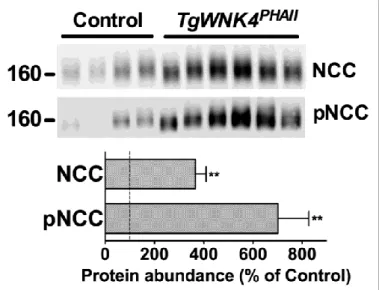
Figure 1. Structure of the nephron………..……….12 Figure 2. Sodium handling along the nephron..……….14 Figure 3. The proximal tubule and its main transport processes………….………….……..16 Figure 4. Transports involved in the thick ascending limb of the loop of Henle ...…..…….18 Figure 5. NaCl reabsorption in the distal convoluted tubule..………..19 Figure 6. Main transport processes in the aldosterone-sensitive distal nephron..………...22 Figure 7. Regulation of NCC by WNKs pathway in normal condition and in PHAII..……..32 Figure 8. Protein abundance of NCC and pNCC is increased in TgWnk4PHAII mice ....…..44 Figure 9. TgWnk4PHAII mice exhibit phenotypic abnormalities characteristic of PHAII …...45
Figure 10. Electroneutral NaCl transport system is activated in the CCD of TgWnk4PHAII
mice ……….47 Figure 11. α-ENaC and the proteolytic cleavage form of γ-ENaC are increased in
TgWnk4PHAII mice ………..……….49
Figure 12. Identical natriuretic response to amiloride observed in TgWnk4PHAII and
control mice ...……….50 Figure 13. Slight increase in α-BKCa and not changes in ROMK protein abundance in
TgWnk4PHAII mice ..………..………..51
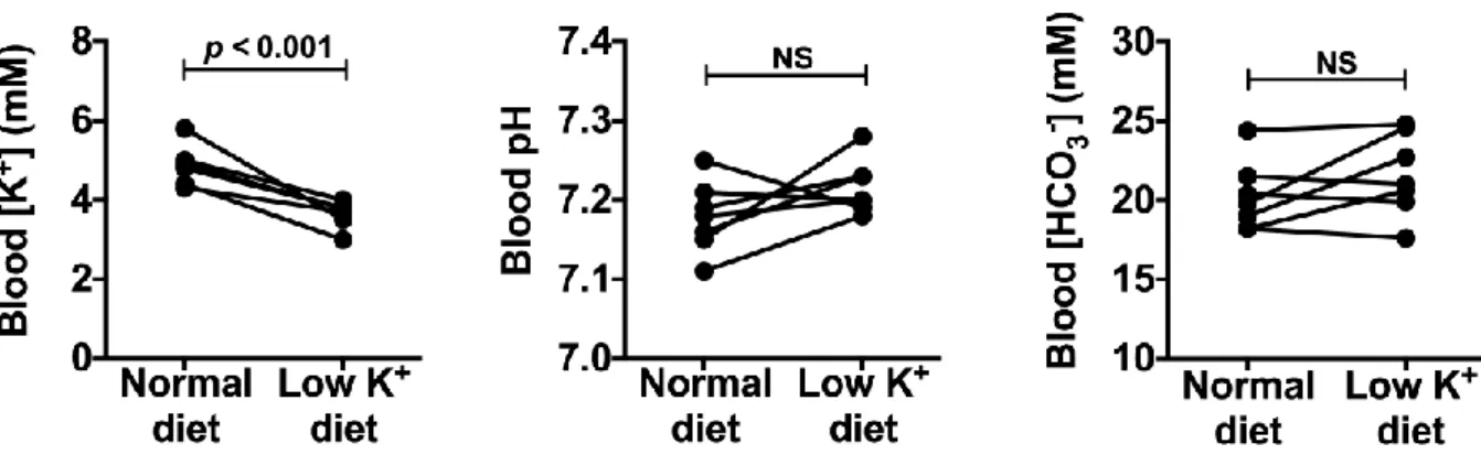
Figure 14. Low potassium diet decreases blood plasma K+ concentration in TgWnk4PHAII
mice without modifying neither blood pH nor blood [HCO3] …...…….……….…….52
Figure 15. Metabolic acidosis in TgWnk4PHAII mice is not caused by impaired acid
excretion ...………...54 Figure 16. Response to an acid load in WNK1PHAII mice indicates a normal acid
excretion ...55 Figure 17. Increased apical Cl-/HCO
3- exchange activity in β-intercalated cells of
TgWnk4PHAII mice ….……….56
Figure 18. The number of β-intercalated cells in TgWnk4PHAII mice is increased ...59
Figure 19. Protein abundances of different Na+ transporters in TgWnk4PHAII and
TgWnk4PHAII/Pds-/-mice ………...62
Figure 20. ENaC-dependent K+ secretion is higher after pendrin disruption in
TgWnk4PHAII mice ………...63
Figure 21. Double transgenicmice (TgWnk4PHAII/Pds-/-) present not changes in ROMK
7
LIST OF TABLES
Table 1. PHAII mutation and their effects on the affected genes ….………...31 Table 2. Blood parameters of TgWNK4PHAII and TgWNK4PHAII; Pds-/- mice .……..60
Table 3. Blood parameters of TgWNK4PHAII before and 6h after thiazide
8
LIST OF ABBREVIATIONS
AE1 Anion Exchanger 1 AE4 Anion Exchanger 4 AKI Acute Kidney Injury AQP2 Aquaporin 2
ASDN Aldosterone-Sensitive Distal Nephron
BKca Large-conductance Ca2+ -activated K+ Channel
CCD Cortical Collecting Duct CD Collecting Duct
CKD Chronic Kidney Disease ClCk Chloride Channel k CNT Connecting Tubule DCT Distal Convoluted Tubule ENaC Epithelial Na+ Channel
ESRD End-Stage Renal Disease
FHHt Familial Hyperkalemic Hypertension HCTZ Hydrochlorothiazide
GFR Glomerular Filtration Rate ICs Intercalated Cells
IMCD Inner Medulla Collecting Duct KCC3 K+-Cl- Contransporter 3
KCC4 K+-Cl- Contransporter 4
Kir4.1 Inward rectifier-type potassium channel 4.1
Kir5.1 Inward rectifier-type potassium channel 5.1
KS-WNK1 Kidney Specific WNK1
KO Knockout
NBCe1 Na+-HCO
3- Cotransporter 1
NCC Na+-Cl- Cotransporter
NDCBE Na+-driven Cl-/HCO
3-exchanger
NHE3 Na+-H+ exchanger 3
NKCC2 Na+-K+-Cl- Cotransporter 2
OMCD Outer Medulla Collecting Duct
9
PCs Principal Cells PT Proximal Tubule
PHAII Pseudohypoaldosteronism type II Pds Pendrin
RTA Renal Tubular Acidosis
ROMK Renal Outer Medullary Potassium Channel SGK1 Serum and Glucocorticoid-induced Kinase 1 SPAK STEP20/SPS1-related Proline/Alanine-rich Kinase TAL Thick Ascending Limb
TASK2 TWIK Related Acid Sensitive K+ Channel 2
vH+-ATPase Vacuolar-type H+-ATPase
WNK With No lysine (K) Kinase WT Wild type
SI units and SI prefixes are used according to the International System of Units. Nomenclature and symbolism for amino acids and peptides follows the guidelines of the IUPAC-IUB Joint Commission on Biochemical Nomenclature (JCBN): “Nomenclature and symbolism for amino acids and peptides. Recommendations 1983.”
10
ABSTRACT
Pseudohypoaldosteronism type II (PHAII) is a rare monogenic disease mainly characterized by the association of hyperkalemia, hyperchloremic metabolic acidosis and hypertension. It is produced by mutations in the WNK1, WNK4,
KLHL3 or CUL3 genes. The clinical manifestations of PHAII are due, among
others, to an increased activity of the thiazide-sensitive Na+-Cl- cotransporter NCC,
expressed in the distal convoluted tubule. Several groups attempted to determine how PHAII-causing mutation led to an increased activity of NCC. However, so far the data do not fully explain the physiopathology of PHAII, giving evidence that other renal transport systems are altered in this disease.
It has been reported that WNK4 is expressed not only in the DCT cells but also in -intercalated cells of the cortical collecting duct. These cells exchange intracellular HCO3- for external Cl- through pendrin, and therefore, account for
renal base excretion. They can also mediate electroneutral thiazide-sensitive NaCl absorption when pendrin-dependent apical Cl- influx is coupled to apical Na+ influx
by the Na+-driven Cl-/HCO3- exchanger NDCBE.
Taking advantage of a mouse model (TgWNK4PHAII) carrying a WNK4 missense
mutation (Q562E) identified in PHAII patients, the purpose of this study was to determine whether the electroneutral Na+-Cl- absorption through pendrin/NDCBE
is involved in the pathogenesis of this disease.
Our results show that renal pendrin activity is markedly increased in TgWNK4PHAII
mice, leading to an increase in thiazide-sensitive NaCl absorption by the collecting duct and contributing to metabolic acidosis. Thus, pendrin genetic ablation in
TgWNK4PHAII mice corrects the metabolic acidosis and also the hyperkalemia
characteristic of PHAII.
Taken together, our data demonstrate the important contribution of pendrin in renal regulation of NaCl, K+ and acid-base homeostasis and in the pathogenesis of
PHAII. Additionally, we identify a renal distal bicarbonate secretion as a novel mechanism of renal tubular acidosis.
11
1. INTRODUCTION
1.1 Overview of kidney structure and function
The kidneys are involved in a variety of physiological processes including regulation of blood pressure, acid-base homeostasis and volume regulation. The maintenance of a relatively constant extracellular environment is necessary for the cells (and organism) to function normally. This is achieved by excretion of some waste products of metabolism such as urea, creatinine, and uric acid, in addition to water and electrolytes, which are derived primarily from dietary intake. The balance is maintained by keeping the rate of excretion equal to the sum of net intake plus endogenous production.
Anatomically two distinct regions can be identified on the cut surface of a bisected kidney: a pale outer region, the cortex, and a darker inner region, the medulla. In humans the medulla is divided into 8 to 18 renal pyramids. The base of each pyramid is positioned at the cortico-medullary boundary and the apex extends toward the renal pelvis to form a papilla (Brenner and Rector's 2012).
The nephron is the functional unit of the kidney and is composed of the glomerulus located in the Bowman’s capsule and the renal tubules: proximal tubule (PT); the thin limbs and the thick ascending limb of loop of Henle (TAL); distal convoluted tubule (DCT) and connecting tubule (CNT). The last segment of the nephron is the collecting duct (CD); which consists of the cortical collecting duct (CCD), outer medullary collecting duct (OMCD) and the inner medullary collecting duct (IMCD) (Figure 1).
12
Figure 1. Structure of the nephron. Schematic representation of a longitudinal section of the kidney (left) and representation of a nephron that expands throughout the cortex and medulla (right). The epithelial elements of the nephron include the Bowman’s capsule, the proximal (convoluted and straight) tubule, the thin descending and ascending limbs of the loop of Henle, the thick ascending limb of the loop of Henle, the distal convoluted tubule, the connecting tubule, the initial collecting tubule and the (cortical and medullary) collecting duct. (From Boron and Boulpaep, Medical Physiology, updated 2th edition: 2012).
13
The cellular constitution and function are different for each renal segment. In general terms, the PT is responsible of the massive reabsorption of water and solutes, around 75%. The thin limbs and TAL are in charge to continue with the reabsorption process but also the setting–up and maintenance of a cortico-papillary gradient of solutes, and hence of osmoles, required for the concentration of final urine. The distal nephron, constituted by DCT, CNT and CD, are responsible of the final reabsorption and secretory processes allowing the organism adjust the final urine according to the body demands.
In summary, in order to maintain a stable milieu intérieur by the selective retention or elimination of water, electrolytes, and other solutes, the kidney is caring out three processes: (1) the filtration of circulating blood from the glomerulus to form an ultra-filtrate of plasma in Bowman’s space; (2) the selective reabsorption across the cells lining the renal tubule (from tubular fluid to blood); and (3) the selective secretion, from peritubular capillary blood to tubular fluid (Boron and Boulpaep 2012).
1.2 Renal physiopathology
Any misbalance in the processes previously described may produce a kidney related pathology. The etiology in many of these diseases has remained puzzling and it is necessary to analyze every case in particular. The three most important clinical problems in nephrology, characterized either by number of patients affected or by rates of associated morbidity and mortality are: end-stage renal disease (ESRD), chronic kidney disease (CKD) and acute kidney injury (AKI) (Brenner and Rector's 2012).
On the other hand, hypertension (high blood pressure) is a major public health problem affecting more than a billion people worldwide with complications, including stroke, heart failure and kidney failure (Trepiccione, Zacchia et al. 2012). According to Arthur Guyton’s hypothesis, the kidney plays a key role in blood pressure control (Guyton, Coleman et al. 1972).
An important aspect in many diseases with renal etiology is the genetic factor. Mutations in some of the major transporters responsible of the reabsorption and excretion of ions along the nephron may produce a diversity of pathologies. The
14
pathophysiological consequences of some of these genetic defects will be discussed in the following part of this thesis.
1.2.1 Sodium handling along the nephron and associated pathologies
Sodium (Na+) being with chloride the principal osmole in the extracellular fluid,
renal excretion of salt (NaCl) is the major determinant of the extracellular fluid volume. For this reason, genetic loss or gain of function in renal NaCl transport can be associated with hypotension or hypertension, respectively.
The reabsorption of Na+ takes place along the entire nephron, and very little is
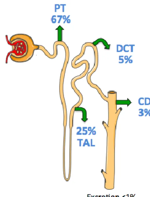
excreted in urine (<1% of the filtered load). The proportion of reabsorption differs between nephron segments; thus, the proximal tubule is responsible for the greatest reabsorption (around 67%). The loop of Henle reabsorbs a smaller but significant fraction of filtered Na+ (~25%). And finally, the “fine tuning” of renal
NaCl absorption occurs in the distal tubule and collecting duct (~8%) (Figure 2).
Figure 2. Sodium handling along the nephron. Arrows indicate reabsorption of Na+. Numbers indicate the percentage of the filtered load of Na+ reabsorbed or
15
The Na+ and Cl− can be reabsorbed through both transcellular and paracellular
pathways. In the transcellular pathway, Na+ and Cl− sequentially cross the apical
and basolateral membranes before entering the blood. In the paracellular pathway, these ions move entirely along an extracellular route through the tight junctions between cells. In the transcellular pathway, transport rates depend on the electrochemical gradients, ion channels and transporters present at the apical and basolateral membranes. In the paracellular pathway, transepithelial electrochemical driving forces and permeability properties of the tight junctions govern ion movement (Boron and Boulpaep 2012) .
In the proximal tubule, the reabsorption of Na+ can be described as a secondary
active transport. Using the Na+ gradient generated by Na+-K+ ATPase pump, the
sodium enters the PT cell via a series of transporters that also transport other solutes such as glucose, amino acids, phosphate and citrate. In addition, Na+ entry
is coupled to the extrusion of H+ through the electroneutral Na+-H+ exchanger 3
(NHE3), providing at the same time a favorable gradient for the apical Cl− uptake by the Cl--Oxalate/formate exchanger (CFEX) (Edwards 2012). On the basolateral
membrane K+ and Cl- are recycled to the blood by the K+-Cl- Cotransporters KCC4
and KCC3, crucial for proximal tubule cell volume regulation (Jentsch 2005). The potassium also is recycled on the basolateral side by K+ channels, such as TASK2,
which has been propose as crucial to maintain electrochemical gradients favorable to bicarbonate reabsorption (Lopez-Cayuqueo, Pena-Munzenmayer et al. 2015). In the reabsorption of bicarbonate apical NHE3 and basolateral Na+-3HCO
3
-cotransporter (NBC1a) are essential. Finally, it should be noted that, in the proximal tubule, a part of Na+, Cl- and water can be reabsorbed through the
paracellular pathway.
Figure 3 represents the main transport processes in the proximal tubule, including the bicarbonate reabsorption, essential for the acid-base homeostasis, which will be further described in the section 1.3.
16
Figure 3. The proximal tubule and its main transport processes. The proximal tubule is the major reabsorptive segment of the nephron and accounts for reabsorption of nearly 80% of filtered water, sodium, and chloride. In addition, the proximal tubule is the segment where the majority of critical organic solutes such as glucose and amino acids are resorbed. This segment also plays an important role in acid-base balance as it is involved in bicarbonate reabsorption and secretion of organic acids. Transport is powered by Na+-K+ ATPase. NHE3: Na+
-H+ exchanger 3; NBCe1: Na+-HCO
3- Cotransporter 1; CFEX: Cl--Oxalate/formate
exchanger; AQP1: Aquaporin 1; KCC4/3: K+-Cl- Contransporter 4 and 3; TASK2:
17
In the loop of Henle, around 25% of filtered NaCl is reabsorbed in the thick ascending limb of the loop of Henle where the apical membrane is impermeable to water. This process is essential for the countercurrent mechanism and urine concentration. In the apical membrane of the TAL, sodium and chloride enter the cell via the electroneutral Na+-K+-2Cl- cotransporter (NKCC2), using the favorable
inward gradient for sodium generated by the constitutive activity of the basolateral Na+-K+-ATPase pump. A large fraction of the K+ brought into the cell through
NKCC2 recycles to the lumen via apical K+ channels, such as ROMK. These
channels are essential for replenishing luminal K+ and maintaining adequate Na+
-K+-Cl- cotransport. The lumen positivity generated by potassium recycling is able
to drive the passive reabsorption of cations (sodium, calcium, and magnesium) between the cells across the tight junction. On the basolateral side, the chloride channel ClC-Kb and its ß-subunit Barttin are essential for Cl- reabsorption
(Estevez, Boettgerr et al. 2001), (see below figure 4).
Inactivating mutations in the genes that encode either NKCC2 (Simon, Karet et al. 1996), ROMK (Simon, Karet et al. 1996), ClC-Kb (Simon, Bindra et al. 1997) or Barttin (Birkenhager, Otto et al. 2001) are responsible for the pathogenesis of Bartter’s syndrome. Bartter’s syndrome is a rare inherited disease characterized by the association of severe hypokalemia, metabolic alkalosis, with low or normal blood pressure, high levels of renin and aldosterone in blood, polyuria and dehydration, with hypomagnesemia (Bartter, Pronove et al. 1962).
18
Figure 4. Transports involved in the thick ascending limb of the loop of Henle. The transport in TAL is characterized by a Na+-2Cl--K+ Cotransporter
(NKCC2) on the luminal surface that allows the reabsorption of large amounts of these ions. Reabsorption is powered by a basolateral Na+-K+ ATPase. Importantly,
the ascending limb is highly impermeable to water and the reabsorption of large amounts of sodium in the absence of water results in significant dilution of the tubular fluid. A small amount of potassium back-leaks into the lumen via the K+
channel ROMK and yields a positive luminal charge that powers the paracellular reabsorption of positive ions, including magnesium and calcium. The Cl- leaves the
cell by the basolateral Cl- channel ClC-Kb. ROMK: Renal Outer Medullary
Potassium Channel; NKCC2: Na+-2Cl--K+ Cotransporter; ClC-Kb: Chloride
Channel Kb; TJ: Tight Junctions.
In the distal tubule, NaCl reabsorption is almost exclusively transcellular. The NaCl uptake in the apical membrane occurs through the electroneutral Na+-Cl
-cotransporter (NCC), highly sensitive to thiazide diuretics. The basolateral step of Na+ reabsorption, as in other cells, is mediated by the Na+-K+ pump and the
chloride channel ClC-Kb is also essential in this part of the nephron for the Cl- exit
through the basolateral membrane(Hennings, Andrini et al. 2017). The transports in a DCT cell are represented in Figure 5.
19
The regulation of NCC is crucial for the understanding of related pathologies. Gitelman’s syndrome is an autosomal recessive condition characterized by hypokalemic metabolic alkalosis, hypocalciuria and hypomagnesemia, can be associated with normal or reduced blood pressure (Gitelman, Graham et al. 1966). This syndrome is due to an inactivating mutation of NCC gene (Simon, Nelson-Williams et al. 1996). On the other hand, Gordon syndrome (also called PHAII) is a condition showing opposite symptoms to Gitelman’s syndrome, in which an over activation of NCC is to blame (O'Shaughnessy 2015). Better understanding of the physiopathology of PHAII is one of the aims in the present work and will be discussed in detail in the next pages.
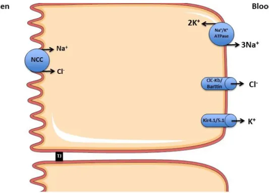
Figure 5. NaCl reabsorption in the distal convoluted tubule. In the distal tubule the filtered sodium load occurs via a luminal Na+-Cl- Cotransporter (NCC) powered
by the basolateral Na+-K+ ATPase. NCC is a target of thiazide diuretics. The
chloride channel ClC-Kb mediates the basolateral recycling of Cl-. The K+ channels
Kir4.1 and Kir5.1 mediate the potassium recycling. NCC: Na+-Cl- Cotransporter;
ClC-Kb: Chloride Channel Kb; Kir4.1: Inward rectifier-type potassium channel 4.1;
20
Finally, the aldosterone-sensitive distal nephron (ASDN) is an important site for the final regulation of urinary Na+ excretion. The ASDN is constituted by the late
distal convoluted tubule (DCT2), the connecting tubule (CNT), and the collecting duct (CD). The DCT2 is the transition segment between the DCT and the CNT, in which appear, besides DCT cells, cells with the characteristics of the CNT/principal cells (PCs). The CNT and CD are comprised of PCs and several types of intercalated cells (ICs). ICs can be characterized according to the expression of different transporters: (1) α-ICs characterized by apical expression of the V-ATPase and basolateral expression of an anion exchanger AE1, (2) β-ICs with the apical expression of pendrin and V-ATPase in the basolateral side, and (3) non α-, non β-ICs with an apical pendrin and V-ATPase expression (Chambrey and Trepiccione 2015).
The PCs express at the apical membrane the amiloride-sensitive Na+ channel
ENaC, formed by 3 homologous subunits: α, β, and γ. ENaC allows the Na+
reabsorption in tandem with the basolateral Na+-K+-ATPase. Consequently, the
generation of a negative lumen drives K+ secretion through the apical K+ channel
ROMK, allowing PCs to operate as a Na+-K+ exchange. Mutations in the genes
encoding ENaC subunits result in rare diseases with either salt loss or retention. Thus, mutations producing gain of function in the β or γ subunit of ENaC are responsible for Liddle’s syndrome, characterized by a severe salt-sensitive hypertension, hypokalemia, and metabolic alkalosis (Warnock 2001). Contrary, loss-of-function mutations in the α, β, or γ subunits of ENaC cause pseudo-hypoaldosteronism 1B, a severe salt-losing syndrome with hyperkalemia and metabolic acidosis (Scheinman, Guay-Woodford et al. 1999).
Research over the past 20 years has established a paradigm in which PCs are the exclusive site of Na+ absorption while ICs are solely dedicated to acid-base
transport. Nevertheless recent studies have revealed the unexpected importance of ICs for NaCl reabsorption. In 2010, Leviel et al. described a novel thiazide-sensitive electroneutral NaCluptake mechanism in renal ICs, where NaCl is taken up from urine by the coordinated action of the Cl--HCO
3- exchanger pendrin and
Na+-driven Cl-/2HCO
3- exchanger NDCBE (Leviel, Hubner et al. 2010). Later on in
2013, Gueutin et al, showed that while a dysfunction in the β1 subunit of the H+
β-21
ICs is responsible for renal loss of NaCl, K+, and water (Gueutin, Vallet et al.
2013). Moreover in 2016, Sinning et al, demonstrated that NDCBE is necessary for maintaining sodium balance and intravascular volume during salt depletion or NCC inactivation in mice (Sinning, Radionov et al. 2017). Interestingly, unlike the other absorptive processes in the kidney, β-ICs are rather energized by the H+
V-ATPase than the Na+-K+ pump (Chambrey, Kurth et al. 2013).
A summary of the main transporters involved in sodium handling in CNT and CCD is represented in figure 6.
22
Figure 6. Main transport processes in the aldosterone-sensitive distal nephron. Principal cells harbor luminal sodium (ENaC) and potassium (ROMK) channels that allow the sodium reabsorption and potassium secretion, respectively. This process is powered by a basolateral Na+-K+ ATPase. Principal
cells also display a regulated transcellular permeability to water in response to vasopressin via aquaporin 2 (AQP2) water channels. The α-intercalated cells are characterized by the capacity to secrete protons via a luminal H+-ATPase and to
reabsorb potassium through the H+-K+ ATPase. Finally, the NaCl reabsorption in
β-intercalated cells is generated by two cycles of pendrin (Pds) coupled with one cycle of NDCBE. This process results in a net uptake of one Na+, one Cl- and two
HCO3- ions, whereas one Cl- ion is recycled across the apical membrane. Then,
one Cl- ion exits the cell through the basolateral chloride channel ClC-Kb, while
Na+ and bicarbonate exit the cell at the basolateral membrane via AE4. ROMK:
Renal Outer Medullary Potassium Channel; ClC-Kb: Chloride Channel Kb; Pds: Pendrin; NDCBE: Na+-driven Cl-/HCO
3- exchanger; AE1: Anion Exchanger 1; AE4:
23
1.2.2 Potassium handling and its physiological consequences
The distribution of potassium (K+) in the body differs strikingly from that of Na+.
Whereas Na+ is largely extracellular, K+ is the most abundant intracellular cation.
Maintenance of extracellular K+ concentration within a narrow range is vital for
numerous cell functions, particularly electrical excitability of cardiac cells and neurons. When plasma K+ concentration deviates from normal, serious adverse
consequences, such as cardiac arrhythmias and death, can result (Palmer 2015). The proximal tubule consistently reabsorbs the bulk of the filtered K+ (~67%),
whereas the distal nephron may reabsorb or secrete K+. Depending, among
others, on dietary K+ intake, aldosterone and insulin levels, adrenergic activity,
osmolality and acid-base status, the K+ excretion can vary widely from 1% to 110%
of the filtered load (Costanzo 2011).
K+ reabsorption in the proximal tubule is largely passive and follows the movement
of Na+ and water. Along the TAL, K+ reabsorption amounts to around 20% of
filtered K+. In part, it is coupled to Na+ absorption and involves the Na+-K+-2Cl
-cotransporter (NKCC2) in the luminal membrane but it is also largely paracellular. In the distal nephron, depending on the body demands, either reabsorption or secretion of K+ can take place. The α-intercalated cells are responsible for K+
absorption, which occurs via an apical K+ uptake mediated by the H+-K+-ATPase,
followed by passive efflux of K+ across the basolateral membrane. This process
occurs only on a low-K+ diet (K+ depletion), when K+ excretion can be as low as
1% of the filtered load. Potassium secretion occurs in the principal cells, where ENaC, the apically-located Na+ channel plays a critical role by providing
intracellular Na+ as substrate for the Na+-K+-ATPase and by depolarizing the apical
membrane, thereby increasing the driving force for K+ efflux into the tubular fluid.
At least two types of apical K+ channels have been implicated in K+ secretion,
ROMK and BK (Welling 2016).
The mechanisms by which aldosterone promotes apparently opposite effects like Na+ reabsorption and K+ secretion, remain poorly understood. Volume depletion
leads to a Na+ retaining state, at least in part, by the action of aldosterone. In this
condition, while reabsorption of salt is increased, K+ secretion remains unchanged.
24
aldosterone is also released, favoring K+ secretion in the distal nephron, without
affecting the salt reabsorption rate. Thus K+ is lost without retaining salt. This is
commonly referred to as the aldosterone paradox (Arroyo, Ronzaud et al. 2011). In brief, hyperaldosteronism increases K+ secretion and causes hypokalemia. And
hypoaldosteronism decreases K+ secretion and causes hyperkalemia.
In addition, acid-base disturbances also have complex effects on renal K+
excretion. A fall in intracellular pH reduces the activity of Na+-K+ ATPase on the
basolateral membrane and therefore diminishes activity of ENaC, ROMK and BK on the apical membrane, contributing to an inhibition of active K+ secretion.
Furthermore, stimulation of electrogenic H+ secretion by the vacuolar ATPase in
intercalated cells during acidemia tend to reduce the lumen-negative, transepithelial potential difference, thus inhibiting K+ secretion. Moreover, acidemia
upregulates apical H+-K+ ATPase in intercalated cells, thereby enhancing K+
reabsorption and reducing net K+ secretion. On the contrary, alkalemia stimulates
K+ secretion in the distal nephron, by increasing activities of ENaC, ROMK, and
BK (Aronson and Giebisch 2011).
From a pharmacological point of view, loop diuretics or thiazide diuretics, increase the driving force for K+ secretion, favoring hypokalemia while K+-sparing diuretics
decrease K+ secretion producing hyperkalemia.
1.3 Acid-base homeostasis
Maintaining systemic acid-base balance is critical because the majority of biological processes are dependent on pH. Plasma pH is controlled by the respiratory and urinary systems, which regulate plasma PCO2 and bicarbonate
(HCO3−) concentrations, respectively. The association between PCO2 and HCO3−
is given by the Henderson-Hasselbalch equation: pH = pK + log ([HCO3-]/s ×
PCO2), where “s” is solubility of CO2 at a given temperature and “pK” the
dissociation constant of the CO2/HCO3− buffer system (Boron and Boulpaep 2012).
The kidneys play a major role in HCO3-handling by reabsorbing filtered HCO3− and
generating new HCO3−, which is absorbed and used to titrate acid yielded by
25
will eventually lead to a disruption in systemic acid-base balance and provoke metabolic acid-base disorders.
The proximal tubule contributes to renal acid-base regulation by absorbing most of filtered bicarbonate (∼85%), generating new bicarbonate via ammoniagenesis, and secreting ammonia into lumen, which ultimately acts as the major base form of buffer (as NH3) used to titrate, trap, and excrete proton into urine (as NH4+). The
thick ascending limb contributes to renal acid-base regulation by absorbing majority of the remain filtered bicarbonate (∼10%) and absorbing luminal ammonia to accumulate sufficiently high ammonia concentration in medullary interstitial fluid (Eladari and Kumai 2015).
The distal parts of the nephron also play a critical role in renal acid excretion, and thus, in acid-base homeostasis. Acid secretion is achieved by α-ICs, where the protons generated from the hydration of CO2 within these cells are extruded
actively across the apical membrane by the vH+-ATPase pump, while bicarbonate
ions, which are also produced by this process, are translocated across the basolateral membrane by the Cl-/HCO
3- exchanger AE1. Dysfunction of either the
pump or the anion exchanger can block proton secretion and produce an accumulation of acid in the organism. This defect is named distal renal tubular acidosis (dRTA) type I (Alper 2002). The β-ICs also contribute to acid-base balance, by secreting bicarbonate through the Cl--HCO
3- exchanger pendrin.
1.4 Pseudohypoaldosteronism type II
Hypertension is one of the most common diseases in the general population. This disorder is a major risk factor for many common causes of morbidity and mortality including stroke, myocardial infarction, congestive heart failure and end-stage renal disease (Lifton, Gharavi et al. 2001). Despite its importance in human pathology, the molecular mechanisms underlying hypertension are still poorly understood in the majority of patients.
However, several rare Mendelian blood pressure syndromes have shed light on the molecular mechanisms involved in deregulated ion transport in the distal kidney. One of them in particular is pseudohypoaldosteronism type II (PHAII). PHAII also known as Familial hyperkalemic hypertension (FHHt) or Gordon
26
syndrome; is an autosomal-dominant disorder characterized by salt-dependent hypertension, hypercalciuria, suppressed plasma renin and variable levels of aldosterone, hyperchloremic metabolic acidosis and hyperkalemia despite normal renal function in terms of an intact glomerular filtration rate (GFR) (Gordon, 1986).
1.4.1 Physiopathology of PHAII
Mutations in two members of the with-no-lysine (K) serine/threonine kinases (WNK) family, WNK1 or WNK4, (Wilson, Disse-Nicodeme et al. 2001) or in their regulators KELCH3 (Louis-Dit-Picard, Barc et al. 2012) and Cul3 (Boyden, Choi et al. 2012), were identified as causing PHAII. All mutations lead to abnormal phosphorylation of the Na+-Cl− Cotransporter (NCC) expressed on DCT,
increasing its expression and activity.
The precise mechanism by which PHAII-causing mutations lead to the different phenotypic abnormalities characteristic of this syndrome is not fully elucidated. The excess reabsorption of salt in the DCT increases vascular volume resulting in thiazide-sensitive hypertension. Patients with PHAII respond to aggressive salt-restriction or relatively small doses of thiazide diuretics, suggesting that indeed NCC gain of function is the major cause of the development of the disease. By contrast, the pathogenesis of hyperkalemia and acidosis in PHAII seems to be more complex and is poorly understood.
It has been proposed that the reduced Na+ delivery to the lumen of downstream
nephron segments (CNT and CD) reduces ENaC activity, and thereby, prevents the development of a lumen negative transepithelial voltage. Simultaneously, it is proposed that PHAII-causing mutations reduce the activity of ROMK in the apical membrane of PCs. Both effects would act synergistically to block potassium secretion and provoke hyperkalemia. The molecular basis responsible of the metabolic acidosis in PHAII is not clear either. Acidosis might result from the absence of ENaC-dependent transepithelial potential difference or could be due to the reduced ammonia production and secretion following hyperkalemia.
27
1.4.1.1 Regulation of NCC
Hormones such as vasopressin (Saritas, Borschewski et al. 2013), parathyroid hormone (Ko, Cooke et al. 2011) or insulin (Chavez-Canales, Arroyo et al. 2013) might participate to NCC regulation. However, it is recognized that the renin– angiotensin–aldosterone system (RAAS) is the major regulatory system of NCC. During NaCl restriction, plasma aldosterone increases, thereby activating both the transcription and protein abundance of ENaC (Masilamani, Kim et al. 1999). This in turn stimulates Na+ absorption by the distal nephron, and Na+ retention. In
parallel, aldosterone also stimulates thiazide-sensitive Na+ reabsorption in the
DCT, an effect correlated with an increase in NCC abundance (Kim, Masilamani et al. 1998). Furthermore, angiotensin II, which production is stimulated during volume depletion to keep blood pressure constant by favoring renal NaCl retention and vascular vasoconstriction stimulates most of renal Na+ transporters, including
NCC (San-Cristobal, Pacheco-Alvarez et al. 2009).
Some studies have established that chronic and acute K+ loading decrease NCC
activity (Sorensen, Grossmann et al. 2013). Therefore NCC is inversely regulated in two situations of elevated aldosterone, i.e., increased during NaCl restriction and decreased during K+ loading. The difference between these two situations
could derive from the level of circulating AngII. While AngII level is high during NaCl restriction, it is low during K+ load (Eladari, Chambrey et al. 2014). The WNK
pathway seems to be the molecular switch coordinating the NCC response to these two physiological states, as we will discuss below.
1.4.1.2 Impact of WNK pathway in NCC regulation and in the pathogenesis of PHAII
The WNK kinases (With No K (lysine)), comprise a serine-threonine kinase subfamily characterized by the lack of a highly conserved lysine residue in subdomain II of the kinase domain, expressing instead a cysteine. Mutations in the genes encoding for two members of the WNK kinase family, WNK1 and WNK4, were shown to cause the human monogenic disease PHAII (Wilson, Disse-Nicodeme et al. 2001). PHAII mutations in the WNK1 gene are large intronic deletions that cause an increase in WNK1 expression while WNK4 mutations are
28
missense mutations clustered in a short acidic segment distal to the kinase domain, also resulting in increased expression of WNK4 due to a decrease in its degradation.
WNK1 and WNK4 together with WNK3 form a complex network involved in the regulation of ion transport in the distal nephron (Murthy, Kurz et al. 2017). WNKs activate NCC by phosphorylating and activating of two homologous kinases, SPAK (STE20 (Sterile 20)/SPS-1 related proline/alanine-rich kinase) and OSR1 (Oxidative Stress-Responsive 1). These, in turn, directly phosphorylate NCC along its amino terminal cytoplasmic domain (Moriguchi, Urushiyama et al. 2005). The phosphorylation events activate the transport protein, leading to increase solute transport, in part by stabilization of NCC in the plasma membrane. However, the role of WNKs pathway in the sodium handling and blood pressure regulation, has been never totally clear because, contradictory results have been obtained with in
vitro experiments. The analysis of genetically modified mouse models has partly
clarified our knowledge on WNKs.
In particular, within the last few years, it has been proposed that chloride plays an important role in the regulation of WNKs. Changes in osmolality and Cl-
concentration activate WNK kinases. When the WNKs are expressed in heterologous systems with NCC, exposure to hypotonic low Cl- medium increases
the abundance of phosphorylated NCC and NCC activity because WNKs activate endogenous SPAK or OSR1. Furthermore, when chloride binds the WNK1 catalytic domain, it inhibits its autophosphorylation and kinase activity (Piala, Moon et al. 2014). According to one model, small changes in plasma K+ concentration
affect NCC via a secondary alteration in the intracellular Cl- concentration, thereby
modulating WNK-SPAK/OSR1-NCC cascade (Terker, Zhang et al. 2016). Thus, the WNK kinase pathway is considered to fine tune the balance between electroneutral NaCl absorption by NCC in the DCT and electrogenic “Na+/K+
exchange” promoted by ENaC in the downstream CNT/CD. This would provide a mean to regulate Na+ balance without disturbing K+ secretion during hypovolemia
or increased K+ secretion without impairing Na+ balance when K+ intake is high
and vice versa (Kahle, Wilson et al. 2005). It has been shown recently that the Kir4.1/Kir5.1 K+ channel are absolutely required for DCT cells to respond to plasma
29
K+ concentration changes and maintain normal K+ homeostasis (Cuevas, Su et al.
2017).
WNK4 is expressed in cells from the thick ascending limb to the collecting duct (Ohno, Uchida et al. 2011), where NCC is its principal target. While WNK4 expression in heterologous systems typically inhibits NCC activity (Chavez-Canales, Zhang et al. 2014), WNK4 dominant effect in vivo is to activate NCC (Ohta, Rai et al. 2009). Thus, WNK4 deletion in a mouse model nearly abrogates NCC activity (Castaneda-Bueno, Cervantes-Perez et al. 2012)
Although WNK1 can also phosphorylate and activate SPAK/OSR1 in vitro, its physiological action along the mammalian distal nephron has been more difficult to resolve. WNK1 deletion is embryonic lethal, and complete deletion of WNK1 in kidney has not been reported. In fact, the predominant WNK1 isoform in the distal nephron lacks a kinase domain; it is called kidney-specific WNK1 (KS-WNK1) (Delaloy, Lu et al. 2003). KS-WNK1 cannot phosphorylate substrates and instead interacts with and modulate the action of other WNKs via a carboxyl terminal HQ domain (Chavez-Canales, Zhang et al. 2014). WNK1-associated PHAII, which results from a large deletion within intron 1, leads to ectopic expression of the full-length WNK1 (L-WNK1) isoform in the DCT. Thus introducing a kinase-active WNK1 isoform in a segment that normally exhibits little WNK1 kinase activity (Vidal-Petiot, Elvira-Matelot et al. 2013),
In 2012, the discovery that mutations in two other genes, cullin 3 (CUL3) and kelch-like 3 (KLHL3), also cause PHAII again has broadened our understanding of the WNK kinase network. These proteins target WNKs by ubiquitination, inducing their degradation through the proteasome. KLHL3 is an adaptor protein, which associates WNK with Cullin 3, part of an E3 ligase that mediates ubiquitination. Disease-causing mutations in KLHL3 impair either KLHL3 binding to WNK1/4 or KLHL3 binding to Cullin 3 (Ohta, Schumacher et al. 2013). When WNKs cannot associate with Cullin 3, they are no longer prone to degradation, they accumulate in cells, and enhance NCC activity. Interestingly, disease-causing mutations within the acidic domain of WNK4 also disrupt its ability to bind KLHL3, suggesting that the fundamental mechanisms for disease caused by WNK4 and KLHL3 mutations are similar (Shekarabi, Zhang et al. 2017). On the other hand, the mechanism by which Cullin 3 mutations cause PHAII is not as clear. In a mouse model carrying
30
the PHAII Cullin 3 producing-mutation (deletion of the exon 9), WNK4, WNK1 and NCC abundance are increased (Schumacher, Siew et al. (2015). However, the mutant protein does not appear to exhibit complete loss of function because deletion of Cullin 3 is embryonic lethal (Singer, Gurian-West et al. 1999). Another group has reported that PHAII-causing mutation in Cullin 3, increased KLHL3 ubiquitination and degradation (McCormick, Yang et al. 2014).
The table 1 shows PHAII-causing mutations and theirs effect on the affected genes, and Figure 7 illustrates the effect of PHAII causing mutation in the regulation of NCC.
31
Table 1. PHAII mutation and their effects on the affected genes
Gene Effect Result Effect on the
encoded protein
WNK1 Deletion of intron 1 Increased WNK1 expression
L-WNK1
WNK4 Missense mutation in the acidic motif
Increased WNK4 expression due to disruption in the KLHL3 recognition site WNK4 WNK4 R1185C mutation in the C-terminal domain
Disruption of regulatory mechanism involving calmodulin binding and SGK1 phosphorylation sites Unknown KLHL3 Missense mutations in the BTB or BACK domain Disruption of the CUL3–KLHL3 interaction WNK1, WNK4, WNK3 KLHL3 Missense mutations in the Kelch propeller blades Disruption of the substrate (WNK) binding WNK1, WNK4, WNK3
CUL3 Exon 9 deletion Increased KLHL3
ubiquitination and degradation.
Altered CUL3 flexibility leading to CUL3 auto-degradation and prevention of WNK ubiquitination KLHL3 WNK1, WNK4, WNK3
32
Figure 7. Regulation of NCC by WNKs pathway in normal condition and in PHAII. Under physiological conditions, protein levels of WNK1 and WNK4 in DCT are maintained by degradation through ubiquitination by the KLHL3–CUL3 E3 ligase complex. However, PHAII-causing mutations in the acidic domain of WNK4 as well as, in the Kelch domains of KLHL3, interrupt binding of the KLHL3–CUL3 E3 ligase complex, resulting in an impaired ubiquitination and degradation of WNK kinases. PHAII-causing CUL3 mutations lack a portion of exon 9 and cause decreased ubiquitination and degradation of WNK kinases. Thus, PHAII-causing mutations in three different genes share a common mechanism: decreased ubiquitination and increased WNK1/WNK4 protein levels in DCT cells. Both WNK1 and WNK4 are increased in the kidneys with PHAII caused by KLHL3 and CUL3 mutations. Ub: ubiquitin; DCT: distal convoluted tubule; NCC: Na+-Cl-
Cotransporter. (From Sohara et al. 2016, Nephrology Dialysis Transplantation (Sohara and Uchida 2016)).
33
1.4.1.3 Influence of the electroneutral NaCl reabsorption in the pathogenesis of PHAII
The WNK4 kinase is expressed in the DCT but interestingly also in β-intercalated cells of the CCD (Ohno, Uchida et al. 2011). These cells are important for renal base excretion via pendrin, which exchanges intracellular HCO3- for external Cl-.
Moreover, β-intercalated cells can also mediate thiazide-sensitive NaCl absorption when pendrin-dependent apical Cl- influx is coupled to apical Na+ influx by the Na+
-driven Cl-/HCO3- exchanger NDCBE.
Overexpression of pendrin in intercalated cells can favour the onset of chloride-dependent hypertension, suggesting a role in the regulation of blood pressure in
vivo for this exchanger (Jacques, Picard et al. 2013). Conversely, pendrin-deficient
mice are protected against mineralocorticoid-induced hypertension (Verlander, Hassell et al. 2003) and develop a lower blood pressure during NaCl restriction (Wall, Kim et al. 2004). Furthermore, an acute inactivation of pendrin results in a low blood pressure (Trepiccione, Soukaseum et al. 2017). Despite all the evidences about the importance of ICs and pendrin in the NaCl balance, thereby in the control of blood pressure, their impact in the pathophysiology of PHAII has not been studied so far.
1.4.1.3.1 Additional information about Pendrin and NDCBE
The anion exchanger Pendrin (Slc26a4) is expressed in the kidney, thyroid and inner ear. Inactivating mutations of pendrin gene in humans lead to Pendred syndrome, an autosomal recessive disease characterized by sensorineural deafness associated with hypothyroidism and goiter (Scott, Wang et al. 1999). In kidney, pendrin is expressed in CD and CNT as well as in a few cells of the DCT2. Within these segments, pendrin localizes in the apical membrane and in apical cytoplasmic vesicles of both β-IC and non-α- and non-β-ICs (Wall and Lazo-Fernandez 2015).
Pendrin-mediated Cl−/HCO3− exchange is greatly upregulated in models of
metabolic alkalosis, such as following aldosterone administration or dietary NaHCO3 loading (Wall 2017). It is also upregulated by angiotensin II. Moreover,
34
abundance and function through a kidney-specific mechanism that does not involve changes in the concentration of a circulating hormone. Instead, pendrin changes ENaC abundance and function at least in part by altering luminal HCO3−
and ATP concentrations. Pendrin has also been shown to participate in the release of catecholamines by adrenal glands (Lazo-Fernandez, Aguilera et al. 2015); this is another mechanism by which pendrin can potentially affect blood pressure. In summary, although pendrin-mediated HCO3- secretion plays a role in the renal
regulation of acid-base balance, pendrin-mediated Cl- absorption is probably more
physiologically relevant by its contribution to the regulation of vascular volume and blood pressure.
NDCBE protein (Na+-driven Cl-/HCO
3- exchanger, encoded by the gene Scl4a8) is
expressed in the brain where it modulates the process of synaptic glutamate release (Sinning, Liebmann et al. 2011). In the kidney, functional data have shown that NDCBE localizes to the intercalated cells (Leviel, Hubner et al. 2010), where during dietary NaCl restriction, it mediates electroneutral NaCl absorption in the CCD together with pendrin-mediated Cl-/HCO3- exchange (Eladari, Chambrey et
al. 2012). The localization of NDCBE in the kidney by immunofluorescence has not been determined but its presence has been demonstrated in CCD by immunoblotting of isolated CCD (Eladari and Hubner 2011).
35
2. AIM OF THE WORK
The pathogenesis of PHAII has remained puzzling for decades. Increased NCC activity per se is not sufficient to explain all the phenotypic characteristics observed in PHA II patients, particularly the metabolic acidosis and hyperkalemia. Since WNK4, one of the genes mutated in PHA II patients is not only expressed in the DCT but also in CCD, where alongside the amiloride-sensitive epithelial Na+
channel ENaC, NaCl reabsorption is performed by the coordinated action of the Cl
-/HCO3- exchanger pendrin/SLC26A4 and the Na+-driven Cl-/2HCO3- exchanger
NDCBE/SLC4A8, we hypothesized that the pendrin/NDCBE system was essential in the pathogenesis of PHAII.
The aim of this study was to investigate whether the pendrin/NDCBE system is upregulated in a mouse model carrying one of the WNK4 missense mutations identified in PHAII patients (WNK4-Q562E; TgWNK4PHAII mice), and in this manner
36
3. MATERIALS AND METHODS
3.1 Animal generation and physiological studies
TgWNK4PHAII mice (Lalioti, Zhang et al. 2006) were provided by Dr. Richard P.
Lifton (Yale University, New Haven Connecticut, USA). Pds-/- mice (Everett,
Belyantseva et al. 2001) were provided by Dr. Andrew Griffith (NIDCD, NIH, Rockville, Maryland, USA). TgWNK4PHAII animals, carrying two copies of the
mutated WNK4 gene, were bred with Pds-/- animals to generate
double-heterozygote animals. Those were then bred with TgWNK4PHAII animals in order to
obtain Pds+/- animals with two copies of the mutated WNK4 gene.
TgWNK4PHAII/Pds+/- were then intercrossed to generate TgWNK4PHAII;Pds+/+ and
TgWNK4PHAII;Pds-/-. In parallel, Pds-/- animals were crossed with C57Bl/6J mice
over two generations in order to generate Pds+/- animals on the same mixed
genetic background than the double-mutant mice, as TgWNK4PHAII animals are
from a C57Bl/6J background. The resulting Pds+/- mice were intercrossed to obtain
Pds+/+ and Pds-/- mice.
The experimental treatments were conducted on 3-4 months-old, TgWnk4PHAII or
TgWnk4PHAII/Pds-/- mice, and their wild-type littermates. The mice were bred and
maintained accordingly to French Ministry of Agriculture (Authorization Executive Order A7515320) for scientific experimentation on animals, European Communities Council Directive, and international ethical standards.
3.2 Blood gas and urinary analysis
Blood was sampled from the retro-orbital sinus of anaesthetized mice (isofluorane 2.5%) in order to measure plasma electrolytes and blood pH. The subsequent blood analyses were done on an ABL 77 pH/Blood-Gas Analyzer (Radiometer). Urinary electrolytes were measured with a flame photometer (IL943; Instruments Laboratory, Lexington, MA) in samples diluted 1:4 in distillated water. Urine aldosterone was measured by RIA (DPC Dade Behring, now Siemens Healthcare, Erlangen, Germany). All urinary values were adjusted by urinary creatinine previously quantified by Jaffé colorimetric method.
37
3.3 Potassium diet and ammonium load
Mice were fed either with normal chow containing 0.32% of Na+, 0.43% of Cl- and
0.81% of K+, or with K+ free diet containing 0% K+, 0.43% of Cl- and 0.32% of Na+
(INRA, Jouy-en-Josas, France). For the acid load treatment, the mice were given free access either to distilled water or water containing 0.28 M NH4Cl for seven
days.
3.4 Intracellular pH measurements on isolated microperfused tubules
During intracellular pH measurement experiments, the average tubule length exposed to bath fluid was limited to 300 – 350 µm in order to prevent motion of the tubule. Two solutions were used, differing in their content in chloride. The composition of the solutions were as follows; chloride-containing solution was composed of 119 mM NMDG-Cl, 23 mM NMDG-HCO3, 2 mM K2HPO4, 1.5 mM
CaCl2, 1.2 mM MgSO4, 10 mM HEPES, and 5.5 mM D-glucose; chloride-free
solution was composed of 119 mM NMDG-gluconate, 23 mM NMDG-HCO3, 2 mM
K2HPO4, 7.5 mM Ca-gluconate, 1.2 mM MgSO4, 10 mM HEPES, and 5.5 mM
D-glucose. All solutions were adjusted to pH 7.4 and continuously bubbled with 95% O2/5% CO2. At the beginning of experiments, tubules were bathed and perfused
with the Cl--containing solution.
To identify principal and intercalated cells, we labeled the apical membrane of intercalated cells by adding fluorescent peanut lectin (PNA, Vector Labs) to the luminal perfusate for 5 minutes and observed which cells were fluorescent. Intracellular pH in CCD cells was assessed with imaging-based, dual excitation-wavelength fluorescence microscopy with use of the fluorescent probe 2',7'-Bis- (2-Carboxyethyl)-5- (And-6)- carboxyfluorescein (BCECF, Molecular probes). Tubules were loaded for ~20 min at room temperature with 5x10⁻⁶mol/l of the
acetoxymethyl ester of BCECF added to the peritubular fluid. Loading was continued until the fluorescence intensity at 440 nm excitation wavelength was at least one order of magnitude higher than background fluorescence. The loading solution was then washed out by initiation of bath flow and the tubule was equilibrated with dye-free solution for 5-10 minutes. Bath solution was delivered at a rate of 20 ml/min and warmed to 37 ± 0.5°C by water jacket immediately
38
upstream to the chamber. During the fluorescence recording, the Cl-containing solution was delivered to the perfusion pipette via a chamber under an inert gas (N2 pressure around 1 bar) connected through a manual 6-way valve. With this
system, opening of the valve instantaneously activated flow of one solution. The majority of the fluid delivery to the pipette exited the rear of the pipette system through a drain port at a rate of 4 ml/min. This method resulted in a smooth and complete exchange of the luminal solution in less than 3 to 4 sec. as measured by the time necessary for appearance of colored dye at the tip of the perfusion pipette. After 3-minutes recording, luminal fluid was instantaneously replaced by the corresponding Cl-free solution for 3 minutes. Finally, the luminal solution was changed again for the Cl-containing solution.
Intracellular dye was excited alternatively every 2 seconds at 440 and 500 nm with a light-emitting diode (Optoled, Cairn Research, Faversham, UK). Emitted light was collected through a dichroïc mirror, passed through a 530 nm filter and focused onto an EM-CCD camera (iXon, Andor Technology, Belfast, Ireland) connected to a computer. The measured light intensities were digitized with 14-bit precision (16384 grey level scale) for further analysis. For each tubule, 3-4 intercalated cells were analyzed and the mean grey level was measured with the Andor IQ software (Andor Technology, Belfast, Ireland). Background fluorescence was subtracted from fluorescence intensity to obtain intensity of intracellular fluorescence.
Intracellular dye was calibrated at the end of each experiment using the high [K+
]-nigericin technique. Tubules were perfused and bathed with a HEPES-buffered, 95-mM K+ solution containing 10 µM of the K+/H+ exchanger nigericine. Four
different calibration solutions, titrated to pH 6.5, 6.8, 7.2 or 7.5 were used.
Initial rates of pHi change (dpHi/dt) were determined by a computer-assisted fitting
of the pHi versus time data to a linear regression line over the first 15-30 seconds.
3.5 Measure of the fractional volume
The fractional volume if CCDs was measure by an in silico analysis, as described by Mutig et al, 2011 (Mutig, Kahl et al. 2011).
39
3.6 Measurements of blood pressure by radiotelemetry
The catheter of the telemeter was inserted into the left carotid artery. The transmitter probe was positioned subcutaneously on the flank. After a one-week recovery period in individual cages, mice were placed on a receiver and blood pressure and locomotor activity were recorded continuously in freely moving mice, in a light/dark-cycled recording room (7am to 7pm), for 3 consecutive days on a normal diet as previously described (Trepiccione, Soukaseum et al. 2017).
3.7 Immunoblotting
Animals were sacrificed by cervical dislocation. Kidneys were removed and placed in ice- cold NaCl 0.9%. Cortex was excised under a stereomicroscope and placed into ice-cold isolation buffer (250 mM sucrose, 20 mM Tris-Hepes, pH 7.4) enriched with protease inhibitor cocktail (Roche), anti-phosphatase inhibitor cocktail (Roche), and pepstatin A (1.5 mg/ml). Cortical minced tissues were homogenized in 1 ml homogenization buffer, by using Fast Prep 24 5G sample preparation system (MP Biomedicals) at 5 m/sec speed for 15 sec, twice.
The homogenate was centrifuged at 4000 g for 15 min at 4°C, and the supernatant was centrifuged at 17,000 g for 30 min at 4°C. The pellet was resuspended in isolation buffer. Protein concentration were determined using the Bradford protein assay (Bio Rad Laboratories, Hercules, CA).
Membrane proteins were solubilized in SDS-loading buffer (62.5 mM Tris HCl, pH 6.8, 2% SDS, 100 mM dithiotreitol, 10% glycerol and bromophenol blue), and incubated at room temperature for 30 min. Electrophoresis was initially performed for all samples on 7.5% polyacrylamide minigels (XCell SureLock Mini-Cell; Invitrogen, Carlsbad, CA), which were stained with Coomassie blue to provide quantitative assessment of loading, as previously described (Gueutin V, J Clin Invest 123: 4219– 4231, 2013).
For immunoblotting, proteins were transferred electrophoretically (XCell II Blot Module, Invitrogen Life Technologies) for 2 h at 4°C from unstained gels to nitrocellulose membranes (Amersham) and then stained with 0.5% Ponceau S in acetic acid to check uniformity of protein transfer into the nitrocellulose membrane.
40
Membranes were first incubated in 5% non-fat dry milk diluted in PBSpH 7.4 for 1 h at room temperature to block nonspecific binding of antibody, followed by overnight at 4°C with the primary antibody (anti-NCC; anti-NCC phospho-Thr53, anti-Pendrin, anti-α and -γ ENaC, were used in a dilution of 1:10000; anti-NDCBE was used 1:250 and anti-ROMK in 1:2000, in PBS containing 1% non-fat dry milk. After five 5 min washes in PBS containing 0.1% Tween-20, membranes were incubated with 1:10000 dilution of goat anti-rabbit IgG (Bio-Rad) conjugated to horseradish peroxidase in PBS containing 5% non-fat dry milk for 2 hours at room temperature. Blots were washed as above, and luminol-enhanced chemiluminescence (ECL, Perkin Elmer Life Science Products) was used to visualize bound antibodies. Detection of chemiluminescence was performed using the mini-LAS imaging system (Fuji). Quantification of each band was performed by densitometry using Multi Gauge software. Densitometric values were normalized to the mean for the control group that was defined as 100% and results were expressed as mean ± SEM.
3.8 Real-time quantitative RT-PCR
Total RNA from whole kidney tissue was extracted in Trizol reagent (Thermo Fisher Scientific, Waltham, MA) according to the manufacturer's instructions. Total RNA was treated with deoxyribonuclease I treatment (Ambion, Life Technologies, Carlsbad, CA), according to the specific protocol and followed by inactivation with DNAse Inactivator (Ambion, Life Technologies, CA). The RNA conectration was measured by spectrophotometer and verified by Qubit fluorometer (Invitrogen, Life Technologies, Carlsbad, CA, USA). 500 ng of total RNA were reverse transcribed using iScript Reverse Transcription Supermix (Biorad, Hercules, CA) and random primers provided in the iSript kit for a complete RNA coverage transciptase. The quantitative Real-time RT-PCR (RT-qPCR) was carried out using SYBRgreen iTaq Universal SYBR® Green Supermix (Biorad, Hercules, CA) on a CFX96 Real Time System thermal cycler (Biorad, Hercules, CA), according to the manufacturer´s instructions. Controls without template were included to verify the specificity on the fluorescence to avoid dimer formation or PCR contaminations. The specificity of each primer was analyzed using the melting curve to confirm that a single amplicon was generated. Standard curves for each gene were generated using